Composition for Use in Treatment of Allergic Diseases
Abstract
The present invention provides a composition and method for treating an allergic disease. The composition comprises a ligand for asialoglycoprotein receptor 1 (Asgr1). The allergic disease may be atopic dermatitis, allergic rhinitis, urticaria, allergic asthma, allergic conjunctivitis, allergic gastrointestinal inflammation, or anaphylactic shock. The allergic disease may be caused by house dust mites. The present invention also provides a method for determining if a test compound activates human Asgr1.
Claims (12)
1 . A method of treating an allergic disease, comprising: administering a composition comprising a ligand for asialoglycoprotein receptor 1 (Asgr1) to a subject in need thereof.
Show 11 dependent claims
2 . The method of claim 1 , wherein the allergic disease is at least one selected from the group consisting of atopic dermatitis, allergic rhinitis, urticaria, allergic asthma, allergic conjunctivitis, allergic gastrointestinal inflammation, and anaphylactic shock.
3 . The method of claim 1 , wherein the allergic disease is caused by a house dust mite.
4 . The method of claim 1 , wherein the subject is a human.
5 . The method of claim 1 , wherein the ligand comprises a polymeric scaffold presenting at least one glycan selected from the group consisting of a T antigen, a Tn antigen, and LeA, provided that the ligand is neither mucin nor a mucin-like protein and does not contain LeX.
6 . The method of claim 5 , wherein the polymeric scaffold comprises at least one polymer selected from the group consisting of polylactic acid, polyacrylamide, polyvinyl, polyvinyl alcohol, polymethyl methacrylate, polyacrylonitrile, polystyrene, polypropylene, polyethylene terephthalate, nylon, collagen, hydroxyethyl methacrylate, chitosan, chitin, polyethylene oxide, polyethylene glycol, polyamino acid, polylactide, polyglycolide, polycaprolactone, and a copolymer thereof.
7 . The method of claim 1 , wherein the composition is in a form of a gel, an emulsion, a cream, a liquid, a paste, a lotion, or a liposome cream.
8 . The method of claim 1 , wherein the composition further comprises an effective amount of a pharmaceutically acceptable additive.
9 . The method of claim 8 , wherein the pharmaceutically acceptable additive is at least one selected from the group consisting of a solvent, a base, a diluent, a volume filler, and an auxiliary.
10 . The method of claim 8 , wherein the pharmaceutically acceptable additive is at least one selected from the group consisting of a dissolution aid, a solubilizer, a buffer, an isotonizing agent, an emulsifier, a suspending agent, a dispersant, a thickener, a gelling agent, a curing agent, an absorbent, an adhesive, an elastic agent, a plasticizer, a sustained release agent, and a propellant.
11 . The method of claim 1 , wherein the ligand positively regulates a downstream signal of asialoglycoprotein receptor 1 (Asgr1) by binding to the receptor.
12 . The method of claim 1 , wherein the ligand comprises at least one glycan selected from the group consisting of a T antigen, a Tn antigen and LeA, and positively regulates a downstream signal of asialoglycoprotein receptor 1 (Asgr1) by binding thereto, provided that the ligand is neither mucin nor a mucin-like protein and does not contain LeX.
Full Description
Show full text →
TECHNICAL FIELD
The present invention relates to a composition for use in treatment of an allergic disease.
BACKGROUND ART
House dust mites (HDMs) are major allergens of allergic diseases such as atopic dermatitis and asthma (Non Patent Literatures 1 and 2). NC/Nga mouse is a mouse strain which is sensitive to HDM and develop more severe dermatitis due to HDM as compared with other strains (Non Patent Literature 3). However, a mechanism of pathogenesis of dermatitis is largely unknown.
CITATION LIST
Patent Literature
•
• Non Patent Literature 1: Bieber, T. N. Engl. J. Med., 358: 1483-1494, 2008 • Non Patent Literature 2: Jacquet, A., Trends Mol. Med., 17: 604-611, 2011 • Non Patent Literature 3: Yamamoto, M. et al., Arch. Dermatol. Res., 301: 739-746, 2009
SUMMARY OF INVENTION
The present invention provides a composition for use in treatment of an allergic disease.
The present inventors discovered that Clec10a is involved in the development and exacerbation of dermatitis due to house dust mites in mice. The present inventors also discovered that, in humans a structural functional counterpart of Clec10a is Asgr1, and also, in humans, Asgr1 is involved in the development and exacerbation of dermatitis by house dust mites. The present inventors further discovered that house dust mites contain a substance which binds to mouse Clec10a and human Asgr1 and which suppresses the development of allergies (e.g., Clec10a ligand or Asgr1 ligand). The present inventors further discovered that the Clec10a ligand includes an O-linked glycan, in particular, a T antigen (Galβ(1-3)GalNAc) or a Tn antigen (αGalNAc), and that ASGR1 binds to both of them. The present invention is based on these findings.
That is, the present invention provides, for example, the following inventions.
•
• (1) A composition for use in treatment of an allergic disease, the composition including a ligand for asialoglycoprotein receptor 1 (Asgr1). • (2) The composition according to (1), wherein the allergic disease is one or more selected from the group consisting of atopic dermatitis, allergic rhinitis, urticaria, allergic asthma, allergic conjunctivitis, allergic gastrointestinal inflammation and anaphylactic shock. • (3) The composition according to (1) or (2), wherein the allergic disease is caused by a house dust mite. • (4) The composition according to any one of (1) to (3), wherein the ligand includes either or both of a T antigen and a Tn antigen. • (5) The composition according to any one of (1) to (3), wherein the ligand is a glycan selected from the group consisting of a T antigen and a Tn antigen. • (6) The composition according to any one of (1) to (4), wherein the ligand is a mucin-like protein or mucin. • (7) The composition according to any one of (1) to (6), wherein the ligand is a ligand for human asialoglycoprotein receptor 1.
The present invention also provides the following inventions.
•
• (1A) A composition for use in treatment of an allergic disease, including a ligand for asialoglycoprotein receptor 1 (Asgr1). • (2A) The composition according to (1A), wherein the allergic disease is one or more selected from the group consisting of atopic dermatitis, allergic rhinitis, urticaria, allergic asthma, allergic conjunctivitis, allergic gastrointestinal inflammation and anaphylactic shock. • (3A) The composition according to (1A) or (2A), wherein the allergic disease is caused by a house dust mite. • (4A) The composition according to any one of (1A) to (3A), wherein the ligand includes at least one glycan selected from the group consisting of a T antigen, a Tn antigen, LeA, and Lex. • (5A) The composition according to any of (1A) to (3A), wherein the ligand includes a polymeric scaffold presenting at least one glycan selected from the group consisting of a T antigen, a Tn antigen, LeA, and Lex. • (6A) The composition according to any one of (1A) to (4A), wherein the ligand is a mucin-like protein or mucin. • (7A) The composition according to any one of (1A) to (6A), wherein the ligand is a ligand for human asialoglycoprotein receptor 1. • (8A) An animal cell expressing a fusion protein including an extracellular region and a transmembrane region of human asialoglycoprotein receptor 1, and an intracellular region of CD3ζ, the animal cell having a gene encoding a reporter operably linked to a promoter activated by a CD3ζ signal. • (9A) A method of testing that a test compound is a compound that activates human asialoglycoprotein receptor 1, the method including: • contacting the cell described in (8A) with a test compound; and determining that the test compound is a compound that binds to human asialoglycoprotein receptor 1 when a reporter expression level is enhanced.
BRIEF DESCRIPTION OF DRAWINGS
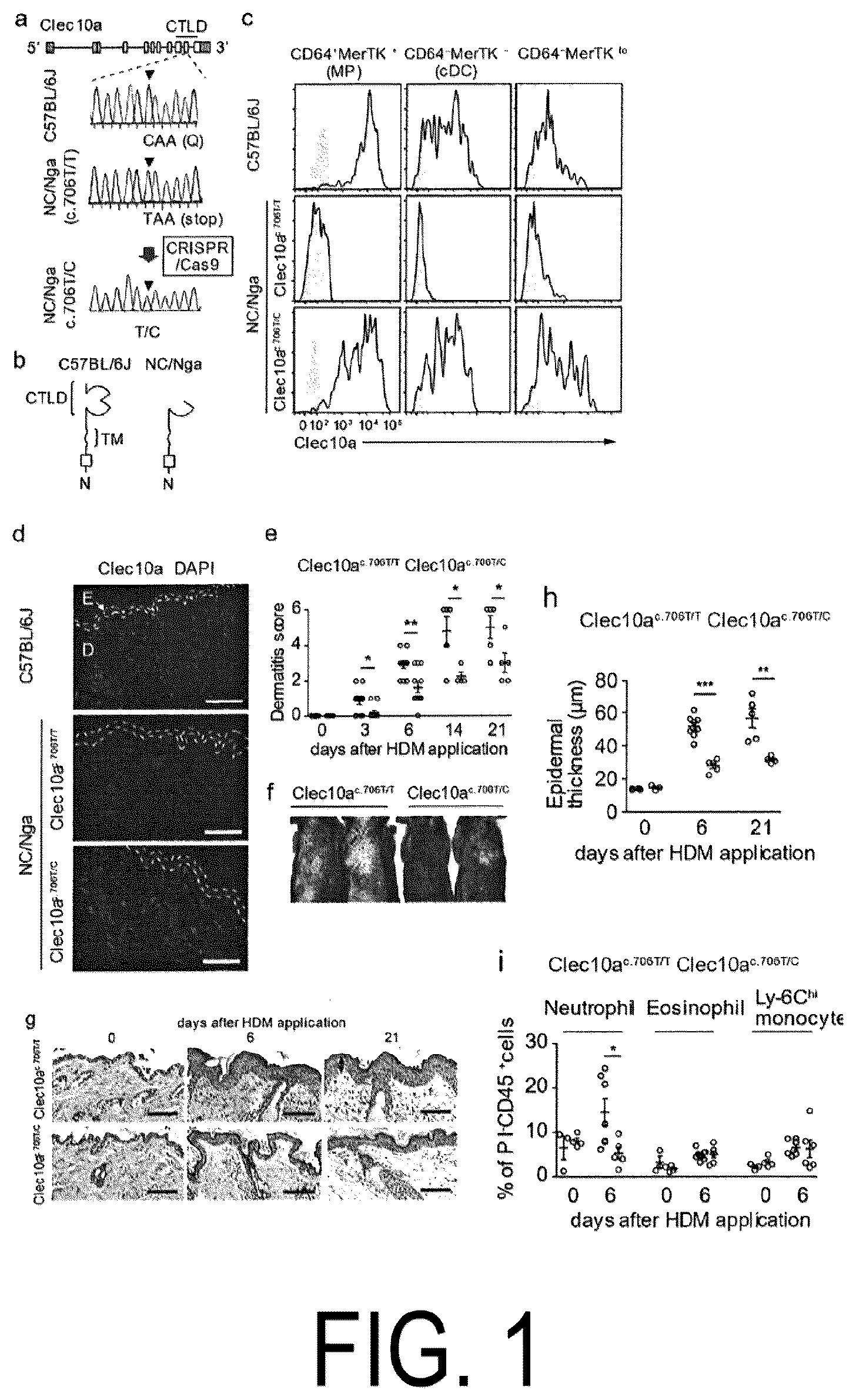
shows that a nonsense mutation in Clec10a in NC/Nga mice causes HDM-induced dermatitis. Panel a shows Clec10a genes and DNA sequences of nonsense mutation sites (c.706) in C57BL/6J Clec10a (NM_010796), NC/Nga-Clec10a c.706T/T (c.706T/T), and NC/Nga-Clec10a c.706 T/C (c.706T/C) mice. The total length of Clec10a is represented by a plurality of open squares and lines therethrough, with open squares representing coding regions of the genes and lines showing introns. CUD represents a c-type lectin-like domain. Panel b shows a schematic representation of Clec10a for each of the C57BL/6J mice and the NC/C57BL/6 mice. TM represents a transmembrane domain. Panel c shows expression of Clec10a on a cell surface of macrophages (MPs) (CD64+MerTK+), known DCs (cDCs) (CD64-MerTK−), and monocyte-derived dendritic cells) (CD64-MerTK lo ) in PI-CD45+MHCII+Lineage (CD3, CD19, NK1.1, and Ly-6G)-EpCAM− cells on the dorsal skin of C57BL/6J mice, NC/Nga-Clec10a c.706T/T mice, and NC/Nga-Clec10a c.706T/C mice. A shaded histogram shows staining with an isotype control antibody (Ab). Panel d shows fluorescence microscopy images of tissue sections of the dorsal skin of C57BL/6J mice, NC/Nga-Clec10a c.706T/T mice, and NC/Nga-Clec10a c.706T/C mice. The tissue sections were stained with an anti-Clec10a monoclonal antibody (mAb) and 4′,6-diamidino-2 phenylindole (DAPI). E represents the epidermis, and D represents the dermis. The scale bar indicates 100 μm. Panels e to i show results of applying an HDM ointment twice a week to the dorsal skin of NC/Nga-Clec10a c.706T/T mice, NC/Nga-Clec10a c.706T/C mice. Panel e shows dermatitis scores, Panel f shows appearance on Day 14, Panels g and h show tissue sections (hematoxylin and eosin stained) and epithelial thickness, respectively. Panel i shows the total number of individuals with neutrophils (CD11b + Ly-6G + ), eosinophils (CD11b + Siglec-F + ), and Ly-6C hi monocytes (CD11b + Ly-6 G -Siglec-F − Ly-6C hi ). * represents p<0.05, ** represents p<0.01, and *** represents p<0.001 (independent two-sided Student's t-test). Data indicates mean±SEM.
shows that Clec10a inhibits HDM-induced immune responses. Panels a-f show results of applying an HDM ointment to the dorsal skin of C57BL/6J wild type mice and Clec10a −/− mice twice weekly. Panels a and b show tissue and epithelial thickness, respectively, at specific time points (n=3, 6, 5). Panels c and d show comparison between the wild type mice and the Clec10a −/− mice. Panel e shows flow cytometry for identifying neutrophils (CD11b + Ly-6g + ) in the skin CD45+ cells of the wild type mice and the Clec10a −/− mice at specific time points. Panel f shows results of quantitative RT-PCR of mRNA obtained from MHCII+ MPs and DCs of the WT and the Clec10a −/− at 3 hours after topical application of HDM. Panel g shows results of cytometric bead array (CBA) analysis of culture supernatants from HDM-stimulated wild-type or Clec10a −/− CD115+ enriched BMMPs (n=3). Panel h shows results of stimulating wild-type, Tlr 4−/−, and Clec10a−/− BMMPs with HDM for a designated time, followed by immunoprecipitation (IP) of lysates thereof with a Clec10a antibody. Immunoblot analysis (IB) was performed using antibodies against phosphorylated tyrosine (pTyr) or Clec10a. Panel i shows results of cytometric bead array (CBA) analysis of culture supernatants from wild-type or Clec10a−/− CD115+ enriched BMMPs pre-treated with 0.5 μM TAK-242 and stimulated with HDM for 6 h (n=3). Panel j shows results of stimulating wild-type or Clec10a−/− CD115+ enriched BMMPs with HDM for a designated time, followed by immunoblot (TB) with phosphorylated Syk (pSyk; Y 519/520 ) or a monoclonal antibody against Syk (mAb). Panel k shows results of stimulating, with HDM, BMW's transfected with wild-type or Y3F Clec10a or with an empty vector (EV), and immunoprecipitating cell lysates with mAb against Clec10a, followed by immunoblot with an antibody against Syk, SHP-1 or Clec10a. Panel 1 shows results of pretreating wild-type and Clec10a−/− BMMPs with Syk inhibitor IV, stimulating the BMMPs with HDM for a designated time, and immunoprecipitating (IP) cell lysates with an antibody against Clec10a, followed by immunoblot with an antibody against pSHP-1, SHP-1 or Clec10a. An arrowhead indicates a molecule of interest (black) or a heavy chain of IP-Ab (white). * represents p<0.05, ** represents p<0.01, and *** represents p<0.001 (independent two-sided Student's t-test). Data indicates mean±SEM.
shows that Clec10a recognizes a mucin-like protein of HDM. Panels a to d show expression of GFP: after stimulation with HDM-coated plates in the presence or absence of rat IgG2a or anti-Clec10a mAb (panels a and b), or galactose (Gal), glucose (Glc) or mannose (Man) (panel c); or after stimulation with HDM-coated plates treated with galactosidase (GALase) or glucosidase (GLCase) or untreated (panel d), in mouse Clec10a-CD3ζ reporter cells or parent reporter cells. Panels e to i show expression of GFP in mouse Clec10a-CD3ζ reporter cells or control reporter cells after stimulation with Clec10a ligand (Clec10a-L) in HDM pulled down (PD) with Clec10a-Fc or control human antibody (panel e), each fraction based on the size of Clec10a-L (panel f), or Clec10a-L before or after treatment with PNGase F or NaOH (panel i). Statistical analysis was performed using PBS-stimulated samples used as controls (panel f). Clec10a-L was immunoblotted using Clec10a-Fc before treatment with PNGase F or NaOH (panels f to h) or after treatment (panel h), or silver-stained with or without alcian blue (panel g). Panel j shows lectin microarray analysis of Clec10a-L. The black bars indicate a lectin binding to Galβ(1-3)GalNAc (T antigen). GalNAc means N-acetylgalactosamine and GlcNAc means N-acetylglucosamine. Panel k shows a schematic representation of Clec10a-L in HDM. T means a T antigen (Galβ(1-3)GalNAc), Tn means a Tn antigen (αGalNAc) and LacNAc means N-acetyl-D-lactosamine (Galβ(1-4)GlcNAc). * represents p>0.05, ** represents p>0.01, and *** represents p>0.001 (one-sided ANOVA test (panels c and f) or independent two-sided Student's t-test (panels b, d, and i)). Data indicates mean±SEM (n=3).
shows that human Asgr1 is a structural and functional counterpart of mouse Clec10a. Panels a and b show expression of GFP in human Asgr1-CD3 reporter cells after stimulation with HDM-coated plates in the absence of galactose, glucose or mannose (panel a) or in the presence thereof (panel b) (n=3). Panel c shows amino acid sequences of intracellular regions of mouse Clec10a, human Asgr1, and human Clec10a. Deduced hemItAM sequences are underlined. Panel d shows GFP expression in human Asgr1-CD3ζ reporter cells stimulated with Clec10a ligand-coated plates obtained by pulling down from HDM with mouse Clec10a-Fc. Panel e shows results of staining tissue sections of human skin with anti-Asgr1 antibodies, anti-CD68 mAbs and DAPI. E means the epidermis, and D means the dermis. The scale bar indicates 100 μm. Panel f shows results of cytometric bead array (CBA) analysis of culture supernatants from human CD14+ monocyte-derived MP treated with siRNA specific for ASGR1 or control siRNA and stimulated with 100 μg/mL HDM for 6 hours (n=3). Panel g shows correlation of ASGR1 expression (GSE5667) in the skin of healthy subjects, psoriasis patients, and atopic dermatitis patients with serum IgE value. Panel h shows a hypothetical model of a function of a C-type lectin receptor during homeostasis of the skin upon exposure to HDM in mice and humans * represents p<0.05, ** represents p <0.01 and *** represents p<0.001 (one-sided ANOVA test (panel b), independent two-sided Student's t-test (panel f) or two-sided Pearson correlation test (panel g)). Data indicates mean±SEM.
shows results of characterization of Clec10a in C57BL/6J mice and NC/C57BL mice. Panel a shows results of gene expression in hematopoietic cells and tissue in C57BL/6J mice based on BioGPS analysis. Although these genes show a nonsense mutation or a frameshift mutation only in NC/Nga mice, such a mutation was not observed in other 19 mouse strains. Panels b to e and i show results of flow cytometry analysis of cells isolated from the dorsal skin (panels b, c, and d) or abdominal (panels e and i) of C57BL/6J mice, NC/Nga mice, and NC/Nga-Clec10a c.706T/C mice. The cells were stained with propidium iodide (Pi), antibodies against designated markers, and anti-Clec10a antibodies or control antibodies. The Lineage indicates CD3, CD19, NK1.1, and Ly-6G. A shaded histogram shows staining with an isotype control antibody. Panels f and g show results of surface analysis for transformants of RAW264.7 expressing Flag-tagged Clec10a c.706C -IRES-GFP or Flag-tagged Clec10a c.706T -IRES-GFP (panel f) and intracellular expression of the Flag tag by flow cytometry (panel g). Panel h shows expression of Clec10a mRNA in the skin of NC/Nga Clec10a c.706T/T (T/T), Clec10a c.706T/C (T/C), and Clec10a c.706C/C (C/C) mice. Panel j shows results of comparison between Clec10a c.706T/T (T/T) and Clec10a c.706T/C (T/C) in terms of serum IgE values at designated time points. * represents p<0.05, ** represents p<0.01, and *** represents p<0.001 (one-sided ANOVA test). Data indicates mean±SEM.
shows a phenotype of HDM-induced dermatitis in Clec10a-deficient mice. A house dust mite (HDM) ointment was applied twice weekly to the dorsal skin of wild type C57BL/6J mice and Clec10a−/− mice. Panel a shows results of flow cytometry analysis of each cell population after staining the skin cells collected at designated time points with an antibody against a Pi, an anti CD45 antibody, or a marker molecule. Markers for each cell were as follows: eosinophils (CD11b + Siglec-F + ) and Ly-6C hi monocytes (CD11b + Ly-6G-Siglec-F-Ly-6C hi ), CD3+CD4+ T cells (CD3 + CD4 + ), CD8+ T cells (CD3+CD8+), epithelial γδ T cells (CD3 hi TCRγδ hi ), dermal γδ T cells (CD3 mid TCRγδ mid ), CD11b + known dendritic cells (cDC) (CD11b + MHCII + CD11c + CD64 − ), MHCII + macrophages (MPs), monocyto-derived DC (CD11b + MHCII + CD11c − CD64 + ). Panel b is an ELISA of serum Ig value at designated time points (n=5). Panel c shows results of quantitative RT-PCR of mRNA expression levels of designated molecules in wild type and Clec10a−/−type CD4+ T cells (CD3 + CD4 + ) sorted from axillary lymph nodes and inguinal lymph nodes on Day 6. ** represents p<0.01 and *** represents p<0.001 (independent two-sided Student's t-test). Data indicates mean±SEM.
shows results of characterization of wild type MP and Clec10a−/−type MP. Panel a shows the gating strategy for sorting of MP (PI-CD45 + MHCII + Lineage (CD3, CD19, NK1.1, and Ly-6G) − EpCAM − CD64 + ) and DC (PI − CD45 + MHCII + Lineage − EpCAM − CD64 − ) obtained from mouse skin 3 hours after topical application of HDM. Panel b shows gate strategy for sorting CD115 + BMMP from the wild type mice and the Clec10a−/−type mice. Panel c shows results of staining of BMMP derived from the wild type mice and the Clec10a−/−type mice with CD115, Clec10a, designated MP markers and antibodies against TLR4. CD115 + cells were gated and the expression of each molecule was analyzed by flow cytometry. A shaded histogram shows staining with an isotype control antibody. Panel d shows results of cytometric bead array (CBA) analysis of culture supernatants of BMMPs derived from Clec10a c.706T/T (T/T) mice and Clec10a c.706T/C (T/C) mice after HDM stimulation (n=3). Panel e shows amino acid sequences of intracellular regions of Clec10a of wild type and Y3F mutant. Deduced hemITAM sequences are underlined. Panels f and g show expression of Clec10a on cell surface of BMMP transfected with cDNA encoding Clec10a of wild-type and Y3F mutant or an empty vector. A shaded histogram shows staining with an isotype control antibody (panel f). Transfected cells were stimulated with HDM, lysed, immunoprecipitated (IP) with anti-Clec10a antibodies, and immunoblotted with anti-phosphorylated tyrosine (pTyr) antibodies and anti-Clec10a antibodies (panel g). An arrowhead indicates a molecule of interest (black) and an antibody (IP-Ab) used in IP (white). Panel h shows results of CBA analysis of cell supernatants from CD115+ BMMP treated with 0.5 μM TAK-242 or DMSO after stimulation with 1 ng/ml LPS or 200 pg/ml Pam2CSK4 for 6 hours. ** represents p<0.01 and *** represents p<0.001 (independent two-sided Student's t-test). Data indicates mean±SEM (n=3).
shows establishment of a Clec10a-CD3ζ reporter cell and a Clec10a-FC chimeric protein. Panels a and b show results of flow cytometry analysis of GFP expression of Clec10a-CD3ζ reporter cells after stimulation with plates coated with Lewis X and Lewis X (10 μg/mL) or Lewis Y (10 μg/mL) at the specified doses (panel a). Panel c shows results of ELISA analysis of Clec10a-Fc bound to the plates coated with Lewis X (LeX) or Lewis Y (LeY). Data indicates mean±SEM (n=3).
shows results of HDM stimulation of human Clec10a-CD3ζ reporter cells and knockdown efficiency of human ASGR1. Panels a and b show expression of GFP in human Clec10a-CD3ζ reporter cells after stimulation with HDM-coated plates in the absence (panel a) or presence (panel b) of galactose (Gal), glucose (Glc), or mannose (Man). Panel c shows expression of Asgr1 on the cell surface of human CD14 + monocyte-derived MPs treated with control siRNA or siRNA specific for ASGR1. A shaded histogram shows staining with an isotype control antibody. *** represents p<0.001 (one-sided ANOVA test). Data indicates mean±SEM (n=3).
is a representation illustrating that a Clec10a ligand (Clec10a-L) improves LPS induced dermatitis. Panels a and b show tissue and epidermal thickness, respectively, on Day 5 after daily application of LPS to the dorsal skin of C57BL/6J wild type (wt) mice and Clec10a −/− mice in the presence or absence of Clec10a-L. Panel c shows the number (/cm 2 ) of neutrophils (CD45 + CD11b + Ly-6G + ) in the skin of WT and Clec10a −/− mice, 6 hours after LPS was applied to the dorsal skin in the presence or absence of Clec10a-L. * represents p<0.05, and ** represents p<0.01 (independent two-sided Student's t-test). Data indicates mean±SEM.
illustrates a scheme for determining Clec10a-L.
shows results of an ELISA assay examining whether Clec10a-Fc binds to plates coated with polymeric scaffolds presenting indicated different glycans.
shows results of a reporter assay using Clec10a reporter cells to examine whether polymeric scaffolds presenting indicated different glycans activated Clec10a.
shows results of a therapeutic experiment examining the effect of administration of polymeric scaffolds presenting T antigens on epidermal inflammation caused by LPS.
shows a structure of the T antigen (Galβ(1-3)GalNAc) and the Tn antigen (aGalNAc).
shows structures of T antigen, Galα1-3LN, Galα1-4LN, LeA, and LeX.
DETAILED DESCRIPTION OF THE INVENTION
Herein, “subject” may be a mammal, including, for example, pets such as dogs, cats, rabbits, hamsters, guinea pigs, and squirrels; livestock such as cows, pigs, horses, sheep, and goats; and primates such as monkeys, chimpanzees, orangutans, gorillas, bonobos, and humans.
“Treatment” is used herein in the sense including therapeutic and prophylactic treatments. Treatment may be used herein in the sense including suppressing a disease or deterioration of a condition, delaying a disease or deterioration of a condition, improving a disease or a condition, or healing of a disease or a condition. Prevention may be used herein in the sense suppressing onset of a disease or a condition or delaying onset of a disease or a condition.
Herein, “allergy” means a systemic or local disorder with respect to a living organism based on an immune response. Allergies are broadly divided into allergies (type I, type II, and type III) based on humoral immune response by blood antibodies and allergies (type IV) based on cellular immunity by sensitized lymphocytes.
Type I allergies are allergies also called immediate allergies or anaphylactic types. IgE is involved in type I allergies, and, when IgE binds with IgE receptors (FcεRI) located on the cell surface of mast cells or basophils in the blood or tissue, and an allergen binds thereto, a chemical mediator such as histamine is released from the mast cells or basophils, thereby causing allergic reactions (e.g., smooth muscle contraction, vascular hyperpermeability, and glandular hypersecretion). Type I allergies include atopic bronchial asthma, allergic rhinitis, urticaria, allergic conjunctivitis, atopic dermatitis, and anaphylactic shock. It is known that, in type I allergies, housing dust, mites, and the like enter the body and cause an allergic reaction (which may enter via routes such as oral route, inhalation route, transdermal route, and transvenous route).
Type II allergies are based on cytotoxicity caused by reaction of IgG or IgM with cells, tissue antigens, and the like and binding of a complement thereto. Antibody-dependent cellular cytotoxicity (ADCC) in which macrophages, killer cells and the like having IgG Fc receptors bind to IgG bound to an antigen of the cell membrane and damage the cells are also included in type II allergies. Type II allergies include hemolytic anemia due to incompatible blood transfusion, autoimmune hemolytic anemia, idiopathic thrombocytopenic purpura, drug-induced hemolytic anemia, granulocytopenia, thrombocytopenia, and Goodpasture's syndrome. Type III allergies are also called immunocomplex type or Arthus type, and are based on tissue damage by the immunocomplex of a soluble antigen with IgG or IgM. Type III allergies include serum disease, autoimmune diseases such as systemic lupus erythematosus and rheumatoid arthritis, glomerulonephritis, hypersensitivity pneumonia, allergic bronchopulmonary aspergillosis. Type IV allergies are also called delayed allergies, and are based on reaction of sensitized T cells with an antigen to release cytokines from the sensitized T cells, resulting in cytotoxicity. Type IV allergies are also based on virus-infected cells by killer T cells, tumor cells, and impairment to grafts. Type IV allergies include allergic contact dermatitis, atopic dermatitis, hypersensitivity pneumonia, tuberculous cavities, leprosy, epithelioid cell granuloma lesions of sarcoidosis, smallpox rash, and measles rash.
Herein, “asialoglycoprotein receptor” is a receptor that binds to a glycoprotein wherein sialic acid at a terminal end of a glycan of a protein is removed and the inner galactose residue is exposed as a terminal group, i.e., an asialoglycoprotein (AGP). The asialoglycoprotein receptor is present on a surface of hepatocytes and binds to AGP in the blood to remove the AGP from the blood. Asialoglycoprotein receptor 1 (ASGR1) is also called C-type lectin domain family member H1 or CLEC4H1. A representative example of human ASGR1 protein can be a protein having an amino acid sequence registered with GenBank under registration number CAG46849.1. “ASGR1”, as used herein, is used in the sense including an ortholog of human ASGR1.
Herein, “Clec10a” is also called C-type lectin domain family 10, member A, which is a molecule that recognizes glycans and functions as a host's biological defense system. Clec10a can specifically bind to galactose or N-acetylgalactosamine.
In the present specification, “house dust mite” is a mite belonging to the genus Dermatophagoides . Main species of house dust mites are Dermatophagoides farinae, Dermatophagoides microceras, Dermatophagoides pteronyssinus , and Euroglyphus maynei.
“Antigen”, as used herein, means a substance that provides an epitope with which a lectin may be reacted in the case of antibodies or sugars. In the context of glycans, “antigen” means a glycan that provides an epitope with which a lectin may be reacted, in accordance with its ordinary word meaning. Thus, when used in the context of glycans, “antigen” means providing a glycan to a skin surface of the glycan as an epitope with which a lectin may be reacted, as is found in natural glycans and glycoproteins.
Herein, “ligand” refers to a counterpart substance to which the receptor binds. The ligand may control a downstream signal of a receptor by binding to the receptor. Herein, substances that positively regulate downstream signals of receptors are called “agonists.” Herein, substances that negatively regulate downstream signals of receptors are called “antagonists.”
Herein, “GalNAc” means N-acetylgalactosamine, and “GlcNAc” means N-acetylglucosamine.
The present inventors discovered that Clec10a is involved in the onset and exacerbation of dermatitis due to house dust mites in mice and that Asgr1 is involved therein in humans. From the results of functional analysis and homology analysis, the present inventors also discovered that human Asgr1 is an original structural and functional counterpart of mouse Clec10a. The present inventors also discovered that the house dust mites include a substance that binds to and activates mouse Clec10a and human Asgr1 and suppresses development of an allergy (e.g., Clec10a ligand or Asgr1 ligand). The present inventors further discovered that the Clec10a ligand includes an O-linked glycan, in particular, a T antigen (Galβ(1-3)GalNAc) or a Tn antigen (αGalNAc), and that ASGR1 binds to both of them. The present inventors also discovered that the Clec10a ligand suppresses TLR4 signals.
The T antigen (Galβ(1-3)GalNAc) and the Tn antigen (αGalNAc) are each a glycan having a structure as shown in . Binding of Asgr1 to a T antigen and a Tn antigen is consistent with Asgr1 having affinity for galactose or N-acetylgalactosamine.
Asgr1 also binds to a glycan selected from the group consisting of a T antigen, LeA, and a LeX, thereby transmitting signals into cells. shows structures of T antigen, Galα1-3LN, Galα1-4LN, LeA, and LeX.
The glycans may be presented on the polymeric scaffold. For example, the glycans may be linked on side chains of polymeric scaffolds having, as their backbone, biocompatible polymers such as polylactic acid, polyacrylamide, polyvinyl, polyvinyl alcohol, polymethyl methacrylate, polyacrylonitrile, polystyrene, polypropylene, polyethylene terephthalate, nylon, collagen, hydroxyethyl methacrylate, chitosan, chitin, polyethylene oxide, polyethylene glycol, polyamino acid, polylactide, polyglycolide, polycaprolactone, and copolymers thereof, and presented to Clec10a. Whether or not the glycan presented on the polymeric scaffold activates Clec10a or Asgr1 can be confirmed using CD3ζ reporter cells which will be described below. In the above, the respective polymers are not particularly limited as long as they have administrable physical properties (e.g., viscosity, osmotic pressure, etc.). For example, biocompatible polymers having a weight average molecular weight from 1 kDa to 100 kDa, from 5 kDa to 50 kDa, from 10 kDa to 40 kDa, or from 20 to 40 kD can be used as their backbone.
According to the present invention, when a nonsense mutation or a frameshift mutation was introduced into Clec10a, which is a mouse counterpart of asialoglycoprotein receptor 1 (Asgr1), the response to HDM became excessive. When extracts (purified) containing a Clec10a ligand were also prepared and contacted with human Asgr1-CD3ζ reporter cells, Asgr1 responded to the Clec10a ligand in a concentration dependent manner. This activation was also offset, in a concentration dependent manner, by the addition of galactose to the system. From this, it can be concluded that the ligand for asialoglycoprotein receptor 1 (Asgr1) can be used to treat a TLR4 signal-induced disease or symptom (e.g., inflammation and an allergic disease) such as a house dust mite-induced allergy.
Accordingly, the present invention provides a composition for use in treatment of an allergic disease, including a ligand for asialoglycoprotein receptor 1 (Asgr1). The present invention provides a composition for use in treatment of a disease or symptom (e.g., inflammation and an allergic disease) induced by activation of a TLR4 signal, including a ligand for asialoglycoprotein receptor 1 (Asgr1). The TLR signal can be activated by a TLR4 ligand. TLR4 ligand includes lipopolysaccharide (LPS) and lipoteichoic acid, and agonists that are analogs thereof. Thus, the ligand for asialoglycoprotein receptor 1 (Asgr1) can be used to treat diseases or conditions (e.g., inflammation and allergic diseases) induced by an allergen including these TLR4 ligands. Accordingly, the present invention provides a composition for use in treatment of a disease or symptom induced by an allergen including a TLR4 ligand, wherein the composition contains a ligand for asialoglycoprotein receptor 1 (Asgr1).
The ligand for asialoglycoprotein receptor 1 (Asgr1) include a glycan from which sialic acid is released and which has Lewis X at its terminal, and a protein having the glycan. The ligand for asialoglycoprotein receptor 1 (Asgr1) include a glycan from which sialic acid is released and which has a T antigen or a Tn antigen at its terminal, and a protein having the glycan. T antigen means Galβ(1-3)GalNAc, and Tn antigen means αGalNAc. The ligand for asialoglycoprotein receptor 1 (Asgr1) may be one, two or all O-linked glycans from which sialic acid is released and which is/are selected from the group consisting of a Lewis X antigen, a T antigen, and a Tn antigen at the terminal, or a protein having the glycan. The mucin-like protein has the O-linked glycan and has a glycan selected from the group consisting of a Lewis X antigen, a T antigen, and a Tn antigen. Thus, the ligand for asialoglycoprotein receptor 1 may be a mucin-like protein or mucin. The protein as the ligand for asialoglycoprotein receptor 1 (Asgr1) can be a mucin-like protein or mucin.
Asgr1 is highly expressed in hepatocytes in vivo, and aged protein (asialoglycoprotein) desialylated in vivo is taken into hepatocytes and removed from the blood. Thus, such desialylated glycoproteins may all be used as the ligand for asialoglycoprotein receptor 1. Asgr1 binds to and reacts with a glycan having at least one or both of a T antigen and a Tn antigen. Thus, a glycan having at least one or both of a T antigen and a Tn antigen or a protein having this glycan can all be used as the ligand for asialoglycoprotein receptor 1. Also, all the ligands for asialoglycoprotein receptor 1 can be obtained by affinity purification of allergen-containing substances (e.g., HDM extracts) based on binding affinity with Clec10a (e.g., mouse Clec10a or human Asgr1). Elution of the Clec10a ligand from Clec10a can be performed, for example, using galactose. A Clec10a ligand eluate may be dialyzed with saline.
Fusion proteins including an extracellular region and a transmembrane region of human asialoglycoprotein receptor 1, and an intracellular region of CD3ζ transmitted a CD3ζ signal to the downstream in the presence of the ligand for asialoglycoprotein receptor 1. Therefore, with a fusion protein including the extracellular region and the transmembrane region of human asialoglycoprotein receptor 1 and the intracellular region of CD3ζ, a substance for use in treatment of an allergic disease can be confirmed. For example, it can be confirmed that a test compound is a compound that binds to human asialoglycoprotein receptor 1 by contacting the compound with an animal cell expressing a fusion protein including the extracellular region and the transmembrane region of human asialoglycoprotein receptor 1 and the intracellular region of CD3ζ, the animal cell having a gene encoding a reporter operably linked to a promoter (for example, an NFAT promoter) that activated by a CD3ζ signal. In addition, it is possible to confirm that the obtained substance is a substance for use in treatment of an allergic disease using control signaling via inhibitory ITAM as an index.
Also, for the allergy suppressive effect of compounds (candidate compounds for the ligand for human Asgr1), for example, compounds can be applied to LPS-induced dermatitis to confirm the effect of suppressing dermatitis.
Thus, in an embodiment of the present invention, there are provided a fusion protein including an extracellular region and a transmembrane region of human asialoglycoprotein receptor 1, and an intracellular region of CD3ζ, and an animal cell that expresses the fusion protein. Also in an embodiment of the present invention, the animal cell may have a gene encoding a reporter operably linked to a promoter activated by a CD3ζ signal. In an embodiment of the present invention, there is provided a method of confirming that a test compound is a compound that binds to human asialoglycoprotein receptor 1 by contacting the compound with an animal cell expressing a fusion protein including an extracellular region and a transmembrane region of human asialoglycoprotein receptor 1, and an intracellular region of CD3ζ, the animal cell having a gene encoding a reporter operably linked to a promoter that activated by a CD3ζ signal.
In an aspect of the invention, the test compound may contain a ligand for asialoglycoprotein receptor 1 (Asgr1). In a certain aspect of the invention, a ligand of the test compound may be at least one glycan selected from the group consisting of a T antigen and a Tn antigen. In a certain aspect of the invention, the ligand may be a mucin-like protein or mucin. In a certain aspect of the invention, the ligand may be a ligand for human asialoglycoprotein receptor 1.
The animal cell expressing a fusion protein including an extracellular region and a transmembrane region of human asialoglycoprotein receptor 1, and an intracellular region of CD3ζ, the animal cell having a gene encoding a reporter operably linked to a promoter that activated by a CD3ζ signal can also be used in compound screening. Therefore, the present invention provides a method for screening for a human Asgr1 ligand or agonist, the method including contacting a test compound with an animal cell expressing a fusion protein including an extracellular region and a transmembrane region of human asialoglycoprotein receptor 1, and an intracellular region of CD3ζ, the animal cell having a gene encoding a reporter operably linked to a promoter that activated by a CD3ζ signal. When the reporter activity is observed, it indicates the test compound is a candidate for the human Asgr1 ligand or agonist.
The animal cell may preferably be a human cell.
The compositions of the present invention may be compositions such as personal care compositions and pharmaceutical compositions.
Pharmaceutical compositions include, for example, pharmaceutical compositions for topical administration and can be used in the present invention. The pharmaceutical composition for topical administration may be a pharmaceutical composition for mucosal or body surface application, and examples thereof include eye drops, eye ointments, sublingual tablets, buccal tablets, troches, gargling agents, sprays, aerosols, and inhalants; solution formulations such as solutions, irrigation agents, glycerin formulations, tartar formulations, aqueous formulations, and coating agents; dispersion formulations such as emulsions, suspensions, liniments, lotions, sprays, and liposomes; semi-solid formulations such as ointments, plasters, patches, adhesive tapes, pastas, cataplasms, cream, oil agents, and sticks; and leaching formulations such as extracts (soft extract, dry extract) and tinctures.
According to the invention, the pharmaceutical composition may contain a pharmaceutically acceptable additive. The pharmaceutically acceptable additive includes solvents, bases, diluents, volume fillers, fillers, and auxiliaries; dissolution aids, solubilizers, buffers, isotonizing agents, emulsifiers, suspending agents, dispersants, thickeners, gelling agents, curing agents, absorbents, adhesives, elastic agents, plasticizers, sustained release agents, and propellants; antioxidants, preservatives, humectants, light blocking agents, antistatic agents, fragrances, flavoring agents, coloring agents, and mitigating agents.
Personal care compositions include, for example, skin care, antiperspirant, deodorant, cosmetic, cosmetic, and hair care products. Personal care compositions include moisturizers, conditioners, anti-aging agents, whitening agents, sunscreens, antiperspirants, shaving compositions, post-shave compositions, foundations, lipsticks, lipsticks, styling compositions, shampoos, cleansers, and lubricants. The personal care composition may be used in personal care products. Personal care products include undergarments, diapers, tissues, wipes, masks, and patches. The composition may contain an additive in addition to the active ingredient. The composition can be in dosage form suitable for administration, such as intravenous administration, transdermal administration, oral administration, enteral administration, and intraperitoneal administration. For the prevention and/or treatment of dermatitis, the composition of the present invention may be administered by transdermal administration, and may be, for example, in the form of a gel, an emulsion, a cream, a liquid, a paste, a lotion, a liposome cream, or the like (for example, a dermatological composition). In an aspect, the composition may be an ointment. In the case of transdermal administration, a dermatologically acceptable additive may be used, and a dosage form suitable therefor can be used. In the case of transmucosal administration, the additive that can be used may be an additive acceptable for mucosal application, and a dosage form suitable therefor may be used.
The present invention provides use of an Asgr1 ligand in the manufacture of a composition for use in treatment of an allergic disease.
The present invention provides a method of treating an allergic disease in a subject in need thereof, including administering to the subject an effective amount of an Asgr1 ligand.
The present invention provides a method of preventing an allergic disease or suppressing development of the allergic disease, including administering to a subject an effective amount of an Asgr1 ligand.
EXAMPLES
Method
(1) Preparation of Skin Cell
Skin cells were minced by pinching the thin-sculpted dorsal skin samples and incubated for 60 minutes in an RPMI-1640 medium containing 200 U/mL collagenase II, 50 U/mL DNase and 10% fetal bovine serum (FBS). Additional dissociation and homogenization were performed using gentleMACS Disociator (Miltenyi Biotec). The resulting cells were filtered through a 55-μm nylon mesh to obtain a single cell suspension.
(2) Flow Cytometry
Flow cytometry and cell sorting were performed using FACS LSRFortessa and FACS Aria (BD Biosciences), respectively. FlowJo software (Tree Star) was used for analysis of data. Dead cells were stained with propidium iodide solution (Sigma-Aldrich, P4864) and removed.
(3) Histology and Immunohistochemical Staining
For histological analysis, dorsal skin samples harvested from mice were formalin-fixed and paraffin-embedded to create 4 μm thick sections. Sections were stained with hematoxylin-eosin and analyzed by optical microscopy. Epithelial thickness was measured in five regions per mouse and at five sites per region.
For immunohistochemical staining, skin samples harvested from mice were embedded in TissueTek Optimal Cutting Temperature Compound (Sakura Finetek Japan) and stored at −80° C. Four (4) μm-thick sections were also used in immunohistochemical staining. The sections were washed with PBS containing 0.05% Tween-20 (PBS-T, pH 7.4), stained and incubated for 10 min at room temperature using Blocking One Histo (Nacalai). The sections were then washed with PBS-T, incubated overnight at 4° C. using anti-Clec10a mAb, washed with PBS-T and incubated using Alexa Fluor 546 labeled anti-rat IgG polyclonal antibody for 1 hour. After washing with PBS-T, the sections were subjected to nuclear staining with DAPI.
Human healthy skin tissue was harvested from the periphery of the patient's tumor region, formalin-fixed, paraffin-embedded into 4 μm-thick sections. The sections were deparaffinized with xylene, and rehydrated with ethanol, and endogenous peroxidase was blocked with methanol. The sections were stained with anti-CD69 antibodies and anti-Asgr1 antibodies according to the manufacturer's manual for Opal 4-Color Automation IHc Kit (PerkinElmer, NEL820001KT). Briefly, the sections were incubated at 95° C. for 15 minutes, washed with TBS (TBS-T) (Takara Bio, T9142) containing 0.05% Tween-20 and treated with a blocking solution at room temperature for 10 minutes. The sections were incubated overnight at 4° C. with anti CD68 antibodies or mouse IgG1 antibodies, washed with TBS-T and treated with Opalpolymer HRP in a wet chamber at room temperature for 30 minutes. After washing with TBS-T, the sections were incubated using Opal Fluorophore Working Solution in a wet chamber at room temperature for 10 minutes and washed with TBS-T. The antibodies for the first staining were removed from the sections by heating at 95° C. The sections were then stained with anti-CD9 antibodies or rabbit IgG antibodies as in the first staining. The sections were subjected to nuclear-staining with a spectral DAPI solution.
(4) House Dust Mite (HDM)-Induced Dermatitis
In the first induction (Day 0), the hair on the skin in the back of anesthetized mice was removed using an electronic clipper, and the remaining hair was epilated using hair removal cream. One hundred (100) mg of HDM ( Dermatophagoides farinae ) ointment (Biostir, Japan) was administered topically to the skin in the shaved back. From the second induction, the skin bather function was disrupted by applying 150 μL 4% sodium dodecyl sulfate to the dorsal skin 2 hours prior to the HDM ointment administration. These procedures were repeated twice weekly. Several factors (erythema/hemorrhage and scar/dryness) were scored on Days 3, 6, 14, and 21 according to an evaluation criterion of 0 (none), 1 (mild), 2 (moderate) or 3 (severe) according to the manufacturer's instructions (Biostir). The sum of the scores was taken as overall dermatitis score.
(5) Establishment of RAW264.7 Transformant
Clec10a cDNA was made from C57BL/6J mice or NC/Nga mice and labeled with a sequence encoding a Flag tag and subcloned into a pMXs-IRES-GFP retroviral vector. A RAW264.7 transformant stably expressing C57BL/6J type or NC/Nga type Clec10a was established based on a routine method.
(6) ELISA Assay
Serum IgE antibodies were measured using capturing antibodies against mouse IgE (R35-72) and biotinylated anti-mouse IgE (R35-118) followed by HRP-labeled streptavidin (Ge Healthcare, RPN1231V). Purified mouse IgE (C38-2, BD Biosciences) was used as a standard. Serum IgG1 was measured using capturing antibodies against mouse IgG1 (A85-3) and HRP-labeled antibodies against mouse IgG1. Purified mouse IgG1 (107.3, BD Biosciences) was used as a standard. Serum IgG2c was detected by mouse ELISA Quantitation Set (Bethyl, E90-136).
(7) Preparation of Bone Marrow Macrophage (BMMP)
Bone marrow cells were cultured on a culture dish (Corning, 430166 or 430167) in an RPMI1640 complete medium containing 10% FBS in the presence of 10 ng/ml GM-CSF (Rd Systems) and 7 ng/mL IL-4 (Wako). On Day 2, 70% of non-adherent cells were removed and a fresh medium containing GM-CSF and IL-4 was added. On Day 5, 100% of non-adherent cells were removed by washing with PBS and a fresh medium containing GM-CSF and IL-4 was added. On Day 7, all non-adherent cells were removed by PBS wash and adherent cells were used in later experiments. For the analysis of cytokine secretion and Syk phosphorylation, CD115 + BMMP was concentrated using anti CD115 antibodies (BioLegend) and anti-rat IgG microbeads (Miltenyi Biotec).
(8) Analysis of Cytokine Secretion
CD115 + BMMP was stimulated with 100 μg/mL HDM extract ( Dermatophagoides farinae ) (COSMO BIO), 1 ng/mL lipopolysaccharide, or 200 pg/mL Pam2CSK4 in the presence or absence of 0.5 μM TAK-242 (Merck) for 15 minutes. After 6-hour stimulation, culture supernatants were collected and the concentration of each cytokine was determined using cytometric bead array analysis (BD Biosciences).
(9) Synthesis of cDNA and Real-Time PCR (RT-PCR)
Total RNA was extracted from skin tissue or cells sorted by flow cytometry using Isogen reagent (Nippon Gene). Skin MP was sorted by CD45 + MHCII + CD3 − CD19 − NK1.1 − Ly-6G − EpCAM − CD64 + and DC was sorted by CD45 + MHCII + CD3 − CD19 − NK1.1 − Ly-6G − EpCAM − CD64 − ). CD3 + CD4 + cells were also obtained. cDNA was synthesized using High Capacity RNA-to-cDNA Kit (Applied Biosystems). Gene expression of Clec10a and inflammatory cytokines was measured by quantitative RT-PCR using SYBR Gree Master Mix (Applied Biosystems) and specific primers. An expression level of Gapdh was used as an internal reference for standardized data. The primer sequences used were as shown in Table 1 below.
TABLE 1
Gene name Forward Reverse
Clec10a 5′-ACCCAAGAGCCTGGTAAAGC-3′ 5′-GATCCAATCACGGAGACGAC-3′
Tnf 5′-GGGCCACCACGCTCTTC-3′ 5′-GGTCTGGGCCATAGAACTGATG-3′
Il6 5′-GAGGATACCACTCCCAACAGACC-3′ 5′-AAGTGCATCATCGTTGTTCATACA-3′
Cxcl1 5′-ACTCAAGAATGGTCGCGAGG-3′ 5′-GTGCCATCAGAGCAGTCTGT-3′
Cxcl2 5′-AAGTTTGCCTTGACCCTGAA-3′ 5′-AGGCACATCAGGTACGATCC-3′
Ifng 5′-ACAGCAAGGCGAAAAAGGATG-3′ 5′-TGGTGGACCACTCGGATGA-3′
Il4 5′-ATCATCGGCATTTTGAACGAGG-3′ 5′-TGCAGCTCCATGAGAACACTA-3′
Il17 5′-TTTAACTCCCTTGGCGCAAAA-3′ 5′-CTTTCCCTCCGCATTGACAC-3′
Il10 5′-GCTGGACAACATACTGCTAACC-3′ 5′-ATTTCCGATAAGGCTTGGCAA-3′
Tbx21 5′-AGCAAGGACGGCGAATGTT-3′ 5′-CGGTGGACATATAAGCGGTTC-3′
Gata3 5′-TTATCAAGCCCAAGCGAAGG-3′ 5′-CATTAGCGTTCCTCCTCCAGAG-3′
Rcrc 5′-GGAGGACAGGGAGCCAAGTT-3′ 5′-CCGTAGTGGATCCCAGATGACT-3′
Foxp3 5′-CCCATCCCCAGGAGTCTTG-3′ 5′-ACCATGACTAGGGGCACTGTA-3′
Gapdh 5′-TGGTGAAGGTCCGTGTGAAC-3′ 5′-ATGAAGGGGTCGTTGATGGC-3′
(10) Retroviral Gene Transfer
Wild-type Clec10a cDNA was subcloned into a pMXs-puro retroviral vector (Cell Biolabs). To create site-specific Clec10a mutants, the PCR primers of the sense strands were designed so as to contain phenylalanine (TTC) instead of tyrosine (TAC). The resulting mutant cDNA was confirmed by sequencing. Retroviruses were obtained by transfecting 293GP packaging cells with wild-type or mutant Y3F cDNA or VSV-G expression vector pCMV-VSV-G. BMMP was infected with viral supernatants added with polybrene (8 μg/ml, Sigma-Aldrich) on Days 2 and 4. After centrifugation, the supernatants containing viruses were removed and the medium was replaced with a fresh BMMP medium. On Day 5, the medium was replaced with a fresh BMMP medium and non-adherent cells were removed by washing with PBS wash on Day 7 and adherent cells were used in the experiment.
(11) Preparation of Clec10a-Fc Chimera
A mouse Clec10a chimeric construct (Clec10a-Fc) was made by cloning the extracellular region of mouse Clec10a into a pME18S expression vector containing the Fc region of human IgG1. Clec10a-Fc was transfected into HEK293T cells using Lipofectamine 2000 (Thermo Fisher Scientific) and the medium was then replaced with GIT medium (KOHJIN BIO). Clec10a-Fc was purified from the culture supernatants using protein a agarose (Bio-Rad Laboratories).
(12) Isolation of Clec10a Ligand
An HDM extract dissolved in a buffer containing 150 mM NaCl, 50 mM Tris, 1 mM CaCl 2 , and 0.01% Tween 20 was subjected to a pull-down assay using Clec10a-Fc. The ligand bound to Clec10a-Fc was eluted using 30 mM EDTA or 200 mM galactose. The eluate was dialyzed with a centrifugal filter unit (Merck, UFC503024) using PBS as an external fluid, named Clec10a-L, and used as one of Clec10a ligands.
(13) Alcian Blue Staining and Silver Staining
The Clec10a ligand was developed by SDS-PAGE. Immediately after electrophoresis, the gel was washed with deionized water and 10% acetate buffer (deionized water with 10% acetic acid and 30% ethanol) and stained for 2 hours at room temperature with or without an alcian blue solution (pH 2.5) (Wako, 013-13801) and then destained with 3% acetate buffer and 10% acetate buffer. The gel was silver-stained according to the manufacturer's instructions (Pierce Silver Stain Kit, Thermo Fisher Scientific).
(14) Fractionation of Clec10a Ligand
The Clec10a ligand was developed by SDS-PAGE and the gel was cut out in a manner of separating according to size. The cut gels were mechanically milled and incubated overnight in PBS with agitation. The supernatant was collected after centrifugation at 17400 g for 10 minutes and dialyzed with a centrifugal filter unit (Merck, UFC503024) using PBS as an external fluid.
(15) Immunoblotting
To analyze phosphorylation of Syk, BMMP was stimulated with an HDM extract (100 μg/mL) at 37° C. for 0, 10, or 30 minutes. The stimulated BMMP was lysed with a 1% NP-40 lysis buffer and separated by SDS-PAGE. It was transferred onto a PVDF membrane by electroblotting, immunoblotted with anti-phosphorylated Syk antibodies and anti-Syk antibodies, and detected using HRP-labeled anti-rabbit IgG antibodies.
To analyze tyrosine phosphorylation of Clec10a, BMMP was stimulated with an HDM extract (100 μg/mL) at 37° C. for 0 min, 10 min, or 30 min. The stimulated BMMP was lysed with a 1% NP-40 lysis buffer and subjected to Immunoprecipitation with anti-Clec10a mAb. An immune precipitate was developed by SDS-PAGE and transferred onto a PVDF membrane and immunoblotted with HRP-labeled anti-phosphorylated tyrosine antibodies or anti-Clec10a antibodies and BRP-labeled anti-rabbit IgG antibodies.
To analyze association of Clec10a with Syk or SHP-1, BMMP was pretreated in the presence or absence of 5 mM Syk inhibitor IV (Merck, 574714) at 37° C. for 30 minutes and stimulated with an HDM extract (100 μg/mL) at 37° C. for 0 min, 10 min, or 30 min. BMMP was lysed with a 0.2% digitonin buffer and immunoprecipitated with anti-Clec10a mAb. An immune precipitate was transferred onto a PVDF membrane as described above and immunoblotted using anti-Syk antibodies, anti-SHP-1 antibodies, or anti-Clec10a antibodies, and then detected with HRP-labeled anti-rabbit IgG antibodies or goat IgG antibodies. All proteins were detected using enhanced chemiluminescence (Thermo Fisher Scientific).
After pretreatment at 37° C. for 16 hours in the presence or absence of peptide N-glycosidase F (PNGase F PRIM™, NZS1, N-Zyme Scientific) and thermal metamorphism at 95° C. for 5 minutes, a Clec10a ligand was pretreated at 40° C. for 16 hours in the presence or absence of 0.05 M NaOH, separated by SDS-PAGE, transferred into a PVDF membrane, and immunoblotted with biotinylated Clec10a-Fc and HRP-labeled streptavidin.
(16) Establishment and Stimulation of Reporter Cell
The intracellular region of human CD3ζ was obtained from a vector provided by LL Lanier (University of California, San Francisco). The extracellular region of mouse or human Clec10a or the extracellular region of human Asgr1 was subcloned into a pMXs-puro retroviral vector. 2B4-NFAT-GFP reporter cells were provided from H. Arase (University of Osaka). 2B4-NFAT-GFP reporter cells stably expressing mouse or human Clec10a were made as described previously. The reporter cells were incubated for 18 hours in the presence or absence of anti-Clec10a mAb, Lewis X (GlycoTech), Lewes Y (GlycoTech), galactose (Sigma-Aldrich), glucose (Sigma-Aldrich) or mannose (Sigma-Aldrich). The reporter cells were also cultured on a galactosidase (R & D Systems, 5704 GH or 5549 GH) or glucosidase (R & D Systems, 8329-GH), or Clec10 ligand-coated plate, or an HDM extract-coated plate treated with size-fractionated Clec10a ligand or untreated. Activation of NFAT-GFP was monitored by flow cytometry.
(17) Lectin Microarray Analysis
Lectin microarrays were made using a non-contact microarray printing robot (MicroSys4000; Genomic Solutions) according to the previous method. Samples were fluorescently labeled with Cy3 Mono-Reactive dye (Ge) and excess Cy3 was removed using a Sephadex g-25 desalting column (Ge). After 10 fold dilution with a probe solution (25 mM Tris-HCl, pH 7.5, 140 mM NaCl) (TBS) containing 2.7 mM KCl, 1 mM CaCl 2 , 1 mM MnCl 2 , and 1% Triton X-100, a Cy3-labeled sample was applied to a lectin microarray and incubated at 20° C. overnight. The sample was washed with a probe solution, and a fluorescence image was obtained using an evanescent-field activated fluorescence scanner (Bio-Rad scan 200, Rexxam Co. Ltd.). Lectin signals in triplicate spots were averaged for each protein sample and normalized to an average value of 96 lectins. The list of lectins was as shown in Table 2.
TABLE 2
Lectin used in lectin microarray
Name Species Origin Source2 Rough specificity1
1 LFA Limax flavus Natural EY Lab. Sia
2 WGA Triticum vulgaris Natural EY Lab. (GlcNAc)n, polySia
3 PVL Psathyrella velutina Natural Wako Sia, GlcNAc
4 MAL Maackia amurensis Natural Seikagaku α2-3Sia
5 MAH Maackia amurensis Natural Vector α2-3Sia
6 ACG Agrocybe cylindracea Natural JOM α2-3Sia
7 rACG Agrocybe cylindracea E . coli AIST α2-3Sia
8 rGal8N Homo sapiens E . coli AIST α2-3Sia
9 SNA Sambucus nigra Natural Seikagaku α2-6Sia
10 SSA Sambucus sieboldiana Natural Vector α2-6Sia
11 TJAI Trichosanthes japonica Natural Vector α2-6Sia
12 rPSL1a Polyporus squamosus E . coli AIST α2-6Sia
13 PHAL Phaseolus vulgaris Natural Seikagaku GlcNAcβ1-6Man
(Tetraantenna)
14 DSA Datura stramonium Natural Seikagaku GlcNAcβ1-6Man
(Tetraantenna)
15 TxLcl Tulipa gesneriana Natural JOM Galactosylated N-glycans
up to triantenna
16 ECA Erythrina cristagalli Natural Seikagaku βGal
17 RCA120 Ricinus communis Natural Vector βGal
18 rGal7 Homo sapiens E . coli AIST Type1 LacNAc,
chondroitin polymer
19 rGal9N Homo sapiens E . coli AIST GalNAcα1-4Gal (A),
PolyLacNAc
20 rGal9C Homo sapiens E . coli AIST PolyLacNAc,
Branched LacNAc
21 rC14 Gallus gallus domesticus E . coli AIST Branched LacNAc
22 rDiscoidinII Dictyostelium dicodeum E . coli AIST LacNAc,
Galβ1-3GalNAc (T),
GalNAc (Tn)
23 BPL Bauhinia purpurea alba Natural Vector Galβ1-3GlcNAc(GalNAc),
α/β GalNAc
24 rCGL2 Homo sapiens E . coli AIST GalNAcα1-3Gal (A),
PolyLacNAc
25 PHAE Phaseolus vulgaris Natural Vector bisecting GlcNAc
26 GSLII Griffonia simplicifolia Natural Vector GlcNAcβ1-4Man
27 rSRL Sclerotium rolfsii E . coli AIST Core1,3, agalacto N-glycan
28 UDA Urtica dioica Natural Vector (GlcNAc)n
29 PWM Phytolacca americana Natural Vector (GlcNAc)n
30 rF17AG Escherichia coli E . coli AIST GlcNAc
31 rGRFT Griffithia sp. E . coli AIST Man
32 NPA Narcissus pseudonarcissus Natural Seikagaku Manα1-3Man
33 ConA Canavalia ensiformis Natural Vector M3, Manα1-2Manα1-
3(Manα1-6)Man,
GlcNAcβ1-2Manα1-
3(Manα 1-6)Man
34 GNA Galαnthus nivalis Natural Vector Manα 1-3Man,
Manα 1-6Man
35 HHL Hippeastrum hybrid Natural Vector Manα 1-3Man,
Manα 1-6Man
36 ASA Allium sativum Natural JOM Galβ 1-4GlcNAcβ1-2Man
37 DBAI Dioscorea batatas Natural JOM High-man
38 CCA Castanea crenata Natural JOM Galactosylated N-
glycans up to triantenna
39 Heltuba Helianthus tuberosus Natural JOM Manα 1-3Man
40 rHeltuba Helianthus tuberosus E . coli AIST Manα 1-3Man
41 ADA Allomyrina dichtoma Natural JOM α2-6Sia, Forssman, A, B
42 VVAII Vicia villosa Natural JOM Man, Agalacto
43 rOrysata Oryza sativa E . coli AIST Manα 1-3Man, Highman,
biantenna
44 rPALa Phlebodium aureum E . coli AIST Man5, biantenna
45 rBanana Musa acuminata E . coli AIST Manα 1-2Manα 1-3(6)Man
46 rCalsepa Calystegia sepium E . coli AIST Biantenna with bisecting
GlcNAc
47 rRSL Ralstonia solanacearum E . coli AIST αMan,α1-2Fuc (H),
α1-3Fuc (Lex),
α1-4Fuc (Lea)
48 rBC2LA Burkholderia cenocepacia E . coli AIST αMan, High-man
49 AOL Aspergillus oryzae Natural Vector α1-2Fuc (H),
α1-3Fuc (Lex),
α1-3Fuc (Lea)
50 AAL Aleuria aurantia Natural Vector α1-2Fuc (H),
α1-3Fuc (Lex),
α1-4Fuc (Lea)
51 rAAL Aleuria aurantia E . coli AIST α1-2Fuc (H),
α1-3Fuc (Lex),
α1-3Fuc (Lea)
52 rPAIIL Pseudomonas aeruginosa E . coli AIST αMan, α1-2Fuc (H),
α1-3Fuc (Lex),
α1-4Fuc (Lea)
53 rRSIIL Ralstonia solanacearum E . coli AIST α1-2Fuc (H),
α1-3Fuc (Lex),
α1-3Fuc (Lea)
54 rPTL Pholiota terrestris E . coli AIST α1-6Fuc
55 PSA Pisum sativum Natural Seikagaku α1-6Fuc up to biantenna
56 LCA Lens culinaris Natural Vector α1-6Fuc up to biantenna
57 rAOL Aspergillus oryzae E . coli AIST α1-2Fuc (H),
α1-3Fuc (Lex),
α1-3Fuc (Lea)
58 rBC2LCN Burkholderia cenocepacia E . coli AIST Fuc α1-2Galβ1-3GlcNAc
(GalNAc)
59 LTL Lotus tetragonolobus Natural Seikagaku Lex, Ley
60 UEAI Ulex europaeus Natural Vector α1-2Fuc
61 TJAII Trichosanthes japonica Natural Vector α1-2Fuc
62 MCA Momordica charantia Natural JOM α1-2Fuc
63 GSLI Griffonia simplicifolia Natural Seikagaku αGalNAc (A, Tn), αGal (B)
64 PTLI Psophocarpus tetragonolobus Natural Tokyo Kasei αGalNAc (A, Tn)
65 GSLIA4 Griffonia simplicifolia Natural EY Lab. αGalNAc (A, Tn)
66 rGC2 Geodia cydonium E . coli AIST α1-2Fuc (H),
αGalNAc (A),
αGal (B)
67 GSLIB4 Griffonia simplicifolia Natural Vector αGal (B)
68 rMOA Marasmius oreades E . coli AIST αGal (B)
69 EEL Euonymus europaeus Natural Vector αGal (B)
70 rPAIL Pseudomonas aeruginosa E . coli AIST α,βGal, αGalNAc (Tn)
71 LEL Lycopersicon esculentum Natural Vector Polylactosamine, (GlcNAc)n
72 STL Solanum tuberosum Natural Seikagaku Polylactosamine, (GlcNAc)n
73 rGal3C Homo sapiens E . coli AIST LacNAc, polylactosamine
74 rLSLN Laetiporus sulphureus E . coli AIST LacNAc, polylactosamine
75 rCGL3 Coprinopsis cinerea E . coli AIST LacDiNAc
76 PNA Arachis hypogaea Natural Vector Galβ 1-3GalNAc (T)
77 ACA Amaranthus caudatus Natural Vector Galβ 1-3GalNAc (T)
78 HEA Hericium erinaceum Natural JOM Galβ 1-3GalNAc (T)
79 ABA Agarics bisporuserinaceum Natural Vector Galβ 1-3GalNAc (T),
GlcNAc
80 Jacalin Artocarpus integrifolia Natural Seikagaku Galβ 1-3GalNAc (T),
GalNAcα(Tn)
81 MPA Maclura pomifera Natural Seikagaku Galβ 1-3GalNAc (T),
GalNAcα(Tn)
82 HPA Helix pomatia Natural Seikagaku αGalNAc (A, Tn)
83 VVA Vicia villosa Natural Vector α, βGalNAc
(A, Tn, LacDiNAc)
84 DBA Dolichos biflorus Natural Vector α, BGalNAc
(A, Tn, LacDiNAc)
85 SBA Glycine max Natural EY Lab. α, βGalNAc
(A, Tn, LacDiNAc)
86 rPPL Pleurocybella porrigens E . coli AIST α, βGalNAc
(A, Tn, LacDiNAc)
87 rCNL Clitocybe nebularis E . coli AIST α, BGalNAc
(A, Tn, LacDiNAc)
88 rXCL Xerocomus chrysenteron E . coli AIST Core1,3,
agalacto N-glycan
89 VVA! Vicia villosa Natural JOM GalNAcβ1-3(4)Gal
90 WFA Wisteria floribunda Natural Vector Terminal GalNAc,
LacDiNAc
91 rABA Agarics bisporus E . coli AIST Galβ1-3GalNAc (T),
GlcNAc
92 rDiscoidinI Dictyostelium Discodeum E . coli AIST Gal
93 DBAIII Dioscorea batatas Natural JOM Maltose
94 rMalectin Homo sapiens E . coli AIST Glcα1-2Glc
95 CSA Oncorhynchus keta Natural JOM Rhamnose, Galα1-4Gal
96 FLAG-EW29Ch-E20K Lumbricus terrestris E . coli AIST 6-sulfo-Gal
Abbreviations:
Gal (D-galactose), GalNAc (N-acetyl-galactosamine), GlcNAc (N-acetyl-glucosamine), Fuc (L-fucose), Sia (Sialic acid), and LacNAc (N-acetyl-lactosamine).
2 Specific data was obtained by frontal affinity chromatography and sugar conjugate microarrays.
3 Abbreviations: JOM (J-OIL MILLS, INC), Vector (VECTOR LABORATORIES), Seikagaku (SEIKAGAKU CORPORATION), EY (EY LABORATORIES, INC), and AIST (National Institute of Advanced Industrial Science and Technology) (18) Sugar Conjugate Microarray Analysis
A sugar conjugate microarray containing 98 sugar conjugates (Table 3) was prepared according to a known method using a non-contact microarray printing robot (MicroSys 4000; Genomic Solutions). Clec10a-Fc (10 μg/mL) was pre-complexed with Cy3-labeled goat anti-human IgG, Fc (Jackson, 109-165-098) (1 μg/mL) in advance and incubated with sugar conjugate microarrays (80 μL/well) overnight at 20° C.
TABLE 3
Glycan used in sugar conjugate microarray
Trivial name Presentation Glycans Source Cat#
αFuc PAA Fucα 1-PAA Glycotech 01-007
Fucα2Gal PAA Fucα 1-2Galβ 1-PAA Glycotech 01-019
Fucα3GlcNAc PAA Fucα 1-3GlcNAcβ 1-PAA Glycotech 01-024
Fucα4GlcNAc PAA Fucα 1-4GlcNAcβ 1-PAA Glycotech 01-025
H type1 PAA Fucα 1-2Galβ 1-3GlcNAcβ 1-PAA Glycotech 01-037
H type2 PAA Fucα 1-2Galβ 1-4GlcNAcβ 1-PAA Glycotech 08-034
H type3 PAA Fucβ 1-2Galβ 1-3GalNAcα 1-PAA Glycotech 08-060
A PAA GalNAc 1-3(Fucα 1-2)Galβ 1- Glycotech 08-091
4GlcNAcβ 1-PAA
B PAA Galα 1-3(Fucα 1-2)Galβ Glycotech 08-092
1-4GlcNAcβ 1-PAA
Le a PAA Galβ 1-3(Fucα 1-4)GlcNAcβ 1-PAA Glycotech 01-035
[3S]Le a PAA (3OSO3)Galβ 1-3(Fucα 1-4) Glycotech 01-040
GlcNAcβ 1-PAA
Le b PAA Fucα 1-2Galβ 1-3(Fucα 1-4) GlcNAcβ Glycotech 08-042
1-PAA
Le x PAA Galβ.1-4(Fucα 1-3)GlcNAcβ 1-PAA Glycotech 01-036
Le y PAA Fucα 1-2Galβ1-4(Fucα 1-3)GlcNAcβ Glycotech 08-043
1-PAA
α Neu5Ac PAA Neu5Acα 2-PAA Glycotech 01-012
α Neu5Gc PAA Neu5Gcα 2-PAA Glycotech 01-051
Sia2 PAA Neu5Acα 2-8Neu5Acα 2-PAA Glycotech 08-064
Sia3 PAA Neu5Acα 2-8Neu5Acα Glycotech 01-081
2-8Neu5Acα 2-PAA
3′SiaLec PAA Neu5Acα 2-3Galβ Glycotech 01-078
1-3GlcNAcβ 1-PAA
3′SL PAA Neu5Acα 2-3Galβ Glycotech 01-038
1-4Glcβ 1-PAA
3′SLN PAA Neu5Acα 2-3Galβ Glycotech 01-077
1-4GlcNAcB 1-PAA
sLe a PAA Neu5Acα 2-3Galβ Glycotech 08-044
1-3(Fucα 1-4)GlcNAcββ 1-PAA
sLe x PAA Neu5Acα 2-3Galβ Glycotech 01-045
1-4(Fucα 1-3)GlcNAcp 1-PAA
6′SL PAA Neu5Acα 2-6Galβ 1-4Glcβ 1-PAA Glycotech 01-039
β Gal PAA Galβ 1-PAA Glycotech 01-004
[3S] β Gal PAA (3OSO3)Galβ 1-PAA Glycotech 01-015
A-di PAA GalNAcα 1-3Galβ 1-PAA Glycotech 01-017
Lac PAA Galβ 1-4Glcβ 1-PAA Glycotech 01-021
Le c PAA Galβ 1-3GlcNAcβ 1-PAA Glycotech 01-020
[3′S]Le c PAA (3OSO3)Galβ 1-3GlcNAcβ 1-PAA Glycotech 01-062
LN PAA Galβ 1-4GlcNAcβ 1-PAA Glycotech 01-022
[3′S]LN PAA (3OSO3)Galβ 1-4GlcNAcβ 1-PAA Glycotech 01-061
[6S]LN PAA Galβ 1-4(6OSO3)GlcNAcβ 1-PAA Glycotech 01-066
[6′S]LN PAA (6OSO3)Galβ 1-4GlcNAcβ 1-PAA Glycotech 01-068
β GalNAc PAA GalNAcβ 1-PAA Glycotech 01-011
di-GalNAcβ PAA GalNAcβ 1-3GalNAcβ 1-PAA Glycotech 01-070
LDN PAA GalNAcβ 1-4GlcNAcβ 1-PAA Glycotech 01-057
GA2 PAA GalNAcβ 1-4Gal 1-4Glcβ 1-PAA Glycotech 08-074
BGlcNAc PAA GlcNAcβ 1-PAA Glycotech 01-009
[6S]β GlcNAc PAA (6OSO3)GlcNAcβ 1-PAA Glycotech 01-016
α Man PAA Manα 1-PAA Glycotech 01-005
βMan PAA Manβ 1-PAA Glycotech 01-050
[6P]Man PAA (6OPO4)Manα 1-PAA Glycotech 01-006
Tn PAA GalNAcα 1-PAA Glycotech 01-010
Core1 PAA Galβ 1-3GalNAcα 1-PAA Glycotech 08-023
Core2 PAA Galβ 1-3(GlcNAcβ 1-6) Glycotech 01-083
GalNAcα 1-PAA
Core3 PAA GlcNAcβ 1-3GalNAcα 1-PAA Glycotech 01-071
Core4 PAA GlcNAcβ 1-3(GlcNAcβ 1-6) Glycotech 01-089
GalNAcα 1-PAA
Forssman PAA GalNAcα 1-3GalNAcβ 1-PAA Glycotech 01-026
Core6 PAA GlcNAcβ 1-6GalNAcα 1-PAA Glycotech 01-072
Core8 PAA Galα 1-3GalNAcα 1-PAA Glycotech 01-028
[3′S]Core1 PAA (3OSO3)Galβ 1-3GalNAcα 1-PAA Glycotech 08-069
Galβ-Core3 PAA Galβ 1-4GlcNAcβ Glycotech 01-116
1-3GalNAcα 1-PAA
STn PAA Neu5Acα 2-6GalNAcα 1-PAA Glycotech 01-059
STn (Gc) PAA Neu5Gcα 2-6GalNAcα 1-PAA Glycotech 01-107
ST PAA Neu5Acα 2-3Galβ Glycotech 01-088
1-3GalNAcα 1-PAA
Siaa 2-6Core1 PAA Galβ 1-3(Neu5Acα 2-6) Glycotech 01-113
GalNAcα 1-PAA
α Gal PAA Galα 1-PAA Glycotech 01-003
Galα 1-2Gal PAA Galα 1-2Galβ 1-PAA Glycotech 01-056
Gaα 1-3Gal PAA Galα 1-3Galβ 1-PAA Glycotech 01-018
Galα 1-3Lac PAA Galα 1-3Galβ 1-4Glcβ 1-PAA Glycotech 01-075
Galα 1-3LN PAA Galα 1-3Galβ 1-4GlcNAcβ 1-PAA Glycotech 01-079
Galα 1-4LN PAA Galα 1-4Galβ 1-4GlcNAcB 1-PAA Glycotech 01-110
Melibiose PAA Galα 1-6Glcβ 1-PAA Glycotech 01-063
α Glc PAA Glcα 1-PAA Glycotech 01-001
β Glc PAA Glcβ 1-PAA Glycotech 01-002
Maltose PAA Glcα 1-4Glcβ 1-PAA Glycotech 01-054
α Rha PAA Rhamnosea 1-PAA Glycotech 01-008
Chitobiose PAA GlcNAcβ 1-4GlcNAcβ 1-PAA Glycotech 08-057
Negative PAA PAA — Glycotech 01-000
(19) Knock Down of Human Asgr1
CD14 + monocytes were concentrated from peripheral blood mononuclear cells using anti CD14 microbeads (Miltenyi Biotec, 130-050-201) and cultured in the presence of GM-CSF for 2 days. The monocytes were treated with siRNA specific for ASGR1 (SMARTpool Accell siRNA, Dharmacon) or a mixture of control siRNA and siRNA delivery agents. The monocytes were then stimulated with 100 μg/mL HDM extract for 6 hours. Concentrations of various cytokines in culture supernatants were determined using cytometric bead array analysis (BD Biosciences).
(20) Human Skin Gene Expression Data
Published microarray data (GSE5667) was used to analyze the expression of human ASGR1 in the pathologic state of human skin and atopic dermatitis.
(21) Statistical Analysis
Statistical analysis was performed using two-sided Student's t-test (GraphPad Prism 5), post-hoc Tukey-Kramer test and ANOVA test (GraphPad Prism 5), or Pearson correlation test (two-sided, GraphPad Prism 5).
Example 1 Exome Analysis of Atopic Dermatitis Model Mouse
In this example, exome analysis of NC/Nga mice as model mice of atopic dermatitis was performed to identify causative genes of atopic dermatitis.
The NC/Nga mice were purchased from Charles River, Japan. DNA was extracted from blood from the NC/Nga mice under conditions suitable for extraction of mouse DNA using QIAamp DNA blood Mini Kit (Qiagen, Venlo, Netherlands). The exome analysis of the resulting DNA was performed as follows. DNA libraries were obtained using SureSelect Library prep kit (post-pool version 4; Agilent Technologies, Santa Clara, CA) and SureSelect Mouse All Exon Kit (Agilent Technologies) according to the manufacturer's manual. The resulting DNA libraries were subjected to emulsion PCR (SOLiD EZ Bead Emulsifier kit; Thermo Fisher Scientific, Waltham, MA) to generate clonal DNA fragments on beads, which were then subjected to bead enrichment (SOLiD EZ Bead Enrichment kit; Thermo Fisher Scientific, Waltham, MA). The resulting beads were sequenced on a SOLiD 5500xl sequencer as single end 60-bp reads. The resulting read were aligned with the mouse reference genome (NCBI37/mm9) using LifeScope version 2.5.1 (Life Sciences) to obtain a BAM file. Variant calls were performed using SAMtool according to the protocol described in Genome Analysis Toolkit, Picard (http://broadinstitute.github.io/picard), and only reads mapped to unique locations on the reference genome were used for analysis. Mutations were annotated using the ANNOVER software. Genetic variation in inbred mice other than NC/Nga was obtained from Release REL-1211 numbered according to the NCBIm 37 assembly (http://www.sanger.ac.uk/science/data/mouse-genomes-project) and Mouse Genome Informatics website (http://www.informatics.jax.org/). Sequencing was performed on an ABI 3130xl Genetic Analyzer (Thermo Fisher Scientific) using BigDye Terminator v1.1 Cycle Sequencing Kit (Thermo Fisher Scientific). The primers used were as shown in Table 4.
TABLE 4
Primer used for confirmation of nonsense mutation or frameshift
mutation in NC/Nga mouse by Sanger sequencing
Gene Primer sequence
Cdh20 Forward TCGGACTCAGAGCAGAGCTT
Reverse CTCTGCTGGGTOCACTCACT
Tlr5 Forward GCCATTCTTCCTTGAACCAC
Reverse ATGGCCGTGTGGGAGTATAA
Tmem141 Forward GATCAGGGACTCCAAAACCA
Reverse TGCTGAGGTAGGAGGGACTG
Qsox2 Forward AGACTCAGCCACGTGAACCT
Reverse TCGGGCTCAGACATTTCACT
Hc Forward TCGTGTTTTTAAATATTTTGCTTCC
Reverse CCCCACCCTCTTCTGGTACT
Tdpoz2 Forward GGTGGAAGTCAATGGTGGAG
Reverse TTGTCTCTGGGACTCAAAGGA
Gm572 Forward GTTTCGGCGCTTTTGTTTTA
Reverse CTTCAGAGGCCAGGACAAAG
Cyp3a57 Forward TGATGTTCTTCTTTGACCTTCC
Reverse TCCCTCTCTGAGTACCATCCA
Try4 Forward GAGGGCTCCACCTAACAACA
Reverse GTACAGACAGGGCCCATCAC
Klri2 Forward TGATGAGCACTCATTTCACACA
Reverse TCCCAGTGCCAACAGTTACA
Kira7 Forward AAAGTTAAAGAGTTGCCCCTTG
Reverse TGAATTATTGCAGGAAACAAATG
Ttc23 Forward GAACTGCTCTAACGCTGTGG
Reverse ACAGTGCCATCCAGGGTTC
Olfml1 Forward GGGCATTCATGGAAGATAGC
Reverse CATCCACAGCAAGGTCAATG
Foxr1 Forward CGCAGTTTCCCCTTCTCAT
Reverse TGGAGGTACAAGGTTCTGTGC
Cilp Forward AAGAGCAATGTGGGAGTTGC
Reverse AGCATCATGAGGCAGAGACA
Slc22a21 Forward GCTTGTTTTGCAACTGATGG
Reverse AGCACTGTTGTCGGTCACTG
Clec10a Forward TGAGGGAGAGGTAACCATGC
Reverse GGGCAAATGTACAGCACACA
Serpina3i Forward GCTGTCAGGACTCAGCAGTG
Reverse GGTCAGGGAGAATGAACAGG
Zkscan4 Forward AATCCACACGGGTGAGAAAC
Reverse CAGTGTGTATTGGCCACACC
Zfp957 Forward TGCAGAGCAAAGTCAAGGTT
Reverse CTTAGCGGCTGCGTTTTT
Timm8a2 Forward CATCCACCACATGACAGAGC
Reverse GTCCATTTCCCCACCTACCT
Pdzd2 Forward ATGCATGCTCGCTTTTTCTT
Reverse GAGGGATGGGGGAAGAGTTA
Cyp2d11 Forward AGGCAGAGTCCAACAGGAAA
Reverse CCTACCTTGGTGACGAGGAA
Kcnk7 Forward CCCCAGCCTCAGTATCAGAA
Reverse ATTTAGCCCAGAGTCGCTTG
In the exon region of the genome of the NC/Nga mice, 70772 mutations were identified relative to the reference genome. The mutations with low quality values obtained from the GATK output were removed and 64518 mutations were obtained. Loss of function mutations, including nonsense mutation and frameshift mutation, were selected using the ANNOVER software. Thirty five (35) nonsense mutations and 48 frameshift mutations (induction of stop codons) were then identified. For each of these 83 genes, 46 mutations in 43 genes were selected for further sequence validation.
Of the 46 mutations, 24 mutations were confirmed by sequencing. The results were as shown in Table 5.
TABLE 5
Nonsense mutation and frameshift mutation in NC/Nga mouse confirmed by Sanger sequencing
Chromo- Last RefSeq Nucleotide Amino acid
Gene some Start End Function exon2 number substitution substitution
Cdh20 1 1 106890959 106890959 stopgain SNV yes NM_011800 c.G2403A p.W801X
Tlr5 1 184902583 184902584 frameshift substitution no NM_016928 c.18_19delinsT
Tmem141 2 25476035 25476038 frameshift deletion yes NM_001109993 c.603_606del
Qsox2 2 26065186 26065186 frameshift deletion yes NM_153559 c.1845delC
Hc 2 34898728 34898729 frameshift deletion no NM_010406 c.646_647del
Tdpoz2 3 93455535 93455535 frameshift insertion no NM_001007222 c.1051_1052insAG
Gm572 4 148045509 148045515 frameshift deletion yes NM_001085505 c.1172_1178del
Cyp3a57 5 146138201 146138205 frameshift deletion no NM_001100180 c.957_961del
Try4 1 6 41253356 41253356 frameshift deletion no NM_011646 c.114delG
Klri2 1 6 129683766 129683766 stopgain SNV no NM_177155 c.G407A p.W136X
Klra7 1 6 130169025 130169029 frameshift deletion yes NM_014194 c.693_697del
Ttc23 1 7 74837804 74837804 frameshift deletion no NM_025905 c.783delT
Olfml1 7 114733919 114733919 frameshift deletion yes NM_172907 c.676delT
Foxr1 9 44243294 44243294 frameshift insertion yes NM_001033469 c.638_639insAA
Cilp 9 65127938 65127938 frameshift deletion yes NM_173385 c.3507delG
Slc22a21 11 53764775 53764775 frameshift insertion yes NM_019723 c.1678_1679insC
Clec10a 1 11 69983716 69983716 stopgain SNV no NM_010796.2 c.C706T p.Q236X
Serpina3i 12 105504794 105504794 frameshift deletion no NM_001199940 c.747delC
Zkscan4 13 21576578 21576578 frameshift insertion yes NM_001039115 c.1416_1417insA
Zfp957 14 79613286 79613286 frameshift insertion yes NM_001033215 c.879_880insG
Timm8a2 14 122434145 122434154 frameshift deletion no NM_001037744 c.237_246del
Pdzd2 1 15 12375345 12375345 stopgain SNV no NM_001081064.1 c.C1054T p.R352X
Cyp2d11 15 82220453 82220453 frameshift substitution no NM_001104531 c.1156_1156delinsCACCC
Kcnk7 19 5706988 5706988 frameshift insertion yes NM_010609 c.1004_1005insT
Gene rs number human homolog Other mouse strains
Cdh20 1 +
Tlr5 + MSM/Ms, JF1/Ms
Tmem141 + MSM/Ms, JF1/Ms
Qsox2 rs260851851 + NZO/HILtJ, PWK/PhJ
Hc rs241579076 + A/J, AKR/J, DBA/2J, FVB/NJ, NOD/ShiLtJ
Tdpoz2 − MSM/Ms, JF1/Ms
Gm572 rs258174385 + 129S1/SvlmJ, A/J, AKR/J, BALB/cJ, C3H/HeJ, CAST/EiJ, CBA/J, NOD/ShiLtJ, PWK/PhJ, WSB/EiJ
Cyp3a57 rs242224866 + 129S1/SvlmJ, AKR/J, CBA/J, LP/J, NZO/HILtJ
Try4 1 +
Klri2 1 −
Klra7 1 −
Ttc23 1 +
Olfml1 rs262610229 + 129S1/SvlmJ, C3H/HeJ, CAST/EiJ, CBA/J, DBA/J, DBA/2J, LP/J, NOD/ShiLtJ, NZO/HlLtJ,
PWK/PhJ, WSB/EiJ, MSM/Ms, JF1/Ms
Foxr1 rs215230760 + 129S1/SvlmJ, CAST/EiJ, LP/J, PWK/PhJ, MSM/Ms, JF1/Ms
Cilp rs262051220 + 129S1/SvlmJ, A/J, AKR/J, BALB/cJ, C3H/HeJ, C57BL/6NJ, CAST/EiJ, CBA/J, DBA/2J, FVB/NJ,
LP/J, NOD/ShiLtJ, NZO/HlLtJ, PWK/PhJ, SPRET/EiJ, WSB/EiJ, MSM/Ms, JF1/Ms
Slc22a21 rs231568920 − NZO/HlLtJ, PWK/PhJ, MSM/Ms, JF1/Ms
Clec10a 1 +
Serpina3i rs242560633 − 129S1/SvlmJ, AKR/J, BALB/cJ, CBA/J, DBA/2J, FVB/NJ, LP/J, NOD/ShiLtJ, WSB/EiJ
Zkscan4 rs249591363 + A/J, AKR/J, BALB/cJ, C3H/HeJ, CAST/EiJ, CBA/J, DBA/2J, FVB/NJ, LP/J, NOD/ShiLtJ, WSB/EiJ
Zfp957 rs249051530 − 129S1/SvlmJ, A/J, AKR/J, BALB/cJ, C3H/HeJ, CBA/J, FVB/NJ, LP/J, NZO/HlLtJ, PWK/PhJ
Timm8a2 rs237108606 − 129S1/SvlmJ
Pdzd2 1 +
Cyp2d11 + 129S1/SvlmJ3, A/J3, AKR/J3, BALB/cJ3, C3H/HeJ3, CBA/J3, DBA/2J3, LP/J3, NOD/ShiLtJ3,
NZO/HlLtJ3
Kcnk7 + A/J, AKR/J, BALB/cJ, C3H/HeJ, NOD/ShiLtJ, PWK/PhJ, SPRET/EiJ, MSM/MS, JF1/Ms
Mutations of 7 genes specifically present in NC/Nga mice are indicated in bold.
“Last Exon” means that a mutation is located in the last exon of the corresponding transcript.
Rs262301704 corresponds to c, 1157_1158insCCCA and a mouse with rs262301704 is indicated by *.
Mutations present in 17 inbred mouse lines and two Japanese pet mice were excluded, and seven mutations in seven genes were noted. (See the bold in Table 5). From the perspective of expression in hematopoietic cells, Clec10a was selected from the seven genes based on BioGPS database information (see panel a in ). Clec10a of the NC/Nga mice was a nonsense mutation (i.e. Q236X) where the 706th C in the base sequence of NM_010769 was T (that is, Q 236X).
Example 2: Clec10a Expression Analysis and Functional Analysis
Clec10a is a member of the type II C-type lectin receptor family and detects galactose moieties at terminals of foreign and endogenous antigens. c.706C>T of Clec10a in the NC/Nga mice is present in the coding region of the C-type lectin-like domain ( , panels a and b). The flow cytometric analysis revealed that Clec10a was expressed on the cell surface of macrophages (MP) (CD64 + MerTK + ), known dendritic cells (cDC) (CD64 − MerTK − ), and mononuclear cell-derived DCs (CD64 − MerTK lo ), as well as macrophages in the peritoneal cavity of C 57BL/6J (not expressed in the peritoneal cavity of NC/Nga mice), in skin CD45 + MHCII + Lineage − EpCAM − cells (see panel c in and panels b and e in ). No expression of Clec10a was observed on non-hematopoietic cells (CD45 − ), CD45 + MHCII− cells, CD45 + MHCII + (Lin, EpCAM) + cells, and neutrophils (CD45 + CD11b + Ly-6G + ) of the skin of the C57BL/6J mice (see panels c and d in ). Similar results were obtained by the immunohistochemistry staining, and expression of Clec10a was observed in skin cells of the C57BL/63 mice, but no expression was observed in the NC/Nga mice (panel d in ).
The characteristics of Clec10a expression in the NC/Nga mice were further analyzed. Specifically, cDNA from Clec10a (from C57BL/6J and NC/Nga mice) with a Flag tag at the 3′end and a sequence encoding IRES-GFP was transfected into RAW264.7 macrophages. The Flag tag was expressed on the cell surface of the macrophages expressing Clec10a derived from the C57BL/6J mice, but not on the cell surface of macrophages expressing Clec10a from the NC/Nga mice. (See panel f in ). However, in the macrophages expressing Clec10a from the NC/Nga mice, expression of Flag in the cells was observed (see panel g in ). These results suggest that mutations in Clec10a negatively affected Clec10a transport to the cell surface.
This Clec10a mutation was then checked whether it is involved in dermatitis due to house dust mites (HDMs). Thus, instead of a native sequence (Clec10a c.706T ), a mutant mouse with Clec10a c.706C (hereafter, sometimes referred to as “NC/Nga mouse Clec10a c.706T>C ”) was made according to a routine method using a CRISPR/Cas9 system (see panel a in ). In the skin of the mutant mice with Clec10a c.706C , Clec10a mRNA expression was significantly increased as compared with the mice with Clec10a c.706T (see panel h in ). In mutant mice heterozygous for Clec10a c.706T and Clec10a c.706C (Clec10a c.706T/C ), Clec10a expression on the surface of macrophages in the skin and abdominal cavity was restored (see panels c and d in , and panel i in ). In addition, symptoms of HDM-induced dermatitis, such as erythema, dryness, and increased skin thickness, were alleviated in the mutant mice (Clec10a c.706T/C ) (panels e to h in ). Furthermore, neutrophil infiltration into the skin after six days of the HDM treatment was reduced in the mutant mice (Clec10a c.706T/C ) as compared with NC/Nga mice with wild type Clec10a (Clec10a c.706T/T ) (panel i in ). In contrast, the IgE serum levels of both the mice were comparable. These results suggest that Clec10a mutants (Clec10a c.706TC ) cause HDM-induced dermatitis and not increase Th2 response in the NC/Nga mice.
To investigate the involvement of cell surface expression of Clec10a in HDM-induced dermatitis, Clec10a-deficient mice (Clec10a −/− mice) were made using C57BL/6J mice as a background. HDM was applied topically to the dorsal skin of the wild type C57BL/6J mice and the Clec10a-deficient mice (Clec10a −/− ). As observed in the skin of the NC/Nga mice, the Clec10a−/− mice showed exacerbations of dermatitis, such as erythema, dryness and skin enhancement on Days 5 to 6 after the HDM treatment, as compared with the wild type mice (see panels a to d in ). In addition, on Days 1 to 3 after the HDM treatment, the Clec10a−/− mice showed an increase in neutrophil infiltration in the skin (see panel e in ). In contrast, the wild type mice and the Clec10a−/− mice were equivalent in number of cells in other bone marrow and lymphocytic systems (see panel a in ).
At two weeks after the HDM treatment, the serum IgG1 level was higher in the Clec10a−/− mice than in the wild type, and the IgE and IgG2c values were comparable between the wild type and the Clec10a−/− mice (see panel b in ). In addition, expression of Il4 and Gata3 CD4 + T cells in the HDM-treated skin draining lymph node was comparable between the wild type and the Clec10a−/− mice (see panel c in ). Therefore, it is believed that Clec10a suppresses dermatitis by reducing neutrophil infiltration rather than reducing the Th2 response.
Since Clec10a was expressed in MHCII + macrophages (MPs) and dendritic cells (DCs) of the skin, these cells were sorted from skin cell samples of topically-HDM-treated wild type and Clec10a−/− mice (see panel a in ). Gene expression levels of Il6, Cxcl1, and Cxcl2 were higher in the Clec10a−/− MP than in the wild type MP, but these gene expression levels were comparable between the wild type DC and the Clec10a−/− DC (see panel f in ). These results suggest that Clec10a in MP is involved in suppression of HDM-induced dermatitis more than in DC. Bone marrow macrophages (BMMPs) were prepared from the wild type mice and the Clec10a−/−C57BL/6J mice, respectively, and characterized (see panel b in ). The expression levels of MP marker and TLR4 in CD115 + BMMP were comparable between the wild type mice and the Clec10a−/−mice. After in vitro HDM stimulation, the Clec10a−/− BMMP secreted a larger amount of neutrophil chemotactic substances, such as IL-6, CXCL1, CCL3 and TNF-α, than the wild type BMMP (see panel g in ). Similarly, BMMP of the Clec10a c.706T/T NC/Nga mice showed a larger amount of cytokine production than BMMP in the Clec10a c.706T/C (see panel d in ). These results demonstrated that the expression of Clec10a on the cell surface inhibits the production of inflammatory cytokines from the skin MP following stimulation with HDM.
HDM stimulation caused activation of spleen tyrosine kinase (Syk) in the wild type BMMP, but no activation of Syk was observed in the Clec10a−/− BMMP (see panel j in ). In addition, HDM stimulation caused the recruitment of Syk and protein tyrosine phosphatase SHP-1 to Clec10a, and the recruitment relied upon Clec10a tyrosine residues (see panel k in ). BMMP treatment with Syk inhibitors suppressed the recruitment of HDM-induced SHP-1 to Clec10a (panel 1 in ), suggesting that the event relies on Syk activation. The results were consistent with control signaling via inhibitory ITAM.
Example 3: Analysis of Clec10a Ligand
To test whether Clec10a recognized glycosylated proteins contained in HDM, NFAT-GFP reporter cells (Clec10a reporter cells) were made which expressed chimeric fusion proteins inch ling an extracellular and transmembrane portions of Clec10a fused to a cytoplasmic portion of CD3. Mouse Clec10a reporter cells expressed GFP in response to Lewis X (Clec10a oligosaccharide ligand) but did not respond to Lewis Y (see panels a and b in ). The mouse Clec10a reporter cells also expressed GFP in response to the HDM-coated plate dose-dependently (panel a in ). In addition, pretreatment of the reporter cells with anti-Clec10a monoclonal antibodies (mAb) or galactose inhibited GFP expression, but glucose or mannose pretreatment did not suppress GFP expression (panels b and c in ). Furthermore, treatment of HDM with galactosidase reduced their ability to cause GFP expression in the reporter cells, but no such reduction was observed when glucosidase was used (panel d in ). These results indicate that Clec10a directly binds to the galactosylated moiety of HDM.
For analysis of the Clec10a ligand (Clec10a-L) in HDM, chimeric fusion proteins including an extracellular portion (Clec10a-Fe) of mouse Clec10a fused to the Fc portion of human IgG1 were made, but the fusion proteins were not bound to Lewis Y but were bound to Lewis X (panel c in ). Pull-down assays from HDM using Clec10a-Fc isolated Clec10a-L having a molecular weight of about 225 kDa, but Clec10a-L was able to induce expression of GFP in the mouse Clec10a reporter cells (see panels e and f in ). Clec10a-L was stained with alcian blue and silver but not stained with silver alone (see panel g in ), indicating that Clec10a-L is a highly sugar-modified protein. Treatment with NaOH to degrade the O-linked glycans inhibited the binding of Clec10a-Fc to the epitope (see panel h in ) and inhibited the induction of GFP expression in the mouse Clec10a reporter cells (see panel i in ). In contrast, PNGase F, which degrades N-linked glycans, did not show any effect on Clec10a-L (see panels h and i in ). These results suggest that Clec10a recognizes the O-linked glycans of Clec10a-L. Analysis by the lectin microarray also showed that Clec10a-L contains a T antigen (Galβ(1-3)GalNAc) and a Tn antigen (αGalNAc) and has an LacNAc epitope (Galβ(1-4)GlcNAc) binding to mucin-type O glycans (panel j in , and Table 6).
TABLE 6
Lectin microarray analysis of Clec10a ligand in HDM
Signal value
Rank Lectin Rough specificity Target 1 Control1 2 Control2 3
1 MPA Galβ1-3GalNAc (T), αGalNAc (Tn) 20283 0 0
2 BPL Galβ1-3GalNAc (T), terminal GalNAc 19440 0 0
3 WFA Terminal GalNAc, LacDiNAc 18081 0 0
4 rSRL Galβ1-3GalNAc (T), GlcNAcβ1-3GalNAc, 17794 0 0
agalacto N-glycan
5 rABA Galβ1-3GalNAc (T), GlcNAc 17018 0 0
6 rxCL Galβ1-3GalNAc (T), GlcNAcβ1-3GalNAc, 16918 0 0
aGalacto N-glycan
7 rCNL Terminal GalNAc (A, Tn, LacDiNAc) 16900 0 0
8 Jacalin Galβ1-3GalNAc (T), αGalNAc (Tn) 15697 0 0
9 UDA (GlcNAc)n 15228 0 0
10 rGC2 α1-2Fuc (H), αGalNAc (A), αGal (B) 15049 0 0
11 RCA120 βGal 14050 0 0
12 rPSL1a α2-6Sia 13311 0 0
13 SBA α, βGalNAc (A, Tn, LacDiNAc) 12337 0 0
14 ABA Galβ-3GalNAc (T), GlcNAc 11636 0 0
15 rCGL2 GalNAcα1-3Gal (A), PolyLacNAc 10999 1370 0
16 PNA Galβ1-3GalNAc (T) 10699 0 0
17 TJAII β1-2Fuc 10518 0 0
18 VVA! GalNAcβ1-3(4)Gal 10377 0 0
19 HEA Galβ1-3GalNAc (T) 9936 0 0
20 rGal9N GalNAcα1-4Gal (A), PolyLacNAc 9093 0 0
21 rLSLN LacNAc, polylactosamine 8962 0 0
22 MCA α1-2Fuc 8239 0 0
23 ACA Galβ1-3GalNAc (T) 8057 0 0
24 rGal9C PolyLacNAc, Branched LacNAc 7990 0 0
25 rAAL α1-2Fuc (H), α1-3Fuc (Lex), α1-3Fuc (Lea) 7764 0 0
26 AAL α1-2Fuc (H), α1-3Fuc (Lex), α1-4Fuc (Lea) 7508 0 0
27 VVA a, βGalNAc (A, Tn, LacDiNAc) 6765 0 0
28 rACG α2-3Sia 6442 0 0
29 rBC2LCN Fucα1-2Galβ1-3GlcNAc (GalNAc) 5909 0 0
30 HPA αGalNAc (A, Tn) 5471 0 0
31 GNA Manα1-3Man, Manα1-6Man 4458 0 0
32 NPA Manα1-3Man 3993 0 0
33 ECA βGal 3871 0 0
34 DBAI High-man 3793 0 0
35 rRC2LA αMan, High-man 3766 0 0
36 PVL Sia, GlcNAc 3729 0 0
37 rRSIIL α1-2Fuc (H), α1-3Fuc (Lex), α1-3Fuc (Lea) 3678 0 0
38 rMOA αGal (B) 3656 0 0
39 rGRFT Man 3502 0 0
40 rAOL α1-2Fuc (H), α1-3Fuc (Lex), α1-3Fuc (Lea) 3194 0 0
41 CCA Galactosylated N-glycans up to triantenna 3093 0 0
42 LEL Polylactosamine, (GlcNAc)n 3054 0 0
43 STL Polylactosamine, (GlcNAc)n 2611 0 0
44 rPPL a, βGalNAc (A, Tn, LacDiNAc) 2579 0 0
45 rPALa Man5, biantenna 2493 0 0
46 GSLIA4 αGalNAc (A, Tn) 2181 0 0
47 HHL Manα1-3Man, Manα1-7Man 1731 0 0
48 rDiscoidinII LacNAc, Galβ1-3GalNAc (T), GalNAc (Tn) 1306 0 0
49 DSA GlcNAcβ1-6Man (Tetraantenna) 1095 0 0
50 SSA α2-6Sia 1013 0 0
51 TxLcl Galαctosylated N-glycans up to triantenna 973 0 0
52 rDiscoidinI Gal 737 0 0
53 rCalsepa Biantenna with bisecting GlcNAc 651 0 0
54 rGal3C LacNAc, polylactosamine 630 0 0
55 WGA (GlcNAc)n, polySia 624 0 0
56 PSA α1-6Fuc up to biantenna 481 0 0
57 AOL α1-2Fuc (H), α1-3Fuc (Lex), α1-3Fuc (Lea) 309 0 0
58 LCA α1-6Fuc up to biantenna 228 0 0
59 rPAIIL αMan, α1-2Fuc (H), α1-3Fuc (Lex), 131 0 0
α1-4Fuc (Lea)
60 TJAI α2-6Sia 113 0 0
61 rBanana Manα1-2Manα1-3(6)Man 10 0 0
62 ASA Galβ1-4GlcNAcβ1-2Man 2 0 0
62 rCGL3 LacDiNAc 2 0 0
64 LFA Sia 0 0 0
64 MAL α2-3Sia 0 0 0
64 MAH α2-3Sia 0 0 0
64 ACG α2-3Sia 0 0 0
64 rGal8N α2-3Sia 0 0 0
64 SNA α2-6Sia 0 0 0
64 ADA α2-6Sia, Forssman, A, B 0 0 0
64 PHAL GlcNAcβ1-6Man (Tetraantenna) 0 0 0
64 rGal7 Type1 LacNAc, chondroitin polymer 0 0 0
64 rC14 Branched LacNAc 0 0 0
64 PHAE bisecting GlcNAc 0 0 0
64 GSLII GlcNAcβ1-4Man 0 0 0
64 PWM (GlcNAc)n 0 0 0
64 rF17AG GlcNAc 0 0 0
64 ConA M3, Manα1-2Manα1-3(Manα1-6)Man, 0 0 0
GlcNAcβ1-2Manα1-3(Manα1-6)Man
64 Heltuba Manα1-3Man 0 0 0
64 rHeltuba Manα1-3Man 0 0 0
64 VVAII Man, agalacto 0 0 0
64 rOrysata Manα1-3Man, Highman, biantenna 0 0 0
64 rRSL αMan, α1-2Fuc (H), α1-3Fuc (Lex), 0 0 0
α1-4Fuc (Lea)
64 rPTL α1-6Fuc 0 0 0
64 LTL Lex, Ley 0 0 0
64 UEAI α1-2Fuc 0 0 0
64 FLAG-EW29Ch Gal 0 0 0
64 PTLI αGalNAc (A, Tn) 0 0 0
64 GSLIB4 αGal (B) 0 0 0
64 EEL αGal (B) 0 0 0
64 rPAIL a, βGal, αGalNAc (Tn) 0 0 0
64 DBA a, βGalNAc (A, Tn, LacDiNAc) 0 0 0
64 DBAIII Maltose 0 0 0
64 rMalectin Glcα1-2Glc 0 0 0
64 CSA Rhamnose, Galα1-4Gal 0 0 0
64 FLAG-EW29Ch- 6-sulfo-Gal 0 0 0
E20K
1 Target: Pull-down assay sample from HDM using Clec10a-Fc
2 Control 1: Pull-down assay sample from buffer using Clec10a-Fc
3 Control 2: Pull-down assay sample from HDM using human IgG1
Clec10a was most strongly bound to the lectins of Maclura pomifera (MPA) recognizing high density of multivalent T and Tn antigens. On the other hand, analysis by glycan microarray revealed that Clec10a-Fc binds to T and Tn antigens (see Table 7) and suggested that Clec10a-L is a mucin-like protein (see panel k in ).
TABLE 7
Analysis of Clec10a-Fc by glycan microarray
Signal value
human Clec10a-Fc
Rank Glycan Structure Clec10a-Fc IgG1 with EDTA
1 αGal Galα1 11934 0 0
2 Lea Galβ1-3(Fucα1-4)GalNAcβ1 5532 0 0
3 Lac Galβ1-4Glcβ1 4850 0 0
4 Core2 Galβ1-3(GlcNAcβ1-6)GalNAcα1 4824 0 0
5 Galα1-3LN Galα1-3Galβ1-4GlcNAcβ1 4342 0 0
6 Lex Galβ1-4(Fucα1-3)GlcNAcβ1 2535 0 0
7 T (Core1) Galβ1-3GalNAcα1 2504 0 0
8 Lec Galβ1-3GlcNAcβ1 2414 0 0
9 βGal Galβ1 1867 0 0
10 Galα1-4LN Galα1-4Galβ1-4GlcNAcβ1 1679 0 0
11 A-di GalNAcα1-3Galβ1 1137 0 0
12 Tn GalNAcα1 682 0 0
13 Galα1-3Lac Galα1-3Galβ1-4Glcβ1 444 0 0
14 Galα1-3Gal Galα1-3Galβ1 144 0 0
15 di-GalNAcβ GalNAcβ1-3GalNAcβ1 65 0 0
16 Melibiose Galα1-6Glcβ1 45 0 0
17 Sia3 Neu5Acα2-8Neu5Acα2-8Neu5Acα2 19 0 3
18 3′SLN Neu5Acα2-3Galβ1-4GlcNAcβ1 7 0 0
18 LN Galβ1-4GlcNAcβ1 7 0 0
20 βGalNAc GalNAcβ1 6 0 0
21 3′SL Neu5Acα2-3Galβ1-4Glcβ1 5 0 0
22 3′SiaLec Neu5Acα2-3Galβ1-3GlcNAcβ1 4 0 0
23 βFuc Fucα1 3 0 0
23 [3S] βGal (3OSO3)Galβ1 3 0 0
23 Galα1-2Gal Galα1-2Galβ1 3 0 0
26 H type1 Fucα1-2Galβ1-3GlcNAcβ1 1 0 0
26 sLex Neu5Acα2-3Galβ1-4(Fucα1-3)GlcNAcβ1 1 0 0
26 6′SL Neu5Acα2-6Galβ1-4Glcβ1 1 0 0
26 STn Neu5Acα2-6GalNAcα1 1 0 0
30 Fucα2Gal Fucα1-2Galβ1 0 0 0
30 Fucα3GlcNAc Fucα1-3GlcNAcβ1 0 0 0
30 Fucα4GlcNAc Fucα1-4GlcNAcβ1 0 0 0
30 H type2 Fucα1-2Galβ1-4GlcNAcβ1 0 0 0
30 H type3 Fucα1-2Galβ1-3GalNAcβ1 0 0 0
30 A GalNAcα1-3(Fucα1-2)Galβ1-4GlcNAcβ1 0 0 0
30 B Galα1-3(Fucα1-2)Galβ1-4GlcNAcβ1 0 0 0
30 [3S]Lea (3OSO3)Galβ1-3(Fucα1-4)GlcNAcβ1 0 0 0
30 Leb Fucα1-2Galβ1-3(Fucα1-4)GlcNAcβ1 0 0 0
30 Ley Fucα1-2Galβ1-4(Fucα1-3)GlcNAcβ1 0 0 0
30 Ne̊u5Ac Neu5Acα2 0 0 0
30 Ne̊u5Gc Neu5Gcα2 0 0 0
30 Siα2 Neu5Acα2-8Neu5Acα2 0 0 0
30 sLea Neu5Acα2-3Galβ1-3(Fucα1-4)GlcNAcβ1 0 0 0
30 [3′S]Lec (3OSO3)Galβ1-3GlcNAcβ1 0 0 0
30 [3′S]LN (3OSO3)Galβ1-4GlcNAcβ1 0 0 0
30 [6S]LN Galβ1-4(6OSO3)GlcNAcβ1 0 0 0
30 [6′S]LN (6OSO3)Galβ1-4GlcNAcβ1 0 0 0
30 LDN GalNAcβ1-4GlcNAcβ1 0 0 0
30 Gα2 GalNAcβ1-4Galβ1-4Glcβ1 0 0 0
30 BGlcNAc GlcNAcβ1 0 205 0
30 [6S]BGlcNAc (6OSO3)GlcNAcβ1 0 318 0
30 αMan Manα1 0 0 0
30 βMan Manβ1 0 0 0
30 [6P]Man (60PO4)Manα1 0 0 0
30 Core3 GlcNAcβ1-3GalNAcα1 0 0 0
30 Core4 GlcNAcβ1-3(GlcNAcβ1-6)GalNAcα1 0 0 0
30 Forssman GalNAcα1-3GalNAcβ 0 0 0
disaccharide
30 Core6 GlcNAcβ1-6GalNAcα1 0 0 0
30 Core8 Galα1-3GalNAcα1 0 0 0
30 [3′S]Core1 (3OSO3)Galβ1-3GalNAcα1 0 0 0
30 Galβ-Core3 Galβ1-4GlcNAcβ1-3GalNAcα1 0 0 0
30 STn (Gc) Neu5Gcα2-6GalNAcα1 0 0 0
30 ST Neu5Acα2-3Galβ1-3GalNAcα1 0 0 0
30 Siaα2-6Core 1 Galβ1-3(Neu5Acα2-6)GalNAcα1 0 0 0
30 αGlc Glcα1 0 0 0
30 βGlc Glcβ1 0 0 0
30 Maltose Glcα1-4Glcβ1 0 0 1
30 αRha Rhamnose 1 0 0 0
30 Chitobiose GlcNAcβ1-4GlcNAcβ1 0 0 0
30 Negative PAA — 0 0 0
51 TxLcl Galαctosylated N-glycans up to triantenna 973 0 0
52 rDiscoidin I Gal 737 0 0
53 rCalsepa Biantenna with bisecting GlcNAc 651 0 0
54 rGal3C LacNAc, polylactosamine 630 0 0
55 WGA (GlcNAc)n, polySia 624 0 0
56 PSA α1-6Fuc up to biantenna 481 0 0
57 AOL α1-2Fuc (H), α1-3Fuc (Lex), α1-3Fuc (Lea) 309 0 0
58 LCA α1-6Fuc up to biantenna 228 0 0
59 rPAIIL αMan, α1-2Fuc (H), α1-3Fuc (Lex), 131 0 0
α1-4Fuc (Lea)
60 TJAI α2-6Sia 113 0 0
61 rBanana Manα1-2Mana1-3(6)Man 10 0 0
62 ASA Galβ1-4GlcNAcβ1-2Man 2 0 0
62 rCGL3 LacDiNAc 2 0
64 LFA Sia 0 0
64 MAL α2-3Sia 0 0 0
64 MAH α2-3Sia 0 0 0
64 ACG α2-3Sia 0 0 0
64 rGal8N α2-3Sia 0 0 0
64 SNA α2-3Sia 0 0 0
64 ADA α2-6Sia, Forssman, A, B 0 0 0
64 PHAL GlcNAcβ1-6Man (Tetraantenna) 0 0 0
64 rGal7 Type 1 LacNAc, chondroitin polymer 0 0 0
64 rC14 Branched LacNAc 0 0
64 PHAE bisecting GlcNAc 0 0
64 GSLII GlcNAcβ1-4Man 0 0 0
64 PWM (GlcNAc)n 0 0 0
64 rF17AG GlcNAc 0 0
64 ConA M3, Manα1-2Manα1-3(Manα1-6)Man, 0 0 0
GlcNAcβ1-2Manα1-3(Manα1-6)Man
64 Heltuba Manα1-3Man 0 0 0
64 rHeltuba Manα1-3Man 0 0 0
64 WAJI Man, agalacto 0 0 0
64 rOryaaia Manα1-3Man, Highman, biantenna 0 0 0
64 rRSL αMan, α1-2Fuc (H), α1-3Fuc (Lex), 0 0 0
α1-4Fuc (Lea)
64 rPTL α1-6Fuc 0 0 0
64 LTL Lex, Ley 0 0 0
64 UEAI α1-2Fuc 0 0 0
64 FLAG-EW29Ch Gal 0 0
64 PTLI αGalNAc (A, Tn) 0 0 0
64 GSLIB4 αGal (B) 0 0 0
64 EEL αGal (B) 0 0 0
64 rPAIL α, βGal, αGalNAc (Tn) 0 0 0
64 DBA α, βGalNAc (A, Tn, LacDiNAc) 0 0 0
64 DBA1J Maltose 0 0 0
64 rMalectin Glcα1-2Glc 0 0 0
64 CSA Rhamnose, Galα1-4Gal 0 0 0
64 FLAG-EW20Ch- 6-sulfo-Gal 0 0 0
E20K
Example 4: Relationship Between Human Asgr1 and Mouse Clec10a
According to Basic Local Alignment Search Tool (BLAST), which is a tool for analyzing amino acid sequence homology and is provided by the National Center for Biotechnology Information (NCBI), in the amino acid sequences of mouse Clec10a and C-type lectin-like domain (CTLD), the human proteins with the highest homology were human Asgr1 and Clec10a (encoded by Gene ID: 432 and 10462, respectively) Human Asgr1 reporter cells and human Clec10a reporter cells expressed GFP in response to HDM stimulation (see panel a in and panel a in ), the addition of galactose inhibited the expression of GFP induced by HDM only in the Asgr1 reporter cells (see panel b in and panel b in ). The hemITAM sequence (YxxL) was not found in human Clec10a and could only be found in human Asgr1 (see panel c in ). These results indicate that the functional structural counterpart of Clec10a in mice is not human Clec10a but human Asgr1. The human Asgr1 reporter cells expressed GFP in response to the ligand (Clec10a-L) for mouse Clec10a in HDM (see panel d in ). Upon confirmation of protein expression, human Asgr1 expressed in MP in the human skin, similarly to Clec10a expression in the mouse skin (see panel e in ). Knockdown of Asgr1 in human mononuclear cell-derived culture MP by siRNA enhanced secretion of inflammatory cytokines in response to HDM (see panel f in , and ). This suggests that Asgr1 controls HDM-induced dermatitis in humans. In addition, transcriptional data (GSE5667) showed a reverse correlation between expression of Asgr1 in the skin and the serum IgE level associated with the sensitivity to HDM, in patients with allergic dermatitis (see panel g in ). From these results, it was understood that C-type lectin receptors such as Asgr1 in humans and Clec10a in mice recognize mucin-like proteins in HDM and play an important role in maintaining skin homeostasis against HDM-induced inflammation (see panel h in ).
Example 5: Treatment of Allergic Symptom with Clec10a Ligand
The results of the previous Examples showed that Clec10a (Asgr1 in humans) is involved in control of HDM-induced allergic symptoms, indicating that ligand stimulation to Clec10a (Asgr1 in humans) is involved in suppression of allergic symptoms. In this Example, allergic symptoms were treated with ligands for Clec10a and the results were observed.
In the first induction (Day 0), the hairs of the dorsal skin of anesthetized mice (C57BL/6J WT) and Clec10a −/− mice) were removed using an electronic clipper, and the remaining hair was epilated using hair removal cream. After tape stripping to the skin in the shaved back, 50 μg LPS was topically administered in the presence or absence of Clec10a-L. The Clec10a ligand was obtained in the manner as described in the above (12) Isolation of Clec10a ligand. The procedures after tape stripping were repeated daily. This induced dermatitis. Epidermal thickness analysis was performed on Day 5 after administration and neutrophil infiltration analysis was performed 6 hours after administration. In this system, TLR4-induced dermatitis was induced.
To observe induced dermatitis, tissue sections were made by a routine method and hematocylin-Eosin staining was performed. Thereafter, the epidermal thickness and the number of neutrophils in the skin were counted.
The results were as shown in . In the skin treated with LPS as shown in panel a in , dermatitis was induced and a trend was observed to increase the epidermal thickness (middle photograph). On the other hand, in the wild type mice administered with the Clec10a ligand, the epidermal thickness was significantly reduced (upper right photograph), and the effect of LPS for increasing the epidermal thickness was suppressed. In contrast, in the Clec10a −/− mice, the effect of LPS for increasing the epidermal thickness was observed, but administration of the Clec10a ligand could not suppress the effect for increasing the epidermal thickness. In panel b in , the epidermal thickness is graphed. In , panel b also shows the same results as those in panel a in . Neutrophil infiltration against skin tissue was observed by a routine method. The results, as shown in panel c in , observed neutrophil infiltration in the LPS-treated group in the wild type (WT) mice but neutrophil infiltration was suppressed in the group treated with the Clec10a ligand in addition to LPS. In contrast, in the Clec10a −/− mice, neutrophil infiltration could not be suppressed by the Clec10a ligand. These results revealed that the Clec10a ligand showed a suppressive effect for dermatitis.
HDM is known to contain a TLR4 ligand, which is believed to induce inflammatory and allergic symptoms, such as dermatitis. It is considered that, even in HDM stimulation, the Clec10a ligand binds to Clec10a (in humans, the Asgr1 ligand binds to its functional counterpart, Asgr1) and suppresses a TLR4 signal, resulting in an alleviation effect for inflammatory symptoms and allergic symptoms.
Example 6: Detection of Human Asgr1 Ligand
shows a scheme for determining the binding profile of Clec10a-L to a lectin by a lectin array and predicting the glycan structure in Clec10a-L (upper left panel in ), and a scheme for determining a binding profile of a glycan to Clec10a by a glycan array (upper right panel in ). shows that five glycans (αGal, βGal, T antigen, LeA, and LeX) can be identified as ligand candidates for Clec10a, considering these results together. In , Galα1-3LN and Galα1-4LN were added, and seven Clec10a ligand candidates were identified.
An ELISA system with Clec10a applied to glycan-coated plates was constructed by a routine method to study the binding of each glycan with Clec10a. In the ELISA system, with the expectation that a multivalent effect is achieved by binding a plurality of glycans to the polymeric scaffold, the glycans were bound to the polymeric scaffold and presented to Clec10a. In this example, polyacrylamide (weight average molecular weight: 30 kDa) was used as the polymeric scaffold. Specifically, the glycans were modified with respect to the OH groups of the polyacrylamide side chains as follows, thereby presenting the glycans to Clec10a. This is as shown in the following formula:
A PBS solution containing 1 μg/well, 0.1 μg/well, or 0.01 μg/well of the polymeric scaffold presenting the glycans at the location “Sugar” of the above formula as described above (the modification rate was 20% as per the above formula) was introduced into a 96 well plate, and incubation was performed overnight at room temperature so that the wells were coated with the glycans. Thereafter, unbound polymer was washed. Subsequently, a 10% fetal bovine serum-containing PBS solution (50 μL/well) containing 1 μg/μl Clec10a-Fc was added to the glycan-coated wells. After 1-hour incubation at room temperature, the unbound Clec10a-Fc was washed, and Clec10a-Fc bound to the plate surface was quantified with peroxidase-labeled anti-Fc antibodies. As a negative object, the polymer described above in which LeY was introduced was used.
The results were as shown in . As shown in , all of the ligand candidate glycans were bound to Clec10a-Fc. That is, all the ligand candidates were confirmed to be ligands.
It was confirmed whether these ligands activated Clec10a, using Clec10a-CD3ζ reporter cells. Then, as shown in , the T antigens, LeA, and LeX activated Clec10a in a dose-dependent manner, enhancing expression of the NFAT reporter.
The above experiments revealed Clec10a-activating glycans. It was examined whether one of them, T antigen, could suppress skin inflammation against lipopolysaccharides (LPS). The polymeric scaffolds presenting 50 μg LPS and 20 μg T antigen were intradermally injected in the mouse back daily and the epidermal thickness was observed on Day 5. The results were as shown in . As shown in , increase in thickness of the skin by LPS was suppressed by the T antigen. In the Clec10a-knockout mice, the epidermal thickness was equal to or greater than that in the WT. Here, since the epidermal thickness suppressive effect of the T antigen was not observed in Clec10a knockout mice, it was found that the inflammation suppressive effect of the T antigen is via Clec10a.
SEQUENCE LISTING
[Final] PT37-9002WO_ST25.txt
Figures (14)
Citations
This patent cites (9)
- US2010/0221265
- US2010/0306863
- US2013/0024961
- US2017/0073413
- US2017/0106051
- US2021/0299224
- US2010-534200
- US2010-534828
- US2017-507945