Fusion Constructs to Express Biopharmaceutical Polypeptides in Cyanobacteria
Abstract
This invention provides compositions and methods for providing high product yield of transgenes encoding biopharmaceutical polypeptides in cyanobacteria and microalgae.
Claims (14)
1 . A cyanobacterial host cell comprising an expression unit comprising: (i) a nucleic acid sequence comprising a transgene that encodes a biopharmaceutical protein, wherein the transgene is fused to the 3′ end of a nucleic acid sequence that encodes a cyanobacteria β-subunit of phycocyanin (cpcB) polypeptide to produce a fusion polypeptide that comprises cpcB and the biopharmaceutical protein, and wherein the biopharmaceutical protein comprises: an interferon polypeptide having at least 95% identity to SEQ ID NO: 1, a tPA polypeptide having at least 95% identity to the region of SEQ ID NO: 2 that lacks the signal peptide or having at least 95% identity to SEQ ID NO: 3, a TTFC polypeptide having at least 95% identity to SEQ ID NO: 15, a Cholera Toxin Fragment B polypeptide having at least 95% identity to SEQ ID NO: 18; an insulin polypeptide having at least 95% identity to SEQ ID NO: 4, or a SARS-CoV2 polypeptide having at least 95% identity to SEQ ID NO: 16 or 17; (ii) a nucleic acid sequence encoding a cyanobacteria α-subunit of phycocyanin (cpcA) polypeptide; and (iii) a nucleic acid sequence encoding a cyanobacterial cpcC1, cpcC2 and cpcD polypeptide.
14 . A nucleic acid encoding a fusion polypeptide that comprises cpcB and a biopharmaceutical protein, and wherein the biopharmaceutical protein comprises: an interferon polypeptide having at least 95% identity to SEQ ID NO: 1, a tPA polypeptide having at least 95% identity to the region of SEQ ID NO: 2 that lacks the signal peptide or having at least 95% identity to SEQ ID NO: 3, a TTFC polypeptide having at least 95% identity to SEQ ID NO: 15, a Cholera Toxin Fragment B polypeptide having at least 95% identity to SEQ ID NO: 18; an insulin polypeptide having at least 95% identity to SEQ ID NO: 4, or a SARS-CoV2 polypeptide having at least 95% identity to SEQ ID NO: 16 or 17.
Show 12 dependent claims
2 . The cyanobacterial host cell of claim 1 , wherein the expression unit is operably linked to an endogenous cyanobacteria cpc promoter.
3 . The cyanobacterial host cell of claim 1 , wherein the fusion polypeptide comprises a protease cleavage site between cpcB and the biopharmaceutical protein encoded by the transgene.
4 . The cyanobacterial host cell of claim 3 , wherein the protease cleavage site is a Factor Xa cleavage site or Tobacco Etch Virus (TEV) cysteine protease cleavage site.
5 . The cyanobacterial host cell of claim 1 , wherein the expression unit comprises an antibiotic resistance gene between the transgene and cpcA.
6 . The cyanobacterial host cell of claim 1 , wherein the cyanobacteria are single-celled cyanobacteria.
7 . The cyanobacterial host cell of claim 6 , where the cyanobacteria are Synechococcus sp., Thermosynechococcus elongatus, Synechocystis sp., or Cyanothece sp.
8 . The cyanobacterial host cell of claim 1 , wherein the cyanobacteria are micro-colonial cyanobacteria.
9 . The cyanobacterial host cell of claim 8 , wherein the cyanobacteria are Gloeocapsa magma, Gloeocapsa phylum, Gloeocapsa alpicola, Gloeocpasa atrata, Chroococcus spp., or Aphanothece sp.
10 . The cyanobacterial host cell of claim 1 , wherein the cyanobacteria are filamentous cyanobacteria.
11 . The cyanobacterial host cell of claim 10 , wherein the cyanobacteria are Oscillatoria spp., Nostoc sp., Anabaena sp., or Arthrospira sp.
12 . A cyanobacterial host cell culture comprising cyanobacteria of claim 1 .
13 . A method of producing a biopharmaceutical protein, the method comprising culturing the cyanobacterial host cell culture of claim 12 to express the protein.
Full Description
Show full text →
CROSS-REFERENCE TO RELATED APPLICATIONS
This application is a U.S. National Phase of International Application No. PCT/US2020/050528, filed Sep. 11, 2020, which claims priority benefit of U.S. Provisional Application No. 62/898,891, filed Sep. 11, 2019, each of which applications is herein incorporated by reference in its entirety for all purposes.
SEQUENCE LISTING PROVIDED AS ASCII FORMAT FILE
This application contains a Sequence Listing submitted electronically in ASCII format and is herein incorporated by reference in its entirety. Said ASCII copy, created on Mar. 10, 2022, is named 086540_1303556_SEQ LST.txt and is 74,632 bytes in size.
BACKGROUND OF THE INVENTION
Efforts to express human therapeutic proteins in photosynthetic microorganisms abound in the literature. In their preponderance, these entail heterologous transformation of a microalgal chloroplast as a synthetic biology platform for the production of biopharmaceutical and therapeutic proteins (Dyo and Purton 2018, and references therein). The vast majority of such efforts have employed transformation of the chloroplast in the model green microalga Chlamydomonas reinhardtii via double homologous recombination of exogenous constructs encoding heterologous proteins (Surzycki et al. 2009; Tran et al. 2009; Coragliotti et al. 2011; Gregory et al. 2013; Jones and Mayfield 2013; Rasala and Mayfield 2015; Baier et al. 2018). A common feature of these efforts is the low yield of the transgenic biopharmaceutical proteins, not exceeding 1% of the total Chlamydomonas reinhardtii protein (Dyo and Purton 2018). In general, there is a need to develop methods that will systematically and reliably over-express eukaryotic, including human therapeutic, proteins in photosynthetic microorganisms, at levels that exceed 1% of the total cell protein.
Cyanobacteria such as Synechocystis and other microalgae can be used as photosynthetic platforms for the heterologous generation of products of interest. For example, bacterial proteins can be heterologously over-expressed in cyanobacteria, reportedly up to 20% of total soluble protein, by using the strong cpc operon promoter and possibly other endogenous or exogenous promoters (Zhou et al. 2014, Kirst et al. 2014; Formighieri and Melis 2017). By way of illustration, Zhou et al. (2014), described the function of a modified (partial) endogenous cyanobacterial promoter (Pcpc560), derived from the native cyanobacterial cpc operon promoter. They examined the efficacy of this promoter to express (i) the trans-enoyl-CoA reductase (Ter) protein from Treponema denticola , a Gram-negative, obligate anaerobic bacterium, and (ii) the D-lactate dehydrogenase (DidhE) protein from Escherichia coli . Both of these bacterial-origin genes and proteins were readily overexpressed in cyanobacteria under the control of the Pcpc. Kirst et al. (2014) showed that Synechocystis readily overexpressed, at the protein level and under the native Pcpc, the nptI gene from Escherichia coli , encoding the neomycin phosphotransferase, a kanamycin resistance conferring protein. Similarly. Xiong et al. (2015) showed overexpression of the Pseudomonas syringae efe gene, encoding an ethylene forming enzyme, in Synechocystis sp. PCC 6803 and enhanced EFE protein accumulation upon transformation of Synechocystis with multiple copies of the P. syringae efe gene (Xiong et al. 2015). Likewise, Chaves and co-workers provided evidence that cyanobacteria will over-express, at the protein level, the cmR gene from Escherichia coli , encoding a chloramphenicol resistance protein (Chaves et al. 2016), and the isopentenyl diphosphate isomerase (fni) gene from Streptococcus pneumoniae , either under the native Pcpc (Chaves et al. 2016) or heterologous Ptrc promoter (Chaves et al. 2018).
In separate work, Desplancq et al. (2005) showed that transgenic Anabaena sp. PCC 7120, a filamentous cyanobacterium, was able to express the Escherichia coli , e.g. bacterial origin, maltose-binding protein (MBP), yielding up to 250 mg MBP per L of culture. In further work, Desplancq et al. (2008) showed that Anabaena was also able to express 100 mg per L of gyrase B (GyrB), a 23 kD Escherichia coli protein. This is consistent with the notion that cyanobacteria easily express other “bacterial” origin proteins.
However, recent experience has also shown that heterologous expression of eukaryotic plant and yeast genes occurs at low protein levels, regardless of the promoter used and mRNA levels achieved in the cyanobacterial cytosol (Formighieri and Melis 2016). For example, plant terpene synthases could not be expressed well in cyanobacteria under the control of different strong endogenous and heterologous promoters (Formighieri and Melis 2014; Englund et al. 2018). Heterologous expression in cyanobacteria of the isoprene synthase (Lindberg et al. 2010; Bentley and Melis 2012), β-phellandrene synthase (Bentley et al. 2013), geranyl diphosphate (GPP) synthase from a higher plant origin (Bentley et al 2014; Formighieri et al 2017; Betterle and Melis 2018), and the alcohol dehydrogenase (ADH1) gene from yeast (Chen et al. 2013), all showed low levels of recombinant protein expression, even under the control of strong endogenous (e.g. psbA2, rbcL, cpc) or strong heterologous promoters (e.g. Ptrc), and even after following a careful codon-use optimization of the target transgene (Lindberg et al. 2010; Bentley and Melis 2012; Ungerer et al. 2012; Bentley et al. 2013; Chen and Melis 2013; Formighieri and Melis 2014a; Englund et al. 2018). Similarly, only low levels of expression were reported for a chimeric complex of plant enzymes, including the ethylene synthase efe gene from Solanum lycopersicum (tomato) (Jindou et al. 2014; Xue et al. 2014), limonene synthase from Mentha spicata (spearmint) (Davies et al. 2014) and Picea sitchensis (Sitka spruce) (Halfmann et al. 2014a), the sesquiterpene famesene and bisabolene synthases from Picea abies (Norway spruce) (Halfmann et al. 2014b) and Abies grandis (grand fir) (Davies et al 2014). In these and other studies, transgenic protein levels were not evident on an SDS-PAGE Coomassie stain of protein extracts and, frequently, shown by sensitive Western blot analysis only.
Animal-origin eukaryotic transgenes are difficult to express in cyanobacteria. Desplancq et al. (2008) showed that the eukaryotic (human) oncogene E6 protein, when expressed in cyanobacteria, is toxic to the cells. To manage the toxicity, they separated in time cell growth from recombinant protein expression. They resorted to using the inducible nitrate assimilation nir promoter of the filamentous cyanobacterium Anabaena , as the promoter for the expression of their transgenes. The latter is repressed in the presence of ammonium (NH4+) salts but induced in the absence of ammonium and presence of nitrate (NO3−). They grew Anabaena to high cell density in the presence of ammonium (NH4+), thereby blocking the expression of the transgenes. By the time cells reached a high density in the culture, the pre-calculated amount of ammonium was either consumed, or experimentally replaced with nitrate salts. Cells then activated the nitrate reductase nir promoter, as they were forced to rely on nitrate nutrients for further growth. This induction process resulted in the accumulation of small amounts of the transgenic eukaryotic (human) oncogene E6 protein, although this product again proved to be lethal to the cells under these conditions. Since efforts to express the oncogene E6 by itself failed due to toxicity of the product, Desplancq et al. (2008) undertook to express it as a fusion-protein with the highly-expressed maltose-binding protein as the leader sequence in a MBP*E6 fusion. This effort resulted in a yield of 1 mg protein per L after 5 days of nir induction, i.e., 0.4% of the amount measured with MBP as the solo recombinant protein. They suggested that the MBP*E6 fusion protein has an inhibitory effect on its own expression and further that this oncoprotein is toxic to Anabaena cells, evidenced from the about 50% inhibition in cell growth observed in the MBP*E6 expressing transformants.
Interferons (IFNs) are a group of signaling proteins made and released by host cells in response to the presence of viruses. Interferon a-2a (IFNA2) is a member of the Type I interferon cytokine family, known for its antiviral and anti-proliferative functions. Recombinant Escherichia coli expression of IFNA2 resulted in inclusion body formation, or required numerous purification steps that decreased the protein yield. Bis et al. (2014) described an expression and purification scheme for IFNA2 using a pET-SUMO bacterial expression system and a single purification step. Using the SUMO protein as the fusion tag increased the soluble protein expression and minimized the amount of inclusion bodies in E. coli . Following protein expression, the SUMO tag was cleaved with the Ulp1 protease leaving no additional amino acids on the fusion terminus following cleavage (Bis et al. 2014). The purified protein had antiviral and anti-proliferative activities comparable to the WHO International Standard, NIBSC 95/650, and the IFNA2 standard available from PBL Assay Science.
Tissue-type plasminogen activator (tPA) is a protein involved in the breakdown of blood clots. Human tPA has a molecular weight of ˜70 kD in the single-chain form. tPA has five peptide domains: The N-terminal finger, epidermal growth factor, serine protease, Kringle 1, and Kringle 2 domain (Youchun et al. 2003). The active part of tPA, the thrombolytic Kringle 2 domain, serine protease domain, two functional regions of protease (176-527 amino acid residues), plus the 1 to 3 amino acids of the N-terminal is known as the “truncated human tissue plasminogen activator” (K2S, reteplase), which has a longer plasma half-life and higher fibrinolytic activity than tPA (Nordt and Bode 2003; Hidalgo et al. 2017). tPA can be manufactured using recombinant DNA technologies based on transgenic microorganism cultures such as Escherichia coli and Saccharomyces cerevisiae in fermentative bioreactors (Demain and Vaishnav 2009). The biotechnological production of recombinant tissue plasminogen activator protein (K2S, reteplase) from transplastomic tobacco cell cultures was also reported (Hidalgo et al. 2017).
Recombinant insulin protein is used as a treatment of diabetic patients. The recombinant protein is produced predominantly in Escherichia col and Saccharomyces cerevisiae.
There is a need to develop additional recombinant DNA technologies for the generation of low-cost biopharmaceutical proteins, without relying on animal systems, and without causing depletion of natural resources, pollution, or other environmental degradation. In this respect, a direct photosynthetic production of such compounds is promising. Recently, fusion constructs were designed as protein overexpression vectors that could be used in cyanobacteria for the over-expression of recalcitrant genes, e.g., plant terpene synthases (WO2016210154). In this approach, highly-expressed endogenous cyanobacteria genes, such as the cpcB gene, encoding the β-subunit of phycocyanin, or highly-expressed heterologous genes, such as the nptI gene, encoding the kanamycin resistance protein, were employed as leader sequences in such fusion constructs, resulting in the accumulation of eukaryotic proteins up to ˜20% of the total cyanobacterial protein.
BRIEF SUMMARY OF SOME ASPECTS OF THE INVENTION
The present invention is based, in part on the discovery of fusion protein constructs that can be used in cyanobacteria as transgenic protein over-expression vectors to provide high levels of transgenic animal protein accumulation and thus provide high rates of production of biopharmaceutical products such as insulin, interferons, or tissue plasminogen activator (tPA), or tPA derivatives, e.g., an active truncated form of tPA.
In one aspect, provided herewith is an expression construct comprising a nucleic acid sequence comprising a transgene that encodes a biopharmaceutical protein, wherein the transgene is fused to the 3′ end of a nucleic acid sequence that encodes a cyanobacteria protein that is expressed in cyanobacteria at a level of at least 1% of the total cellular protein or encodes an exogenous protein that is over-expressed in cyanobacteria at a level of at least 1% of the total cellular protein. In some embodiments, the transgene is fused to the 3′ end of a nucleic acid sequence that encodes a cyanobacteria protein that is expressed in cyanobacteria at a level of at least 1% of the total cellular protein. In some embodiments, the cyanobacteria protein is a β-subunit of phycocyanin (cpcB), an α-subunit of phycocyanin (cpcA), a phycoerythrin subunit (cpeA or cpeB), an allophycocyanin subunit (apcA or apcB), a large subunit of Rubisco (rbcL), a small subunit of Rubisco (rbcS), a D1/32 kD reaction center protein (psbA) of photosystem-II, a D2/34 kD reaction center protein (psbD) of photosystem-II, a CP47 (psbB) or CP43 (psbC) reaction center protein of photosystem-II, a psaA or psaB reaction center protein of photosystem-I, a psaC or psaD reaction center protein of photosystem-I, an rpl ribosomal RNA protein, or an rps ribosomal RNA protein. In some embodiments, the transgene encode insulin, e.g., human insulin. In some embodiments the transgene encode an interferon, e.g., a human interferon alpha, such as IFNA2. In some embodiments, the transgene encodes a human tissue plasminogen activator, for example, a truncated human tissue plasminogen activator (K2S, reteplase), which includes the Kringle 2 domain and the serine protease domain. In some embodiments, the transgene encodes a SARS-CoV2 receptor binding domain. In other embodiments, the transgene encodes a a Tetanus Toxin Fragment C polypeptide. In some embodiments, the transgene is fused to the 3′ end of a nucleic acid sequence that encodes an exogenous protein that is over-expressed in cyanobacteria at a level of at least 1% of the total cellular protein. For example, the exogenous protein may be an antibiotic resistance protein such as kanamycin, chloramphenicol, streptomycin, erythromycin, zeocin, or spectinomycin. In some embodiments, the transgene encode insulin, e.g., human insulin. In some embodiments the transgene encode an interferon, e.g., a human interferon alpha, such as IFNA2. In some embodiments, the transgene encodes a human tissue plasminogen activator, for example, a truncated human tissue plasminogen activator (K2S, reteplase), which includes the Kringle 2 domain and the serine protease domain. In some embodiments, the transgene encodes a SARS-CoV2 receptor binding domain. In other embodiments, the transgene encodes a a Tetanus Toxin Fragment C polypeptide.
In another aspect, the disclosure provide a host cell comprising an expression construct as described herein, e.g., in the preceeding paragraph. In some embodiments, the host cell is a cyanobacteria host cell, such as a single celled cyanobacteria, e.g., a Synechococcus sp., a Thermosynechococcus elongatus , a Synechocystis sp., or a Cyanothece sp. In some embodiments, the cyanobacteria are micro-colonial cyanobacteria, e.g., a Gloeocapsa magma, Gloeocapsa phylum, Gloeocapsa alpicola, Gloeocapsa atrata, Chroococcus spp., or Aphanothece sp. In some embodiments, the cyanobacteria is a filamentous cyanobacteria, such as an Oscillatoria spp., a Nostoc sp., an Anabaena sp., or an Arthrospira sp.
In further aspects, provided a cyanobacterial cell culture comprising cyanobacteria genetically modified as described herein to produce a biopharmaceutical protein, e.g., as described in the preceding paragraph. In some embodiments, the disclosure provide a photobioreactor containing such a cyanobacterial cell culture.
In an addition as expect, the disclosure provides a method of expressing a transgene at high levels, the method comprising culturing a cyanobacterial cell culture as described herein, e.g. in the preceding paragraph under conditions in which the transgene is expressed.
In a further aspect provided herein is a method of modifying a cyanobacterial cell to express a transgene at high levels, the method comprising introducing an expression construct as described herein, e.g., in the preceding paragraphs, into the cell.
In other aspective provided herein is an isolated fusion protein comprising a biopharmaceutical protein to be expressed in cyanobacteria fused to the 3′ end of a heterologous protein that is expressed in cyanobacteria at a level of at least 1% of the total cellular protein. In some embodiments, the heterologous protein is a native cyanobacteria protein.
In a further aspect, provided herein is a cyanobacterial host cell comprising an expression unit comprising: (i) a nucleic acid sequence comprising a transgene that encodes a biopharmaceutical protein, wherein the transgene is fused to the 3′ end of a nucleic acid sequence that encodes a cyanobacteria β-subunit of phycocyanin (cpcB) polypeptide to produce a fusion polypeptide comprises cpcB and the biopharmaceutical protein; (ii) a nucleic acid sequence encoding a cyanobacteria α-subunit of phycocyanin (cpcA) polypeptide; and (iii) a nucleic acid sequence encoding a cyanobacterial cpcC1, cpcC2 and cpcD polypeptide. In some embodiments, the recombinant expression unit is operably linked to an endogenous cyanobacteria cpc promoter. In some embodiments, the transgene encodes a native human interferon polypeptide. In some embodiments, the transgene encodes an interferon polypeptide having at least 95% identity to SEQ ID NO:1. In some embodiments, the fusion protein comprises a protease cleavage site, e.g., a Factor Xa cleavage site, between cpcB and the interferon polypeptide. In some embodiments, the transgene encodes a native human tissue plasminogen activator (tPA) polypeptide or truncated native human tPA polypeptide. In some embodiments, the transgene encodes a tPA polypeptide having at least 95% identity to the region of SEQ ID NO:2 that lacks the signal peptide or having at least 95% identity to SEQ ID NO:3. In some embodiments, the fusion protein comprises a protease cleavage site, e.g., a Factor Xa cleavage site, between cpcB and the tPA polypeptide. In some embodiments, the transgene encodes a native Tetanus Toxin Fragment C (TTFC) polypeptide or a TTFC polypeptide having at least 95% identity to SEQ ID NO:15. In some embodiments, the fusion protein comprises a protease cleavage site, e.g., a Tobacco Etch virus (TEV) cleavage site, between cpcB and the TTFC polypeptide. In some embodiments, the transgene encodes a native Cholera Toxin Fragment B polypeptide or a Cholera Toxin Fragment B polypeptide having at least 95% identity to SEQ ID NO:18. In some embodiments, the fusion protein comprises a protease cleavage site, e.g., a TEV cleavae site, between cpcB and the Cholera Toxin Fragment B polypeptide. In some embodiments, the transgene encodes a native human insulin polypeptide. In some embodiments, the transgene encodes an insulin polypeptide having at least 95% identity to SEQ ID NO:4. In some embodiments, the fusion protein comprises a protease cleavage site, e.g., a Factor Xa cleavage site, between the cpcB and insulin polypeptide. In some embodiments, the transgene encodes a SARS-CoV2 polypeptide having at least 95% identity to SEQ ID NO:16 or 17. In some embodiments, the fusion protein comprises a protease cleavage site between cpcB and the SARS-CoV2 polypeptide. In some embodiments, an expression unit as provided herein comprises an antibiotic resistance gene, e.g., a chloramphenicol or streptomycin antibiotic resistance gene, between the transgene and cpcA.
BRIEF DESCRIPTION OF THE DRAWINGS
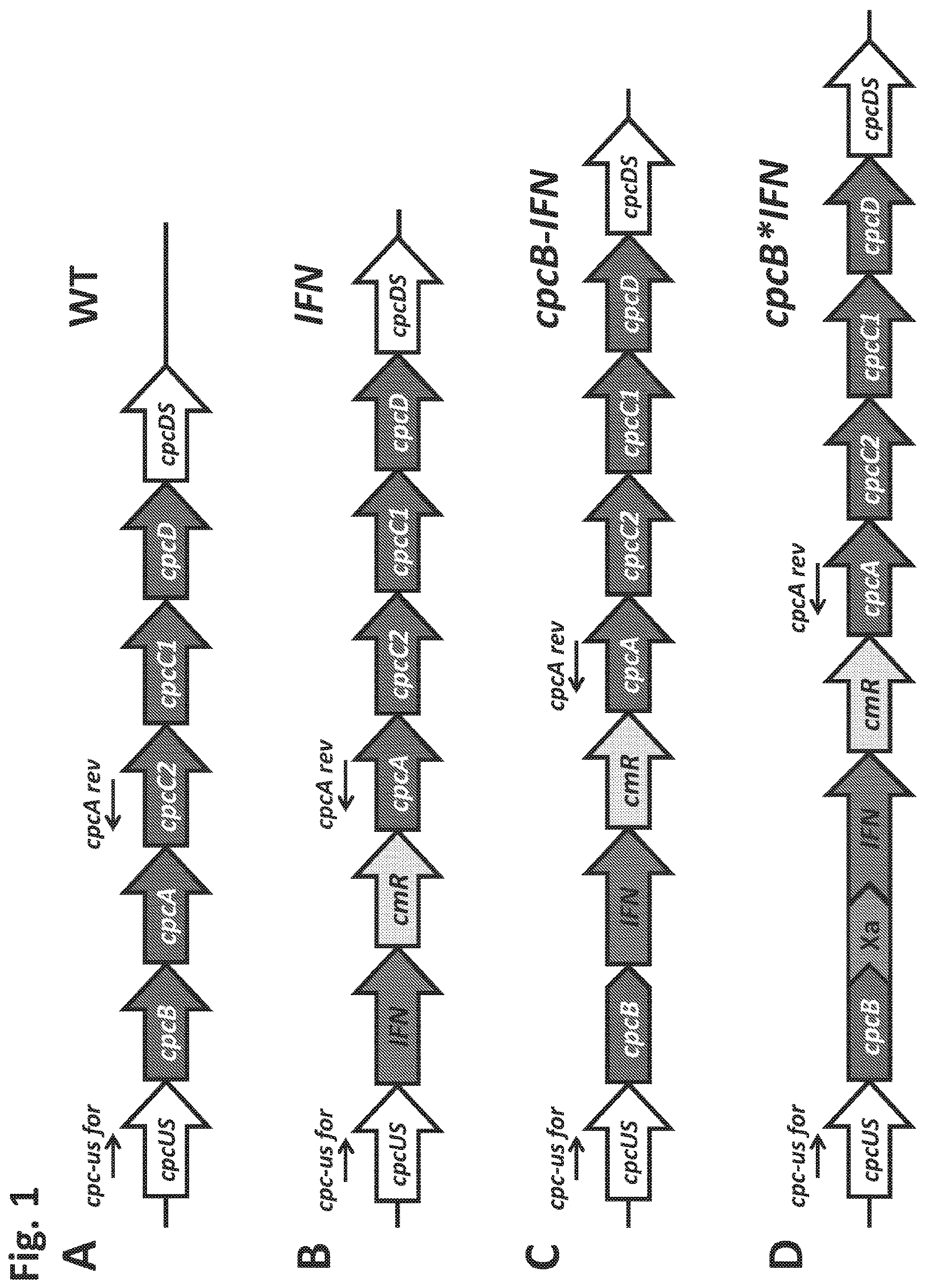
. Schematic overview of DNA constructs designed for the transformation of the Synechocystis PCC 6803 ( Synechocystis ) genome. (A) The native cpc operon, as it occurs in wild type Synechocystis . This DNA sequence and associated strain are referred to as the wild type (WT). (B) Insertion in the cpc operon of the codon-optimized human interferon (IFN) gene followed by the chloramphenicol (cmR) resistance cassette in an operon configuration, replacing the phycocyanin-encoding β-subunit cpcB gene of Synechocystis . This DNA construct is referred to as IFN; (C) Insertion in the cpc operon of the codon-optimized IFN gene immediately downstream of the phycocyanin-encoding β-subunit cpcB gene of Synechocystis , followed by the cmR resistance cassette, in an operon configuration. This DNA construct is referred to as cpcB-IFN; (D) Insertion in the cpc operon of the codon-optimized IFN gene as a fusion construct with the phycocyanin-encoding β-subunit cpcB gene, with the latter in the leader sequence position. The fusion construct cpcB*IFN was followed by the cmR resistance cassette in an operon configuration, cpcB and IFN genes were linked by the DNA sequence encoding the Factor Xa cleavage site. The latter comprises the Ile-Glu/Asp-Gly-Arg amino acid sequence. This DNA construct is referred to as the cpcB*IFN.
. Genomic DNA PCR analysis testing for transgenic DNA copy homoplasmy in Synechocystis transformants. Wild type and transformant strains were probed in genomic DNA PCR reactions for product generation and transgenic DNA segregation. Primers <cpc-us for> and <cpcA rev> showed substantially different and unique products in the wild type and the different transformants comprising the constructs of . Wild type PCR products had a 1,289 bp size, whereas the IFN, cpcB-IFN, and the cpcB*IFN transformants generated 2,094, 2,723, and 2.619 bp size products, respectively. Absence of wild type products from the latter was evidence of DNA copy homoplasmy for the transformants. (The cpcB-IFN construct generated a product size slightly larger than that of the cpcB*IFN because it contained the Synechocystis native cpcB-cpcA intergenic DNA sequence. Please see gene nucleotide sequences in the Supplementary Materials.)
. Coloration of cells from photoautotrophically-grown liquid cultures showing a blue-green wild type (WT) phenotype, and greenish phenotype for the IFN, CpcB-IFN, and CpcB*IFN-containing transformants. The latter did not assemble phycocyanin rods, hence the absence of the distinct blue cyanobacterial coloration from the cells.
. Protein expression analysis of Synechocystis wild type and transformants. (A) Total cellular protein extracts were resolved by SDS-PAGE and visualized by Coomassie-stain. Two independent replicates of total protein extracts from wild type (WT), and IFN, CpcB-IFN and CpcB*IFN transformant cells were loaded onto the SDS-PAGE. Individual native and heterologous proteins of interest are indicated to the right of the gel. Sample loading corresponds to 0.25 μg of chlorophyll. Note the clear presence of a heterologous protein migrating to ˜36 kD in the CpcB*IFN fusion extracts. (B) Total protein extracts of (A) were subjected to Western-blot analysis with loading of the lanes as per A . Specific polyclonal antibodies against the human IFN protein were used to probe target proteins. Sample loading corresponds to 0.25 μg of chlorophyll. Note the specific antibody cross-reaction with proteins migrating to ˜36 and ˜108 kD in the cpcB*IFN fusion and the absence of a cross reaction with any protein from the IFN and cpcB-IFN transformant cells. The latter do not seem to make/accumulate IFN.
. Protein expression analysis of Synechocystis wild type (WT) and transformants harboring the cpcB*IFN fusion construct. Total cellular protein extracts were resolved by SDS-PAGE and visualized by Coomassie-stain. Two different versions of the IFN gene were used: the human native IFN′ and the Synechocystis codon-optimized IFN gene. Note the presence of heterologous proteins migrating to ˜36 kD (CpcB*IFN) and ˜23 kD (CmR) in the transformants but not in the wild type. Also note the presence of the ˜19 kD CpcB β-subunit and the ˜17 kD CpcA α-subunit of phycocyanin in the wild type but not in the transformants. Sample loading corresponds to 0.5 μg of chlorophyll. Quantification of the CpcB*IFN protein accumulation relative to that of the Rubisco large subunit (RbcL) is given in the results of Table 1.
. Protein expression analysis of Synechocystis wild type (WT) and transformants harboring the cpcB*IFN fusion construct. Total cellular protein extracts were resolved by SDS-PAGE and visualized by Coomassie-stain. Two different versions of the fusion construct were used comprising the CpcB*IFN fusion and the more extensive cpcB*His*Xa*IFN fusion configuration, followed by the cmR resistance cassette. Equivalent amount of the CpcB*IFN and the CpcB*His*Xa*IFN fusion proteins were expressed in Synechocystis . Individual native and heterologous proteins of interest are indicated to the right of the gel. Sample loading corresponds to 0.25 μg of chlorophyll.
. Batch-scale purification of the recombinant CpcB*His*Xa*IFN protein through cobalt affinity chromatography. Protein purification was conducted employing a small amount of resin as solid phase. The latter was mixed and incubated with the cell extracts. The resin was pelleted and washed repeatedly with buffers containing imidazole at different concentrations.
Lane 1 shows the cell extracts (upper panel) and the resin pellet (lower panel) of the wild type, CpcB*IFN, and CpcB*His*Xa*IFN fusion construct cells prior to incubation with the resin. Note the natural pink coloration of the latter.
Lane 2 shows the cell extracts (upper panel) and the resin pellet (lower panel) of the wild type, CpcB*IFN, and CpcB*His*Xa*IFN fusion construct cells following a 5-min incubation with the resin in the presence of 10 mM imidazole. Note the blue coloration of the resin and the green coloration of the supernatant.
Lanes 3-5 show the remaining cell extracts (upper panel) and the resin pellet (lower panel) of the wild type, CpcB*IFN, and CpcB*His*Xa*IFN fusion construct cells following a consecutive wash of the resin three times with a buffer containing 10 mM of imidazole. Note the resulting clear supernatant and the pink coloration of the resin after the third wash (lane 5) for the wild type and CpcB*IFN, suggesting absence of His-tagged proteins. Also note the blue coloration of the resin in the CpcB*His*Xa*IFN sample, which was retained in this pellet (lanes 3-5) in spite of the repeated wash, suggesting the presence of resin-bound blue-colored His-tagged proteins.
Lanes 6-8 show the subsequent extracts (upper panel) and the resin pellet (lower panel) of the wild type, CpcB*IFN, and CpcB*His*Xa*IFN fusion construct cells following a wash three times with a buffer containing 250 mM of imidazole, designed to dissociate His-tagged proteins from the resin. Note the bluish supernatant in lanes 6 and 7 and the corresponding loss of the blue color from the resin pellet, suggesting the specific removal of His-tagged proteins from the resin.
. Coomassie-stained SDS-PAGE gel analysis of fractions eluted with different imidazole concentrations. Fractions were obtained upon affinity chromatography purification as shown in . Samples were loaded on a per volume basis. Note the ˜108, ˜38, and ˜17 kD proteins eluted from the CpcB*His*Xa*IFN extract (marked with arrows).
. Absorbance spectra of purified Synechocystis complexes. (A) Absorbance spectra of eluent E1 fractions from wild type, CpcB*IFN, and CpcB*His*Xa*IFN samples, as shown in . (B) Absorbance spectra of cellular protein extracts from wild type, Δcpc deletion mutant (Kirst et al., 2014) and CpcB*His*Xa*IFN transformant cells.
. Column-based purification of the CpcB*His*Xa*IFN fusion protein through cobalt affinity chromatography.
Lane 1, upper panel, shows the CpcB*His*Xa*IFN cell extracts in the presence of 5 mM imidazole prior to resin application. Lane 1, lower panel, shows the SDS-PAGE protein profile of these extracts, indicating presence of all Synechocystis proteins.
Lane 2, upper panel, shows the CpcB*His*Xa*IFN cell extracts after incubation with the resin but prior to washing with additional imidazole applications. Lane 2, lower panel, shows the SDS-PAGE protein profile of these extracts, obtained upon a prior removal of the resin from the mix, indicating presence of all Synechocystis proteins.
Lanes 3-6, upper panel, show the CpcB*His*Xa*IFN cell extracts that passed through the resin upon four consecutive washes with 5 mM imidazole and, lower panel, the SDS-PAGE protein profile of these extracts, showing a steep depletion (from lane 3 to lane 6) of total protein.
Lanes 7-9, upper panel, show the further removal of resin-bound proteins from the CpcB*His*Xa*IFN cell extracts that eluted upon three consecutive washes with 250 mM imidazole and, lower panel, the SDS-PAGE protein profile of these extracts, showing substantial enrichment in mainly four proteins with apparent molecular weights of 108, 36, 27, and 17 kD. The majority of these proteins were eluted with the first application of the 250 mM imidazole solution.
. (A) SDS-PAGE and Coomassie-staining analysis of Synechocystis wild type, CpcB*IFN, and CpcB*His*Xa*IFN total cell extract, and of proteins eluted from the resin column upon application of 250 mM imidazole. (B) Western blot analysis with specific IFN polyclonal antibodies of the proteins resolved in (A). Note the heterologous ˜36 kD CpcB*His*Xa*IFN and the ˜108 kD putative CpcB*His*Xa*IFN trimer (marked by arrowheads).
. (A) SDS-PAGE and Coomassie-stain analysis of Synechocystis wild type, CpcB*His*Xa*IFN, and resin-eluted proteins. (B) SDS-PAGE and Zinc-stain analysis of Synechocystis wild type, CpcB*His*Xa*IFN, and resin-eluted proteins. Zn-staining is designed to highlight the presence of bilin tetrapyrrole pigments. Individual native and heterologous proteins of interest are indicated to the right of the gels.
. (A) Map of the nptI*IFN fusion construct in the cpc operon locus. Note the presence of the His-tag and the Xa protease cleavage site in-between the two genes in the fusion. (B) SDS-PAGE and Coomassie staining of the protein extracts from wild type (WT), the cpcB*His*Xa*IFN, and two independent lines of the nptI*His*Xa*IFN transformants. (C) Western blot analysis of a duplicate gel as the one shown in (B). Specific anti-IFN polyclonal antibodies were used in this analysis. Note the specific antibody cross reactions with protein bands migrating to ˜36 kD (CpcB*His*Xa*IFN) and ˜46 kD (NptI*His*Xa*IFN). Also note the antibody cross reactions with protein bands of higher molecular mass.
. Efficacy of interferon in preventing encephalomyocarditis virus (EMC) infection of human lung cells (A549), as performed by a PBL Assay Science, Piscataway, N.J. USA test. (Diamonds) IFN titration curve using a standard recombinant interferon. (Squares) IFN titration curve using the cyanobacterial CpcB*His*Xa*IFN fusion interferon. The analysis showed that 0.002 ng/mL of a standard recombinant interferon was needed to cause 50% inhibition in EMC infection, whereas 0.0875 ng/mL of cyanobacterial CpcB*His*Xa*IFN fusion interferon was required to cause 50% inhibition in EMC infection.
. (A) Map of the cpcB*His*Xa*K2S fusion construct in the cpc operon locus. Note the presence of the His-tag and the Xa protease cleavage site in-between the two genes in the fusion. (B) SDS-PAGE and Coomassie stain of the protein extracts from wild type (WT), and three independent lines of the cpcB*His*Xa*K2S transformant. (C) Western blot analysis of a duplicate gel as the one shown in (B). tissue-Plasminogen Activase recognizing polyclonal antibodies were used in this assay. Note the specific antibody cross reactions with protein bands migrating to ˜58.9 kD protein band in the K2S transformants.
. (A) Map of the cpcB*INS fusion construct in the cpc operon locus. (B) SDS-PAGE and Coomassie stain of the protein extracts from wild type (WT), a CpcB*INS (insulin) containing transformant and, for comparison purposes, a CpcB*PHLS (β-phellandrene synthase) transformant. Note the 19 kD β-subunit and 17 kD α-subunit of phycocyanin in the wild type, the ˜27 kD CpcB*INS (insulin) in the cpcB*INS transformant, and the ˜84 kD CpcB*PHLS protein in the cpcB*PHLS transformant.
. (A) Map of the cpcB*L7*His*TEV*TTFC fusion construct in the cpc operon locus, including a linker of seven aminoacids (L7) and a His×6-tag (His). (B, left panel) SDS-PAGE and Coomassie stain analysis of the protein extracts from wild type (WT), the LTV recipient strain, and three Synechocystis transformant lines of the cpcB*L7*His*TEV*TTFC (Tetanus Toxin Fragment C). Note the presence of the 19 kD CpcB β-subunit and 17 kD CpcA α-subunit of phycocyanin in the wild type only, the ˜72 kD cpcB*L7*His*TEV*TTFC protein (denoted as cpcB*TTFC) in the TTFC transformants, and the ˜55 kD RBCL (large subunit of Rubisco) protein in all strains. Hashtag (#) denotes the electrophoretic mobility position of the cpcB*L7*TEV*ISPS fusion protein from the respective isoprene synthase (ISPS)-containing strain that was used as the recipient strain of the cpcB*L7*His*7EV*TTFC construct. Densitometric analysis of the SDS-PAGE Coommassie stain showed that the cpcB*L7*His*TEV*TTFC fusion protein accounted for about 28% of the total cell protein. (B, right panel) Western blot analysis of the protein profile shown in B (left panel), probed with specific polyclonal antibodies against the TTFC polypeptide. Note the antibody cross reaction with the 72 kD CpcB*L7*His*TEV*TTFC fusion protein, the ˜290 kD putative trimeric [CpcB*L7*His*TEV*TTFC]×3 undissolved fusion protein complex, plus some lower molecular size putative proteolysis fragments.
. (A) Map of the cpcB*L7*His*TEV*RBD fusion construct in the cpc operon locus, including a linker of seven amino acids (L7), a His×6-tag (His) and the TEV cleavage site (TEV), followed by the Receptor Binding Domain (RBD) of the S1 protein from the SARS-CoV-2. (B, left panel) SDS-PAGE and Coomassie stain of the protein extracts from wild type (WT), the LTV recipient strain, and a Synechocystis transformant line harboring the cpcB*L7*His*TEV*RBD fusion protein (RBD). The arrow points to the electrophoretic mobility of the 45 kD RBD fusion protein. (B, center panel). Western blot analysis of the protein profile shown in B (left panel), probed with specific polyclonal antibodies against the leader CpcB protein in the fusion construct. Note the antibody cross reaction with the 45 kD cpcB*L7*His*TEV*RBD fusion protein. (B, right panel) SDS-PAGE and Zinc-stain analysis of Synechocystis expressing the LTV and RBD fusion construct phenotypes. Zn-staining is designed to highlight the presence of bilin tetrapyrrole pigments. Note the Zn-staining of a band at 45 kD in the RBD expressing transformant, and the staining of a band migrating to ˜85 kD in the LTV (cpcB*L7*REV*ISPS) transformant.
. Panels A-D provide schematics of illustrative expression constructs.
DETAILED DESCRIPTION OF THE INVENTION
The term “naturally-occurring” or “native” as used herein as applied to a nucleic acid, a protein, a cell, or an organism, refers to a nucleic acid, protein, cell, or organism that is found in nature. For example, a polypeptide or polynucleotide sequence that is present in an organism that can be isolated from a source in nature and which has not been intentionally modified by a human in the laboratory is naturally occurring.
The term “heterologous nucleic acid,” as used herein, refers to a nucleic acid wherein at least one of the following is true: (a) the nucleic acid is foreign (“exogenous”) to (i.e., not naturally found in) a given host microorganism or host cell; (b) the nucleic acid comprises a nucleotide sequence that is naturally found in (e.g., is “endogenous to”) a given host microorganism or host cell (e.g., the nucleic acid comprises a nucleotide sequence endogenous to the host microorganism or host cell. In some embodiments, a “heterologous” nucleic acid may comprise a nucleotide sequence that differs in sequence from the endogenous nucleotide sequence but encodes the same protein (having the same amino acid sequence) as found endogenously; or two or more nucleotide sequences that are not found in the same relationship to each other in nature, e.g., the nucleic acid is recombinant. An example of a heterologous nucleic acid is a nucleotide sequence encoding a fusion protein comprising two proteins that are not joined to one another in nature.
The term “recombinant” polynucleotide or nucleic acid refers to one that is not naturally occurring, e.g., is made by the artificial combination of two otherwise separated segments of sequence through human intervention. This artificial combination is often accomplished by either chemical synthesis means, or by the artificial manipulation of isolated segments of nucleic acids, e.g., by genetic engineering techniques. A “recombinant” protein is encoded by a recombinant polynucleotide. In the context of a genetically modified host cell, a “recombinant” host cell refers to both the original cell and its progeny.
As used herein, the term “genetically modified” refers to any change in the endogenous genome of a cyanobacteria cell compared to a wild-type cell. Thus, changes that are introduced through recombinant DNA technology and/or classical mutagenesis techniques are both encompassed by this term. The changes may involve protein coding sequences or non-protein coding sequences such as regulatory sequences as promoters or enhancers.
An “expression construct” or “expression cassette” as used herein refers to a recombinant nucleic acid construct, which, when introduced into a cyanobacterial host cell in accordance with the present invention, results in increased expression of a fusion protein encoded by the nucleic acid construct. The expression construct may comprise a promoter sequence operably linked to a nucleic acid sequence encoding the fusion protein or the expression cassette may comprise the nucleic acid sequence encoding the fusion protein where the construct is configured to be inserted into a location in a cyanobacterial genome such that a promoter endogenous to the cyanobacterial host cell is employed to drive expression of the fusion protein. An “expression unit” as used herein refers to a minimal region of a polynucleotide that is expressed that provided for high level protein expression, which comprises the polynucleotide that encodes the fusion protein, as well as other genes, e.g., cpcA and cpc operon genes encoding cpc linker polypeptides CpcC2, CpcC1, and CpcD. In some embodiments, the expression unit additionally include a gene encoding an antibiotic resistance polypeptide, such as a chloramphenicol resistance gene or streptomycin resistance gene. The expression unit may also comprise additional sequences, such as nucleic acid sequences encoding a protease cleavage sites, a linker polypeptide, or a polypeptide tagging sequence, such as a His tag.
By “construct” is meant a recombinant nucleic acid, generally recombinant DNA, which has been generated for the purpose of the expression of a specific nucleotide sequence(s), or is to be used in the construction of other recombinant nucleotide sequences.
As used herein, the term “exogenous protein” refers to a protein that is not normally or naturally found in and/or produced by a given cyanobacterium, organism, or cell in nature. As used herein, the term “endogenous protein” refers to a protein that is normally found in and/or produced by a given cyanobacterium, organism, or cell in nature.
An “endogenous” protein or “endogenous” nucleic acid is also referred to as a “native” protein or nucleic acid that is found in a cell or organism in nature.
The terms “nucleic acid” and “polynucleotide” are used synonymously and refer to a single or double-stranded polymer of deoxyribonucleotide or ribonucleotide bases read from the 5′ to the 3′ end. A nucleic acid of the present invention will generally contain phosphodiester bonds, although in some cases, nucleic acid analogs may be used that may have alternate backbones, comprising, e.g., phosphoramidate, phosphorothioate, phosphorodithioate, or O-methylphophoroamidite linkages (see Eckstein, Oligonucleotides and Analogues: A Practical Approach, Oxford University Press); and peptide nucleic acid backbones and linkages. Other analog nucleic acids include those with positive backbones; non-ionic backbones, and non-ribose backbones. Thus, nucleic acids or polynucleotides may also include modified nucleotides, that permit correct read through by a polymerase. “Polynucleotide sequence” or “nucleic acid sequence” may include both the sense and antisense strands of a nucleic acid as either individual single strands or in a duplex. As will be appreciated by those in the art, the depiction of a single strand also defines the sequence of the complementary strand; thus the sequences described herein also provide the complement of the sequence. Unless otherwise indicated, a particular nucleic acid sequence also implicitly encompasses conservatively modified variants thereof (e.g., degenerate codon substitutions) and complementary sequences, as well as the sequence explicitly indicated. The nucleic acid may be DNA, both genomic and cDNA, RNA or a hybrid, where the nucleic acid may contain combinations of deoxyribo- and ribo-nucleotides, and combinations of bases, including uracil, adenine, thymine, cytosine, guanine, inosine, xanthine hypoxanthine, isocytosine, isoguanine, etc.
The term “promoter” or “regulatory element” refers to a region or sequence determinants located upstream or downstream from the start of transcription that are involved in recognition and binding of RNA polymerase and other proteins to initiate transcription. A “cyanobacteria promoter” is a promoter capable of initiating transcription in cyanobacteria cells. Such promoters need not be of cyanobacterial origin, for example, promoters derived from other bacteria or plant viruses, can be used in the present invention.
A polynucleotide sequence is “heterologous to” a second polynucleotide sequence if it originates from a foreign species, or, if from the same species, is modified by human action from its original form. For example, a promoter operably linked to a heterologous coding sequence refers to a coding sequence from a species different from that from which the promoter was derived, or, if from the same species, a coding sequence which is different from any naturally occurring allelic variants.
Two nucleic acid sequences or polypeptides are said to be “identical” if the sequence of nucleotides or amino acid residues, respectively, in the two sequences is the same when aligned for maximum correspondence as described below. The term “complementary to” is used herein to mean that the sequence is complementary to all or a portion of a reference polynucleotide sequence.
Optimal alignment of sequences for comparison may be conducted by the local homology algorithm of Smith and Waterman Add. APL. Math. 2:482 (1981), by the homology alignment algorithm of Needle man and Wunsch. J. Mol. Biol. 48:443 (1970), by the search for similarity method of Pearson and Lipman Proc. Natl. Acad. Sci. (U.S.A.) 85: 2444 (1988), by computerized implementations of these algorithms (GAP, BESTFIT, BLAST, FASTA, and TFASTA in the Wisconsin Genetics Software Package, Genetics Computer Group (GCG), 575 Science Dr., Madison, Wis.), or by inspection.
“Percentage of sequence identity” is determined by comparing two optimally aligned sequences over a comparison window, wherein the portion of the polynucleotide sequence in the comparison window may comprise additions or deletions (i.e., gaps) as compared to the reference sequence (which does not comprise additions or deletions) for optimal alignment of the two sequences. The percentage is calculated by determining the number of positions at which the identical nucleic acid base or amino acid residue occurs in both sequences to yield the number of matched positions, dividing the number of matched positions by the total number of positions in the window of comparison and multiplying the result by 100 to yield the percentage of sequence identity.
The term “substantial identity” in the context of polynucleotide or polypeptide sequences means that a polynucleotide or polypeptide comprises a sequence that has at least 50% sequence identity to a reference nucleic acid or polypeptide sequence. Alternatively, percent identity can be any integer from 40% to 100%. Exemplary embodiments include at least: 50%, 55%, 60%, 65%, 70%, 75%, 80%, 85%, 90%, 95%, or 99% compared to a reference sequence using the programs described herein; preferably BLAST using standard parameters, as described below.
Another indication that nucleotide sequences are substantially identical is if two molecules hybridize to each other, or a third nucleic acid, under stringent conditions. Stringent conditions are sequence dependent and will be different in different circumstances. Generally, stringent conditions are selected to be about 5° C. lower than the thermal melting point (Tm) for the specific sequence at a defined ionic strength and pH. The Tm is the temperature (under defined ionic strength and pH) at which 50% of the target sequence hybridizes to a perfectly matched probe. Typically, stringent conditions will be those in which the salt concentration is about 0.02 molar at pH 7 and the temperature is at least about 60° C.
The term “isolated”, when applied to a nucleic acid or protein, denotes that the nucleic acid or protein is essentially free of other cellular components with which it is associated in the natural state. It is preferably in a homogeneous state and may be in either a dry or aqueous solution. Purity and homogeneity are typically determined using analytical chemistry techniques such as polyacrylamide gel electrophoresis or high-performance liquid chromatography. A protein which is the predominant species present in a preparation is substantially purified. In particular, an isolated gene is separated from open reading frames which flank the gene and encode a protein other than the gene of interest.
The term “reactor” as used herein refers to the vessel in which cyanobacteria are grown.
Introduction
The present invention is based, in part, on the discovery of fusion protein constructs that can be used in cyanobacteria as transgenic protein over-expression vectors to provide high levels of transgenic animal proteins, e.g., interferons, insulin, or tPA polypeptides. Expression of transgenes in cyanobacteria using such vectors results in high levels of accumulation of a protein encoded by the transgene.
A fusion protein of the present invention comprises a protein that is to be expressed in cyanobacteria, typically a non-native protein that is not expressed in cyanobacteria, e.g., a plant protein fused to a protein that is expressed at high levels in cyanobacteria. In the context of the present invention, a protein that is “expressed at high levels in cyanobacteria” refers to a protein that accumulates to at least 1%. Such proteins, when fused at the N-terminus of a protein of interest to be expressed in cyanobacteria, are also referred to herein as “leader proteins”, “leader peptides”, or “leader sequences”. A nucleic acid encoding a leader protein is typically referred to herein as a “leader polynucleotide” or “leader nucleic acid sequence” or “leader nucleotide sequence”.
In some embodiments, a protein that is expressed at high levels is a naturally occurring protein that is expressed at high levels in wild-type cyanobacteria, and is used as endogenous “leader polypeptide sequence” in the cyanobacterial strain of origin. Such proteins include, e.g., a phycocyanin β-subunit (cpcB), a phycocyanin α-subunit (cpcA), a phycoerythrin α-subunit (cpeA), a phycoerythrin β-subunit (cpeB), an allophycocyanin α-subunit (apcA), an allophycocyanin β-subunit (apcB), a large subunit of Rubisco (rbcL), a small subunit of Rubisco (rbcS), a photosystem II reaction center protein, a photosystem 1 reaction center protein, or a rpl or rps cyanobacterial ribosomal RNA protein. In some embodiments, a protein that is expressed at high levels is a naturally occurring protein that is expressed at high levels in wild-type cyanobacteria, and it is used as heterologous leader sequence in a different cyanobacterial strain.
In some embodiments, a protein that is expressed at high levels is an exogenous protein that the cyanobacteria have been genetically modified to express at high levels. For example, proteins that provide for antibiotic resistance that are expressed to high levels in cyanobacteria, e.g., a bacterial kanamycin resistance protein, NPT, or a bacterial chloramphenicol resistance protein, CmR, may be used as a leader sequence.
The invention additionally provides nucleic acids encoding a fusion protein as described herein, as well as expression constructs comprising the nucleic acids and host cells that have been genetically modified to express such fusion proteins. In further aspects, the invention provides methods of modifying a cyanobacterial cell to overexpress a protein of interest using an expression construct of the invention and methods of producing the protein of interests and products generated by the proteins using such genetically modified cyanobacterial cells.
The invention employs various routine recombinant nucleic acid techniques. Generally, the nomenclature and the laboratory procedures in recombinant DNA technology described below are those commonly employed in the art. Many manuals that provide direction for performing recombinant DNA manipulations are available, e.g., Sambrook, Molecular Cloning, A Laboratory Manual (4th Ed, 2012); and Current Protocols in Molecular Biology (Ausubel et al., eds., 1994-2015).
Proteins Expressed at High Levels in Cyanobacteria
In the present invention, nucleic acid constructs are created in which a polynucleotide sequence encoding a protein of interest is fused to the C-terminal end of a polynucleotide that encodes a leader protein, i.e., a protein that is expressed at high levels in cyanobacteria as described herein. The protein of interest is then also expressed at high levels in conjunction with the leader sequence. In the context of the invention, a protein that is “expressed at high levels” in cyanobacteria refers to a protein that is at least 1%, typically at least 2%, at least 3%, at least 4%, at least 5%, or at least 10%, or greater, of the total protein expressed in the cyanobacteria. Expression levels in cyanobacteria may be evaluated in cells that are logarithmically growing, but may be alternatively determined in cells in a stationary phase of growth. The level of protein expression can be assessed using various techniques. In the present invention, high level expression is typically determined using SDS PAGE analysis. Following electrophoresis, the gel is stained and the level of proteins assessed by scanning the gel and quantifying the amount of protein using an image analyzer.
In some embodiments, a leader sequence in accordance with the invention encodes a naturally occurring cyanobacteria protein that is expressed at high levels in native cyanobacteria. Thus, in some embodiments, the protein is endogenous to cyanobacteria. Examples of such proteins include cpcB, cpcA, cpeA, cpeB, apcA, apcB, rbcL, rbcS, psbA, rpl, or rps. In some embodiments, the leader sequence encodes less than the full-length of the protein, but typically comprises a region that encodes at least 25%, typically at least 50%, or at least 75%, or at least 90%, or at least 95%, or greater, of the length of the protein. As appreciated by one of skill in the art, use of an endogenous cyanobacterial polynucleotide sequence for constructing an expression construct in accordance with the invention provides a sequence that need not be codon-optimized, as the sequence is already expressed at high levels in cyanobacteria. Examples of cyanobacterial polynucleotides that encode cpcB, cpcA, cpeA, cpeB, apcA, apcB, rbcL, rbcS, psbA, rpl, or rps are available at the website www.genome.microbedb.jp/cyanobase under accession numbers, as follows:
•
• cpcA: Synechocystis sp. PCC6803 sll1578, Anabaena sp. PCC7120 arl0529, Thermosynechococcus elongatus BP-1 tlr1958 , Synechococcus elongatus PCC6301 syc0495_c, syc0500_c • cpcB: Synechocystis sp. PCC6803 sll1577, Anabaena sp. PCC7120 arl0528, Thermosynechococcus elongatus BP-1 tlr1957 , Synechococcus elongatus PCC6301 syc0496_c, syc0501_c • cpeA: Prochlorococcus marinus SS120 Pro0337 , Synechococcus sp. WH8102 SYNW2009, SYNW2016 • cpeB: Prochlorococcus marinus SS120 Pro0338 , Synechococcus sp. WH8102 SYNW2008, SYNW2017 • apcA: Synechocystis sp. PCC 6803, slr2067: Anabaena sp. PCC 7120, all0450, alr0021 ; Synechococcus elongatus PCC 6301, syc1186_d • apcB: Synechocystis sp. PCC 6803, slr1986, Anabaena sp. PCC 7120, alr0022 , Synechococcus elongatus PCC 6301, syc1187_d • rbcL RubisCO large subunit: Synechocystis sp. PCC 6803 slr0009 • rbcS RubisCO small subunit: Synechocystis sp. PCC 6803 slr0012 • rpl: 50S ribosomal protein of Synechocystis , e.g. sll1803; sll1810; ssr1398 and • rps: 30S ribosomal protein of Synechocystis , e.g. sll1804; slr1984.
The polynucleotide sequence that encodes the leader protein need not be 100% identical to a native cyanobacteria polynucleotide sequence. A polynucleotide variant having at least 50% identity or at least 60% identity, or greater, to a native cyanobacterial polynucleotide sequence, e.g., a native cpcB, cpcA, cpeA, cpeB, rbcL, rbcS, psbA, rpl, or rps cyanobacteria polynucleotide sequence, may also be used, so long as the codons that vary relative to the native cyanobacterial polynucleotide are codon optimized for expression in cyanobacteria and the codons that vary relative to the wild type sequence do not substantially disrupt the structure of the protein. In some embodiments, a polynucleotide variant that has at least 70% identity, at least 75% identity, at least 80% identity, or at least 85% identity, or greater to a native cyanobacterial polynucleotide sequence, e.g., a native cpcB, cpcA, cpeA, cpeB, rbcL, rbcS, psbA, rpl, or rps cyanobacteria polynucleotide sequence, is used, again maintaining codon optimization for cyanobacteria. In some embodiments, a polynucleotide variant that has least 90% identity, or at least 95% identity, or greater, to a native cyanobacterial polynucleotide sequence, e.g., a native cpcB, cpcA, cpeA, cpeB, rbcL, rbcS, psbA, rpl, or rps cyanobacteria polynucleotide sequence, is used. The percent identity is typically determined with reference the length of the polynucleotide that is employed in the construct, i.e., the percent identity may be over the full length of a polynucleotide that encodes the leader polypeptide sequence, or may be over a smaller length, e.g., in embodiments where the polynucleotide encodes at least 25%, typically at least 50%, or at least 75%, or at least 90%, or at least 95%, or greater, of the length of the protein. The protein encoded by a variant polynucleotide sequence as described need not retain a biological function, however, a codon that varies from the wild-type polynucleotide is typically selected such that the protein structure of the native cyanobacterial sequence is not substantially altered by the changed codon, e.g., a codon that encodes an amino acid that has the same charge, polarity, and/or is similar in size to the native amino acid is selected.
In some embodiments, a polynucleotide variant of a naturally over-expressed (more than 1% of the total cellular protein) cyanobacterial gene is employed, that encodes for a polypeptide sequence that has at least 70%, or 80%, or at least 85% or greater identity to the protein encoded by the wild-type gene. In some embodiments, the polynucleotide encodes a protein that has 90% identity, or at least 95% identity, or greater, to the protein encoded by the wild-type gene. Variant polynucleotides may also be codon optimized for expression in cyanobacteria.
In some embodiments, a protein that is expressed at high levels in cyanobacteria is not native to cyanobacteria in which a fusion construct in accordance with the invention is expressed. For example, polynucleotides from bacteria or other organisms that are expressed at high levels in cyanobacteria may be used as leader sequences. In some embodiments, the polynucleotides from other organisms may be codon-optimized for expression in cyanobacteria. In some embodiments, codon optimization is performed such that codons used with an average frequency of less than 12% by Synechocystis are replaced by more frequently used codons. Rare codons can be defined, e.g., by using a codon usage table derived from the sequenced genome of the host cyanobacterial cell. See, e.g., the codon usage table obtained from Kazusa DNA Research Institute, Japan (website wwtw.kazusa.or.jp/codon/) used in conjunction with software, e.g., “Gene Designer 2.0” software, from DNA 2.0 (website www.dna20.com/) at a cut-off thread of 15%.
In some embodiments, a leader sequence in accordance with the present invention encodes a protein that confers antibiotic resistance. For example, in some embodiments, the leader sequence encodes neomycin phosphotransferase e.g., NPT1, which confers neomycin and kanamycin resistance. Other polynucleotides that may be employed include a chloramphenicol acetyltransferase polynucleotide, which confers chloramphenicol resistance; or a polynucleotide encoding a protein that confers streptomycin, ampicillin, erythromycin, zeocin, or tetracycline resistance, or resistance to another antibiotic. In some embodiments, the leader sequence encodes less than the full-length of the protein, but typically comprises a region that encodes at least 25%, typically at least 50%, or at least 75%, or at least 90%, or at least 95%, or greater, of the length of the protein. In some embodiments, a polynucleotide variant of a naturally occurring antibiotic resistance gene is employed. As noted above, a variant polynucleotide need not encode a protein that retains the native biological function. A variant polynucleotide typically encodes a protein that has at least 80% identity, or at least 85% or greater, identity to the protein encoded by the wild-type antibiotic resistance gene. In some embodiments, the polynucleotide encodes a protein that has 90% identity, or at least 95% identity, or greater, to the wild-type antibiotic resistance protein. Such variant polynucleotides employed as leader sequence may also be codon-optimized for expression in cyanobacteria. The percent identity is typically determined with reference to the length of the polynucleotide that is employed in the construct, i.e., the percent identity may be over the full length of a polynucleotide that encodes the leader polypeptide sequence, or may be over a smaller length, e.g., in embodiments where the polynucleotide encodes at least 25%, typically at least 50%, or at least 75%, or at least 90%, or at least 95%, or greater, of the length of the protein. A protein encoded by a variant polynucleotide sequence need not retain a biological function, however, codons that are present in a variant polynucleotide are typically selected such that the protein structure relative to the wild-type protein structure is not substantially altered by the changed codon, e.g., a codon that encodes an amino acid that has the same charge, polarity, and/or is similar in size to the native amino acid is selected.
Other leader proteins can be identified by evaluating the level of expression of a candidate leader protein in cyanobacteria. For example, a leader polypeptide that does not occur in wild type cyanobacteria may be identified by measuring the level of protein expressed from a polynucleotide codon optimized for expression in cyanobacteria that encodes the candidate leader polypeptide. A protein may be selected for use as a leader polypeptide if the protein accumulates to a level of at least 1%, typically at least 2%, at least 3%, at least 4%, at least 5%, or at least 10%, or greater, of the total protein expressed in the cyanobacteria when the polynucleotide encoding the leader polypeptide is introduced into cyanobacteria and the cyanobacteria cultured under conditions in which the transgene is expressed. The level of protein expression is typically determined using SDS PAGE analysis. Following electrophoresis, the gel is scanned and the amount of protein determined by image analysis.
Transgenes
A fusion construct of the invention may be employed to provide high level expression in cyanobacteria for any desired biopharmaceutical protein. Thus, for example, cyanobacteria can be engineered to express an animal biopharmaceutical polypeptide such as an antibody, hormone, cytokine, therapeutic enzyme and the like, as a fusion polypeptide with a protein expressed at a high level in cyanobacteria, e.g. a cpcB or other protein encoded by the Cpc operon. In some embodiments the biopharmaceutical polypeptide is expressed at a level of at least 1%, or at least 5%, or at least 10%, or at least 15%, or at least 20%, of total cellular protein as described herein.
In some embodiments, the nucleic acid sequence encoding the animal, e.g., mammalian, biopharmaceutical polypeptide is codon-optimized for expression in cyanobacteria. Alternatively, the nucleic acid sequence need not be codon-optimized, as high-level expression of the fusion polypeptide does not require codon optimization.
In some embodiments, the mature form of the biopharmaceutical polypeptide lacking the native signal sequence is expressed.
In some embodiments, the transgene that is expressed encodes an interferon, e.g., an interferon alpha, such as IFNA2. In some embodiments, the interferon is interferon-alpha, such as human interferon α-2. An illustrative polypeptide sequence is available under uniprot number P01563. The amino acid sequence of a mature form of human interferon alpha-2, which lacks the signal polypeptide, is provided in SEQ ID NO:1. In some embodiments, the IFNA2 protein is expressed as a fusion construct with cpcB, e.g., by replacing the cpcB gene in the cpc operon with a transgene encoding a cpcB*interferon fusion construct. In some embodiments, the transgene encodes an interferon polypeptide fused to an antibiotic resistance polypeptide, such as Npt1. In some embodiments, such a fusion polypeptide is introduced into the cpc operon for expression. In some embodiments, the gene encoding the Npt1*interferon fusion polypeptides is inserted to replace the cpcb gene in the cpc operon. In some embodiments, the fusion polypeptide comprises a protease cleavage site such as a Factor Xa cleavage site or alternative cleavage site, e.g., a Tobacco Etch Virus (TEV) cysteine protease cleavage site. Alternatively, the fusion polypeptide may comprise an Enteropeptidase, Thrombin, Protease 3C, Sortase A, Genase I, Intein, or a Snac-tag cleavage site (e.g., Kosobokova et al. 2016; Dang et al. 2019). In some embodiments, the fusion polypeptide may comprise a protein purification tag, such as a 6×His tag.
In some embodiments, the transgene that is expressed encodes a tPA, e.g., a human tPA lacking a native signal sequence. Human tPA has a molecular weight of about 70 kDa in the single-chain form. The tPA polypeptide had five domains: an N-terminal finger domain, an epidermal growth factor domain, a serine protease domain, and Kringle 1 and Kringle 2 domains. In some embodiments, the tPA polypeptide that is expressed is a truncated human tissue plasminogen activator (K2S, reteplase), which includes the Kringle 2 domain and the serine protease domain. Illustrative examples of tPA polypeptide sequences that can be expressed in accordance with the invention are shown in SEQ ID NOS:2 and 3. In some embodiments, the tPA that is expressed lacks the signal polypeptide. In some embodiments, the tPA incorporated into the fusion polypeptide has the amino acid sequence of SEQ ID NO:3. In some embodiments, the IFNA2 protein is expressed as a fusion construct with cpcB, e.g., by replacing the cpcB gene in the cpc operon with a transgene encoding a cpcB*tPA fusion construct. In some embodiments, the transgene encodes a tPA polypeptide fused to an antibiotic resistance polypeptide, such as Npt1. In some embodiments, such a fusion polypeptide is introduced into the cpc operon for expression. In some embodiments, the gene encoding the Npt1*tPA fusion polypeptides is inserted to replace the cpcb gene in the cpc operon. In some embodiments, the fusion polypeptide comprises a protease cleavage site such as a Factor Xa cleavage site or alternative cleavage site, e.g., a TEV cysteine protease cleavage site. Alternatively, the fusion polypeptide may comprise an Enteropeptidase, Thrombin, Protease 3C, Sortase A, Genase I, Intein, or a Snac-tag cleavage site (e.g., Kosobokova et al. 2016; Dang et al. 2019). In some embodiments, the fusion polypeptide may comprise a protein purification tag, such as a 6×His tag.
In some embodiments, the transgene that is expressed encodes an insulin e.g., a human insulin. An illustrative polypeptide sequence is available under uniprot number P01308. The amino acid sequence of a mature form of human insulin, which lacks the signal polypeptide, is provided in SEQ ID NO:4. In some embodiments, the insulin protein is expressed as a fusion construct with cpcB, e.g., by replacing the cpcB gene in the cpc operon with a transgene encoding a cpcB*insulin fusion construct. In some embodiments, the transgene encodes an insulin polypeptide fused to an antibiotic resistance polypeptide, such as Npt1. In some embodiments, such a fusion polypeptide is introduced into the cpc operon for expression. In some embodiments, the gene encoding the Npt1*insulin fusion polypeptides is inserted to replace the cpcb gene in the cpc operon. In some embodiments, the fusion polypeptide comprises a protease cleavage site such as a Factor Xa cleavage site or alternative cleavage site, e.g., a TEV cysteine protease cleavage site. Alternatively, the fusion polypeptide may comprise an Enteropeptidase, Thrombin, Protease 3C, Sortase A, Genase I, Intein, or a Snac-tag cleavage site (e.g., Kosobokova et al. 2016; Dang et al. 2019). In some embodiments, the fusion polypeptide may comprise a protein purification tag, such as a 6×His tag.
As noted above, in some embodiments, the transgene portion of a fusion construct in accordance with the invention may be codon optimized for expression in cyanobacteria. For example, in some embodiments, codon optimization is performed such that codons used with an average frequency of less than 12% by Synechocystis are replaced by more frequently used codons. Rare codons can be defined, e.g., by using a codon usage table derived from the sequenced genome of the host cyanobacterial cell. See, e.g., the codon usage table obtained from Kazusa DNA Research Institute, Japan (website www.kazusa.or.jp/codon/) used in conjunction with software, e.g., “Gene Designer 2.0” software, from DNA 2.0 (website www.dna20.com/) at a cut-off thread of 15%; or the software available at the website, idtdna.com/CodonOpt.
Preparation of Recombinant Expression Constructs
Recombinant DNA vectors suitable for transformation of cyanobacteria cells are employed in the methods of the invention. Preparation of suitable vectors and transformation methods can be prepared using any number of techniques, including those described, e.g., in Sambrook, Molecular Cloning, A Laboratory Manual (4th Ed, 2012); and Current Protocols in Molecular Biology (Ausubel et al., eds., 1994-2015). For example, a DNA sequence encoding a fusion protein of the present invention will be combined with transcriptional and other regulatory sequences to direct expression in cyanobacteria.
In some embodiments, the vector includes sequences for homologous recombination to insert the fusion construct at a desired site in a cyanobacterial genome, e.g., such that expression of the polynucleotide encoding the fusion construct will be driven by a promoter that is endogenous to the organism. A vector to perform homologous recombination will include sequences required for homologous recombination, such as flanking sequences that share homology with the target site for promoting homologous recombination.
Regulatory sequences incorporated into vectors that comprise sequences that are to be expressed in the modified cyanobacterial cell include promoters, which may be either constitutive or inducible. In some embodiments, a promoter for a nucleic acid construct is a constitutive promoter. Examples of constitutive strong promoters for use in cyanobacteria include, for example, the psbD1 gene or the basal promoter of the psbD2 gene, or the rbcLS promoter, which is constitutive under standard growth conditions. Various other promoters that are active in cyanobacteria are also known. These include the strong cpc operon promoter, the cpe operon and apc operon promoters, which control expression of phycobilisome constituents. The light inducible promoters of the psbA1, psbA2, and psbA3 genes in cyanobacteria may also be used, as noted below. Other promoters that are operative in plants, e.g., promoters derived from plant viruses, such as the CaMV35S promoters, or bacterial viruses, such as the T7, or bacterial promoters, such as the PTrc, can also be employed in cyanobacteria. For a description of strong and regulated promoters, e.g., active in the cyanobacterium Anabaena sp. strain PCC 7120 and Synechocystis 6803, see e.g., Elhai. FEMS Microbiol Lett 114:179-184, (1993) and Formighieri, Planta 240:309-324 (2014).
In some embodiments, a promoter can be used to direct expression of the inserted nucleic acids under the influence of changing environmental conditions. Examples of environmental conditions that may affect transcription by inducible promoters include anaerobic conditions, elevated temperature, or the presence of light. Promoters that are inducible upon exposure to chemicals reagents are also used to express the inserted nucleic acids. Other useful inducible regulatory elements include copper-inducible regulatory elements (Mett et al., Proc. Natl. Acad. Sci. USA 90:4567-4571 (1993); Furst et al., Cell 55:705-717 (1988)); copper-repressed petJ promoter in Synechocystis (Kuchmina et al. 2012 , J Biotechn 162:75-80); riboswitches, e.g. theophylline-dependent (Nakahira et al. 2013 , Plant Cell Physiol 54:1724-1735; tetracycline and chlor-tetracycline-inducible regulatory elements (Gatz et al., Plant J. 2:397-404 (1992); Röder et al., Mol. Gen. Genet. 243:32-38 (1994); Gatz, Meth. Cell Biol. 50:411-424 (1995)); ecdysone inducible regulatory elements (Christopherson et al., Proc. Natl. Acad. Sci. USA 89:6314-6318 (1992); Kreutzweiser et al., Ecotoxicol. Environ. Safety 28:14-24 (1994)); heat shock inducible promoters, such as those of the hsp70/dnaK genes (Takahashi et al., Plant Physiol. 99:383-390 (1992); Yabe et al., Plant Cell Physiol. 35:1207-1219 (1994); Ueda et al., Mol. Gen. Genet. 250:533-539 (1996)); and lac operon elements, which are used in combination with a constitutively expressed lac repressor to confer, for example, IPTG-inducible expression (Wilde et al., EMBO J. 11:1251-1259 (1992)). An inducible regulatory element also can be, for example, a nitrate-inducible promoter, e.g., derived from the spinach nitrite reductase gene (Back et al., Plant Mol. Biol. 17:9 (1991)), or a light-inducible promoter, such as that associated with the small subunit of RuBP carboxylase or the LHCP gene families (Feinbaum et al., Mol. Gen. Genet. 226:449 (1991); Lam and Chua, Science 248:471 (1990)).
In some embodiments, the promoter may be from a gene associated with photosynthesis in the species to be transformed or another species. For example, such a promoter from one species may be used to direct expression of a protein in transformed cyanobacteria cells. Suitable promoters may be isolated from or synthesized based on known sequences from other photosynthetic organisms. Preferred promoters are those for genes from other photosynthetic species, or other photosynthetic organism where the promoter is active in cyanobacteria.
A vector will also typically comprise a marker gene that confers a selectable phenotype on cyanobacteria transformed with the vector. Such marker genes, include, but are not limited to those that confer antibiotic resistance, such as resistance to chloramphenicol, kanamycin, spectinomycin, G418, bleomycin, hygromycin, and the like.
Cell transformation methods and selectable markers for cyanobacteria are well known in the art (Wirth, Mol. Gen. Genet., 216(1):175-7 (1989); Koksharova, Appl. Microbiol. Biotechnol., 58(2): 123-37 (2002); Thelwell et al., Proc. Natl. Acad. Sci. U.S.A., 95:10728-10733 (1998)).
Any suitable cyanobacteria may be employed to express a fusion protein in accordance with the invention. These include unicellular cyanobacteria, micro-colonial cyanobacteria that form small colonies, and filamentous cyanobacteria. Examples of unicellular cyanobacteria for use in the invention include, but are not limited to, Synechococcus and Thermosynechococcus sp., e.g., Synechococcus sp. PCC 7002 , Synechococcus sp. PCC 6301, and Thermosynechococcus elongatus ; as well as Synechocystis sp., such as Synechocystis sp. PCC 6803; and Cyanothece sp., such as PCC 8801. Examples of micro-colonial cyanobacteria for use in the invention, include, but are not limited to, Gloeocapsa magma, Gloeocapsa phylum, Gloeocapsa alpicola, Gloeocapsa atrata, Chroococcus spp., and Aphanothece sp. Examples of filamentous cyanobacteria that can be used include, but are not limited to, Oscillatoria spp., Nostoc sp., e.g., Nostoc sp. PCC 7120, and Nostoc sphaeroides; Anabaena sp., e.g., Anabaena variabilis and Arthrospira sp. (“ Spirulina ”), such as Arthrospira platensis and Arthrospira maxima , and Mastigocladus laminosus . Cyanobacteria that are genetically modified in accordance with the invention may also contain other genetic modifications, e.g., modifications to the terpenoid pathway, to enhance production of a desired compound.
Cyanobacteria can be cultured to high density, e.g., in a photobioreactor (see, e.g., Lee et al., Biotech. Bioengineering 44:1161-1167, 1994; Chaumont, J Appl. Phycology 5:593-604, 1990) to produce the protein encoded by the transgene. In some embodiments, the protein product of the transgene is purified. In many embodiments, the cyanobacteria culture is used to produce a desired, non-protein product, e.g., isoprene, a hemiterpene; β-phellandrene, a monoterpene; famesene, a sesquiterpene; or other products. The product produced from the cyanobacteria may then be isolated or collected from the cyanobacterial cell culture.
EXAMPLES
The following examples illustrate the over-expression of illustrative biopharmaceutical polypeptides in cyanobacteria.
Example 1. Expression of an Interferon in Cyanobacteria
cpcB*IFN Fusion Constructs
This example demonstrates the expression of the mature human interferon α-2 protein (Uniprot No. P01563), referred to in this example as IFN, in the cyanobacteria Synechocystis sp. PCC 6803 ( Synechocystis ). To validate the fusion constructs approach, three different DNA constructs were designed for the transformation of wild type (WT) Synechocystis through double homologous DNA recombination in the cpc operon locus ( A ). The nucleic acid construct IFN ( B ) was codon optimized for expression in Synechocystis , and designed to replace the cpcB gene in the cpc operon. IFN was followed by the chloramphenicol resistance cassette (cmR) in an operon configuration. Construct cpcB-IFN ( C ) was designed to insert both the IFN and the cmR genes after the cpcB gene in an operon configuration. Finally, construct cpcB*IFN ( D ) was designed to replace the cpcB gene in the cpc operon with the fusion construct cpcB*IFN, followed by the cmR gene in an operon configuration. A Factor Xa cleavage-encoding sequence was inserted between the cpcB and IFN genes in the construct of D .
PCR analysis to determine whether transgenic DNA copy homoplasmy was achieved. Primers cpc-us for and cpcA rev were designed on the flanking regions of the transgenic DNA insertion sites ( ). PCR amplification using WT genomic DNA as a template generated a product of 1,289 bp ( ). PCR amplification using DNA from the transformant IFN, CpcB-IFN, and CpcB*IFN strains generated the expected product sizes of 2,094 bp, 2,723 bp and 2,619 bp, respectively. DNA copy homoplasmy was evidenced by the absence of WT PCR products in the PCR amplification reactions of the IFN transformants.
After DNA copy homoplasmy was achieved, WT and transformant strains were grown photo-autotrophically in liquid BG-11 cultures. The visual phenotype ( ) was noticeably different between the WT and transformant strains. The WT cells had a blue-green coloration, consistent with the presence of blue phycocyanin and green chlorophyll pigments in their functional light-harvesting antennae. All transformant strains showed a yellow-green pigmentation, suggesting lack of phycocyanin, which is responsible for the blue pigmentation of the cells. This is consistent with previously reported results (Kirst et al. 2014; Formighieri and Melis 2015; Chaves and Melis 2016) and underscores the absence of assembled phycocyanin rods in the transformants.
Protein analysis of total cell extracts from WT and transformant Synechocystis was performed using SDS-PAGE followed by Coomassie blue staining and Western blot analysis ( ). Two replicate samples of WT protein extracts showed the presence of CpcB β-subunit and CpcA α-subunit of phycocyanin as the dominant protein bands, migrating to ˜19 and ˜17 kD, respectively. Another dominant band in the SDS-PAGE profile was the large subunit of Rubisco (RbcL), migrating to about ˜56 kD ( A ). The latter was used as a normalization factor in protein quantification and as a loading control of the gels.
CpcB and CpcA subunits were not evident in the protein extracts of the transformants because of inability of these transformants to assemble the phycobilisome-peripheral phycocyanin rods. The IFN and cpcB-IFN transformants failed to show accumulation of recombinant IFN protein in the expected ˜19 kD region, both in the SDS-PAGE and the associated Western blot ( B , IFN and CpcB-IFN), suggesting either very-low levels or absence of the recombinant IFN protein from these samples. These results show that the powerful cpc promoter was not sufficient to support IFN (˜19 kD) protein expression/accumulation in Synechocystis . In contrast, protein extracts from the cpcB*IFN fusion transformants showed a clear presence of an abundant protein with electrophoretic mobility to ˜36 kD. This band was attributed to accumulation of the CpcB*IFN fusion protein ( A , CpcB*IFN). Identification of the ˜36 kD protein was tested by Western blot analysis with specific polyclonal antibodies raised against the human IFN protein ( B , CpcB*IFN). A strong reaction between the polyclonal antibodies and a protein band migrating to ˜36 kD suggested that this band is the recombinant CpcB*IFN protein. Moreover, binding was also detected with protein bands at a higher MW, suggesting the formation/presence of complexes (˜108 kD) containing the CpcB*IFN fusion protein.
To evaluate the effect of DNA codon-use optimization on the IFN protein expression level, CpcB*IFN fusion DNA constructs were designed using the Synechocystis codon optimized IFN as well as the native unoptimized human DNA sequence (termed IFN′) for comparative expression measurements in Synechocystis . The latter construct harbored the same elements of the CpcB*IFN fusion, with the exception of the IFN gene that was replaced by the human native IFN′ sequence (no codon-use optimization). Wild type (WT). cpcB*IFN′, and cpcB*IFN transformant strains were grown in parallel, and total cell proteins were extracted and subjected to SDS-PAGE analysis. Upon Coomassie staining of the SDS-PAGE ( ), the WT protein extract showed as main subunits the 56 kD RbcL, 19 kD CpcB, and 17 kD CpcA. The latter two subunits were missing from the extract of the transformant cells, shown in three independent replicates per transformant in . Densitometric analysis of Coomassie stained SDS-PAGE ( ) showed the presence of RbcL to ˜12.5% of total cellular protein. Fusion constructs accumulated to ˜10.2% in the cpcB*IFN′ and ˜11.8% in cpcB*IFN codon-optimized transformant strains. Validation of the Coomassie stained SDS-PAGE protein assignments was obtained through Western blot analysis with specific polyclonal antibodies (not shown).
The above results showed that IFN successfully accumulated in Synechocystis only when expressed in a fusion construct configuration with the native highly-expressed CpcB subunit of phycocyanin, regardless of whether the IFN gene was codon-optimized or not. In order to isolate the recombinant fusion protein, we designed a new DNA construct referred to as the cpcB*His*Xa*IFN, based on the previous CpcB*IFN construct ( ). A DNA fragment encoding the domain of six histidines and the Factor Xa cleavage-site was inserted between the cpcB and the IFN genes in the fusion construct. Protein analysis was then conducted on the transformant lines. Coomassie staining of the SDS-PAGE profile ( ) showed the abundant RbcL, CpcB and CpcA subunits in the wild type extracts ( , WT).
The cpcB*IFN transformants lacked the CpcB and CpcA proteins but accumulated the CpcB*IFN as a ˜36 kD protein ( , CpcB*IFN). The cpcB*His*Xa*IFN transformants also lacked the CpcB and CpcA proteins but accumulated an abundant protein band with a slightly higher apparent molecular mass than that of the CpcB*IFN ( , CpcB*His*Xa*IFN). This band was attributed to the CpcB*His*Xa*IFN protein. The fact that CpcB*His*Xa*IFN protein band showed a similar abundance as that of the CpcB*IFN construct suggested that the His*Xa addition to the CpcB*IFN fusion did not adversely affect the expression level of this recombinant protein.
Batch-Based Purification of the cpcB*His*Xa*IFN Recombinant Protein
We initially applied a “batch” purification procedure to the recombinant CpcB*His*Xa*IFN protein using a His-Select resin (Sigma) and by following the manufacturer's instructions. The procedure was conducted in Eppendorf tubes, thereby minimizing the amount of resin and cell extract used. Total cell extracts from WT, cpcB*IFN, and cpcB*His*Xa*IFN fusion construct transgenic cells were employed in a side-by-side comparative resin treatment and purification analysis. Prior to incubation with the resin, cellular extracts were incubated on ice for 20 min in the presence of 1% Triton X-100 to disperse cellular aggregates that appeared to interfere with the precipitation of the resin upon centrifugation. Un-solubilized cell debris were pelleted and discarded following a brief centrifugation. The supernatant, containing the cellular protein extracts, was incubated with the resin for 5 min, followed by centrifugation to pellet the resin and any His-tagged proteins bound to it.
Lane 1 in shows the cell extracts (upper panel) and the resin (lower panel) of the wild type, cpcB*IFN, and cpcB*His*Xa*IFN fusion construct transgenic cells prior to incubation with the resin. The resin had a natural pink coloration.
Lane 2 in shows the cell extracts (upper panel) and the resin pellet (lower panel) of the wild type, cpcB*IFN, and cpcB*His*Xa*IFN cell lines following a 5-min incubation with the resin and a subsequent centrifugation. There was a blue coloration of the resin pellet and green coloration of the supernatant.
Lanes 3-5 in show the remaining extracts (upper panels) and the resin pellet (lower panels) of the wild type, cpcB*IFN, and cpcB*His*Xa*IFN cell lines following a consecutive wash of the resin with a buffer containing 10 mM imidazole to remove non-target proteins. The supernatant was clear and there was a pink coloration of the resin after the third wash (lane 5) for the wild type and cpcB*IFN transformants, suggesting absence of His-tagged proteins. There was a blue coloration of the resin in the cpcB*His*Xa*IFN sample, which was retained in this pellet (lanes 3-5) in spite of the repeated 10 mM imidazole wash, suggesting the presence and binding to the resin of blue-colored His-tagged proteins.
Lanes 6-8 in show the subsequent extracts (upper panel) and the resin pellet (lower panel) of the wild type, cpcB*IFN, and cpcB*His*Xa*IFN cell lines following a wash of the resin three times with a buffer containing 250 mM of imidazole, designed to dissociate His-tagged proteins from the resin. There was a bluish color to the supernatant in lanes 6 and 7 and a corresponding loss of the blue color from the resin pellet, suggesting the specific removal of His-tagged proteins from the resin under these conditions.
Fractions eluted from the resin upon application of 250 mM imidazole were analyzed by SDS-PAGE ( ). Elution fractions from both WT and the cpcB*IFN transgenic extracts showed no protein bands in the Coomassie stained gels ( , left and middle panels), whereas eluent 1 (E1) from the cpcB*His*Xa*IFN extracts clearly showed the presence of protein bands, with the most abundant migrating to ˜36 kD, attributed to the CpcB*His*Xa*IFN fusion protein. Secondary bands migrating to ˜17 kD, ˜27 kD, and ˜108 kD were also noted ( , right panel). The ˜17 kD protein was attributed to the CpcA α-subunit of phycocyanin. The ˜27 kD protein could be the CpcG1 subunit of the phycobilisome, a phycocyanin rod-core linker polypeptide (Kondo et al., 2005), and the ˜108 kD band is tentatively attributed to a CpcB*His*Xa*IFN trimer, as it was shown to contain the CpcB*His*Xa*IFN fusion protein (see below).
The nature of the pigmentation of proteins from eluent 1 of the cell extracts was investigated through spectrophotometric analysis ( A ). The spectra of E1 from the WT and CpcB*IFN extracts did not show any absorbance features, consistent with absence of coloration in lanes 6-8 ( ) of these samples. Eluent 1 from the CpcB*His*Xa*IFN sample showed a distinct absorbance band with a peak at ˜625 nm and a secondary broad band peaking in the UV-A region of the spectrum. This closely resembled the absorbance spectrum of phycocyanin from Synechocystis (Kirst et al. 2014), suggesting the presence of bilin pigment covalently-bound to the CpcB*His*Xa*IFN fusion protein. To further investigate this observation, absorbance spectra of total protein extracts from WT and cpcB*His*Xa*IFN transformant cells were also measured. These were compared with the absorbance spectrum of cells lacking phycocyanin due to a Δcpc operon deletion (Kirst et al. 2014). The spectrum of WT cells showed typical absorbance bands of chlorophyll at 680 nm and phycocyanin at 625 nm ( B ). The extract from the Δcpc transformants showed the specific Chl absorbance peak at 680 nm, whereas the phycocyanin absorbance peak at around 625 nm was missing ( B ). The absorbance spectrum from the cpcB*His*Xa*IFN transformant cells showed a substantially lower absorbance at about 625 nm due to depletion of phycocyanin, but this lowering was not as extensive as that observed with the Δcpc cells ( B ). The difference, and apparent low-level absorbance of the cpcB*His*Xa*IFN cells at 625 nm, suggests that the CpcB protein, albeit in a fusion construct configuration with the IFN, and/or the CpcA protein that apparently accompanies this recombinant protein, covalently bind at least some of the phycobilin pigment that is naturally associated with it, and which is manifested in the blue coloration of the E1 eluent.
Column-Based Purification of the cpcB*his*Xa*IFN Recombinant Proteins
Based on the initial encouraging results obtained with the “batch” purification approach, we proceeded to conduct a “column-based” purification of the His-tagged proteins ( ). This experimental work was conducted as an alternative method in an attempt to elute a greater amount of the CpcB*His*Xa*IFN protein. Total protein extract from the cpcB*His*Xa*IFN transformant cells, mixed with 5 mM imidazole, was loaded onto the resin. Four subsequent washing steps were conducted with 5 mM imidazole to remove non-target proteins from the resin. After these washing steps, elution of the target protein with 250 mM imidazole was undertaken. The pigmentation pattern of the resulting fractions was in accordance with the results obtained with the “batch-based” purification (please see below).
Lane 1 in , upper panel, shows the cpcB*His*Xa*IFN cell extracts that were incubated in the presence of 5 mM imidazole prior to loading on the resin. Lane 1 in , lower panel, shows the SDS-PAGE protein profile of these extracts, indicating presence of all expected Synechocystis proteins.
Lane 2 in , upper panel, shows the cpcB*His*Xa*IFN cell extracts after incubation with the resin but prior to washing with additional imidazole. Lane 2 in , lower panel, shows the SDS-PAGE protein profile of these extracts, obtained upon removal of the resin from the mix, again indicating presence of all expected Synechocystis proteins
Lanes 3-6 in (upper panel) show the cpcB*His*Xa*IFN cell extracts that were removed from the resin upon four consecutive washes with 5 mM imidazole and ( , lower panel) the SDS-PAGE protein profile of these extracts, showing removal of the majority of cellular proteins in the first wash ( , lane 3) and the virtual absence of cell proteins (lane 4 to lane 6) in three additional wash steps with 5 mM imidazole.
Lanes 7-9 in (upper panel) show the further removal of bound His-tagged proteins from the cpcB*His*Xa*IFN cell extracts. These eluted from the resin upon three consecutive washes with 250 mM imidazole. (lower panel) is the SDS-PAGE protein profile of these extracts, showing substantial enrichment in mainly four proteins with apparent molecular weights of ˜108, 36, 27, and 17 kD. The majority of these proteins were eluted upon the first application of the 250 mM imidazole ( , lane 7), as subsequent elution treatments ( , lanes 8 and 9) produced much lower levels of protein eluent. Western blot analysis with specific anti-IFN antibodies showed strong cross reactions with the 36 and 108 kD protein bands only ( ). The ˜17 kD protein was attributed to the CpcA α-subunit of phycocyanin, as it reacted with CpcA-specific antibodies (not shown, but see also below), whereas the 27 kD protein was attributed to the CpcG1 linker polypeptide (Kondo et al. 2005) that helped to bind the CpcA α-subunit to the CpcB*His*Xa*IFN fusion complex, thereby explaining the simultaneous elution of all three proteins from the resin.
Blue Coloration of the Target Proteins
The blue coloration of the target proteins ( ) and the absorbance spectral evidence of A , suggested the presence of bilin in association with the recombinant CpcB*His*Xa*IFN protein. This finding was surprising as CpcB*fusion constructs are known to abolish the assembly of the phycocyanin peripheral rods of the phycobilisome (Formighieri and Melis 2015; 2016; Chaves et al. 2017; Betterle and Melis 2018; 2019), leading to the assumption of a CpcB inability to bind bilin. To further test the spectrophotometric suggestion of bilin presence ( A ), SDS-PAGE analysis of protein extracts from wild type, the cpcB*His*Xa*IFN transformant, and the resin column-based 1 st eluent proteins of the latter ( A ) were subjected to “zinc-staining” (please see Materials and methods). Zinc-staining is designed to specifically label the open tetrapyrroles that are covalently bound to Synechocystis proteins. B shows the result of the Zn-staining of proteins in a duplicate gel, as the one shown in A . In the WT, Zn-staining occurred for proteins migrating to ˜19 and ˜17 kD, attributed to the native CpcB and CpcA phycocyanin subunits. Zn-staining of the total CpcB*His*Xa*IFN transformant cell extract occurred for protein bands migrating to ˜36 and ˜17 kD, attributed to the CpcB*His*Xa*IFN and the CpcA proteins, respectively. Zn-staining of the first resin eluent (E1) fraction occurred for protein bands migrating to ˜108, ˜36 and ˜17 kD, putatively attributed to a CpcB*His*Xa*IFN trimer, the CpcB*His*Xa*IFN monomer and the CpcA proteins, respectively. These results corroborate the evidence based on spectrophotometry and Western blot analysis, clearly showing the presence of bilin in association with the CpcB*His*Xa*IFN fusion and residual CpcA proteins.
nptI*IFN Fusion Constructs
To further evaluated fusion constructs in the expression and accumulation of biopharmaceutical proteins, two different fusion constructs were designed for the transformation of wild type (WT) Synechocystis , based on the nptI gene serving as the leader sequence in a nptI*IFN configuration and through homologous DNA recombination in the cpc operon or glgA1 locus sites ( A ). In such constructs, the NptI protein served as the antibiotic selection marker, in addition to being the leader protein sequence in the fusion construct (Betterle and Melis 2018; 2019). SDS-PAGE profile of Synechocystis protein extracts showed absence of IFN from the wild type, as expected ( B , WT). The cpcB*His*Xa*IFN transformant showed the expected accumulation of a protein band migrating to about 36 kD ( B , cpcB*His*Xa*IFN), whereas two different lines of a transformant expressing the nptI*His*Xa*IFN construct in the cpc operon locus showed the presence of a 46 kD protein attributed to this fusion. Positive identification of these assignments was offered by the Western blot analysis of duplicate gels as the one shown in C , further confirming the relative abundance of the fusion constructs expressed in the different Synechocystis genome loci.
Antiviral Activity of the Native and CpcB*IFN Fusion Protein
Activity the cyanobacterial recombinant CpcB*His*Xa*IFN protein was compared with that of commercially-available native interferon provided by the PBL Assay Science. Piscataway, N.J., USA ( ). The results showed that 0.0875 ng/mL of CpcB*His*Xa*IFN fusion interferon was needed to cause a 50% inhibition in encephalomyocarditis (EMC) virus infection, whereas the commercial control required 0.002 ng/mL to cause a 50% inhibition in EMC infection. Part of the difference in sensitivity is probably due to the presence of the CpcB leader sequence in the CpcB*His*Xa*IFN fusion protein, which may have slowed the activity of the fusion IFN. This assumption was validated upon measurements with the cyanobacterial recombinant IFN protein, from which the CpcB leader sequence was removed (Xa excision function).
Example 2. Expression of Tissue Plasminogen Activator Derivative K2S Protein in Cyanobacteria
K2S Fusion Constructs
The fusion constructs approach was also implemented with the tissue plasminogen activator derivative K2S protein. The modified cpc operon with the cpcB*His*Xa*K2S construct was coupled with the chloramphenicol (cmR) resistance cassette and expressed under the control of the cpc promoter ( A ). A similar construct was made in which the Factor Xa protease cleavage domain was replaced by the Tobacco Etch Virus (TEV) cysteine protease cleavage site. SDS-PAGE analysis of the total protein content of wild type, cpcB*His*Xa*K2S, and cpcB*His*TEV*K2S are shown in B . A single WT and three independent lines of each the cpcB*His*Xa*K2S, and cpcB*His*TEV*K2S transformants are shown in this figure. Western blot analysis of the same protein profile was conducted with polyclonal antibodies raised against the CpcA α-subunit of phycocyanin, which also recognize the CpcB β-subunit ( C ). The results clearly show that dominant in the wild type 19 kD CpcB β-subunits and CpcA α-subunits of phycocyanin are absent in the cpcB*His*Xa*K2S, and cpcB*His*TEV*K2S transformants. This is consistent with previous results on the protein phenotype of cpcB*fusion transformants, and it serves as evidence that the cpcB*His*Xa*K2S, and cpcB*His*TEV*K2S transformants have reached a state of transgenic DNA homoplasmy, underlined by the absence of wild type products in the CpcB and CpcA electrophoretic mobility region. The results also show expression of the CpcB*His*Xa*K2S, and CpcB*His*TEV*K2S transgenic proteins, evidenced by the presence of 58.9 kD protein bands in the gels and the corresponding Western blots ( C ).
Example 3. Expression of Insulin in Cyanobacteria
Insulin Fusion Construct
The fusion constructs approach was further implemented with the human pro-insulin protein expression. The modified cpc operon with the cpcB*INS construct was coupled with the kanamycin (nptI) resistance cassette and expressed under the control of the cpc promoter ( A ). SDS-PAGE profile analysis of the total protein content of wild type, cpcB*INS, and an earlier transformant carrying the β-phellandrene synthase gene (PHLS) from lavender were compared ( B ). The results clearly showed that dominant in the wild type ˜19 kD CpcB β-subunit and ˜17 kD CpcA α-subunit of phycocyanin are absent in the cpcB*INS, as they are also absent from the cpcB*PHLS transformants. This is consistent with previous results on the protein phenotype of “cpcB*fusion” transformants, and serves as evidence that the cpcB*INS transformants have reached a state of transgenic DNA homoplasmy, underscored by the absence of wild type products in the CpcB and CpcA electrophoretic mobility region. The results also showed expression of the CpcB*INS transgenic protein, evidenced by the presence of ˜28 kD protein band specifically in the respective gel lanes ( B , CpcB*INS).
Example 4. Expression of the Tetanus Toxin Fragment C (TTFC) in Cyanobacteria
TTFC Fusion Construct
The fusion construct approach was also reduced to practice with the over-expression of the Tetanus Toxin Fragment C (TTFC) protein in cyanobacteria. The modified cpc operon, in this case with the cpcB*L7*His*TEV*TTFC construct, was coupled with the streptomycin (smR) resistance cassette and expressed under the control of the cpc promoter ( A ). The work compared the SDS-PAGE profile of the total protein content of wild type, the recipient LTV strain (a transformant carrying the isoprene synthase gene from lavender), and the cpcB*L7*His*TEV*TTFC fusion construct ( B , left panel). In this configuration, presence of the His-tag allowed for a subsequent isolation and purification of the fusion protein. The SDS-PAGE Coomassie stain results clearly showed that the dominant in the wild type ˜19 kD CpcB β- and ˜17 kD CpcA α-subunits of phycocyanin are absent from the TTFC (cpcB*L7*His*TEV*TTFC) transformant, as they are also absent from the LTV (cpcB*L7*TEV*ISPS) transformant. This is consistent with previous results on the protein phenotype of “cpcB*fusion” transformants, and serves as evidence that the cpcB*L7*His*TEV*TTFC transformants have reached a state of transgenic DNA homoplasmy, underscored by the absence of wild type products in the CpcB and CpcA electrophoretic mobility region. Importantly, densitometric analysis of the SDS-PAGE Coommassie stain showed that the 72 kD cpcB*L7*His*TEV*TTFC fusion protein accounted for about 28% of the total cell protein. These results were validated by Western blot analysis, probed with specific polyclonal antibodies against the TTFC polypeptide ( , right panel). Noted was the antibody cross reaction with the 72 kD cpcB*L7*His*TEV*TTFC fusion protein, but also with a ˜290 kD putative trimeric [cpcB*L7*His*TEV*TTFC]×3 undissolved fusion protein complex, plus some lower molecular size putative proteolysis fragments of the cpcB*L7*His*TEV*TTFC fusion protein.
Example 5. Expression of the Receptor Binding Domain (RBD) of the SARS-CoV-2 Virus in Cyanobacteria
RBD Fusion Construct
The fusion construct approach was also reduced to practice with the over-expression of a viral protein, the Receptor Binding Domain (RBD) of the spike (S) protein from the SARS-CoV-2, which causes the coronavirus disease 2019 (COVID-19). Map of the modified cpc operon expressing the cpcB*L7*His*TEV*RBD fusion construct, including a linker of seven amino acids (L7), a His×6-tag (His) and the TEV cleavage factor, followed by the Receptor Binding Domain (RBD) of the spike (S1) protein from the SARS-CoV-2 virus is shown in (A). SDS-PAGE and Coomassie stain of the protein extracts from the LTV recipient strain (LTV), and a transformant line harboring the cpcB*L7*His*TEV*RBD fusion protein (RBD) are shown in (B, left panel). The arrow points to the electrophoretic mobility of the 45 kD RBD fusion protein, which partially overlaps a native Synechocystis 44 kD protein. Western blot analysis of the electropheretically-resolved protein profile for the LTV and RBD Synechocystis strains, probed with specific polyclonal antibodies against the leader CpcB protein, showed an antibody cross reaction with the 45 kD cpcB*L7*His*TEV*RBD fusion protein ( B, middle panel). Further identification of the 45 kD protein in the RBD sample was achieved by Zinc-stain analysis of the electophretically-separated proteins from Synechocystis expressing the LTV and RBD fusion construct phenotypes ( B, right panel). Zn-staining is designed to highlight the presence of bilin tetrapyrrole pigments. Note the specific Zn-staining of a band at 45 kD in the RBD expressing transformant, attributed to the presence of the bilin-binding CpcB protein in the cpcB*L7*His*TEV*RBD fusion protein. (A protein band migrating to about 85 kD is also stained with Zn, and is attributed to the bilin-binding CpcB protein in the cpcB*L7*His*TEV*ISPS expressing construct, which is larger than the RBD-containing one.)
Summary of Examples
Eukaryotic transgenes of plant and animal origin are not always expressed to significant levels in cyanobacteria (Desplancq et al. 2005; 2008; Jindou et al. 2014; Formighieri and Melis 2015). Based on these results, the choice of a strong promoter, such as cpc, was necessary but not sufficient to provide high levels of terpene synthase expression in cyanobacteria. Previous investigations pointed to the importance of efficient translation for protein accumulation. This also appears to be the case in the illustrative examples provided above.
The cpc operon promoter controls expression of the abundant phycocyanin subunits and their associated linker polypeptides of the phycobilisome light-harvesting antenna ( A ). This endogenous strong promoter was employed in an effort to drive heterologous expression of the codon-optimized IFN gene. However, of the three IFN construct configurations ( b , 1 c , and 1 d ), only the fusion construct cpcB*Xa*IFN produced substantial amounts of the transgenic IFN protein ( d ). Earlier real time RT-qPCR analysis compared transcript levels of plant-origin transgenes, under the same different configurations as those depicted in . The analysis revealed that such transgene constructs resulted in about equal rates of transcription and showed comparable steady-state levels of eukaryotic transgene mRNA (Formighieri and Melis 2016). Hence, the rate of transcription does not appear to be the determinant of recombinant protein abundance in this case.
Protein synthesis was later investigated by analyzing the polyribosomes distribution profile associated with the various transcripts (Formighieri and Melis 2016). A high density of polyribosomes in prokaryotes, such as cyanobacteria, was attributed to a ribosome pileup, when a slower ribosome migration rate on the mRNA causes multiple ribosomes to associate with the same mRNA molecule (Qin and Fredrick 2013). This was observed to be the case for the b - and 1 c -type constructs resulting in low transgenic protein accumulation (Formighieri and Melis 2016). Conversely, a low density of polyribosomes is attributed to efficient ribosome migration on the mRNA, resulting in efficient translation and high levels of protein accumulation (Qin and Fredrick 2013). This was observed to be the case for the d -type constructs of high transgenic protein accumulation (Formighieri and Melis 2016).
It is of interest that elution of the CpcB*His*Xa*IFN protein from the corresponding cell lysates showed a bluish coloration, which was attributed to the binding of the blue bilin to both the CpcB protein in the CpcB*His*Xa*IFN transformant and to the small amounts of the phycocyanin α-subunit present. Both of these apparently carry the tetrapyrrole chromophore, as evidenced by the typical phycocyanin absorbance spectra of these extracts ( a ) and by the Zn-staining of the proteins ( ). However, unlike the in vivo situation when about equal amounts of CpcB and CpcA are noted ( , WT), there appeared to be no stoichiometry of CpcB*His*Xa*IFN and CpcA in the transformants ( , IFN). The role of small amounts of CpcA in stabilizing the CpcB*His*Xa*IFN recombinant protein is not known at present.
Materials and Methods
Synechocystis Strains, Recombinant Constructs, and Culture Conditions.
The cyanobacterium Synechocystis sp. PCC 6803 ( Synechocystis ) was used as the experimental strain in this work and referred to as the wild type (WT). Gene sequences encoding the human interferon α-2 protein (referred to in the Examples as IFN) and human pro-insulin protein, both without the corresponding N-terminal signal peptides, were codon optimized for protein expression in Synechocystis using an open software system available on website, idtdna.com/CodonOpt. Gene sequence encoding the tissue plasminogen activator derivative K2S protein (sequence variable at www site drugbank.ca/drugs/DB00015) was codon optimized using the same above-cited open software. DNA constructs for Synechocystis transformation were synthesized by Biomatik USA (Wilmington, Del.). Sequences of the DNA constructs are shown in the Supplemental Materials.
Synechocystis transformations were carried out according to established protocols (Eaton-Rye, 2011; Williams, 1988; Lindberg et al., 2010). Wild type and transformants were maintained on BG11 media supplemented with 1% agar, 10 mM TES-NaOH (pH 8.2) and 0.3% sodium thiosulfate. Liquid cultures of BG11 were buffered with 25 mM sodium bicarbonate, pH 8.2, and 25 mM dipotassium hydrogen phosphate, pH 9, and incubated in the light upon slow continuous bubbling with air at 26° C. Transgenic DNA copy homoplasmy in the cells was achieved upon transformant incubation on agar in the presence of increasing concentrations of chloramphenicol (3-25 μg/mL). Growth of the cells was promoted by using a balanced combination of white LED bulbs supplemented with incandescent light to yield a final visible light (PAR) intensity of ˜100 μmol photons m −2 s −1 .
Genomic DNA PCR Analysis of Synechocystis Transformants.
Genomic DNA templates were prepared, as previously described (Formighieri and Melis, 2014a). A 20 μL culture aliquot was provided with an equal volume of 100% ethanol followed by brief vortexing. A 200 μL aliquot of a 10% (w/v) Chelex®100 Resin (BioRad) suspension in water was added to the sample prior to mixing and heating at 98° C. for 10 min to lyse the cells. Following centrifugation at 16,000 g for 10 min to pellet cell debris, 5 μL of the supernatant was used as a genomic DNA template in a 25 μL PCR reaction mixture. Q5® DNA polymerase (New England Biolabs) was used to perform the genomic DNA PCR analyses. Transgenic DNA copy homoplasmy in Synechocystis was tested using suitable primers listed in the Supplemental Materials. The genomic DNA location of these primers is indicated in for the appropriate DNA constructs.
Protein Analysis
Cells in the mid exponential growth phase (OD 730 ˜1) were harvested by centrifugation at 4,000 g for 10 min. The pellet was resuspended in a solution buffered with 25 mM Tris-HCl, pH 8.2, also containing a cOmplete™ mini protease inhibitor cocktail (Roche; one 50 mg tablet was added per 50 mL suspension). Cells were broken by passing the suspension through a French press cell at 1,500 psi. A slow speed centrifugation (350 g for 3 min) was applied to remove unbroken cells. For protein electrophoretic analysis, sample extracts were solubilized upon incubation for 1 h at room temperature in the presence of 125 mM Tris-HCl, pH 6.8, 3.5% SDS, 10% glycerol, 2 M urea, and 5% β-mercaptoethanol. SDS-PAGE was performed using Mini-PROTEAN TGX precast gels (BIORAD). Densitometric quantification of target proteins was performed using the BIORAD (Hercules, CA) Image Lab software. A subsequent Western blot analysis entailed transfer of the SDS-resolved proteins to a 0.1 μm pore size PVDF membrane (Life Technologies, Carlsbad, CA). Protein transfer to PVDF was followed by protein probing with rabbit-raised CpcA specific polyclonal antibodies (Abbiotec, San Diego, CA), as previously described (Formighieri and Melis, 2015;), or IFN-specific polyclonal antibodies (Abcam, Cambridge, MA).
Recombinant Protein Purification
Total cellular extracts (concentration 100 μg dcw mL −1 ) from wild-type and transformant strains of Synechocystis were gently solubilized upon incubation with 1% Triton X-100 at 0° C. for 20 min. Solubilization of the extracts was conducted in an ice-water bath, upon gentle shaking. Following this solubilization treatment, samples were centrifuged at 10,000 g for 10 min to remove cell debris and insoluble material. His-Select resin (Sigma, Saint Louis, MO) was employed as a solid phase for protein binding and purification through cobalt affinity chromatography. Manufacturer's instructions were followed for both batch-type and column-based binding and purification. The washing solution was buffered with 20 mM Hepes, pH 7.5, and contained 150 mM NaCl and 10 mM imidazole to help remove non-target proteins. The elution solution was buffered with 20 mM Hepes, pH 7.5, and contained 150 mM NaCl and 250 mM imidazole to elute target protein from the resin.
Zn-Staining
SDS-PAGE was incubated in 5 mM zinc sulfate for 30 min X (Li et al. 2016). To detect covalent chromophore-binding polypeptides, zinc induced fluorescence was monitored by Chemidoc imaging system (BIORAD), employing UV light as a light source. Loading of total protein extracts was the same as for the Coomassie-stained SDS-PAGE.
Interferon Activity
Viruses replicate by co-opting normal host cell functions, turning cells into viral factories. Interferon protects cells by binding to extracellular receptors activating a cascade of signals that shuts down both de novo protein and DNA synthesis, depriving the invader the means to replicate. This puts the cells into a semi dormant state, preventing the production of new virus. This is most evident in the life cycle of lytic viruses which normally burst or lyse target cells, but fail to do so when cells are in an interferon-induced antiviral state. One can assess interferon activity by visually comparing the number of intact/lysed cells for a particular concentration of interferon added.
To assess interferon activity, we contracted the services of PBL Assay Science, Piscataway, NJ, USA, a commercial biomedical testing company, to impartially compare a commercially-available interferon against our own cyanobacterially-generated fusion IFN using the cytopathic effect (CPE) assay.
The PBL test entailed cells that were (1) untreated; (2) incubated with the encephalomyocarditis (EMC) virus alone; (3) pre-incubated with increasing concentrations of commercial interferon (provided by PBL Assay Science, Piscataway, N.J., USA); or (4) pre-incubated with our cyanobacteria-derived interferon at various concentrations of protein ranging from 1×10 −3 to 1×10 −7 μg/mL.
Samples were titrated in % well plates, and protection against the EMC virus was determined in comparison to the virus (no IFN) and cell (no virus) controls. The samples were run in duplicate alongside Human Interferon Alpha (INF-α) in a viral challenge assay using the encephalomyocarditis virus (EMC) on A549 human cells.
After maturation of the viral cytopathic effect (CPE), the live cells were fixed and stained using a mixture of 2 mL of 4% formaldehyde, 5% glycerol and 0.5% crystal violet stains per well and allowed to sit at for 60 min at room temperature. Plates were then washed 6-times in running water and dried upside down on filter paper. The dye was subsequently solubilized and assayed by absorbance readings at 570 nm.
All references, including publications, accession numbers, patent applications, and patents, cited herein are hereby incorporated by reference for the purpose for which it is cited to the same extent as if each reference were individually and specifically indicated to be incorporated by reference.
LISTING OF REFERENCES CITED BY IN SPECIFICATION BY AUTHOR, PUBLICATION YEAR
• Baier T, Kros D, Feiner R C, Lauersen K J, Müller K M, Kruse O (2018) Engineered fusion proteins for efficient protein secretion and purification of a human growth factor from the green microalga Chlamydomonas reinhardiii . ACS Synth Biol. 7(11):2547-2557. doi: 10.1021/acssynbio.8b00226. • Bentley F K, Melis A (2012) Diffusion-based process for carbon dioxide uptake and isoprene emission in gaseous/aqueous two-phase photobioreactors by photosynthetic microorganisms. Biotech Bioeng 109:100-109 doi:10.1002/bit.23298 • Bentley F K, Garcia-Cerdán J G, Chen H-C, Melis A (2013) Paradigm of monoterpene (β-phellandrene) hydrocarbons production via photosynthesis in cyanobacteria. Bioenergy Res 6, 917-929. doi: 10.1007/s12155-013-9325-4 • Bentley F K, Zurbriggen A, Melis A (2014) Heterologous expression of the mevalonic acid pathway in cyanobacteria enhances endogenous carbon partitioning to isoprene. Molecular Plant 7:71-86; doi:10.1093/mp/sst134 • Betterle N, Melis A (2018) Heterologous leader sequences in fusion constructs enhance expression of geranyl diphosphate synthase and yield of β-phellandrene production in cyanobacteria ( Synechocystis ). ACS Synth Biol 7:912-921 • Bis R L, Stauffer T M, Singh S M, Lavoie T B, Krishna M. G. Mallela K M G (2014) High yield soluble bacterial expression and streamlined purification of recombinant human interferon a-2a. Protein Expression and Purification 99, 138-146 • Chaves J E, Rueda Romero P, Kirst H, Melis A (2016) Role of isopentenyl-diphosphate isomerase in heterologous cyanobacterial ( Synechocystis ) isoprene production. Photosynth Res 130:517-527. doi:10.1007/s11120-016-0293-3 • Chaves J E, Melis A (2018) Biotechnology of cyanobacterial isoprene production. Appl Microbiol Biotechnol 102(15):6451-6458 • Chen H-C, Melis A (2013) Marker-free genetic engineering of the chloroplast in the green microalga Chlamydomonas reinhardtii . Plant Biotech J. 11, 818-828; DOI: 10.1111/pbi.12073 • Clark E D (2001) Protein refolding for industrial processes. Curr Opin Biotechnol 12, 202-207 • Coragliotti A T, Beligni M V, Franklin S E, Mayfield S P (2011) Molecular factors affecting the accumulation of recombinant proteins in the Chlamydomonas reinhardtii chloroplast. Mol Biotechnol 48:60-75 • Dang B, Mravic M, Hu H. Schmidt N, Mensa B, DeGrado W F (2019) Nat Methods 16(4):319-322. doi: 10.1038/s41592-019-0357-3. Epub 2019 Mar. 25. • Demain A L, Vaishna P (2009) Production of recombinant proteins by microbes and higher organisms. Biotechnol. Adv. 27, 297-306 • Desplancq D, Rinaldi A-S, Horzer H, Ho Y, Nierengarten H, R Andrew Atkinson R A, Kieffer B, Weiss E (2005) Combining inducible protein overexpression with NMR-grade triple isotope labeling in the cyanobacterium Anabaena sp. PCC 7120. BioTechniques 39, 405-411 • Desplancq D, Rinaldi A-S. Horzer H, Ho Y, Nierengarten H, R. Andrew Atkinson R A, Kieffer B, Weiss E (2008) Automated overexpression and isotopic labelling of biologically active oncoproteins in the cyanobacterium Anabaena sp. PCC 7120. Biotechnol Appl Biochem 51, 53-61 doi:10.1042/BA20070276 • Davies F K, Work V H, Beliaev A S, Posewitz M C (2014) Engineering limonene and bisabolene production in wild type and a glycogen-deficient mutant of Synechococcus sp. PCC7002. Front. Bioeng. Biotechnol. 2, 21. • Dyo Y M, Purton S (2018) The algal chloroplast as a synthetic biology platform for production of therapeutic proteins. Microbiol 164(2):113-121. doi: 10.1099/mic.0.000599. • Englund E, Shabestary K, Hudson E P, Lindberg P (2918) Systematic overexpression study to find target enzymes enhancing production of terpenes in Synechocystis PCC 6803, using isoprene as a model compound. Metab Eng. 49:164-177. doi: 10.1016/j.ymben.2018.07.004. Epub 2018 Jul. 17 • Formighieri C, Melis A (2014a) Regulation of β-phellandrene synthase gene expression, recombinant protein accumulation, and monoterpene hydrocarbons production in Synechocystis transformants. Planta 240, 309-324. doi: 10.1007/s00425-014-2080-8 • Gregory J A, Topol A B, Doemer D Z, Mayfield S (2013) Alga-produced cholera toxin-Pfs25 fusion proteins as oral vaccines. Appl Environ Microbiol 79:3917-3925. • Halfmann C. Gu L, Zhou R (2014a) Engineering cyanobacteria for the production of a cyclic hydrocarbon fuel from CO 2 and H 2 O. Green Chem. 16, 3175-3185 • Halfmann C, Gu L, Gibbons W, Ruanbao Zhou R (2014b) Genetically engineering cyanobacteria to convert CO 2 , water, and light into the long-chain hydrocarbon famesene. Appl Microbiol Biotechnol 98:9869-9877 • Hidalgo D, Abdoli-Nasab M, Jalali-Javaran M, Bru-Martinez R. Cusido R M, Corchete P, Palazon J (2017) Biotechnological production of recombinant tissue plasminogen activator protein (reteplase) from transplastomic tobacco cell cultures. Plant Physiol Biochem 118, 130-137. http://dx.doi.org/10.1016/j.plaphy.2017.06.013 • Kondo K, Geng X, Katayama M. Ikeuchi M (2005) Distinct roles of CpcG1 and CpcG2 in phycobilisome assembly in the cyanobacterium Synechocystis sp. PCC 6803. Photosyn Res • 84:269-73 • Jindou S, Ito Y, Mito N, Uematsu K. Hosoda A, Tamura H (2014) Engineered platform for bioethylene production by a cyanobacterium expressing a chimeric complex of plant enzymes. ACS Synth. Biol. 37, 487-496 • Jones C S, Mayfield S P (2013) Steps toward a globally available malaria vaccine: harnessing the potential of algae for future low cost vaccines. Bioengineered 4:164-167 • Kosobokova E N, Skrypnik K A, Kosorukov V S (2016) Overview of fusion tags for recombinant proteins. Biochemistry (Mosc). 81(3):187-200. doi: 10.1134/S0006297916030019. • Lin Y, Garvey C J, Birch D, Corkery R W, Loughlin P C, Scheer H, Willows R D, Chen M (2016) Characterization of red-shifted phycobilisomes isolated from the chlorophyll f-containing cyanobacterium Halomicronema hongdechloris . Biochim Biophys Acta. 1857, 107-114 • Lindberg P, Park S, Melis A (2010) Engineering a platform for photosynthetic isoprene production in cyanobacteria, using Synechocystis as the model organism. Metab Eng 12:70-79. doi: 10.1016/j.ymben.2009.10.001 • Luo X-G, Tian W-J, Ni M, Jing X-L, Lv L-H, Wang N, Jiang Y, Zhang T-C (2011) Soluble expression of active recombinant human tissue plasminogen activator derivative (K2S) in Escherichia coli . Pharmaceutical Biology 49:653-657 • Nordt T K, Bode C (200). Thrombolisys: newer trombolytic agents and their role in clinical medicine. Hearth 89, 1358-1362 • Parkin J, Cohen B (2001) An overview of the immune system. Lancet. 357 (9270): 1777-1789. doi:10.1016/S0140-6736(00)04904-7 • Qin D, Fredrick K (2013) Analysis of polysomes from bacteria. Methods Enzymol 530:159-172 • Qiu J I, Swartz J R, Georgiou G (1998) Expression of active human tissue-type plasminogen activator in Escherichia coli . Appl Environ Microbiol 64:4891-4896 • Rasala B A, Mayfield S P (2015) Photosynthetic biomanufacturing in green algae; production of recombinant proteins for industrial, nutritional, and medical uses. Photosynth Res 123:227-239 • Sonksen P, Sonksen J (2000) Insulin understanding its action in health and disease. British Journal of Anaesthesia 85(1): 69-79 doi:10.1093/bja/85.1.69. PMID 10927996 • Stryer L (1995) Biochemistry (Fourth ed.). New York: W.H. Freeman and Company. pp. 773-774. ISBN 0 7167 2009 4 • Surzycki R, Greenham K, Kitayama K, Dibal F, Wagner R, Rochaix J-D, Ajam T. Surzycki S (2009) Factors effecting expression of vaccines in microalgae. Biologicals 37:133-138 • Tran M, Zhou B, Pettersson P L, Gonzalez M J, Mayfield S P (2009) Synthesis and assembly of a full-length human monoclonal antibody in algal chloroplasts. Biotechnol Bioeng 104:663-673. • Ungerer J, Tao L, Davis M, Ghirardi M. Maness P-C. Yu J (2012) Sustained photosynthetic conversion of CO 2 to ethylene in recombinant cyanobacterium Synechocystis 6803. Energy Environ Sci 5:8998-9006 • Vijay D, M Akhtar, M K, Hess W R (2019) Genetic and metabolic advances in the engineering of cyanobacteria. Current Opinion in Biotechnology 59:150-156. • Voet D, Voet J G (2011) Biochemistry (4th ed.). New York: Wiley. • Wilson S A, Roberts S C (2012) Recent advances towards development and commercialization of plant cell culture processes for the synthesis of biomolecules. Plant Biotech. J. 10, 249-268. • Xiong W. Morgan J A, Ungerer J, Wang B. Maness P-C, Yu J (2015) The plasticity of cyanobacterial metabolism supports direct CO 2 conversion to ethylene. Nature Plants 1, Article Number 15053. • Youchun Z, Ge W, Kong Y, Zhang C (2003) Cloning, expression and renaturation studies of reteplase. J. Microbiol. Biotechnol. 13 (6), 989-992. • Zhou J. Zhang H, MengH, Zhu Y, Bao G, Zhang Y, Li Y, Ma Y (2014) Discovery of a super-strong promoter enables efficient production of heterologous proteins in cyanobacteria. Scientific Rep 4(1), 4500
TABLE 1
Quantification of the RbcL and CpcB*IFN fusion proteins as percent of the total
Synechocystis proteins loaded onto the SDS-PAGE lanes of . RbcL levels were
measured to account for ~12.5% ± 0.5, CpcB*IFN’ accounted for 10.2% ± 0.2, whereas the
CpcB*IFN accounted for 11.8% ± 0.1 of the total cellular proteins.
Protein
measured IFN' 1 IFN’ 2 IFN’ 3 IFN 1 IFN 2 IFN 3
RbcL 12.1 12.4 13.2 11.9 12.9 12.6
CpcB*IFN 10.4 9.9 10.2 11.8 11.9 11.7
ILLUSTRATIVE SEQUENCES
SEQ ID NO:1 Human Interferon Alpha-2 (165 Amino Acids in Length)
CDLPQTHSLGSRRTLMLLAQMRKISLFSCLKDRHDFGFPQEEFGNQFQKA
ETIPVLHEMIQQIFNLFSTKDSSAAWDETLLDKFYTELYQQLNDLEACVI
QGVGVTETPLMKEDSILAVRKYFQRITLYLKEKKYSPCAWEVVRAEIMRS
FSLSTNLQESLRSKE SEQ ID NO:2 Human Tissue-Type Plasminogen Activator (562 Amino Acids in Length). The signal peptide is underlined.
MDAMKRGLCCVLLLCGAVFVSP SQEIHARFRRGARSYQVICRDEKTQMIY
QQHQSWLRPVLRSNRVEYCWCNSGRAQCHSVPVKSCSEPRCFNGGTCQQA
LYFSDFVCQCPEGFAGKCCEIDTRATCYEDQGISYRGTWSTAESGAECTN
WNSSALAQKPYSGRRPDAIRLGLGNHNYCRNPDRDSKPWCYVFKAGKYSS
EFCSTPACSEGNSDCYFGNGSAYRGTHSLTESGASCLPWNSMILIGKVYT
AQNPSAQALGLGKHNYCRNPDGDAKPWCHVLKNRRLTWEYCDVPSCSTCG
LRQYSQPQFRIKGGLFADIASHPWQAAIFAKHRRSPGERFLCGGILISSC
WILSAAHCFQERFPPHHLTVILGRTYRVVPGEEEQKFEVEKYIVHKEFDD
DTYDNDIALLQLKSDSSRCAQESSVVRTVCLPPADLQLPDWTECELSGYG
KHEALSPFYSERLKEAHVRLYPSSRCTSQHLLNRTVTDNMLCAGDTRSGG
PQANLHDACQGDSGGPLVCLNDGRMTLVGIISWGLGCGQKDVPGVYTKVT
NYLDWIRDNMRP SEQ ID NO:3 Truncated Human Tissue Plasminogen Activator (K(2S Reteplase) Amino Acid Sequence (355 Amino Acids in Length)
SYQGNSDCYFGNGSAYRGTHSLTESGASCLPWNSMILIGKVYTAQNPSAQ
ALGLGKHNYCRNPDGDAKPWCHVLKNRRLTWEYCDVPSCSTCGLRQYSQP
QFRIKGGLFADIASHPWQAAIFAKHRRSPGERFLCGGILISSCWILSAAH
CFQERFPPHHLTVILGRTYRVVPGEEEQKFEVEKYIVHKEFDDDTYDNDI
ALLQLKSDSSRCAQESSVVRTVCLPPADLQLPDWTECELSGYGKHEALSP
FYSERLKEAHVRLYPSSRCTSQHLLNRTVTDNMLCAGDTRSGGPQANLHD
ACQGDSGGPLVCLNDGRMTLVGIISWGLGCGQKDVPGVYTKVTNYLDWIR
DNMRP SEQ ID NO:4 Human Pro-Insulin Amino Acid Sequence (86 Amino Acids in Length)
FVNQHLCGSHLVEALYLVCGERGFFYTPKTRREAEDLQVGQVELGGGPGA
GSLQPLALEGSLQKRGIVEQCCTSICSLYQLENYCN SEQ ID NO: 15 TTFC, Tetanus Toxin Fragment C (451 Amino Acids in Length)
KNLDCWVDNEEDIDVILKKSTILNLDINNDHISDISGFNSSVITYPDAQL
VPGINGKAIHLVNNESSEVIVHKAMDIEYNDMFNNFTVSFWLRVPKVSAS
HLEQYDTNEYSIISSMKKYSLSIGSGWSVSLKGNNLIWTLKDSAGEVRQI
TFRDLSDKFNAYLANKWVFITITNDRLSSANLYINGVLMGSAEITGLGAI
REDNNITLKLDRCNNNNQYVSIDKFRIFCKALNPKEIEKLYTSYLSITFL
RDFWGNPLRYDTEYYLIPVAYSSKDVQLKNITDYMYLTNAPSYTNGKLNI
YYRRLYSGLKFIIKRYTPNNEIDSFVRSGDFIKLYVSYNNNEHIVGYPKD
GNAFNNLDRILRVGYNAPGIPLYKKMEAVKLRDLKTYSVQLKLYDDKDAS
LGLVGTHNGQIGNDPNRDILIASNWYFNHLKDKTLTCDWYFVPTDEGWTN
D SEQ ID NO: 16 Receptor Binding Domain (RBD) of the S1-spike protein from the SARS-CoV-2 virus (223 amino acids in length)
RVQPTESIVRFPNITNLCPFGEVFNATRFASVYAWNRKRISNCVADYSVL
YNSASFSTFKCYGVSPTKLNDLCFTNVYADSFVIRGDEVRQIAPGQTGKI
ADYNYKLPDDFTGCVIAWNSNNLDSKVGGNYNYLYRLFRKSNLKPFERDI
STEIYQAGSTPCNGVEGFNCYFPLQSYGFQPTNGVGYQPYRVVVLSFELL
HAPATVCGPKKSTNLVKNKCVNF SEQ ID NO: 17 the S1-Spike Protein from the SARS-CoV-2 Virus (673 Amino Acids in Length
SQCVNLTTRTQLPPAYTNSFTRGVYYPDKVFRSSVLHSTQDLFLPFFSNV
TWFHAIHVSGTNGTKRFDNPVLPFNDGVYFASTEKSNIIRGWIFGTTLDS
KTQSLLIVNNATNVVIKVCEFQFCNDPFLGVYYHKNNKSWMESEFRVYSS
ANNCTFEYVSQPFLMDLEGKQGNFKNLREFVFKNIDGYFKIYSKHTPINL
VRDLPQGFSALEPLVDLPIGINITRFQTLLALHRSYLTPGDSSSGWTAGA
AAYYVGYLQPRTFLLKYNENGTITDAVDCALDPLSETKCTLKSFTVEKGI
YQTSNFRVQPTESIVRFPNITNLCPFGEVFNATRFASVYAWNRKRISNCV
ADYSVLYNSASFSTFKCYGVSPTKLNDLCFTNVYADSFVIRGDEVRQIAP
GQTGKIADYNYKLPDDFTGCVIAWNSNNLDSKVGGNYNYLYRLFRKSNLK
PFERDISTEIYQAGSTPCNGVEGFNCYFPLQSYGFQPTNGVGYQPYRVVV
LSFELLHAPATVCGPKKSTNLVKNKCVNFNFNGLTGTGVLTESNKKFLPF
QQFGRDIADTTDAVRDPQTLEILDITPCSFGGVSVITPGTNTSNQVAVLY
QDVNCTEVPVAIHADQLTPTWRVYSTGSNVFQTRAGCLIGAEHVNNSYEC
DIPIGAGICASYQTQTNSPRRAR SEQ ID NO: 18 CtxB, Cholera Toxin B (103 Amino Acids in Length)
TPQNITDLCAEYHNTQIHTLNDKIFSYTESLAGKREMAIITFKNGATFQV
EVPGSQHIDSQKKAIERMKDTLRIAYLTEAKVEKLCVWNNKTPHAIAAIS
MAN
ILLUSTRATIVE EXPRESSION CONSTRUCT SEQUENCES
1. cpc _us . . . optIFN-cmR . . . cpcA construct (see, , panel B)
CTCGAG - XhoI DNA restriction site
AGATCT - BglII DNA restriction site
GGATCC - BamHI DNA restriction site
Lower case - cpc upstream
5′ RECOMBINATION
UPPER CASE - Codon-optimized human interferon (501 nt)
lower case - intergenic sequence in construct
lower case bold - cmR
lower case underlined - Transcription terminator
3′ RECOMBINATION
lower case italics - cpcB-cpcA intergenic sequence
lower case bold - cpcA (partial)
SEQ ID NO: 5 (2336 nt) nucleic acid sequence
CTCGAG taggctgtggttccctaggcaacagtcttccctaccccactggaaactaaaaaaacgagaaaagttcgcaccgaa
catcaattgcataattttagccctaaaacataagctgaacgaaactgg+tgtcttcccttcccaatccaggacaatctgagaatcccc
tgcaacattacttaacaaaaaagcaggaataaaattaacaagatgtaacagacataagtcccatcaccgttgtataaagttaact
gtgggattgcaaaagcattcaagcctaggcgctgagctgtttgagcatcccggtggcccttgtcgctgcctccgtgtttctccctggat
ttatttaggtaatatctctcataaatccccgggtagttaacgaaagttaatggagatcagtaacaataactctagggtcattactttgg
actccctcagtttatccgggggaattgtgtttaagaaaatcccaactcataaagtcaagtaggagattaattca AAGTGTGA
CTTGCCTCAGACGCATTCTTTGGGAAGCCGACGCACACTGATGCTGCTCGCCCAA
ATGCGCCGGATCTCCTTATTCTCCTGTCTCAAGGATCGGCATGACTTCGGCTTCCC
TCAGGAGGAGTTTGGAAATCAGTTCCAAAAGGCCGAAACCATTCCGGTCCTCCAT
GAAATGATTCAACAGATCTTTAACTTATTCAGTACCAAAGACAGCAGTGCGGCCT
GGGACGAAACATTACTCGATAAATTCTACACGGAATTATACCAACAGTTGAACG
ACTTAGAAGCCTGTGTAATCCAAGGTGTTGGTGTCACTGAGACTCCATTAATGAA
AGAAGACTCTATTCTGGCCGTCCGCAAGTATTTCCAGCGAATCACACTGTATTTG
AAAGAGAAAAAGTATTCTCCGTGTGCGTGGGAGGTAGTACGGGCTGAAATCATG
CGGTCCTTCTCTTTAAGCACAAACCTCCAGGAATCTCTGCGCTCCAAAGAATGAA
GATCTgcggccgcgttgatcggcacgtaagaggttccaactttcaccataatgaaataagatcactaccgggcgtattttttgagtta
tcgagattttcaggagctaaggaagctaaaa tggagaaaaaaatcactggatataccaccgttgatatatcccaatggcatcgta
aagaacattttgaggcatttcagtcagttgctcaatgtacctataaccagaccgttcagctggatattacggcctttttaaagacc
gtaaagaaaaataagcacaagttttatocggcctttattcacattcttgcccgcctgatgaatgctcatccggaattccgtatgg
caatgaaagacggtgagctggtgatatgggatagtgttcacccttgttacaccgttttccatgagcaaactgaaacgttttcatc
gctctggagtgaataccacgacgatttccggcagtttctacacatatattcgcaagatgtggcgtgttacggtgaaaacctggc
ctatttccctaaagggtttattgagaatatgtttttcgtctcagccaatccctgggtgagtttcaccagttttgatttaaacgtggcc
aatatggacaacttcttcgcccccgttttcaccatgggcaaatattatacgcaaggcgacaaggtgctgatgccgctggcgatt
caggttcatcatgccgtctgtgatggcttccatgtcggcagaatgcttaatgaattacaacagtactgcgatgagtggcagggc
ggggcgtaa tttttttaaggcagttattggtgcccttaaacgcctgg GGATCCtctggttattttaaaaaccaactttactcaggttcc
atacccgagaaaatccagcttaaagctgacatatctaggaaaattttcacattctaacgggagataccagaaca atgaaaacc
ctttaactgaagccgtttccaccgctgactctcaaggtcgctttctgagcagcaccgaattgcaaattgctttcggtcgtctacgt
caagctaatgctggtttgcaagccgctaaagctctgaccgacaatgcccagagcttggtaaatggtgctgcccaagccgtttat
aacaaattcccctacaccacccaaacccaaggcaacaactttgctgcggatcaacggggtaaagacaagtgtgcccgggac
atcggctactacctccgcatcgttacctactgcttagttgctggtggtaccggtcctttggatgagtacttgatcgccggtattgat
gaaatcaaccgcacctttgacctctcccccagctggtatgtt CTCGAG
2. cpcB . . . optIFN-cmR . . . cpcA construct (see, , panel C):
CTCGAG - XhoI DNA restriction site
AGATCT - BglII DNA restriction site
GGATCC - BamHI DNA restriction site
Lower case - partial cpcB
UPPER CASE - intergenic sequence cpcB-cpcA
5′ RECOMBINATION
UPPER CASE - Codon-optimized human interferon (501 nt)
lower case - intergenic sequence
lower case bold - cmR
lower case underlined - Transcription terminator
3′ RECOMBINATION
lower case italics - cpcB-cpcA intergenic sequence
lower case bold - cpcA (partial)
SEQ ID NO: 6 cpcB . . . optIFN-cmR . . . cpcA (2340 nt) nucleic acid sequence
CTCGAGccgcatcaccggtaatgcttccgctatcgtttccaacgctgctcgtgctttgttcgccgaacagccccaattaatccaacc
cggtggaaacgcctacaccagccgtcgtatggctgcttgtttgcgtgacatggaaatcatcctccgctatgttacctacgcaaccttcac
cggcgacgcttccgttctagaagatcgttgcttgaacggtctccgtgaaacctacgttgccctgggtgttcccggtgcttccgtagctgct
ggcgttcaaaaaatgaaagaagctgccctggacatcgttaacgatcccaatggcatcacccgtggtgattgcagtgctatcgttgctga
aatcgctggttacttcgaccgcgccgctgctgccgtagcctag TCTGGTTATTTTAAAAACCAACTTTAC
TCAGGTTCCATACCCGAGAAAATCCAGCTTAAAGCTGACATATCTAGGAAAA
TTTTCACATTCTAACGGGAGATACCAGAACA ATGTGTGACTTGCCTCAGACGC
ATTCTTTGGGAAGCCGACGCACACTGATGCTGCTCGCCCAAATGCGCCGGATCTC
CTTATTCTCCTGTCTCAAGGATCGGCATGACTTCGGCTTCCCTCAGGAGGAGTTTG
GAAATCAGTTCCAAAAGGCCGAAACCATTCCGGTCCTCCATGAAATGATTCAAC
AGATCTTTAACTTATTCAGTACCAAAGACAGCAGTGCGGCCTGGGACGAAACATT
ACTCGATAAATTCTACACGGAATTATACCAACAGTTGAACGACTTAGAAGCCTGT
GTAATCCAAGGTGTTGGTGTCACTGAGACTCCATTAATGAAAGAAGACTCTATTC
TGGCCGTCCGCAAGTATTTCCAGCGAATCACACTGTATTTGAAAGAGAAAAAGT
ATTCTCCGTGTGCGTGGGAGGTAGTACGGGCTGAAATCATGCGGTCCTTCTCTTT
AAGCACAAACCTCCAGGAATCTCTGCGCTCCAAAGAATGAAGATCTgcggccgcgttga
tcggcacgtaagaggttccaactttcaccataatgaaataagatcactaccgggcgtattttttgagttatcgagattttcaggagctaagg
aagctaaa atggagaaaaaaatcactggatataccaccgttgatatatcccaatggcatcgtaaagaacattttgaggcatttc
agtcagttgctcaatgtacctataaccagaccgttcagctggatattacggcctttttaaagaccgtaaagaaaaataagcaca
agttttatccggcctttattcacattcttgcccgcctgatgaatgctcatccggaattccgtatggcaatgaaagacggtgagctg
gtgatatgggatagtgttcacccttgttacaccgttttccatgagcaaactgaaacgttttcatcgctctggagtgaataccacga
cgatttccggcagtttctacacatatattcgcaagatgtggcgtgttacggtgaaaacctggcctatttccctaaagggtttattg
agaatatgtttttcgtctcagccaatccctgggtgagtttcaccagttttgatttaaacgtggccaatatggacaacttcttcgccc
ccgttttcaccatgggcaaatattatacgcaaggcgacaaggtgctgatgccgctggcgattcaggttcatcatgccgtctgtg
atggcttccatgtcggcagaatgcttaatgaattacaacagtactgcgatgagtggcagggccgggcgtaa tttttttaaggcagt
tattggtgcccttaaacgcctgg GGATCCtctggttattttaaaaaccaactttactcaggttccatacccgagaaaatccagctta
aagctgacatatctaggaaaattttcacattctaacgggagataccagaaca atgaaaacccctttaactgaagccgtttccacc
gctgactctcaaggtcgctttctgagcagcaccgaattgcaaattgctttcggtcgtctacgtcaagctaatgctggtttgcaagc
cgctaaagctctgaccgacaatgcccagagcttggtaaatggtgctgcccaagccgtttataacaaattcccctacaccaccca
aacccaaggcaacaactttgctgcggatcaacggggtaaagacaagtgtgcccgggacatcggctactacctccgcatcgtt
acctactgcttagttgctggtggtaccggtcctttggatgagtacttgatcgccggtattgatgaaatcaaccgcacctttgacct
ctcccccagctggtatgtt CTCGAG
3. cpc us . . . cpcB*Xa*IFN-cmR . . . cpcA construct (see, , panel D):
CTCGAG - XhoI DNA restriction site
AGATCT - BglII DNA restriction site
GGATCC - BamHI DNA restriction site
Lower case - cpcB
5′ RECOMBINATION
UPPER CASE - Factor Xa cleavage site (IEGR)
UPPER CASE - codon-optimized human interferon
lower case - intergenic sequence
lower case bold - cmR
lower case underline - Transcription terminator
3′ RECOMBINATION
lower case italics - cpcB-cpcA intergenic sequence
lower case bold - cpcA (partial)
SEQ ID NO: 7 (2361 nt) nucleic aic squence
CTCGAGatgttcgacgtattcactcgggttgtttcccaagctgatgctcgcggcgagtacctctctggttctcagttagatgctttgag
cgctaccgttgctgaaggcaacaaacggattgattctgttaaccgcatcaccggtaatgcttccgctatcgtttccaacgctgctcgtgct
ttgttcgccgaacagccccaattaatccaacccggtggaaacgcctacaccagccgtcgtatggctgcttgtttgcgtgacatggaaat
catcctccgctatgttacctacgcaaccttcaccggcgacgcttccgttctagaagatcgttgcttgaacggtctccgtgaaacctacgtt
gccctgggtgttcccggtgcttccgtagctgctggcgttcaaaaaatgaaagaagctgccctggacatcgttaacgatcccaatggcat
cacccgtggtgattgcagtgctatcgttgctgaaatcgctggttacttcgaccgcgccgctgctgccgtagccATCGAAGGGC
GATGTGACTTGCCTCAGACGCATTCTTTGGGAAGCCGACGCACACTGATGCTGCT
CGCCCAAATGCGCCGGATCTCCTTATTCTCCTGTCTCAAGGATCGGCATGACTTC
GGCTTCCCTCAGGAGGAGTTTGGAAATCAGTTCCAAAAGGCCGAAACCATTCCG
GTCCTCCATGAAATGATTCAACAGATCTTTAACTTATTCAGTACCAAAGACAGCA
GTGCGGCCTGGGACGAAACATTACTCGATAAATTCTACACGGAATTATACCAAC
AGTTGAACGACTTAGAAGCCTGTGTAATCCAAGGTGTTGGTGTCACTGAGACTCC
ATT AATGAAAGAAGACTCTATTCTGGCCGTCCGCAAGTATTTCCAGCGAATCACA
CTGTATTTGAAAGAGAAAAAGTATTCTCCGTGTGCGTGGGAGGTAGTACGGGCT
GAAATCATGCGGTCCTTCTCTTTAAGCACAAACCTCCAGGAATCTCTGCGCTCCA
AAGAATGAAGATCTgcggccgcgttgatcggcacgtaagaggttccaactttcaccataatgaaataagatcactaccgg
gcgtattttttgagttatcgagattttcaggagctaaggaagctaaa atggagaaaaaaatcactggatataccaccgttgatatatc
ccaatggcatcgtaaagaacattttgaggcatttcagtcagttgctcaatgtacctataaccagaccgttcagctggatattacg
gcctttttaaagaccgtaaagaaaaataagcacaagttttatccggcctttattcacattcttgcccgcctgatgaatgctcatcc
ggaattccgtatggcaatgaaagacggtgagctggtgatatgggatagtgttcacccttgttacaccgttttccatgagcaaact
gaaacgttttcatcgctctggagtgaataccacgacgatttccggcagtttctacacatatattcgcaagatgtggcgtgttacg
gtgaaaacctggcctatttccctaaagggtttattgagaatatgtttttcgtctcagccaatccctgggtgagtttcaccagttttg
atttaaacgtggccaatatggacaacttcttcgcccccgttttcaccatgggcaaatattatacgcaaggcgacaaggtgctga
tgccgctggcgattcaggttcatcatgccgtctgtgatggcttccatgtcggcagaatgcttaatgaattacaacagtactgcga
tgagtggcagggcggggcgtaa tttttttaaggcagttattggtgcccttaaacgcctgg GGATCCtctggttattttaaaaacca
actttactcaggttccatacccgagaaaatccagcttaaagctgacatatctaggaaaattttcacattctaacgggagataccaga
aca atgaaaacccctttaactgaagccgtttccaccgctgactctcaaggtcgctttctgagcagcaccgaattgcaaattgctt
tcggtcgtctacgtcaagctaatgctggtttgcaagccgctaaagctctgaccgacaatgcccagagcttggtaaatggtgctg
cccaagccgtttataacaaattcccctacaccacccaaacccaaggcaacaactttgctgcggatcaacggggtaaagacaa
gtgtgcccgggacatcggctactacctccgcatcgttacctactgcttagttgctggtggtaccggtcctttggatgagtacttga
tcgccggtattgatgaaatcaaccgcacctttgacctctcccccagctggtatgtt CTCGAG
4. cpc us . . . cpcB*Xa*IFN′-cmR . . . cpcA construct (see, ):
CTCGAG - XhoI DNA restriction site
AGATCT - BglII DNA restriction site
GGATCC - BamHI DNA restriction site
Lower case - cpcB
5′ RECOMBINATION
UPPER CASE - Factor Xa cleavage site (IEGR)
UPPER CASE - Native human interferon
lower case - intergenic sequence in Cinzia′s construct
lower case bold - cmR
lower case underlined - Transcription terminator
3′ RECOMBINATION
lower case italics - cpcB-cpcA intergenic sequence
lower case bold - cpcA (partial)
SEQ ID NO: 8 (2361 nt) nucleic acid sequence
CTCGAGatgttcgacgtattcactcgggttgtttcccaagctgatgctcgcggcgagtacctctctggttctcagttagatgctttgag
cgctaccgttgctgaaggcaacaaacggattgattctgttaaccgcatcaccggtaatgcttccgctatcgtttccaacgctgctcgtgct
ttgttcgccgaacagccccaattaatccaacccggtggaaacgcctacaccagccgtcgtatggctgcttgtttgcgtgacatggaaat
catcctccgctatgttacctacgcaaccttcaccggcgacgcttccgttctagaagatcgttgcttgaacggtctccgtgaaacctacgtt
gccctgggtgttcccggtgcttccgtagctgctggcgttcaaaaaatgaaagaagctgccctggacatcgttaacgatcccaatggcat
cacccgtggtgattgcagtgctatcgttgctgaaatcgctggttacttcgaccgcgccgctgctgccgtagccATCGAAGGGC
GATGTGATCTGCCTCAAACCCACAGCCTGGGTAGCAGGAGGACCTTGATGCTCCT
GGCACAGATGAGGAGAATCTCTCTTTTCTCCTGCTTGAAGGACAGACATGACTTT
GGATTTCCCCAGGAGGAGTTTGGCAACCAGTTCCAAAAGGCTGAAACCATCCCT
GTCCTCCATGAGATGATCCAGCAGATCTTCAATCTCTTCAGCACAAAGGACTCAT
CTGCTGCTTGGGATGAGACCCTCCTAGACAAATTCTACACTGAACTCTACCAGCA
GCTGAATGACCTGGAAGCCTGTGTGATACAGGGGGTGGGGGTGACAGAGACTCC
CCTGATGAAGGAGGACTCCATTCTGGCTGTGAGGAAATACTTCCAAAGAATCACT
CTCTATCTGAAAGAGAAGAAATACAGCCCTTGTGCCTGGGAGGTTGTCAGAGCA
GAAATCATGAGATCTTTTTCTTTGTAACAAACTTGCAAGAAAGTTTAAGAAGTA
AGGAATGAAGATCTgcggccgcgttgatcggcacgtaagaggttccaactttcaccataatgaaataagatcactaccgg
gcgtattttttgagttatcgagattttcaggagctaaggaagctaaa atggagaaaaaaatcactggatataccaccgttgatatatc
ccaatggcatcgtaaagaacattttgaggcatttcagtcagttgctcaatgtacctataaccagaccgttcagctggatattacg
gcctttttaaagaccgtaaagaaaaataagcacaagttttatccggcctttattcacattcttgcccgcctgatgaatgctcatcc
ggaattccgtatggcaatgaaagacggtgagctggtgatatgggatagtgttcacccttgttacaccgttttccatgagcaaact
gaaacgttttcatcgctctggagtgaataccacgacgatttccggcagtttctacacatatattcgcaagatgtggcgtgttacg
gtgaaaacctggcctatttccctaaagggtttattgagaatatgtttttcgtctcagccaatccctgggtgagtttcaccagttttg
atttaaacgtggccaatatggacaacttcttcgcccccgttttcaccatgggcaaatattatacgcaaggcgacaaggtgctga
tgccgctggcgattcaggttcatcatgccgtctgtgatggcttccatgtcggcagaatgcttaatgaattacaacagtactgcga
tgagtggcagggcggggcgtaa tttttttaaggcagttattggtgcccttaaacgcctgg GGATCCtctggttattttaaaaacca
actttactcaggttccatacccgagaaaatccagcttaaagctgacatatctaggaaaattttcacattctaacgggagataccaga
aca atgaaaacccctttaactgaagccgtttccaccgctgactctcaaggtcgctttctgagcagcaccgaattgcaaattgctt
tcggtcgtctacgtcaagctaatgctggtttgcaagccgctaaagctctgaccgacaatgcccagagcttggtaaatggtgctg
cccaagccgtttataacaaattcccctacaccacccaaacccaaggcaacaactttgctgcggatcaacggggtaaagacaa
gtgtgcccgggacatcggctactacctccgcatcgttacctactgcttagttgctggtggtaccggtcctttggatgagtacttga
tcgccggtattgatgaaatcaaccgcacctttgacctctcccccagctggtatgtt CTCGAG
5. cpc us . . . cpcB*HisTag*Xa*IFN-cmR . . . cpcA construct (see, ):
CTCGAG - XhoI DNA restriction site
AGATCT - BglII DNA restriction site
GGATCC - BamHI DNA restriction site
Lower case - cpcB
5′ RECOMBINATION
UPPER CASE - Histag 6x
UPPER CASE - Factor Xa cleavage site (IEGR)
UPPER CASE - synechocystis-optimized human interferon
lower case - intergenic sequence in Cinzia′s construct
lower case bold - cmR
lower case underlined - Transcription terminator
3′ RECOMBINATION
lower case italics - cpcB-cpcA intergenic sequence
lower case bold - cpcA (partial)
SEQ ID NO: 9 cpc us . . . cpcB*HisTag*Xa*IFN-cmR . . . cpcA (2379 nt) nucleic acid
sequence
CTCGAGatgttcgacgtattcactcgggttgtttcccaagctgatgctcgcggcgagtacctctctggttctcagttagatgctttgag
cgctaccgttgctgaaggcaacaaacggattgattctgttaaccgcatcaccggtaatgcttccgctatcgtttccaacgctgctcgtgct
ttgttcgccgaacagccccaattaatccaacccggtggaaacgcctacaccagccgtcgtatggctgcttgtttgcgtgacatggaaat
catcctccgctatgttacctacgcaaccttcaccggcgacgcttccgttctagaagatcgttgcttgaacggtctccgtgaaacctacgtt
gccctgggtgttcccggtgcttccgtagctgctggcgttcaaaaaatgaaagaagctgccctggacatcgttaacgatcccaatggcat
cacccgtggtgattgcagtgctatcgttgctgaaatcgctggttacttcgaccgcgccgctgctgccgtagccCACCATCACC
ATCACCATATCGAAGGGCGATGTGACTTGCCTCAGACGCATTCTTTGGGAAGCCG
ACGCACACTGATGCTGCTCGCCCAAATGCGCCGGATCTCCTTATTCTCCTGTCTCA
AGGATCGGCATGACTTCGGCTTCCCTCAGGAGGAGTTTGGAAATCAGTTCCAAAA
GGCCGAAACCATTCCGGTCCTCCATGAAATGATTCAACAGATCTTTAACTTATTC
AGTACCAAAGACAGCAGTGCGGCCTGGGACGAAACATTACTCGATAAATTCTAC
ACGGAATTATACCAACAGTTGAACGACTTAGAAGCCTGTGTAATCCAAGGTGTTG
GTGTCACTGAGACTCCATTAATGAAAGAAGACTCTATTCTGGCCGTCCGCAAGTA
TTTCCAGCGAATCACACTGTATTTGAAAGAGAAAAAGTATTCTCCGTGTGCGTGG
GAGGTAGTACGGGCTGAAATCATGCGGTCCTTCTCTTTAAGCACAAACCTCCAGG
AATCTCTGCGCTCCAAAGAATGAAGATCTgcggccgcgttgatcggcacgtaagaggttccaactttcacc
ataatgaaataagatcactaccgggcgtattttttgagttatcgagattttcaggagctaaggaagctaaa atggagaaaaaaatcact
ggatataccaccgttgatatatcccaatggcatcgtaaagaacattttgaggcatttcagtcagttgctcaatgtacctataacc
agaccgttcagctggatattacggcctttttaaagaccgtaaagaaaaataagcacaagttttatccggcctttattcacattctt
gcccgcctgatgaatgctcatccggaattccgtatggcaatgaaagacggtgagctggtgatatgggatagtgttcacccttgt
tacaccgttttccatgagcaaactgaaacgttttcatcgctctggagtgaataccacgacgatttccggcagtttctacacatata
ttcgcaagatgtggcgtgttacggtgaaaacctggcctatttccctaaagggtttattgagaatatgtttttcgtctcagccaatc
cctgggtgagtttcaccagttttgatttaaacgtggccaatatggacaacttcttccgcccccgttttcaccatgggcaaatattat
acgcaaggcgacaaggtgctgatgccgctggcgattcaggttcatcatgccgtctgtgatggcttccatgtcggcagaatgctt
aatgaattacaacagtactgcgatgagtggcagggcggggcgtaa tttttttaaggcagttattggtgcccttaaacgcctgg GG
ATCCtctggttattttaaaaaccaactttactcaggttccatacccgagaaaatccagcttaaagctgacatatctaggaaaatttt
cacattctaacgggagataccagaaca atgaaaacccctttaactgaagccgtttccaccgctgactctcaaggtcgctttctga
gcagcaccgaattgcaaattgctttcggtcgtctacgtcaagctaatgctggtttgcaagccgctaaagctctgaccgacaatg
cccagagcttggtaaatggtgctgcccaagccgtttataacaaattcccctacaccacccaaacccaaggcaacaactttgctg
cggatcaacggggtaaagacaagtgtgcccgggacatcggctactacctccgcatcgttacctactgcttagttgctggtggta
ccggtcctttggatgagtacttgatcgccggtattgatgaaatcaaccgcacctttgacctctcccccagctggtatgtt CTCG
AG
6. cpc-US . . . nptI*IFN . . . cpcA + cpc genes-DS construct (see. ):
UPPER CASE - upstream cpc operon FLANKING SITE (506 nt)
nptI *(His 6x *Xa)* IFN (acts also as the resistance cassette) (1,341 nt)
lower case underlined - Transcription terminator
UPPER CASE - cpcB-cpcA intergenic sequence
UPPER CASE - gene FLANKING SITE (517 nt including UPPER
CASE intergenic sequence)
CTCGAG - XhoI restriction site
AGATCT - BglII restriction site
GGATCC - BamHI restriction site
SEQ ID NO: 10 (2420 nt) nucleic acid
sequence
CTCGAGGGAAAGTAGGCTGTGGTTCCCTAGGCAACAGTCTTCCCTACCCCACTGG
AAACTAAAAAAACGAGAAAAGTTCGCACCGAACATCAATTGCATAATTTTAGCC
CTAAAACATAAGCTGAACGAAACTGGTTGTCTTCCCTTCCCAATCCAGGACAATC
TGAGAATCCCCTGCAACATTACTTAACAAAAAAGCAGGAATAAAATTAACAAGA
TGTAACAGACATAAGTCCCATCACCGTTGTATAAAGTTAACTGTGGGATTGCAAA
AGCATTCAAGCCTAGGCGCTGAGCTGTTTGAGCATCCCGGTGGCCCTTGTCGCTG
CCTCCGTGTTTCTCCCTGGATTTATTTAGGTAATATCTCTCATAAATCCCCGGGTA
GTTAACGAAAGTTAATGGAGATCAGTAACAATAACTCTAGGGTCATTACTTTGGA
CTCCCTCAGTTTATCCGGGGGAATTGTGTTTAAGAAAATCCCAACTCATAAAGTC
AAGTAGGAGATTAATTCA atgagtcacatccagagagaaactagttgttcccgacctcgtttgaatagcaatatgg
atgcagatctgtacggatataaatgggcgcgagataacgtaggccaatctggggccactatttatcggttatatggcaaaccagat
gctcccgaactgtttctcaaacatggcaaagggtctgtggccaatgatgttaccgatgaaatggtgcggttgaactggttgacaga
atttatgcccctcccgaccatcaaacattttatcaggactccagacgatgcatggctattaactacggccattcctgggaaaactgcc
tttcaggtgttggaagaatatcccgattctggtgagaatatcgtcgatgcgttagcggtttttctaagacgtctacatagcattcccgttt
gcaattgtccctttaattcggaccgggtgttccgcttggcgcaggctcagtcccggatgaataacggtttggtagatgcctcggacttt
gatgatgaacggaacggctggcccgttgaacaggtttggaaagagatgcataagctgctgcccttctcccccgacagcgttgttac
tcatggagatttttctctcgataatctgattttcgacgaaggcaagctaattggctgtatcgatgtgggacgggtagggattgcggac
cggtatcaagacctagcaattttgtggaactgcctaggtgaattttcccccagcctacaaaaacggctgtttcaaaaatacggaatc
gataatcccgacatgaacaaattacaatttcatctgatgctagatgagttctttcaccatcaccatcaccat atcgaagggcgatgtg
acttgcctcagacgcattctttgggaagccgacgcacactgatgctgctcgcccaaatgcgccggatctccttattctcctgtctc
aaggatcggcatgacttcggcttccctcaggaggagtttggaaatcagttccaaaaggccgaaaccattccggtcctccatga
aatgattcaacagatctttaacttattcagtaccaaagacagcagtgcggcctgggacgaaacattactcgataaattctacac
ggaattataccaacagttgaacgacttagaagcctgtgtaatccaaggtgttggtgtcactgagactccattaatgaaagaag
actctattctggccgtccgcaagtatttccagcgaatcacactgtatttgaaagagaaaaagtattctccgtgtgcgtgggaggt
agtacgggctgaaatcatgcggtccttctctttaagcacaaacctccaggaatctctgcgctccaaagaatga tttttttaaggca
gttattggtgcccttaaacgcctggg GATCCTCTGGTTATTTTAAAAACCAACTTTACTCAGGTTC
CATACCCGAGAAAATCCAGCTTAAAGCTGACATATCTAGGAAAATTTTCACATTC
TAACGGGAGATACCAGAACA ATGAAAACCCCTTTAACTGAAGCCGTTTCCACC
GCTGACTCTCAAGGTCGCTTTCTGAGCAGCACCGAATTGCAAATTGCTTTCG
GTCGTCTACGTCAAGCTAATGCTGGTTTGCAAGCCGCTAAAGCTCTGACCGA
CAATGCCCAGAGCTTGGTAAATGGTGCTGCCCAAGCCGTTTATAACAAATTC
CCCTACACCACCCAAACCCAAGGCAACAACTTTGCTGCGGATCAACGGGGT
AAAGACAAGTGTGCCCGGGACATCGGCTACTACCTCCGCATCGTTACCTACT
GCTTAGTTGCTGGTGGTACCGGTCCTTTGGATGAGTACTTGATCGCCGGTAT
TGATGAAATCAACCGCACCTTTGACCTCTCCCCCAGCTGGTATGTTGAAGCT
CTGAAATACAT CTCGAG
7. glgA1-US . . . P TRC -nptI*IFN . . . glgA1-DS construct: (see, , panel A)
UPSTREAM glgA1 FLANKING SITE (540 nt)
UPPER CASE lower case combination TRC (101 nt)
nptI*(HiS6x*Xa)*IFN (acts also as the resistance cassette) (1,341 nt)
UPPER CASE T (terminator ) (193)
DOWNSTREAM glgA1 FLANKING SITE (512)
CTCGAG - XhoI restriction site
GGATCC - BamHI restriction site
SEQ ID NO: 11 (2705 nt) nucleic acid sequence
CTCGAGGCCATGTCCCAAATTCTTGATCCCATCCCCAACAACCAGCCATCAGCCT
TATTCTGTTGCTACGTCAATGCCACCAATCAAATCCAAGTGGCCCGCATTACCAA
TGTCCCTAATTGGTATTTTGAAAGAGTTGTGTTCCCTGGTCAACGGTTAGTATTTG
AGGCAGTGCCCAGCGCTCAGTTAGAAATTCATACTGGCATGATGGCCAGCTCGAT
TATTTCGGACACCATTCCCTGCGAACAACTGAGTATTGATCCCGACGGATTAGCA
GCGGGCGGTTTCATCTCTCCAGAAAAAGAACACGAGTCCGAGGATATGACTTCC
CAATCCTTAGTGGCTTAGCAATGAATTAATGAATTGGAATACTTAGGCCATGCCA
CCGGCCGGCAATGGATAGTCCACGGACAAAGCACTAAGAAAAAGGTATAGGGAT
GGAAAGCAGAAACTGTTAATTACTCTCTCCGATGGGTAACCACCACCGTCATATA
ATTGAGCGGAAAGTATGGCAACCAGGCCCTGAACTCAATTAGTGGAATAACG CG
GTCCTGCAGGATTCTGAAATGAGCTGTTGACAATTAATCATCCGGCTCGTAT
AAtgtgtggaAATTGTGAGCGGATAACAATTAGGAGGTTAATTAACA atgagtcacatcc
agagagaaactagttgttcccgacctcgtttgaatagcaatatggatgcagatctgtacggatataaatgggcgcgagataacgta
ggccaatctggggccactatttatcggttatatggcaaaccagatgctcccgaactgtttctcaaacatggcaaagggtctgtggcc
aatgatgttaccgatgaaatggtgcggttgaactggttgacagaatttatgcccctcccgaccatcaaacattttatcaggactccag
acgatgcatggctattaactacggccattcctgggaaaactgcctttcaggtgttggaagaatatcccgattctggtgagaatatcgt
cgatgcgttagcggtttttctaagacgtctacatagcattcccgtttgcaattgtccctttaattcggaccgggtgttccgcttggcgcag
gctcagtcccggatgaataacggtttggtagatgcctcggactttgatgatgaacggaacggctggcccgttgaacaggtttggaa
agagatgcataagctgctgcccttctcccccgacagcgttgttactcatggagatttttctctcgataatctgattttcgacgaaggca
agctaattggctgtatcgatgtgggacgggtagggattgcggaccggtatcaagacctagcaattttgtggaactgcctaggtgaat
tttcccccagcctacaaaaacggctgtttcaaaaatacggaatcgataatcccgacatgaacaaattacaatttcatctgatgctag
atgagttctttcaccatcaccatcaccatatcgaagggcgatgtgacttgcctcagacgcattctttgggaagccgacgcacactgat
gctgctcgcccaaatgcgccggatctccttattctcctgtctcaaggatcggcatgacttcggcttccctcaggaggagtttggaaat
cagttccaaaaggccgaaaccattccggtcctccatgaaatgattcaacagatctttaacttattcagtaccaaagacagcagtgc
ggcctgggacgaaacattactcgataaattctacacggaattataccaacagttgaacgacttagaagcctgtgtaatccaaggtg
ttggtgtcactgagactccattaatgaaagaagactctattctggccgtccgcaagtatttccagcgaatcacactgtatttgaaaga
gaaaaagtattctccgtgtgcgtgggaggtagtacgggctgaaatcatgcggtccttctctttaagcacaaacctccaggaatctct
gcgctccaaagaaga GGATCC TCCTTGGTGTAATGCCAACTGAATAATCTGCAAATT
GCACTCTCCTTCAATGGGGGGTGCTTTTTGCTTGACTGAGTAATCTTCTGAT
TGCTGATCTTGATTGCCATCGATCGCCGGGGAGTCCGGGGCAGTTACCATT
AGAGAGTCTAGAGAATTAATCCATCTTCGATAGAGGAATTATGGGGGAAGA
ACC CTAGGCAATTGATGGCCATGCGTTATGGCTGTATCCCCATTGTGCGGCGGAC
AGGGGGTTTGGTGGATACGGTATCCTTCTACGATCCTATCAATGAAGCCGGCACC
GGCTATTGCTTTGACCGTTATGAACCCCTGGATTGCTTTACGGCCATGGTGCGGG
CCTGGGAGGGTTTCCGTTTCAAGGCAGATTGGCAAAAATTACAGCAACGGGCCA
TGCGGGCAGACTTTAGTTGGTACCGTTCCGCCGGGGAATATATCAAAGTTTATAA
GGGCGTGGTGGGGAAACCGGAGGAATTAAGCCCCATGGAAGAGGAAAAAATCG
CTGAGTTAACTGCTTCCTATCGCTAACAATCTCCCGGCAGTGAAGTAAAATCCTG
AACCCTAATCCCGCTCCACTGCCGACCCCAATTCTCCTTGCCTAGGCAAATTTGA
AAATTTTTTCTGATCAATGCTTGTGGTGAAGCAAAAGCTATGTTAACGTTATAAA
TCGTGCCAATGAAGCACAACGGGCTCGAG
8. cpc us . . . cpcB*HisTag*Xa*optK2S-cmR . . . cpcA construct (see. )
5′ RECOMBINATION
CTCGAG - XhoI DNA restriction site
Lower case - CpcB
UPPER CASE - Histag 6x
UPPER CASE - Factor Xa cleavage site (IEGR)
UPPER CASE - Synechtxystis -optimized K2S (without first methionine, plus
stop codon)
AGATCT - BglII DNA restriction site
GGATCC - BamHI DNA restriction site
lower case - intergenic sequence in Cinzia′s construct
Lower case bold - cmR
lower case underlined - Transcription terminator
GGATCC - BamHI DNA restriction site
lower case italics - CpcB-CpcA intergenic sequence
lower case bold - CpcA (partial)
CTCGAG - XhoI DNA restriction site
3′ RECOMBINATION
SEQ ID NO: 12 cpc us . . . cpcB*HisTag*Xa*optK2S-cmR . . . cpcA (2949 nt) nucleic acid
sequence
CTCGAGatgttcgacgtattcactcgggttgtttcccaagctgatgctcgcggcgagtacctctctggttctcagttagatgctttgag
cgctaccgttgctgaaggcaacaaacggattgattctgttaaccgcatcaccggtaatgcttccgctatcgtttccaacgctgctcgtgct
ttgttcgccgaacagccccaattaatccaacccggtggaaacgcctacaccagccgtcgtatggctgcttgtttgcgtgacatggaaat
catcctccgctatgttacctacgcaaccttcaccggcgacgcttccgttctagaagatcgttgcttgaacggtctccgtgaaacctacgtt
gccctgggtgttcccggtgcttccgtagctgctggcgttcaaaaaatgaaagaagctgccctggacatcgttaacgatcccaatggcat
cacccgtggtgattgcagtgctatcgttgctgaaatcgctggttacttcgaccgcgccgctgctgccgtagccCACCATCACC
ATCACCATATCGAAGGGCGATCCTATCAAGGCAATTCCGATTGTTATTTTGGCAA
TGGCTCCGCCTATCGGGGCACCCATTCCTTGACCGAATCCGGCGCCTCCTGTTTG
CCCTGGAATTCCATGATTTTGATTGGCAAAGTGTATACCGCCCAAAATCCCTCCG
CCCAAGCCTTGGGCTTGGGCAAACATAATTATTGTCGGAATCCCGATGGCGATGC
CAAACCCTGGTGTCATGTGTTGAAGAATCGGCGGTTGACCTGGGAATATTGTGAT
GTGCCCTCCTGTTCCACCTGTGGCTTGCGGCAATATTCCCAACCCCAATTTCGGAT
TAAAGGCGGCTTGTTTGCCGATATTGCCTCCCATCCCTOGCAAGCCGCCATCTTT
GCCAAACATCGGCGGTCTCCCGGCGAACGGTTCTTGTGTGGCGGCATTTTGATTT
CCTCCTGTTGGATTTTGTCCGCCGCCCATTGTTTTCAAGAACGGTTTCCTCCCCAT
CATTTGACCGTGATTTTGGGCCGGACCTATCGGGTGGTGCCCGGCGAAGAAGAA
CAGAAATTTGAAGTGGAGAAATATATTGTGCATAAAGAATTTGATGATGATACCT
ATGATAATGATATTGCCTTGTTGCAATTGAAATCCGATTCCTCCCGGTGTGCCCA
AGAATCCTCCGTGGTGCGGACCGTGTGTTTGCCTCCCGCCGATTTGCAATTGCCC
GATTGGACCGAATGTGAATTGTCCGGCTATGGCAAACATGAAGCCTTGTCTCCCT
TTTATTCCGAACGGTTGAAAGAAGCCCATGTGCGGTTGTATCCCTCCTCCCGGTG
TACCTCCCAACATTTGTTGAATCGGACCGTGACCGATAATATGTTGTGTGCCGGC
GATACCCGGTCCGGCGGCCCCCAAGCCAATTTGCATGATGCCTGTCAAGGCGATT
CCGGCGGCCCCTTGGTGTGTTTGAATGATGGCCGGATGACCTTGGTGGGCATTAT
TTCCTGGGGCTTGGGCTGTGGCCAGAAAGATGTGCCCGGCGTGTATACCAAAGTG
ACCAATTATTTGGATTGGATTCGGGATAATATGCGGCCCTAAAGATCTgcggccgcgtt
gatcggcacgtaagaggttccaactttcaccataatgaaataagatcactaccgggcgtattttttgagttatcgagattttcaggagctaa
ggaagctaaa atggagaaaaaaatcactggatataccaccgttgatatatcccaatggcatcgtaaagaacattttgaggcatt
tcagtcagttgctcaatgtacctataaccagaccgttcagctggatattacggcctttttaaagaccgtaaagaaaaataagca
caagttttatccggcctttattcacattcttgcccgcctgatgaatgctcatccggaattccgtatggcaatgaaagacggtgagc
tggtgatatgggatagtgttcacccttgttacaccgttttccatgagcaaactgaaacgttttcatcgctctggagtgaataccac
gacgatttccggcagtttctacacatatattcgcaagatgtggcgtgttacggtgaaaacctggcctatttccctaaagggtttat
tgagaatatgtttttcgtctcagccaatccctgggtgagtttcaccagttttgatttaaacgtggccaatatggacaacttcttcgc
ccccgttttcaccatgggcaaatattatacgcaaggcgacaaggtgctgatgccgctggcgattcaggttcatcatgccgtctgt
gatggcttccatgtcggcagaatgcttaatgaattacaacagtactgcgatgagtggcagggcggggcgtaa tttttttaaggca
gttattggtgcccttaaacgcctgg GGATCCtctggttattttaaaaaccaactttactcaggttccatacccgagaaaatccagct
taaagctgacatatctaggaaaattttcacattctaacgggagataccagaaca atgaaaacccctttaactgaagccgtttcca
ccgctgactctcaaggtcgctttctgagcagcaccgaattgcaaattgctttcggtcgtctacgtcaagctaatgctggtttgcaa
gccgctaaagctctgaccgacaatgcccagagcttggtaaatggtgctgcccaagccgtttataacaaattcccctacaccacc
caaacccaaggcaacaactttgctgcggatcaacggggtaaagacaagtgtgcccgggacatcggctactacctccgcatcg
ttacctactgcttagttgctggtggtaccggtcctttggatgagtacttgatcgccggtattgatgaaatcaaccgcacctttgac
ctctcccccagctggtatgtt CTCGAG
9. cpc-US . . . nptI*HisTag*Xa*K2S . . . cpcA + cpc genes-DS construct: (see, , panel B)
UPPER CASE - upstream cpc operon FLANKING SITE (506 nt)
nptI *(His 6x *Xa)* K2S (acts also as the resistance cassette) (2,478 nt)
lower case underlined - Transcription terminator
UPPER CASE - cpcB-cpcA intergenic sequence plus gene FLANKING
SITE (517 nt)
CTCGAG - XhoI restriction site
AGATCT - BglII restriction site
GGATCC - BamHI restriction site
SEQ ID NO: 13 (2990 nt) nucleic
acid sequence
CTCGAGGGAAAGTAGGCTGTGGTTCCCTAGGCAACAGTCTTCCCTACCCCACTGG
AAACTAAAAAAACGAGAAAAGTTCGCACCGAACATCAATTGCATAATTTTAGCC
CTAAAACATAAGCTGAACGAAACTGGTTGTCTTCCCTTCCCAATCCAGGACAATC
TGAGAATCCCCTGCAACATTACTTAACAAAAAAGCAGGAATAAAATTAACAAGA
TGTAACAGACATAAGTCCCATCACCGTTGTATAAAGTTAACTGTGGGATTGCAAA
AGCATTCAAGCCTAGGCGCTGAGCTGTTTGAGCATCCCGGTGGCCCTTGTCGCTG
CCTCCGTGTTTCTCCCTGGATTTATTTAGGTAATATCTCTCATAAATCCCCGGGTA
GTTAACGAAAGTTAATGGAGATCAGTAACAATAACTCTAGGGTCATTACTTTGGA
CTCCCTCAGTTTATCCGGGGGAATTGTGTTTAAGAAAATCCCAACTCATAAAGTC
AAGTAGGAGXTTAATTCA atgagtcacatccagagagaaactagttgttcccgacctcgtttgaatagcaatatgg
atgcagatctgtacggatataaatgggcgcgagataacgtaggccaatctggggccactatttatcggttatatggcaaaccagat
gctcccgaactgtttctcaaacatggcaaagggtctgtggccaatgatgttaccgatgaaatggtgcggttgaactggttgacaga
atttatgcccctcccgaccatcaaacattttatcaggactccagacgatgcatggctattaactacggccattcctgggaaaactgcc
tttcaggtgttggaagaatatcccgattctggtgagaatatcgtcgatgcgttagcggtttttctaagacgtctacatagcattcccgttt
gcaattgtccctttaattcggaccgggtgttccgcttggcgcaggctcagtcccggatgaataacggtttggtagatgcctcggacttt
gatgatgaacggaacggctggcccgttgaacaggtttggaaagagatgcataagctgctgcccttctcccccgacagcgttgttac
tcatggagatttttctctcgataatctgattttcgacgaaggcaagctaattggctgtatcgatgtgggacgggtagggattgcggac
cggtatcaagacctagcaattttgtggaactgcctaggtgaattttcccccagcctacaaaaacggctgtttcaaaaatacggaatc
gataatcccgacatgaacaaattacaatttcatctgatgctagatgagttcttt caccatcaccatcaccat atcgaagggcga TCC
TATCAAGGCAATTCCGATTGTTATTTTGGCAATGGCTCCGCCTATCGGGGCACCCATTCCTTGACCGAATC
CGGCGCCTCCTGTTTGCCCTGGAATTCCATGATTTTGATTGGCAAAGTGTATACCGCCCAAAATCCCTCC
GCCCAAGCCTTGGGCTTGGGCAAACATAATTATTGTCGGAATCCCGATGGCGATGCCAAACCCTGGTGTC
ATGTGTTGAAGAATCGGCGGTTGACCTGGGAATATTGTGATGTGCCCTCCTGTTCCACCTGTGGCTTGCG
GCAATATTCCCAAGCCCAATTTCGGATTAAAGGCGGCTTGTTTGCCGATATTGCCTCCCATCCCTGGCAAG
CCGCCATCTTTGCCAAACATCGGCGGTCTCCCGGCGAACGGTTCTTGTGTGGCGGCATTTTGATTTCCTC
CTGTTGGATTTTGTCCGCCGCCCATTGTTTTCAAGAACGGTTTCCTCCCCATCATTTGACCGTGATTTTGG
GCCGGACCTATCGGGTGGTGCCCGGCGAAGAAGAACAGAAATTTGAAGTGGAGAAATATATTGTGCATAA
AGAATTTGATGATGATACCTATGATAATGATATTGCCTTGTTGCAATTGAAATCCGATTCCTCCCGGTGTGC
CCAAGAATCCTCCGTGGTGCGGACCGTGTGTTTGCCTCCCGCCGATTTGCAATTGCCCGATTGGACCGA
ATGTGAATTGTCCGGCTATGGCAAACATGAAGCCTTGTCTCCCTTTTATTCCGAACGGTTGAAAGAAGCCC
ATGTGCGGTTGTATCCCTCCTCCCGGTGTACCTCCCAACATTTGTTGAATCGGACCGTGACCGATAATATG
TTGTGTGCCGGCGATACCCGGTCCGGCGGCCCCCAAGCCAATTTGCATGATGCCTGTCAAGGCGATTCC
GGCGGCCCCTTGGTGTGTTTGAATGATGGCCGGATGACCTTGGTGGGCATTATTTCCTGGGGCTTGGGC
TGTGGCCAGAAAGATGTGCCCGGCGTGTATACCAAAGTGACCAATTATTTGGATTGGATTCGGGATAATAT
GCGGCCCTAA tttttttaaggcagttattcgtgcccttaaacgcctggg GATCCTCTGGTTATTTTAAAAACC
AACTTTACTCAGGTTCCATACCCGAGAAAATCCAGCTTAAAGCTGACATATCTAG
GAAAATTTTCACATTCTAACGGGAGATACCAGAACA ATGAAAACCCCTTTAACT
GAAGCCGTTTCCACCGCTGACTCTCAAGGTCGCTTTCTGAGCAGCACCGAAT
TGCAAATTGCTTTCGGTCGTCTACGTCAAGCTAATGCTGGTTTGCAAGCCGC
TAAAGCTCTGACCGACAATGCCCAGAGCTTGGTAAATGGTGCTGCCCAAGC
CGTTTATAACAAATTCCCCTACACCACCCAAACCCAAGGCAACAACTTTGCT
GCGGATCAACGGGGTAAAGACAAGTGTGCCCGGGACATCGGCTACTACCTC
CGCATCGTTACCTACTGCTTAGTTGCTGGTGGTACCGGTCCTTTGGATGAGT
ACTTGATCGCCGGTATTGATGAAATCAACCGCACCTTTGACCTCTCCCCCAG
CTGGTATGTTGAAGCTCTGAAATACAT CTCGAG
10. cpc us . . . cpcB*INS - cmR + cpc genes . . . cpc ds construct (see, ):
CTCGAG - XhoI DNA restriction site
AGATCT - BglII DNA restriction site
GGATCC - BamHI DNA restriction site
Lower case - cpcB
5′ RECOMBINATION
UPPER CASE - Factor Xa cleavage site (IEGR)
lower case - Human proinsulin, codon-optimized for expression in
Synechocystis PCC.6803
lower case - intergenic sequence in Cinzia′s construct
lower case - cmR
lower case underlined - Transcription terminator
3′ RECOMBINATION
lower case italics - cpcB-cpcA intergenic sequence
lower case bold - cpcA (partial)
SEQ ID NO: 14 (2112 nt) nucleic acid
sequence
atgttcgacgtattcactcgggttgtttcccaagctgatgctcgcggcgagtacctctctggttctcagttagatgctttgagcgctaccgtt
gctgaaggcaacaaacggattgattctgttaaccgcatcaccggtaatgcttccgctatcgtttccaacgctgctcgtgctttgttcgccg
aacagccccaattaatccaacccggtggaaacgcctacaccagccgtcgtatggctgcttgtttgcgtgacatggaaatcatcctccgc
tatgttacctacgcaaccttcaccggcgacgcttccgttctagaagatcgttgcttgaacggtctccgtgaaacctacgttgccctgggtg
ttcccggtgcttccgtagctgctggcgttcaaaaaatgaaagaagctgccctggacatcgttaacgatcccaatggcatcacccgtggt
gattgcagtgctatcgttgctgaaatcgctggttacttcgaccgcgccgctgctgccgtagccATCGAAGGGCGAttcgtga
accagcacttgtgcggtagtcacttagtcgaagcgctctatctagtctgtggtgaacgaggtttcttctatactccctaagactcgacgtga
ggctgaggacctccaagtaggacaggtagaactaggaggcggaccaggagccgggtctttgcagccgttggcactagaagggagc
ctccagaagcgagggatcgtggagcagtgctgcacatccatctgtagcttataccaattagagaattactgcaattagAGATCTgc
ggccgcgttgatcggcacgtaagaggttccaactttcaccataatgaaataagatcactaccgggcgtattttttgagttatcgagattttc
aggagctaaggaagctaaa atggagaaaaaaatcactggatataccaccgttgatatatcccaatggcatcgtaaagaacattt
tgaggcatttcagtcagttgctcaatgtacctataaccagaccgttcagctggatattacggcctttttaaagaccgtaaagaaa
aataagcacaagttttatccggcctttattcacattcttgcccgcctgatgaatgctcatccggaattccgtatggcaatgaaag
acggtgagctggtgatatgggatagtgttcacccttgttacaccgttttccatgagcaaactgaaacgttttcatcgctctggagt
gaataccacgacgatttccggcagtttctacacatatattcgcaagatgtggcgtgttacggtgaaaacctggcctatttcccta
aagggtttattgagaatatgtttttcgtctcagccaatccctgggtgagtttcaccagttttgatttaaacgtggccaatatggac
aacttcttcgcccccgttttcaccatgggcaaatattatacgcaaggcgacaaggtgctgatgccgctggcgattcaggttcatc
atgccgtctgtgatggcttccatgtcggcagaatgcttaatgaattacaacagtactgcgatgagtggcagggcggggcgtaa t
ttttttaaggcagttattggtgcccttaaacgcctgg GGATCCtctggttattttaaaaaccaactttactcaggttccatacccgaga
aaatccagcttaaagctgacatatctaggaaaattttcacattctaacgggagataccagaaca atgaaaacccctttaactgaa
gccgtttccaccgctgactctcaaggtcgctttctgagcagcaccgaattgcaaattgctttcggtcgtctacgtcaagctaatgc
tggtttgcaagccgctaaagctctgaccgacaatgcccagagcttggtaaatggtgctgcccaagccgtttataacaaattccc
ctacaccacccaaacccaaggcaacaactttgctgcggatcaacggggtaaagacaagtgtgcccgggacatcggctactac
ctccgcatcgttacctactgcttagttgctggtggtaccggtcctttggatgagtacttgatcgccggtattgatgaaatcaaccg
cacctttgacctctcccccagctggtatgtt
11. cpcB*L7*His*TTFC-smR + cpc (3243 nt) (see , panel A)
TTFC: Tetanus Toxin Fragment C
UPPER CASE, cpcB gene + L7 linker ( underlined ) for homologous recombination (537 nt)
Lower case <caccatcaccatcaccatgataatttgtatttacaaggc>: His-tag + TEV cleavage site (39 nt)
UPPER CASE BOLD , Tetanus Toxin Fragment C (TTFC) + STOP CODON (1356 nt)
Lower case bold RBS (18 nt)
UPPER CASE ITALICS, smR gene for antibiotic selection (792 nt)
Lower case italics , transcription terminator + intergenic seq + partial cpcA gene for
homologous recombination (501 nt)
ATGTTCGACGTATTCACTCGGGTTGTTTCCCAAGCTGATGCTCGCGGCGAGTACCTCTCTG
GTTCTCAGTTAGATGCTTTGAGCGCTACCGTTGCTGAAGGCAACAAACGGATTGATTCTG
TTAACCGCATCACCGGTAATGCTTCCGCTATCGTTTCCAACGCTGCTCGTGCTTTGTTCGC
CGAACAGCCCCAATTAATCCAACCCGGTGGAAACGCCTACACCAGCCGTCGTATGGCTG
CTTGTTTGCGTGACATGGAAATCATCCTCCGCTATGTTACCTACGCAACCTTCACCGGCG
ACGCTTCCGTTCTAGAAGATCGTTGCTTGAACGGTCTCCGTGAAACCTACGTTGCCCTGG
GTGTTCCCGGTGCTTCCGTAGCTGCTGGCGTTCAAAAAATGAAAGAAGCTGCCCTGGACA
TCGTTAACGATCCCAATGGCATCACCCGTGGTGATTGCAGTGCTATCGTTGCTGAAATCG
CTGGTTACTTCGACCGCGCCGCTGCTGCCGTAGCC CCCATGCCTTGGCGCGTGATT caccatc
accatcaccatgataatttgtatttacaaggc AAGAACTTAGACTGTTGGGTCGATAATGAGGAGGATAT
CGATGTCATTCTAAAGAAGTCTACCATCCTAAATCTGGACATTAACAATGATATCAT
TAGTGATATTTCTGGTTTTAATTCTTCTGTTATCACATACCCCGACGCCCAATTAGTT
CCAGGAATTAATGGGAAGGCTATTCATCTAGTAAATAATGAGAGCAGCGAAGTGAT
CGTCCACAAGGCGATGGACATTGAGTATAATGATATGTTCAACAACTTTACTGTGTC
CTTTTGGTTGCGCGTCCCCAAAGTGTCTGCCAGTCACCTGGAACAATACGACACGA
ATGAATATAGTATCATTAGCAGTATGAAAAAGTATAGTTTAAGTATTGGGTCTGGGT
GGTCCGTCTCTCTCAAAGGAAACAACCTCATCTGGACCCTCAAGGATTCTGCAGGC
GAAGTGCGTCAAATTACATTCCGCGACTTGTCCGATAAATTCAATGCGTACCTCGCT
AACAAATGGGTTTTCATCACCATCACGAACGACCGGCTGAGTAGCGCTAACCTCTA
CATTAATGGCGTGTTGATGGGGAGTGCGGAGATCACCGGCCTGGGGGCAATTCGC
GAGGACAACAACATCACACTCAAGTTGGACCGTTGCAATAACAACAACCAATATGT
CTCTATCGACAAATTTCGTATTTTCTGTAAGGCGCTAAACCCAAAGGAGATCGAAAA
GTTATATACTAGTTATTTGAGCATCACGTTTTTACGCGATTTTTGGGGCAACCCACT
GCGTTATGACACTGAATATTATCTCATTCCCGTTGCGTACAGCAGTAAAGACGTCCA
ATTAAAGAATATCACGGATTATATGTATCTGACTAATGCTCCCAGTTACACGAACGG
GAAATTAAACATTTACTACCGCCGTCTGTACTCTGGTCTGAAGTTTATTATCAAACG
CTACACCCCCAACAATGAAATCGACTCTTTTGTTCGGTCTGGTGACTTTATTAAACT
GTACGTAAGTTACAACAACAATGAACACATCGTGGGATACCCTAAAGACGGGAATG
CGTTCAATAACTTAGATCGGATCCTCCGAGTAGGGTATAATGCACCCGGTATTCCTC
TGTATAAGAAGATGGAAGCGGTAAAGCTCCGTGACCTCAAAACTTATAGCGTGCAA
CTCAAACTGTACGACGACAAAGATGCGTCTCTAGGGTTGGTGGGTACCCACAACGG
ACAAATCGGGAATGACCCTAACCGCGATATTCTAATCGCTTCTAATTGGTATTTTAA
CCACTTAAAAGATAAGACCCTCACCTGCGACTGGTATTTCGTCCCAACCGACGAGG
GATGGACTAATGATTGAggaattaggaggtaatat ATGAGGGAAGCGGTGATCGCCGAAGTATCGA
CTCAACTATCAGAGGTAGTTGGCGTCATCGAGCGCCATCTCGAACCGACGTTGCTGGCCGTAC
ATTTGTACGGCTCCGCAGTGGATGGCGGCCTGAAGCCACACAGTGATATTGATTTGCTGGTTAC
GGTGACCGTAAGGCTTGATGAAACAACGCGGCGAGCTTTGATCAACGACCTTTTGGAAACTTC
GGCTTCCCCTGGAGAGAGCGAGATTCTCCGCGCTGTAGAAGTCACCATTGTTGTGCACGACGA
CATCATTCCGTGGCGTTATCCAGCTAAGCGCGAACTGCAATTTGGAGAATGGCAGCGCAATGA
CATTCTTGCAGGTATCTTCGAGCCAGCCACGATCGACATTGATCTGGCTATCTTGCTGACAAAA
GCAAGAGAACATAGCGTTGCCTrGGTAGGTCCAGCGGCGGAGGAACTCTTTGATCCGGTTCCr
GAACAGGATCTATTTGAGGCGCTAAATGAAACCTTAACGCTATGGAACTCGCCGCCCGACTGG
GCTGGCGATGAGCGAAATGTAGTGCTTACGTTGTCCCGCATTTGGTACAGCGCAGTAACCGGC
AAAATCGCGCCGAAGGATGTCGCTGCCGACTGGGCAATGGAGCGCCTGCCGGCCCAGTATCA
GCCCGTCATACTTGAAGCTAGACAGGCTTATCTTGGACAAGAAGAAGATCGCTTGGCCTCGCG
CGCAGATCAGrrGGAAGAAnTGTCCACTACGTGAAAGGCGAGATCACCAAGGTAGTCGGCAA
ATAAtttttttaaggcagttattggtgcccttaaacgcctgggGATCCtctggttattttaaaaaccaactttactcaggttccatacccgagaa
aatccagcttaaagctgacatatctaggaaaattttcacattctaacgggagataccagaaca
12. cpcB*L7*His*TEV*RBD S1 -smR + cpc (2559 nt) (see, , panel A)
RBD S1 of S protein from SARS-CoV-2,
website http covid-19.uniprot.org/uniprotkb/P0DTC2
UPPER CASE, cpcB gene + L7 linker ( underlined ) for homologous recombination (537 nt)
Lower case <caccatcaccatcaccatgataatttgtatttacaaggc>: His-tag + TEV cleavage site (39 nt)
UPPER CASE BOLD , Receptor Binding Domain (RBD) of the Sl-protein from SARS-
CoV-2 + STOP CODON (672 nt)
Lower case bold RBS (ggaattaggaggtaatat) , (18 nt)
UPPER CASE ITALICS , smR gene for antibiotic selection (792 nt)
Lower case italics , transcription terminator + intergenic seq + partial cpcA gene for
homologous recombination (501 nt)
ATGTTCGACGTATTCACTCGGGTTGTTTCCCAAGCTGATGCTCGCGGCGAGTACCTCTCTG
GTTCTCAGTTAGATGCTTTGAGCGCTACCGTTGCTGAAGGCAACAAACGGATTGATTCTG
TTAACCGCATCACCGGTAATGCTTCCGCTATCGTTTCCAACGCTGCTCGTGCTTTGTTCGC
CGAACAGCCCCAATTAATCCAACCCGGTGGAAACGCCTACACCAGCCGTCGTATGGCTG
CTTGTTTGCGTGACATGGAAATCATCCTCCGCTATGTTACCTACGCAACCTTCACCGGCG
ACGCTTCCGTTCTAGAAGATCGTTGCTTGAACGGTCTCCGTGAAACCTACGTTGCCCTGG
GTGTTCCCGGTGCTTCCGTAGCTGCTGGCGTTCAAAAAATGAAAGAAGCTGCCCTGGACA
TCGTTAACGATCCCAATGGCATCACCCGTGGTGATTGCAGTGCTATCGTTGCTGAAATCG
CTGGTTACTTCGACCGCGCCGCTGCTGCCGTAGCC CCCATGCCTTGGCGCGTGATT caccatc
accatcaccatcataatttgtatttacaaggc CGGGTGCAACCCACCGAATCCATTGTGCGGTTTCCCAAT
ATTACCAATTTGTGTCCCTTTGGCGAAGTGTTTAATGCCACCCGGTTTGCCTCCGTG
TATGCCTGGAATCGGAAACGGATTTCCAATTGTGTGGCCGATTATTCCGTGTTGTAT
AATTCCGCCTCCTTTTCCACCTTTAAATGTTATGGCGTGTCCCCCACCAAATTGAAT
GATTTGTGTTTTACCAATGTGTATGCCGATTCCTTTGTGATTCGGGGCGATGAAGTG
CGGCAAATTGCCCCCGGCCAAACCGGCAAAATTGCCGATTATAATTATAAATTGCC
CGATGATTTTACCGGCTGTGTGATTGCCTGGAATTCCAATAATTTGGATTCCAAAGT
GGGCGGCAATTATAATTATTTGTATCGGTTGTTTCGGAAATCCAATTTGAAACCCTT
TGAACGGGATATTTCCACCGAAATTTATCAAGCCGGCTCCACCCCCTGTAATGGCG
TGGAAGGCTTTAATTGTTATTTTCCCTTGCAATCCTATGGCTTTCAACCCACCAATG
GCGTGGGCTATCAACCCTATCGGGTGGTGGTGTTGTCCTTTGAATTGTTGCATGCC
CCCGCCACCGTGTGTGGCCCCAAAAAATCCACCAATTTGGTGAAAAATAAATGTGT
GAATTTTTGAggaattaggaggtaatat ATGAGGGAAGCGGTGATCGCCGAAGTATCGACTCAACTAT
CAGAGGTAGTTGGCGTCATCGAGCGCCATCTCGAACCGACGTTGCTGGCCGTACATTTGTATG
GCTCCGCAGTGGATGGCGGCCTGAAGCCACACAGTGATATTGATTTGCTGGTTACGGTGACCG
TAAGGCTTGATGAAACAACGCGGCGAGCTTTGATCAACGACCTTTTGGAAACTTCGGCTTCCCC
TGGAGAGAGCGAGATTCTCCGCGCTGTAGAAGTCACCATTGTTGTGCACGACGACATCATTCC
GTGGCGTTATCCAGCTAAGCGCGAACTGCAATTTGGAGAATGGCAGCGCAATGACATTCTTGC
AGGTATCTTCGAGCCAGCCACGATCGACATTGATCTGGCTATCTTGCTGACAAAAGCAAGAGAA
CATAGCGTTGCCTTGGTAGGTCCAGCGGCGGAGGAACTCTTTGATCCGGTTCCTGAACAGGAT
CTATTTGAGGCGCTAAATGAAACCTTAACGCTATGGAACTCGCCGCCCGACTGGGCTGGCGAT
GAGCGAAATGTAGTGCTTACGTTGTCCCGCATTTGGTACAGCGCAGTAACCGGCAAAATCGCG
CCGAAGGATGTCGCTGCCGACTGGGCAATGGAGCGCCTGCCGGCCCAGTATCAGCCCGTCAT
ACTTGAAGCTAGACAGGCTTATCTTGGACAAGAAGAAGATCGCCTTGGCCTCGCGCGCAGATCA
GTTGGAAGAATTTGTCCACTACGTGAAAGGCGAGATCACCAAGGTAGTCGGCAAATAAtttttttaag
gcagttattggtgcccttaaacgcctgggCTATCCtctggttattttaaaaaccaactttactcagttccatacccgagaaaatccagcttaaa
gctgacatatctaggaaaattttcacattctaacgggagataccagaaca
13. cpcB*L7*His*TEV*S1-smR + cpc (3909 nts) (see, , panel C)
S1 domain of S protein from SARS-CoV-2
website http covid-19.uniprot.org/uniprotkb/P0DTC2
UPPER CASE, cpcB gene + L7 linker ( underlined ) for homologous recombination (537 nt)
Lower case <caccatcaccatcaccatgataatttgtatttacaaggc>: His-tag + TEV cleavage site (39 nt)
UPPER CASE BOLD , S1 domain of spike S-protein from SARS-CoV-2 virus + STOP
CODON (2022 nt)
Lower case bold RBS (ggaattaggaggtaatat) (18 nt)
UPPER CASE ITALICS , smR gene for antibiotic selection (792 nt)
Lower case italics , transcription terminator + intergenic seq + partial cpcA gene for
homologous recombination (501 nt)
ATGTTCGACGTATTCACTCGGGTTGTTTCCCAAGCTGATGCTCGCGGCGAGTACCTCTCTG
GTTCTCAGTTAGATGCTTTGAGCGCTACCGTTGCTGAAGGCAACAAACGGATTGATTCTG
TTAACCGCATCACCGGTAATGCTTCCGCTATCGTTTCCAACGCTGCTCGTGCTTTGTTCGC
CGAACAGCCCCAATTAATCCAACCCGGTGGAAACGCCTACACCAGCCGTCGTATGGCTG
CTTGTTTGCGTGACATGGAAATCATCCTCCGCTATGTTACCTACGCAACCTTCACCGGCG
ACGCTTCCGTTCTAGAAGATCGTTGCTTGAACGGTCTCCGTGAAACCTACGTTGCCCTGG
GTGTTCCCGGTGCTTCCGTAGCTGCTGGCGTTCAAAAAATGAAAGAAGCTGCCCTGGACA
TCGTTAACGATCCCAATGGCATCACCCGTGGTGATTGCAGTGCTATCGTTGCTGAAATCG
CTGGTTACTTCGACCGCGCCGCTGCTGCCGTAGCC CCCATGCCTTGGCGCGTGATT caccatc
accatcaccatcataatttgtatttacaagg TCCCAATGTGTGAATTTGACCACCCGGACCCAATTGCCC
CCCGCCTATACCAATTCCTTTACCCGGGGCGTGTATTATCCCGATAAAGTGTTTCGG
TCCTCCGTGTTGCATTCCACCCAAGATTTGTTTTTGCCCTTTTTTTCCAATGTGACCT
GGTTTCATGCCATTCATGTGTCCGGCACCAATGGCACCAAACGGTTTGATAATCCC
GTGTTGCCCTTTAATGATGGCGTGTATTTTGCCTCCACCGAAAAATCCAATATTATT
CGGGGCTGGATTTTTGGCACCACCTTGGATTCCAAAACCCAATCCTTGTTGATTGTG
AATAATGCCACCAATGTGGTGATTAAAGTGTGTGAATTTCAATTTTGTAATGATCCC
TTTTTGGGCGTGTATTATCATAAAAATAATAAATCCTGGATGGAATCCGAATTTCGG
GTGTATTCCTCCGCCAATAATTGTACCTTTGAATATGTGTCCCAACCCTTTTTGATG
GATTTGGAAGGCAAACAAGGCAATTTTAAAAATTTGCGGGAATTTGTGTTTAAAAAT
ATTGATGGCTATTTTAAAATTTATTCCAAACATACCCCCATTAATTTGGTGCGGGAT
TTGCCCCAAGGCTTTTCCGCCTTGGAACCCTTGGTGGATTTGCCCATTGGCATTAAT
ATTACCCGGTTTCAAACCTTGTTGGCCTTGCATCGGTCCTATTTGACCCCCGGCGAT
TCCTCCTCCGGCTGGACCGCCGGCGCCGCCGCCTATTATGTGGGCTATTTGCAACC
CCGGACCTTTTTGTTGAAATATAATGAAAATGGCACCATTACCGATGCCGTGGATTG
TGCCTTGGATCCCTTGTCCGAAACCAAATGTACCTTGAAATCCTTTACCGTGGAAAA
AGGCATTTATCAAACCTCCAATTTTCGGGTGCAACCCACCGAATCCATTGTGCGGTT
TCCCAATATTACCAATTTGTGTCCCTTTGGCGAAGTGTTTAATGCCACCCGGTTTGC
CTCCGTGTATGCCTGGAATCGGAAACGGATTTCCAATTGTGTGGCCGATTATTCCGT
GTTGTATAATTCCGCCTCCTTTTCCACCTTTAAATGTTATGGCGTGTCCCCCACCAA
ATTGAATGATTTGTGTTTTACCAATGTGTATGCCGATTCCTTTGTGATTCGGGGCGA
TGAAGTGCGGCAAATTGCCCCCGGCCAAACCGGCAAAATTGCCGATTATAATTATA
AATTGCCCGATGATTTTACCGGCTGTGTGATTGCCTGGAATTCCAATAATTTGGATT
CCAAAGTGGGCGGCAATTATAATTATTTGTATCGGTTGTTTCGGAAATCCAATTTGA
AACCCTTTGAACGGGATATTTCCACCGAAATTTATCAAGCCGGCTCCACCCCCTGTA
ATGGCGTGGAAGGCTTTAATTGTTATTTTCCCTTGCAATCCTATGGCTTTCAACCCA
CCAATGGCGTGGGCTATCAACCCTATCGGGTGGTGGTGTTGTCCTTTGAATTGTTG
CATGCCCCCGCCACCGTGTGTGGCCCCAAAAAATCCACCAATTTGGTGAAAAATAA
ATGTGTGAATTTTAATTTTAATGGCTTGACCGGCACCGGCGTGTTGACCGAATCCAA
TAAAAAATTTTTGCCCTTTCAACAATTTGGCCGGGATATTGCCGATACCACCGATGC
CGTGCGGGATCCCCAAACCTTGGAAATTTTGGATATTACCCCCTGTTCCTTTGGCGG
CGTGTCCGTGATTACCCCCGGCACCAATACCTCCAATCAAGTGGCCGTGTTGTATC
AAGATGTGAATTGTACCGAAGTGCCCGTGGCCATTCATGCCGATCAATTGACCCCC
ACCTGGCGGGTGTATTCCACCGGCTCCAATGTGTTTCAAACCCGGGCCGGCTGTTT
GATTGGCGCCGAACATGTGAATAATTCCTATGAATGTGATATTCCCATTGGCGCCG
GCATTTGTGCCTCCTATCAAACCCAAACCAATTCCCCCCGGCGGGCCCGGTGAggaatt
aggaggtaatat ATGAGGGAAGCGGTGATCGCCGAAGTATCGACTCAACTATCAGAGGTAGTTGGC
GTCATCGAGCGCCATCTCGAACCGACGTTGCTGGCCGTACAATTGTACGGCTCCGCAGTGGAA
GGCGGCCTGAAGCCACACAGTGATATTGATTTGCTGGTTACGGTGACCGTAAGGCTTGATGAA
ACAACGCGGCGAGCTTTGATCAACGACCTTTTGGAAACTTCGGCTTCCCCTGGAGAGAGCGAG
ATTCTCCGCGCTGTAGAAGTCACCATTGTTGTGCACGACGACATCATTCCGTGGCGTTATCCAG
CTAAGCGCGAACTGCAATTTGGAGAATGGCAGCGCAATGACATTCTTGCAGGTATCTTCGAGCC
AGCCACGATCGACATTGATCTGGCTATCTTGCTGACAAAAGCAAGAGAACATAGCGTTGCCTTG
GTAGGTCCAGCGGCGGAGGAACTCTTTGATCCGGTTCCTGAACAGGATCTATTTGAGGCGCTA
AATGAAACCTTAACGCTATGGAACTCGCCGCCCGACTGGGCTGGCGAIGAGCGAAATGTAGTG
CTTACGTTGTCCCGCATTTGGTACAGCGCAGTAACCGGCAAAATCGCGCCGAAGGATGTCGCT
GCCGACTGGGCAATGGAGCGCCTGCCGGCCCAGTATCAGCCCGTCATACTTGAAGCTAGACA
GGCTTATCTTGGACAAGAAGAAGATCGCTTGGCCTCGCGCGCAGATCAGTTGGAAGAATTTGT
CCACTACGTGAAAGGCGAGATCACCAAGGTAGTCGGCAAATAAtttttttaaggcagttattggtgcccttaaacg
cctgggGATCCtctggttattttaaaaaccaactttactcaggttccatacccgagaaaatccagcttaaagctgacatatctaggaaaatttt
cacattctaacgggagataccagaaca
14. cpcB*L7*His*TEV*ctxB-smR + cpc (2199 nt) (see, , panel D)
ctxB from Vibrio cholerae , website http www.uniprot.org/uniprot/Q57193
UPPER CASE, cpcB gene + L7 linker ( underlined ) for homologous recombination (537 nt)
Lower case <caccatcaccatcaccatgataatttgtatttacaaggc>: His-tag + TEV cleavage site (39 nt)
UPPER CASE BOLD , ctxB gene + STOP CODON (312 nt)
Lower case bold RBS (ggaattaggaggtaatat) (18 nt)
UPPER CASE ITALICS , smR gene for antibiotic selection (792 nt)
Lower case italics , transcription terminator + intergenic seq + partial cpcA gene for
homologous recombination (501 nt)
ATGTTCGACGTATTCACTCGGGTTGTTTCCCAAGCTGATGCTCGCGGCGAGTACCTCTCTG
GTTCTCAGTTAGATGCTTTGAGCGCTACCGTTGCTGAAGGCAACAAACGGATTGATTCTG
TTAACCGCATCACCGGTAATGCTTCCGCTATCGTTTCCAACGCTGCTCGTGCTTTGTTCGC
CGAACAGCCCCAATTAATCCAACCCGGTGGAAACGCCTACACCAGCCGTCGTATGGCTG
CTTGTTTGCGTGACATGGAAATCATCCTCCGCTATGTTACCTACGCAACCTTCACCGGCG
ACGCTTCCGTTCTAGAAGATCGTTGCTTGAACGGTCTCCGTGAAACCTACGTTGCCCTGG
GTGTTCCCGGTGCTTCCGTAGCTGCTGGCGTTCAAAAAATGAAAGAAGCTGCCCTGGACA
TCGTTAACGATCCCAATGGCATCACCCGTGGTGATTGCAGTGCTATCGTTGCTGAAATCG
CTGGTTACTTCGACCGCGCCGCTGCTGCCGTAGCC CCCATGCCTTGGCGCGTGATT caccatc
accatcaccatgataatttgtatttacaaggc ACCCCCCAAAATATTACCGATTTGTGTGCCGAATATCAT
AATACCCAAATTCATACCTTGAATGATAAAATTTTTTCCTATACCGAATCCTTGGCC
GGCAAACGGGAAATGGCCATTATTACCTTTAAAAATGGCGGCACCTTTCAAGTGGA
AGTGCCCGGCTCCCAACATATTGATTCCCAAAAAAAAGCCATTGAACGGATGAAAG
ATACCTTGCGGATTGCCTATTTGACCGAAGCCAAAGTGGAAAAATTGTGTGTGTGG
AATAATAAAACCCCCCATGCCATTGCCGCCATTTCCATGGCCAATTGAggaattaggaggta
atat ATGAGGGAAGCGGTGATCGCCGAAGTATTCGACTCAACTATCAGAGGTAGTTGGCGTCATC
GAGCGCCATCTCGAACCGACGTTGCTGGCCGTACATTTGTACGGCTCCGCAGTGGATGGCGG
CCTGAAGCCACACAGTGATATrGATTTGCTGGTTACGGTGACCGTAAGGCTTGATGAAACAACG
CGGCGAGCTTTGATCAACGACCTTTTGGAAACTTCGGCTTCCCCTGGAGAGAGCGAGATTCTC
CGCGCTGTAGAAGTCACCATrGTrGTGCACGACGACATCATTCCGTGGCGTTATCCAGCTAAGC
GCGAACTGCAATTTGGAGAATGGCAGCATCAATGACATTCTTGCAGGTATCTTCGAGCCAGCCA
CGATCGACATTGATCTGGCTATCTTGCTGACAAAAGCAAGAGAACATAGCGTTGCCTTGGTAGG
TCCAGCGGCGGAGGAACTCTTTGATCCGGTTCCTGAACAGGATCTATTTGAGGCGCTAAATGAA
ACCTTAACGCTATGGAACTCGCCGCCCGACTGGGCTGGCGATGAGCGAAATGTAGTGCTTACG
TTGTCCCGCATTTGGTACAGCGCAGTAACCGGCAAAATCGCGCCGAAGGATGTCGCTGCCGAC
TGGGCAATGGAGCGCCTGCCGGCCCAGTATCAGCCCGTCATACTTGAAGCTAGACAGGCTTAT
CTTGGACAAGAAGAAGATCGCTTGGCCTCGCGCGCAGATCAGTTGGAAGAATTTGTCCACTAC
GTGAAAGGCGAGATCACCAAGGTAGTCGGCAAATAAtttttttaaggcagttattggtgcccttaaacscctgggGAT
CCtctggttattttaaaaaccaactttactcaggttccatacccgagaaaatccagcttaaagctgacatatctaggaaaattttcacattctaac
gggagataccagaaca
Figures (19)
Citations
This patent cites (19)
- US7947478
- US8133708
- US8993290
- US9951354
- US10385310
- US10563228
- US10876124
- US10889835
- US11884927
- US12391950
- US2004/0175359
- US2009/0011995
- US2018/0171342
- US2024/0376174
- US85076
- US2011/143557
- US2013/010048
- US2016/210154
- US2017/205788