Fusion Polypeptide Comprising Polypeptide Region That Can Be O-glycosylated
Abstract
Disclosed are a fusion polypeptide comprising a target polypeptide and a hinge region of an immunoglobulin; a pharmaceutical composition containing the fusion polypeptide; and a method for increasing the in vivo period of a target polypeptide, comprising a step of fusing a hinge region of an immunoglobulin with the target polypeptide.
Claims (10)
1 . A fusion polypeptide comprising the structure of the following formula: N′—(Z) n -Y—(Z) m -C′ in the above formula, N′ is the N-terminus of the fusion polypeptide, C′ is the C-terminus of the fusion polypeptide, Y is a target polypeptide, Z is an O-glycosylatable polypeptide region, n is the number of O-glycosylatable polypeptide regions bound to the N-terminus of the target polypeptide, and is an integer of 1, and m is the number of O-glycosylatable polypeptide regions bound to the C-terminus of the target polypeptide, and is an integer of 2 or 3, wherein each of the O-glycosylatable polypeptide regions is independently selected from the group consisting of: (1) a hinge region of Immunoglobulin D (IgD), (2) a hinge region of Immunoglobulin A (IgA), (3) a polypeptide comprising the amino acid sequence of SEQ ID NO: 1, (4) a polypeptide comprising 5 or more consecutive amino acids of the amino acid sequence of SEQ ID NO: 1 and containing 3 to 7 O-glycosylated residues, (5) a polypeptide comprising 34 or more consecutive amino acids of Immunoglobulin D (IgD) and containing the polypeptide of (3) or (4), (6) a polypeptide comprising the amino acid sequence of SEQ ID NO: 2, (7) a polypeptide comprising 8 or more consecutive amino acids of the amino acid sequence of SEQ ID NO: 2 and containing 3 to 8 O-glycosylated residues, and (8) a polypeptide comprising 19 or more consecutive amino acids of Immunoglobulin A (IgA) and containing the polypeptide of (6) or (7), the fusion polypeptide is O-glycosylated, and the total number of O-glycans contained in the fusion polypeptide is 13 or more per the target polypeptide.
Show 9 dependent claims
2 . The fusion polypeptide according to claim 1 , wherein each of the O-glycosylatable polypeptide regions is independently selected from the group consisting of: (1) a polypeptide comprising the amino acid sequence of SEQ ID NO: 1, (2) a polypeptide comprising 5 or more consecutive amino acids of the amino acid sequence of SEQ ID NO: 1 and containing SEQ ID NO: 9, or 7 or more consecutive amino acids of the amino acid sequence of SEQ ID NO: 1 and containing SEQ ID NO: 10, (3) a polypeptide comprising 34 or more consecutive amino acids of Immunoglobulin D (IgD) and containing the polypeptide of (1) or (2), (4) a polypeptide comprising the amino acid sequence of SEQ ID NO: 2, (5) a polypeptide comprising 8 or more consecutive amino acids of the amino acid sequence of SEQ ID NO: 2 and containing SEQ ID NO: 12, and (6) a polypeptide comprising 19 or more consecutive amino acids of Immunoglobulin A (IgA) and containing the polypeptide of (4) or (5).
3 . The fusion polypeptide according to claim 1 , wherein an in vivo half-life of the target polypeptide bound to the O-glycosylatable polypeptide regions in the fusion polypeptide increases by 1.5 times as compared with the target polypeptide that is not bound to the O-glycosylated polypeptide regions.
4 . A pharmaceutical composition for treatment of a disease associated with a deficiency or functional abnormality of a target polypeptide, comprising the fusion polypeptide of claim 1 .
5 . A nucleic acid molecule encoding the fusion polypeptide of claim 1 .
6 . A recombinant vector comprising the nucleic acid molecule of claim 5 .
7 . A recombinant cell comprising the recombinant vector of claim 6 .
8 . A method for producing the fusion polypeptide of claim 1 , the method comprising the step of culturing a recombinant cell comprising a recombinant vector containing a nucleic acid molecule encoding the fusion polypeptide.
9 . A method of enhancing an in-vivo stability of a target polypeptide comprising the step of linking O-glycosylatable polypeptide regions to both the N- and C-termini of the target polypeptide, to generate the fusion polypeptide of claim 1 , wherein the fusion polypeptide comprises the structure of the following formula: N′—(Z) n -Y—(Z) m -C′ in the above formula, N′ is the N-terminus of the fusion polypeptide, C′ is the C-terminus of the fusion polypeptide, Y is a target polypeptide, Z is an O-glycosylatable polypeptide region, n is the number of O-glycosylatable polypeptide regions bound to the N-terminus of the target polypeptide, and is an integer of 1, and m is the number of O-glycosylatable polypeptide regions bound to the C-terminus of the target polypeptide, and is an integer of 2 or 3, and wherein each of the O-glycosylatable polypeptide regions is independently selected from the group consisting of: (1) a hinge region of Immunoglobulin D (IgD), (2) a hinge region of Immunoglobulin A (IgA), (3) a polypeptide comprising the amino acid sequence of SEQ ID NO: 1, (4) a polypeptide comprising 5 or more consecutive amino acids of the amino acid sequence of SEQ ID NO: 1 and containing 3 to 7 O-glycosylated residues, (5) a polypeptide comprising 34 or more consecutive amino acids of Immunoglobulin D (IgD) and containing the polypeptide of (3) or (4), (6) a polypeptide comprising the amino acid sequence of SEQ ID NO: 2, (7) a polypeptide comprising 8 or more consecutive amino acids of the amino acid sequence of SEQ ID NO: 2 and containing 3 to 8 O-glycosylated residues, and (8) a polypeptide comprising 19 or more consecutive amino acids of Immunoglobulin A (IgA) and containing the polypeptide of (6) or (7), the fusion polypeptide is O-glycosylated, and the total number of O-glycans contained in the fusion polypeptide is 13 or more per the target polypeptide.
10 . The method of enhancing an in-vivo stability of a target polypeptide according to claim 9 , wherein each of the O-glycosylatable polypeptide regions is independently selected from the group consisting of: (1) a polypeptide comprising the amino acid sequence of SEQ ID NO: 1, (2) a polypeptide comprising 5 or more consecutive amino acids of the amino acid sequence of SEQ ID NO: 1 and containing SEQ ID NO: 9, or 7 or more consecutive amino acids of the amino acid sequence of SEQ ID NO: 1 and containing SEQ ID NO: 10, (3) a polypeptide comprising 34 or more consecutive amino acids of Immunoglobulin D (IgD) and containing the polypeptide of (1) or (2), (4) a polypeptide comprising the amino acid sequence of SEQ ID NO: 2, (5) a polypeptide comprising 8 or more consecutive amino acids of the amino acid sequence of SEQ ID NO: 2 and containing SEQ ID NO: 12, and (6) a polypeptide comprising 19 or more consecutive amino acids of Immunoglobulin A (IgA) and containing the polypeptide of (4) or (5).
Full Description
Show full text →
CROSS-REFERENCE TO RELATED APPLICATION
This application is a 35 U.S.C. 371 National Phase Entry Application from PCT/KR2019/011409, filed on Sep. 4, 2019 and, designating the United States, which claims the benefit of Korean Patent Application No. 10-2018-0105741 filed on September 5, with the Korean Intellectual Property Office, the disclosures of which are herein incorporated by reference in their entirety.
The present application includes a Sequence Listing filed in electronic format. The Sequence Listing is entitled “US17_273591_SEQ_revised_20240903.txt” created on Sep. 20, 2024 and is 492,514 bytes in size. The information in the electronic format of the Sequence Listing is part of the present application and is incorporated herein by reference in its entirety.
TECHNICAL FIELD
The present disclosure relates to a fusion polypeptide including a target polypeptide and an O-glycosylatable polypeptide region, a pharmaceutical composition containing the fusion polypeptide; and a method for increasing the in vivo sustained period of a target polypeptide, including a step of fusing an O-glycosylatable polypeptide region.
BACKGROUND OF THE INVENTION
Most protein or peptide drugs shorten the period of maintaining the in vivo activity, and has a low absorption rate when administered by methods other than intravenous administration. When long-term drug treatment is required, there is an inconvenience that these drugs must be repeatedly and continuously injected at short dosage intervals. In order to eliminate such inconvenience, there is a need to develop a technique that continuously releases the drug in a single administration. In an attempt to meet these needs, sustained-release formulations for continuous release are being developed.
For example, research on sustained-release dosage forms is being actively conducted in which fine particles in the form of enclosing a protein or peptide drug with a biodegradable polymer matrix are prepared, and the drug is gradually released at the time of administration while the matrix substance is gradually decomposed and removed in the body.
For example, U.S. Pat. No. 5,416,017 discloses a sustained-release injection of erythropoietin using a gel with a hyaluronic acid concentration of 0.01 to 3%, Japanese Unexamined Patent Publication No. (Hei) 1-287041 discloses a sustained-release injection containing insulin in a gel with a hyaluronic acid concentration of 1%, and Japanese Unexamined Patent Publication No. (Hei) 2-213 discloses a sustained-release formulation containing calcitonin, elkatonin, or a human target polypeptide in 5% concentration of hyaluronic acid. In such a formulation, the protein drug dissolved in the hyaluronic acid gel passes at a low speed through the gel matrix having a high viscosity, and thus can exhibit a sustained release effect. However, there is a disadvantage that it is not easy to administer the drug by injection due to the high viscosity, the gel is easily diluted or decomposed by body fluids after injection, so that it is difficult to sustainably release the drug longer than a day.
Meanwhile, there are examples in which solid microparticles are prepared by an emulsion solvent extraction method using a hyaluronic acid derivative (e.g., hyaluronic acid-benzyl ester) having hydrophobicity (N. S. Nightlinger, et al., Proceed. Intern. Symp. Control. Rel. Bioact. Mater., 22nd, Paper No. 3205 (1995); L. Ilum, et al., J. Controlled Rel., 29, 133(1994)). When the drug release formulation particles are produced using a hydrophobic hyaluronic acid derivative, an organic solvent must be used, and thus, the protein drug may come into contact with the organic solvent to be denatured, and there is a high possibility of denaturing proteins due to the hydrophobicity of the hyaluronic acid derivative.
Therefore, in order to improve the in vivo sustained period of protein or peptide drugs, approach to aspects different from existing studies is required.
DETAILED DESCRIPTION OF THE INVENTION
Provided herein is a technique in which an O-glycosylatable polypeptide (e.g., a hinge region of immunoglobulin, or the like) is linked to a target polypeptide to form a fusion polypeptide, thereby increasing the in vivo half-life of a target polypeptide and thus enhancing the in vivo sustained period, and increasing the dosage interval, as compared with the case that is not fused with an O-glycosylatable polypeptide region.
One example provides a fusion polypeptide comprising a target polypeptide and an 0-glycosylatable polypeptide region.
In the fusion polypeptide, the O-glycosylatable polypeptide region may be included at the N-terminus, C-terminus, or both the N- and C-termini of the target polypeptide.
The total number of O-glycosylatable polypeptide regions contained in the fusion polypeptide may be 1 or more, for example, 1 to 10, 1 to 8, 1 to 6, 1 to 4, 2 to 10, 2 to 8, 2 to 6, 2 to 4 (e.g., 1, 2, 3, 4, 5, 6, 7, 8, 9 or 10).
In one embodiment, the fusion polypeptide may be represented by the following general formula: N′—(Z) n -Y—(Z) m —C′ [General Formula]
•
• in the above formula, • N′ is the N-terminus of the fusion polypeptide, C′ is the C-terminus of the fusion polypeptide, • Y is the target polypeptide, • Z is an O-glycosylatable polypeptide region, • n is the number of O-glycosylatable polypeptide regions (bound to the N-terminus of the target polypeptide) located at the N-terminus of the fusion polypeptide, and is an integer of 0 to 10 (i.e., 0, 1, 2, 3, 4, 5, 6, 7, 8, 9, 10), 0 to 7, 0 to 5, 1 to 10, 1 to 7, 1 to 5, or 1 to 3, • m is the number of O-glycosylatable polypeptide regions (bound to the C-terminus of the target polypeptide) located at the C-terminus of the fusion polypeptide, and is an integer of 0 to 10 (i.e., 0, 1, 2, 3, 4, 5, 6, 7, 8, 9, 10), 0 to 7, 0 to 5, 1 to 10, 1 to 7, 1 to 5, or 1 to 3, • at least one of n and m is not zero, and • n+m is the total number of O-glycosylatable polypeptide regions contained in the fusion polypeptide, and is an integer of 1 to 10, 1 to 8, 1 to 6, 1 to 4, 2 to 10, 2 to 8, 2 to 6, or 2 to 4.
The n+m O-glycosylatable polypeptide regions contained in the fusion polypeptide may each independently be selected from polypeptide moieties including O-glycosylatable amino acid residues. For example, the polypeptide moiety comprising O-glycosylatable amino acid residues may be a hinge region of immunoglobulin. In one embodiment, the O-glycosylatable polypeptide region may each independently be selected from a group consisting of a hinge region of immunoglobulin D (IgD) and a hinge region of immunoglobulin A (IgA, such as IgA1) (That is, the hinge regions of n+m immunoglobulins may be the same or different from each other).
In the fusion polypeptide, the stability (sustained period) in the body (or blood) of the target polypeptide fused with an O-glycosylatable polypeptide region is increased as compared with a target polypeptide not fused with an O-glycosylatable polypeptide region (for example, increase of the half-life in the body or blood).
Another embodiment provides a nucleic acid molecule encoding the fusion polypeptide.
Another embodiment provides a recombinant vector comprising the nucleic acid molecule.
Another embodiment provides a recombinant cell comprising the recombinant vector.
Another embodiment provides a method for producing a target polypeptide having an increased half-life in the body (or blood), comprising the step of expressing the recombinant vector in cells, or a method for producing a fusion polypeptide containing the target polypeptide having an increased half-life in the body (or blood).
Another embodiment provides a method of increasing the in vivo sustained period of a target polypeptide including the step of fusing (or linking or binding) a target polypeptide with an O-glycosylatable polypeptide region, or a method of increasing the in vivo (or blood) stability and/or increasing the in vivo (or blood) half-life of the target polypeptide (protein or peptide) drug. In one embodiment, the fusing step may include a step of fusing (or linking or binding) one or more O-glycosylatable polypeptide regions to the N-terminus, C-terminus, or both the N- and C-termini of the target polypeptide via a linker or without through the linker. The fusing (or linking or binding) step may be performed in vitro.
Another embodiment provides a pharmaceutical composition comprising at least one selected from the group consisting of the fusion polypeptide, a nucleic acid molecule encoding the fusion polypeptide, a recombinant vector containing the nucleic acid molecule, and a recombinant cell containing the recombinant vector.
Another embodiment provides a method for producing a pharmaceutical composition using at least one selected from the group consisting of the fusion polypeptide, a nucleic acid molecule encoding the fusion polypeptide, a recombinant vector containing the nucleic acid molecule, and a recombinant cell containing the recombinant vector.
Another embodiment provides an application thereof for use in the manufacture of a pharmaceutical composition comprising at least one selected from the group consisting of the fusion polypeptide, a nucleic acid molecule encoding the fusion polypeptide, a recombinant vector containing the nucleic acid molecule, and a recombinant cell containing the recombinant vector.
Another embodiment provides a use of the O-glycosylatable polypeptide region for promoting the in vivo (or blood) stability and/or increasing the in vivo (or blood) half-life of the target polypeptide (protein or peptide) drug. Specifically, one embodiment provides a composition for enhancing the in vivo (or blood) stability and/or increasing the vivo (or blood) half-life of the target polypeptide (protein or peptide) drug comprising an O-glycosylatable polypeptide region.
The present disclosure provides the form of a fusion polypeptide in which an O-glycosylatable polypeptide region, such as an immunoglobulin hinge region, is fused to a target polypeptide, and thereby, provides a technique capable of enhancing the stability in the body (or blood) and/or the sustained period in the body (or blood) and increasing the dosage interval, when the target polypeptide is applied in vivo.
One embodiment provides a fusion polypeptide comprising a target polypeptide and an O-glycosylatable polypeptide region.
In the fusion polypeptide, the O-glycosylatable polypeptide region may be included at the N-terminus, C-terminus, or both the N- and C-termini of the target polypeptide.
The total number of O-glycosylatable polypeptide regions contained in the fusion polypeptide may be 1 or more, for example, 1 to 10, 1 to 8, 1 to 6, 1 to 4, 2 to 10, 2 to 8, 2 to 6, 2 to 4 (e.g., 1, 2, 3, 4, 5, 6, 7, 8, 9 or 10).
In one embodiment, the fusion polypeptide may be represented by the following general formula: N′—(Z) n -Y—(Z) m -C′ [General Formula]
•
• in the above formula, • N′ is the N-terminus of the fusion polypeptide, C′ is the C-terminus of the fusion polypeptide, • Y is the target polypeptide, • Z is an O-glycosylatable polypeptide region, • n is the number of O-glycosylatable polypeptide regions (bound to the N-terminus of the target polypeptide) located at the N-terminus of the fusion polypeptide, and is an integer of 0 to 10 (i.e., 0, 1, 2, 3, 4, 5, 6, 7, 8, 9, 10), 0 to 7, 0 to 5, 1 to 10, 1 to 7, 1 to 5, or 1 to 3, • m is the number of O-glycosylatable polypeptide regions (bound to the C-terminus of the target polypeptide) located at the C-terminus of the fusion polypeptide, and is an integer of 0 to 10 (i.e., 0, 1, 2, 3, 4, 5, 6, 7, 8, 9, 10), 0 to 7, 0 to 5, 1 to 10, 1 to 7, 1 to 5, or 1 to 3, • at least one of n and m is not zero (for example, if n is 0, m is not 0, and if m is 0, n is not 0), and • n+m is the total number of O-glycosylatable polypeptide regions contained in the fusion polypeptide, and is an integer of 1 to 10, 1 to 8, 1 to 6, 1 to 4, 2 to 10, 2 to 8, 2 to 6, or 2 to 4.
In one embodiment, when the active site of the target polypeptide is located at the N-terminus, the O-glycosylatable polypeptide region may be fused to the C-terminus (i.e., n is 0, and m is not 0), and when the active site is located at the C-terminus, the O-glycosylatable polypeptide region can be fused to the N-terminus (i.e., n is not 0, and m is 0).
The n+m O-glycosylatable polypeptide regions contained in the fusion polypeptide may each independently be selected from polypeptides containing O-glycosylatable amino acid residues. For example, the polypeptide moiety containing O-glycosylatable amino acid residues may be a hinge region of immunoglobulin. In one embodiment, the O-glycosylatable polypeptide region may each independently be selected from a group consisting of a hinge region of immunoglobulin D (IgD) and a hinge region of immunoglobulin A (IgA, such as IgA1). The hinge regions of n+m immunoglobulins may be the same or different from each other.
In one embodiment, when the n+m O-glycosylatable polypeptide regions contained in the fusion polypeptide are located at both the N-terminus and C-terminus of the fusion polypeptide (that is, when one or more O-glycosylatable polypeptide regions each independently exist at the N-terminus and C-terminus of the fusion polypeptide), the type and number of the O-glycosylatable polypeptide region located at the N-terminus and the 0-glycosylatable polypeptide region located at the C-terminus may be the same or different from each other. In one embodiment, one or more (e.g., 1, 2, 3, 4, 5, 6, 7, 8, 9, or 10) 0-glycosylatable polypeptide regions located at the N-terminus all are hinge regions of IgD or hinge regions of IgA (e.g., IgA1), or one or more (e.g., 1, 2, 3, 4, 5, 6, 7, 8, 9, or 10) hinge regions of IgD and one or more (e.g., 1, 2, 3, 4, 5, 6, 7, 8, 9, or 10) hinge regions of IgA (e.g., IgA1) may be included in various orders. The one or more hinge regions of immunoglobulins located at the C-terminus all are hinge regions of IgD or hinge regions of IgA (e.g., IgA1), or one or more hinge regions of IgD and one or more hinge regions of IgA (e.g., IgA1) may be included in various orders.
In another embodiment, when all the n+m O-glycosylatable polypeptide regions contained in the fusion polypeptide are located only at the N-terminus of the fusion polypeptide (i.e., when one or more O-glycosylatable polypeptide regions exist only at the N-terminus of the fusion polypeptide), the one or more (e.g., 1, 2, 3, 4, 5, 6, 7, 8, 9, or 10) O-glycosylatable polypeptide regions all are hinge regions of IgD or hinge regions of IgA, or one or more (e.g., 1, 2, 3, 4, 5, 6, 7, 8, 9, or 10) hinge regions of IgD and one or more (e.g., 1, 2, 3, 4, 5, 6, 7, 8, 9, or 10) hinge regions of IgA may be included in various orders.
In another embodiment, when all the n+m O-glycosylatable polypeptide regions contained in the fusion polypeptide are located only at the C-terminus (i.e., when one or more O-glycosylatable polypeptide regions exist only at the C-terminus of the fusion polypeptide), the one or more (e.g., 1, 2, 3, 4, 5, 6, 7, 8, 9, or 10) O-glycosylatable polypeptide regions all are hinge regions of IgD or hinge regions of IgA (e.g., IgA1), or one or more (e.g., 1, 2, 3, 4, 5, 6, 7, 8, 9, or 10) hinge regions of IgD and one or more (e.g., 1, 2, 3, 4, 5, 6, 7, 8, 9, or 10) hinge regions of IgA (e.g., IgA1) may be included in various orders.
The O-glycosylatable polypeptide region (each region when there are two or more 0-glycosylatable polypeptide regions) may include 1 or more, 2 or more, 3 or more, 4 or more, 5 or more, 6 or more, or 7 or more (the upper limit is 100, 50, 25, 20, 19, 18, 17, 16, 15, 14, 13, 12, 11, 10, 9, or 8) (e.g., 1, 2, 3, 4, 5, 6, 7, or 8) O-glycosylatable polypeptide residues (0-glycosylatable amino acid residues). For example, the O-glycosylatable polypeptide region (each region when there are two or more O-glycosylatable polypeptide regions) may include 1 to 10 or 3 to 10 O-glycosylated residues (O-glycosylatable amino acid residues).
In one embodiment, the O-glycosylatable polypeptide region may be selected from one or more hinge regions of immunoglobulins (e.g., human immunoglobulins), and for example, it may be an IgD hinge region, an IgA hinge region, or a combination thereof.
The IgD may be human IgD (e.g., UniProKB P01880 (constant region; SEQ ID NO: 7), etc.), and the hinge region of IgD may be at least one selected from the group consisting of:
•
• a polypeptide (“IgD hinge”) comprising an amino acid sequence of “N′-ESPKAQASSVPTAQPQAEGSLAKATTAPATTRNT-C′ (SEQ ID NO: 1); the amino acid residues shown in bold are O-glycosylated residues (7 in total)”, or consisting essentially of the amino acid sequence, • a polypeptide comprising 5 or more, 7 or more, 10 or more, 15 or more, 20 or more, 22 or more, or 24 or more (the upper limit is 34 or 33) consecutive amino acids containing 1 or more, 2 or more, 3 or more, 4 or more, 5 or more, 6 or more, or 7 O-glycosylated residues in the amino acid sequences of SEQ ID NO: 1, or consisting essentially of the amino acids (“a part of IgD hinge”; for example, a polypeptide comprising 5 or more consecutive amino acids containing “SSVPT” (SEQ ID NO: 9) in SEQ ID NO: 1 or a polypeptide comprising 7 or more consecutive amino acids containing “TTAPATT” (SEQ ID NO: 10)), and • a polypeptide comprising 34 or more or 35 or more consecutive amino acids containing an amino acid sequence of SEQ ID NO: 1 (IgD hinge) in IgD (e.g., SEQ ID NO: 7), or 7 or more, 10 or more, 15 or more, 20 or more, 22 or more, or 24 or more consecutive amino acids containing a part of the IgD hinge, or consisting essentially of the amino acids (“extension of IgD hinge”; for example, SEQ ID NO: 1 in “ESPKAQASS VPTAQPQAEG SLAKATTAPA TTRNTGRGGE EKKKEKEKEE QEERETKTP” (SEQ ID NO: 11) among IgD (SEQ ID NO: 7) or comprising 34 or more or 35 or more consecutive amino acids containing a part of the IgD hinge.
The IgA may be human IgA (e.g., IgA1 (UniProKB P01876, constant region; SEQ ID NO: 8), etc.), and the hinge region of the IgA may be at least one selected from the group consisting of:
•
• a polypeptide (“IgA hinge”) comprising an amino acid sequence of “N′-VPSTPPTPSPSTPPTPSPS-C′ (SEQ ID NO: 2); the amino acid residues shown in bold are O-glycosylated residues (8 in total)”, or consisting essentially of the amino acid sequence, • a polypeptide comprising 5 or more, 6 or more, 7 or more, 8 or more, 9 or more, 10 or more, 12 or more, 15 or more, 17 or more, or 18 consecutive amino acids containing 1 or more, 2 or more, 3 or more, 4 or more, 5 or more, 6 or more, 7 or more, or 8 O-glycosylated residues in the amino acid sequence of SEQ ID NO: 2, or consisting essentially of the amino acids (“a part of IgA hinge”; for example, a polypeptide comprising 8 or more or 9 or more consecutive amino acids containing “STPPTPSP” (SEQ ID NO: 12) in SEQ ID NO: 2, and • a polypeptide (“extension of IgA hinge”) comprising 19 or more or 20 or more consecutive amino acids containing the amino acid sequence of SEQ ID NO: 2 in IgA (e.g., IgA1) hinge) in IgA (e.g., IgA1 (SEQ ID NO: 8)), or 7 or more, 10 or more, 12 or more, 15 or more, 17 or more, or 18 consecutive amino acids containing a part of IgA (e.g., IgA1) hinge, or consisting essentially of the amino acid sequence.
In another embodiment, the O-glycosylatable polypeptide region may be a polypeptide region comprising 5 or more, 7 or more, 10 or more, 12 or more, 15 or more, 17 or more, 20 or more, 22 or more, 25 or more, 27 or more, 30 or more, 32 or more or 35 or more consecutive amino acids (the upper limit is 40, 50, 60, 70, 80, 90, 100, 150, 200, 250, 300, or the total number of amino acids in each protein) containing 1 or more, 2 or more, 5 or more, 7 or more, 10 or more, 12 or more, 15 or more, 17 or more, 20 or more, or 22 or more (e.g., 1 to 10, 3 to 10; or 1, 2, 3, 4, 5, 6, 7, 8, 9, 10, 11, 12, 13, 14, 15, 16, 17, 18, 15, 16, 17, 18, 19, 20, 21, 22, 23, 24 or 25) M-glycosylatable amino acid residues (3-glycosylation site) in the proteins exemplified in Table 1 below (for example, a protein comprising an amino acid sequence selected from the group consisting of SEQ ID NOs: 23 to 113), or consisting essentially of the amino acid sequences. It is preferable that the O-glycosylatable polypeptide region as used herein does not affect the function of the target polypeptide. The O-glycosylatable polypeptide region of the proteins exemplified in Table 1 below may be selected from regions that are not involved in the intrinsic function of the full-length protein. This allows the O-glycosylatable polypeptide region to serve only to increase the half-life without affecting the function of the target polypeptide:
TABLE 1
UniProtKB UniProtKB Gene O-Glycosylation SEQ
Entry No. Entry name Protein names names Length (site) ID NO
Q96DR8 MUCL1_HUMAN Mucin-like protein MUCL1 90 23T, 24T, 30T, 34T, 23
1 SBEM 46T, 47T, 51T, 52T,
UNQ590/ 54T, 55T, 59T, 60T,
PRO1160 62T, 63T, 66S, 67T,
68T
Q0VAQ4 SMAGP_HUMAN Small cell adhesion SMAGP 97 2T, 3S, 6T, 7T, 9S, 24
glycoprotein 16T, 17T, 23T
P04921 GLPC_HUMAN Glycophorin-C GYPC 128 3S, 4T, 6S, 9S, 10T, 25
GLPC 15S, 24S, 26S, 27T,
GPC 28T, 31T, 32T, 33T,
42S
P16860 ANFB_HUMAN Natriuretic peptides NPPB 134 62T, 63S, 70S, 74T, 26
B 79S, 84T, 97T
P04141 CSF2_HUMAN Granulocyte- CSF2 144 22S, 24S, 26S, 27T 27
macrophage colony- GMCSF
stimulating factor
P02724 GLPA_HUMAN Glycophorin-A GYPA 150 21S, 22T, 23T, 29T, 28
GPA 30S, 31T, 32S, 36T,
38S, 41S, 44T, 52T,
56T, 63S, 66S, 69T
P10124 SRGN_HUMAN Serglycin SRGN 158 94S, 96S, 100S, 29
PRG 102S, 104S, 106S,
PRG1 108S, 110S
Q86YL7 PDPN_HUMAN Podoplanin PDPN 162 25T, 32T, 34T, 35T, 30
GP36 52T, 55T, 65T, 66T,
PSEC0003 76T, 85T, 86S, 88S,
PSEC0025 89T, 96S, 98S, 100T,
102S, 106T, 107S,
109S, 110T, 117T,
119T, 120T
P0DN87 CGB7_HUMAN Choriogonadotropin CGB7 165 139S, 141S, 147S, 31
subunit beta 7 150S, 152S, 158S
P0DN86 CGB3_HUMAN Choriogonadotropin CGB3 165 139S, 141S, 147S, 32
subunit beta 3 CGB; 150S, 152S, 158S
CGB5;
CGB8
P01344 IGF2_HUMAN Insulin-like growth IGF2 180 96T, 99T, 163T 33
factor II PP1446
P07498 CASK_HUMAN Kappa-casein CSN3 182 133T, 143T, 148T, 34
CASK 151T, 157T, 167T,
CSN10 169T, 178T
CSNK
P31431 SDC4_HUMAN Syndecan-4 SDC4 198 39S, 61S, 63S 35
P34741 SDC2_HUMAN Syndecan-2 SDC2 201 41S, 55S, 57S, 101T 36
HSPG1
Q99075 HBEGF_HUMAN Proheparin-binding HBEGF 208 37T, 38S, 44T, 47T, 37
EGF-like growth DTR 75T, 85T
factor DTS
HEGFL
P13727 PRG2_HUMAN Bone marrow PRG2 222 23T, 24S, 25T, 34T, 38
proteoglycan MBP 62S
(BMPG)
P24592 IBP6_HUMAN Insulin-like growth IGFBP6 240 126T, 144S, 145T, 39
factor-binding IBP6 146T, 152S
protein 6 (IBP-6)
Q9UHG2 PCSK1_HUMAN ProSAAS PCSK1N 260 53T, 228S, 247T 40
(Proprotein
convertase
subtilisin/kexin type
1 inhibitor)
P01589 IL2RA_HUMAN Interleukin-2 IL2RA 272 218T, 224T, 229T, 41
receptor subunit 237T
alpha (IL-2 receptor
subunit alpha)
P21583 SCF_HUMAN Kit ligand (Mast KITLG 273 167S, 168T, 180T 42
cell growth factor) MGF
(MGF) SCF
A1E959 ODAM_HUMAN Odontogenic ODAM 279 115T, 119T, 244T, 43
ameloblast- APIN 249S, 250T, 251T,
associated protein 255T, 256S, 261T,
(Apin) 263T, 273T, 275S
P10451 OSTP_HUMAN Osteopontin SPP1 314 134T, 138T, 143T, 44
BNSP 147T, 152T
OPN
PSEC0156
P21815 SIAL_HUMAN Bone sialoprotein 2 IBSP 317 119T, 122T, 227T, 45
(Bone sialoprotein BNSP 228T, 229T, 238T,
II) (BSP II) 239T
P02649 APOE_HUMAN Apolipoprotein E APOE 317 26T, 36T, 212T, 46
(Apo-E) 307T, 308S, 314S
Q99645 EPYC_HUMAN Epiphycan EPYC 322 60T, 64S, 96S 47
(Dermatan sulfate DSPG3
proteoglycan 3) PGLB
SLRR3B
Q6UXG3 CLM9_HUMAN CMRF35-like CD300L 332 137T, 143T, 144T, 48
molecule 9 (CLM- G CLM9 155T, 161T, 170T,
9) TREM4 171T, 177T, 187T,
UNQ422/ 195T, 196S, 199T,
PRO846 201T, 202S, 207T,
208S, 213S, 214S,
222S, 223T, 224S,
228T, 229S, 237S
Q9GZM5 YIPF3_HUMAN Protein YIPF3 YIPF3 350 333T, 334T, 339T, 49
(Killer lineage C6orf109 346T
protein 1) KLIP1
P51681 CCR5_HUMAN C-C chemokine CCR5 352 6S, 7S, 16T, 17S 50
receptor type 5 (C- CMKBR5
C CKR-5)
P40225 TPO_HUMAN Thrombopoietin (C- THPO 353 22S, 58T, 131T, 51
mpl ligand) (ML) MGDF 179T, 180T, 184S,
213T, 265S
P01876 IGHA1_HUMAN Immunoglobulin IGHA1 353 105S, 106T, 109T, 8
heavy constant 111S, 113S, 117T,
alpha 1 (Ig alpha-1 119S, 121S
chain C region)
P02765 FETUA_HUMAN Alpha-2-HS- AHSG 367 270T, 280S, 293S, 52
glycoprotein FETUA 339T, 341T, 346S
(Alpha-2-Z- PRO2743
globulin)
P21810 PGS1_HUMAN Biglycan BGN 368 42S, 47S, 180S, 198S 53
SLRR1A
P01860 IGHG3_HUMAN Immunoglobulin IGHG3 377 122T, 137T, 152T 54
heavy constant
gamma 3 (HDC)
P80370 DLK1_HUMAN Protein delta DLK1 383 94S, 143T, 163S, 55
homolog 1 (DLK-1) DLK 214S, 222T 251S
256T, 260S
P01880 IGHD_HUMAN Immunoglobulin IGHD 384 109S, 110S, 113T, 7
heavy constant delta 126T, 127T, 131T,
(Ig delta chain C 132T
region)
P15529 MCP_HUMAN Membrane cofactor CD46 392 290S, 291S, 292T, 56
protein (TLX) MCP 298S, 300S, 302S,
MIC10 303T, 304S, 305S,
306T, 307T, 309S,
312S, 313S, 315S,
320T, 326S
P04280 PRP1_HUMAN Basic salivary PRB1 392 40S, 87S, 150S, 330S 57
proline-rich protein
1
P78423 X3CL1_HUMAN Fractalkine (C-X3- CX3CL1 397 183T, 253S, 329T 58
C motif chemokine FKN
1) NTT
SCYD1
A-152E5.2
P16150 LEUK_HUMAN Leukosialin SPN 400 21T, 22T, 26T, 28T, 59
(GPL115) CD43 29S, 35S, 36T, 37S,
41S, 42S, 46T, 47T,
48S, 50T, 58T, 69T,
99S, 103S, 109T,
113T, 114S, 136T,
137T, 173T, 178T
P13473 LAMP2_HUMAN Lysosome- LAMP2 410 195S, 196T, 200T, 60
associated 203T, 204T, 207S,
membrane 209T, 210T,
glycoprotein 2 211T, 213T
(LAMP-2)
P11279 LAMP1_HUMAN Lysosome- LAMP1 417 197S, 199T, 200T, 61
associated 207S, 209S, 211S,
membrane
glycoprotein 1
(LAMP-1)
P21754 ZP3_HUMAN Zona pellucida ZP3 424 156T, 162T, 163T 62
sperm-binding ZP3A
protein 3 (Sperm ZP3B
receptor) ZPC
P05783 K1C18_HUMAN Keratin, type I KRT18 430 30S, 31S, 49S 63
cytoskeletal 18 CYK18
PIG46
Q08629 TICN1_HUMAN Testican-1 (Protein SPOCK1 439 228T, 383S, 388S 64
SPOCK) SPOCK
TIC1
TICN1
O75056 SDC3_HUMAN Syndecan-3 SDC3 442 80S, 82S, 84S, 91S, 65
(SYND3) KIAA0468 314S, 367S
P10645 CMGA_HUMAN Chromogranin-A CHGA 457 181T, 183T, 251T 66
(CgA)
P15169 CBPN_HUMAN Carboxypeptidase N CPN1 458 400T, 402T, 409T 67
catalytic chain ACBP
(CPN)
P00740 FA9_HUMAN Coagulation factor F9 461 85T, 99S, 107S 68
IX (EC 3.4.21.22)
P20333 TNR1B_HUMAN Tumor necrosis TNFRSF1B 461 30T, 206T, 221S, 69
factor receptor TNFBR 222T, 224S, 230T,
superfamily TNFR2 234S, 235T, 239T,
member 1B 240S, 248S
P08670 VIME_HUMAN Vimentin VIM 466 7S, 33T, 34S 70
Q8WXD2 SCG3_HUMAN Secretogranin-3 SCG3 468 216T, 231T, 359S 71
(Secretogranin III) UNQ2502/
(SgIII) PRO5990
Q16566 KCC4_HUMAN Calcium/calmodulin- CAMK4 473 57T, 58S, 137S, 72
dependent protein CAMK 189S, 344S, 345S,
kinase type IV CAMK-GR 356S
(CaMK IV) (EC CAMKIV
2.7.11.17)
P31749 AKT1_HUMAN RAC-alpha AKT1 480 126S, 129S, 305T, 73
serine/threonine- PKB 312T, 473S
protein kinase (EC RAC
2.7.11.1)
P31751 AKT2_HUMAN RAC-beta AKT2 481 128S, 131S, 306T, 74
serine/threonine- 313T
protein kinase (EC
2.7.11.1)
O60883 G37L1_HUMAN G-protein coupled GPR37L1 481 79T, 85T, 86S, 95T, 75
receptor 37-like 1 ETBRLP2 107T
Q9BXF9 TEKT3_HUMAN Tektin-3 TEKT3 490 7T, 9T, 10T 76
P05155 IC1_HUMAN Plasma protease C1 SERPIN 500 47T, 48T, 64S, 71T, 77
inhibitor (C1 Inh) G1 C1IN 83T, 88T, 92T, 96T
C1NH
P11831 SRF_HUMAN Serum response SRF 508 277S, 307S, 309S, 78
factor (SRF) 316S, 383S
P0DOX3 IGD_HUMAN Immunoglobulin 512 238S, 255T, 256T, 79
delta heavy chain 260T, 261T,
O75487 GPC4_HUMAN Glypican-4 (K- GPC4 556 494S, 498S, 500S 80
glypican) UNQ474/
PRO937
P35052 GPC1_HUMAN Glypican-1 GPC1 558 486S, 488S, 490S 81
P78333 GPC5_HUMAN Glypican-5 GPC5 572 441S, 486S, 495S, 82
507S, 509S
Q8N158 GPC2_HUMAN Glypican-2 GPC2 579 55S, 92S, 155S, 83
500S, 502S
P00748 FA12_HUMAN Coagulation factor F12 615 109T, 299T, 305T, 84
XII (EC 3.4.21.38) 308S, 328T, 329T,
337T
P01042 KNG1_HUMAN Kininogen-1 KNG1 644 401T, 533T, 542T, 85
(Alpha-2-thiol BDK 546T, 557T, 57IT,
proteinase inhibitor) KNG 577S, 628T
P51693 APLP1_HUMAN Amyloid-like APLP1 650 215T, 227S, 228T 86
protein 1 (APLP)
(APLP-1)
Q9NQ79 CRAC1_HUMAN Cartilage acidic CRTAC1 661 608T, 618T, 619T, 87
protein 1 (68 kDa ASPIC1 621T, 626T
chondrocyte- CEP68
expressed protein)
(CEP-68) (ASPIC)
Q14515 SPRL1_HUMAN SPARC-like protein SPARCL1 664 31T, 40T, 44S, 116T 88
1 (High endothelial
venule protein)
(Hevin) (MAST 9)
Q16820 MEP1B_HUMAN Meprin A subunit MEP1B 701 593S, 594T, 599T, 89
beta (EC 3.4.24.63) 603S
P17600 SYN1_HUMAN Synapsin-1 (Brain SYN1 705 55S, 87T, 96S, 103S, 90
protein 4.1) 261S, 432S, 526T,
(Synapsin I) 564T, 578S
P19835 CEL_HUMAN Bile salt-activated CEL 753 558T, 569T, 579T, 91
lipase (BAL) (EC BAL 607T, 618T, 629T,
3.1.1.13) (EC 640T, 651T,
3.1.1.3) 662T, 673T
Q9HCU0 CD248_HUMAN Endosialin (Tumor CD248 757 60T, 401T, 428T, 92
endothelial marker CD164L1 448T, 456T, 459T,
1) (CD antigen TEM1 472T, 519T, 541T,
CD248) 543T, 544T, 545T,
587T, 593T, 594T,
595T, 598S, 601S,
612T, 619T, 623S,
625S, 627T, 630T,
631S, 636T, 640S,
P05067 A4_HUMAN Amyloid-beta APP A4 770 633T, 651T, 652T, 93
precursor protein AD1 656S, 659T, 663T,
(APP) 667S,
Q9NR71 ASAH2_HUMAN Neutral ceramidase ASAH2 780 62T, 67S, 68T, 70T, 94
(N-CDase) HNAC1 73S, 74T, 76T, 78S,
(NCDase) (EC 79S, 80T, 82T, 84T
3.5.1.—) (EC
3.5.1.23)
P08047 SP1_HUMAN Transcription factor SP1 785 491S, 612S, 640T, 95
Sp1 TSFP1 641S, 698S, 702S
Q17R60 IMPG1_HUMAN Interphotoreceptor IMPG1 797 403T, 421T, 432T, 96
matrix proteoglycan IPM150 442T
1 SPACR
P19634 SL9A1_HUMAN Sodium/hydrogen SLC9A1 815 42T, 56S, 61T, 62T, 97
exchanger 1 APNH1 68T
(APNH) NHE1
P12830 CADH1_HUMAN Cadherin-1 (CAM CDH1 882 280S, 285T, 358T, 98
120/80) CDHE 470T, 472T, 509T,
UVO 576T, 578T, 580T
Q14118 DAG1_HUMAN Dystroglycan DAG1 895 63T, 317T, 319T, 99
(Dystrophin- 367T, 369T, 372T,
associated 379T, 388T, 455T
glycoprotein 1)
Q14624 ITIH4_HUMAN Inter-alpha-trypsin ITIH4 930 719T, 720T, 722T 100
inhibitor heavy IHRP
chain H4 (ITI heavy ITIHL1
chain H4) (ITI- PK120
HC4) PRO1851
P19823 ITIH2_HUMAN Inter-alpha-trypsin ITIH2 946 666T, 673S, 675T, 101
inhibitor heavy IGHEP2 691T
chain H2 (ITI heavy
chain H2) (ITI-
HC2)
Q9UPV9 TRAK1_HUMAN Trafficking kinesin- TRAK1 953 447S, 680S, 719S, 102
binding protein 1 KIAA1042 935T
OIP106
P15941 MUC1_HUMAN Mucin-1 (MUC-1) MUC1 1255 131T, 139T, 140S, 103
PUM 144T
Q7Z589 EMSY_HUMAN BRCA2-interacting EMSY 1322 228S, 236S, 271T, 104
transcriptional C11orf30 501T, 506T, 557S,
repressor EMSY GL002 1120T
Q92954 PRG4_HUMAN Proteoglycan 4 PRG4 1404 123S, 136S, 240T, 105
(Lubricin) MSF 253T, 277T, 291T,
SZP 305T, 306S, 310T,
317S, 324T, 332T,
338T, 367T, 373S,
376T, 384T, 385T,
388S, 391T, 399T,
400T, 407T, 408T,
415T, 423T, 427S,
430T, 438T, 439T,
446T, 447T, 454T,
455T, 477T, 478T,
485T, 493T, 494T,
501T, 502T, 509T,
525T, 529S, 532T,
540T, 541T, 553S,
555T, 563T, 564T,
571T, 572T, 579T,
580T, 587T, 588T,
595T, 603T, 604T,
611T, 612T, 616T,
619T, 627T, 676T,
683T, 684T, 691T,
692T, 699T, 700T,
704T, 707T, 723T,
724T, 736T, 768T,
769T, 776T, 777T,
792T, 793T, 805T,
812S, 829T, 837T,
838T, 892S, 900T,
930T, 931T, 962S,
963T, 968T, 975T,
978T, 979T, 980T,
1039T, 1161T
Q76LX8 ATS13_HUMAN A disintegrin and ADAMTS13 1427 399S, 698S, 106
metalloproteinase C9orf8 757S, 907S, 965S,
with UNQ6102/ 1027S, 1087S
thrombospondin PRO20085
motifs 13 (ADAM-
TS 13)
P49790 NU153_HUMAN Nuclear pore NUP153 1475 534S, 544S, 908S, 107
complex protein 909S, 1113S, 1156T
Nup153 (153 kDa
nucleoporin)
(Nucleoporin
Nup153)
P31327 CPSM_HUMAN Carbamoyl- CPS1 1500 537S, 1331S, 1332T 108
phosphate synthase
[ammonia],
mitochondrial (EC
6.3.4.16)
Q8N6G6 ATL1_HUMAN ADAMTS-like ADAMTSL1 1762 48T, 312T, 391S, 109
protein 1 ADAMTSR1 451T
(ADAMTSL-1) C9orf94
(Punctin-1) UNQ528/
PRO1071
P46531 NOTC1_HUMAN Neurogenic locus NOTCH1 2555 65S, 73T, 116T, 110
notch homolog TAN1 146S, 194T, 232T,
protein 1 (Notch 1) 311T, 341S, 349T,
(hN1) 378S, 435S, 458S,
466T, 496S, 534S,
609S, 617T, 647S,
692T, 722S, 759S,
767T, 784S, 797S,
805T, 921S, 951S,
997T, 1027S, 1035T,
1065S, 1159T,
1189S, 1197T,
1273S, 1362T,
1379T, 1402T,
P04275 VWF_HUMAN von Willebrand VWF 2813 1248T, 1255T, 111
factor (vWF) F8VWF 1256T, 1263S,
1468T, 1477T,
1486S, 1487T,
Q9UPA5 BSN_HUMAN Protein bassoon BSN 3926 1343T, 1384T, 112
(Zinc finger protein KIAA0434 2314T, 2691T, 2936T
231) ZNF231
Q86WI1 PKHL1_HUMAN Fibrocystin-L PKHD1L1 4243 122T, 445T, 1803T, 113
(Polycystic kidney 1839T, 2320T, 3736T
and hepatic disease
1-like protein 1)
(PKHD1-like
protein 1)
In the fusion polypeptide, the total number of O-glycans actually contained may be 13 or more, 14 or more, 15 or more, 16 or more, 17 or more, 18 or more, 19 or more, 20 or more, or 21 or more (the maximum value is determined by the number of O-glycosylatable polypeptide regions described above and the number of O-glycosylated residues contained in respective O-glycosylatable polypeptide regions), or the total number of O-glycans contained theoretically may be 20 or more, 21 or more, 23, or 24 or more (the maximum value is determined by the number of O-glycosylatable polypeptide regions described above and the number of O-glycosylated residues contained in respective O-glycosylatable polypeptide regions). Further, the total number of O-glycans actually contained in the fusion polypeptide may be associated with the stability when administered in vivo (e.g., in blood). Specifically, as the total number of O-glycans actually contained in the fusion polypeptide increases, the in vivo stability of the fusion polypeptide or the target polypeptide contained in the fusion polypeptide may increase (that is, increased half-life in the body (in blood) and/or increased concentration in the body (blood) and/or decreased degradation rate in the body (in blood), etc.).
The fusion polypeptide may further comprise a peptide linker between the target polypeptide and the O-glycosylatable polypeptide region, and/or between O-glycosylatable polypeptide regions when the fusion polypeptide includes two or more O-glycosylatable polypeptide regions. In one embodiment, the peptide linker may be a GS linker that repeatedly contains one or more Gly (G) and one or more Ser (S), and for example, it may be (GGGGS) n (where n is an integer of 1 to 10 or 1 to 5 as the number of repetitions of GGGGS (SEQ ID NO: 13) (e.g., 1, 2, 3, 4, or 5)), without being limited thereto.
In the fusion polypeptide, the stability (sustained period) in the body (or blood) of the target polypeptide fused with an O-glycosylatable polypeptide region is increased as compared with a target polypeptide not fused with an O-glycosylatable polypeptide region (for example, increase of the half-life in the body or blood).
Another embodiment provides a nucleic acid molecule encoding the fusion polypeptide.
Another embodiment provides a recombinant vector comprising the nucleic acid molecule.
Another embodiment provides a recombinant cell comprising the recombinant vector.
Another embodiment provides a method for producing a target polypeptide having an increased half-life in the body (or blood), comprising the step of expressing the recombinant vector in cells, or a method for producing a fusion polypeptide containing the target polypeptide having an increased half-life in the body (or blood).
Another embodiment provides a method of increasing the in vivo sustained period of a target polypeptide including the step of fusing (or linking or binding) a target polypeptide with an O-glycosylatable polypeptide region. In one embodiment, the fusing step may include a step of fusing (or linking or binding) one or more O-glycosylated polypeptide regions to the N-terminus, C-terminus, or both the N- and C-termini of the target polypeptide via a linker or without through the linker. The fusing (or linking or binding) step may be performed in vitro.
Another embodiment provides a pharmaceutical composition comprising at least one selected from the group consisting of the fusion polypeptide, a nucleic acid molecule encoding the fusion polypeptide, a recombinant vector comprising the nucleic acid molecule, and a recombinant cell containing the recombinant vector.
Another embodiment provides an application thereof for use in the manufacture of a pharmaceutical composition containing at least one selected from the group consisting of the fusion polypeptide, a nucleic acid molecule encoding the fusion polypeptide, a recombinant vector containing the nucleic acid molecule, and a recombinant cell containing the recombinant vector.
Another embodiment provides the use of the O-glycosylatable polypeptide region for enhancing the in vivo (or blood) stability and/or increasing the in vivo (or blood) half-life of the target polypeptide (protein or peptide) drug. Specifically, one embodiment provides a composition for enhancing the in vivo (or blood) stability and/or increasing the in vivo (or blood) half-life of the polypeptide (protein or peptide) drug comprising an O-glycosylatable polypeptide region. As used herein, enhancing the stability and/or increasing the half-life means that the stability is improved and/or the half-life is increased as compared with a polypeptide (protein or peptide) that does not contain an O-glycosylatable polypeptide region.
Hereinafter, the present disclosure will be described in more detail: The target polypeptide (Y) may be at least one selected from all soluble proteins. In one embodiment, the target polypeptide is a protein and/or peptide having a desired in vivo activity (for example, preventive, alleviating, and/or therapeutic activity of a particular disease or condition, and/or activity as a marker, or activity of replacing substances necessary for living organisms) (for example, including about 100 or less or about 50 or less amino acids). For example, it may be at least one selected from the group consisting of an enzymatically active protein or peptide (e.g., proteases, kinases, phosphatases, etc.), a receptor protein or peptide, a transporter protein or peptide, a sterile and/or endotoxin-binding polypeptide, a structural protein or peptide, an immunogenic polypeptide, an antibody-mimetic protein (e.g., protein scaffolds, fc-fusion protein, etc.), toxins, antibiotics, hormones, growth factors, vaccines, and the like.
In one embodiment, the target polypeptide may be at least one selected from the group consisting of hormone, cytokine, tissue plasminogen activator, immunoglobulin, and the like (for example, antibodies or antigen binding fragments or variants thereof), antibody-mimetic protein (e.g., protein scaffold, fc-fusion protein, etc.).
In another embodiment, the target polypeptide may include at least one selected from the group consisting of: growth hormone (e.g., human growth hormone (hGH)), p40, BMP-1 (bone morphogenetic protein-1), growth hormone-releasing hormone, growth hormone-releasing peptide, interferons (e.g., interferon-alpha, -beta, -gamma, etc.), interferon receptors (e.g., water-soluble type I interferon receptors, etc.), G-CSF (granulocyte colony stimulating factor), GM-CSF (granulocyte-macrophage colony stimulating factor), glucagon-like peptides (e.g., GLP-1, etc.), insulin-like growth factor (IGF), G-protein-coupled receptor, interleukins (e.g., interleukin-1, -2, -3, -4, -5, -6, -7, -8, -9, -10, -11, -12, -13, -14, -15, -16, -17, -18, -19, -20, -21, -22, -23, -24, -25, -26, -27, 28, -29, -30, etc.), interleukin receptors (e.g., IL-1 receptor, IL-4 receptor, etc.), enzymes (e.g. glucocerebrosidase), iduronate-2-sulfatase, alpha-galactosidase-A, agalsidase alpha and beta, alpha-L-iduronidase, butyrylcholinesterase, chitinase, glutamate decarboxylase, imiglucerase, lipase, uricase, platelet-activating factor acetylhydrolase, neutral endopeptidase, myeloperoxidase, etc.), interleukin or cytokine binding protein (e.g., IL-18 bp, TNF-binding protein, etc.), macrophage activating factor, macrophage peptide, B cell factor, T cell factor, protein A, allergy inhibitor, cell necrosis glycoproteins, immunotoxin, lymphotoxin, tumor necrosis factor, tumor suppressors, metastasis growth factor, alpha-1 antitrypsin, albumin, alpha-lactalbumin, apolipoprotein-E, erythropoietin, highly glycosylated erythropoietin, angiopoietins; hemoglobin, thrombin, thrombin receptor activating peptide, thrombomodulin, blood factor VII, blood factor VIIa, blood factor IX, blood factor IX, blood factor XIII, plasminogen activating factor, fibrin-binding peptide, urokinase, streptokinase, hirudin, protein C, C-reactive protein, renin inhibitor, collagenase inhibitor, superoxide dismutase, leptin, platelet-derived growth factor, epithelial growth factor, epidermal growth factor, angiostatin, angiotensin, bone growth factor, bone stimulating protein, calcitonin, insulin, atriopeptin, cartilage inducing factor, elcatonin, connective tissue activating factor, tissue factor pathway inhibitor, follicle stimulating hormone, luteinizing hormone, luteinizing hormone releasing hormone, nerve growth factor (e.g., nerve growth factor, ciliary neurotrophic factor, AF-1 (axogenesis factor-1), brain-natriuretic peptide, glial derived neurotrophic factor, netrin, neutrophil inhibitor factor, neurotrophic factor, nuturin, etc.), parathyroid hormone, relaxin, secretin, somatomedin, adrenocortical hormone, glucagon, cholecystokinin, pancreatic polypeptide, gastrin releasing peptide, corticotropin releasing factor, thyroid stimulating hormone, autotaxin, lactoferrin, myostatin, receptor (e.g., TNF receptor (e.g., TNFR(p75), TNFR(p55), etc.)), IL-1 receptor, VEGF receptor, EGF receptor, B cell activating factor receptor, etc.), receptor antagonists (IL1-Ra, etc.), cell surface antigen (e.g., CD2, 3, 4, 5, 7, 11a, 11b, 18, 19, 20, 23, 25, 33, 38, 40, 45, 69, etc.), virus vaccine antigen, antibody (e.g., monoclonal antibody, polyclonal antibody), antibody fragment (e.g. scFv, Fab, Fab′, F(ab′)2 and Fd), virus-derived vaccine antigen, and variants/fragments thereof (e.g., variants/fragments that maintain the desired function and/or structure), antibody-mimetic protein (e.g., protein scaffold, fc-fusion protein, etc.), and the like, without being limited thereto.
The antibody may be of any isotype (e.g., IgA (IgA1, IgA2, etc.), IgD, IgG (IgG1, IgG2, IgG3, IgG4, etc.), IgM or IgE), and the antibody fragment is an antigen-binding fragment that retains the antigen-binding ability of the original antibody, and may be any fragment of an antibody comprising at least about 20 amino acids, such as at least about 100 amino acids (e.g., CDR, Fab, Fab′, F(ab)2, Fd, Fv, scFv, scFv-Fc, etc.). The Fab fragment includes a variable domain (VL) and a constant domain (CL) of the light chain and a variable domain (VH) and a first constant domain (CH1) of the heavy chain. The Fab′ fragment differs from Fab fragments in that an amino acid residue containing at least one cysteine residue has been added from the hinge region to the carboxyl terminal of the CH1 domain. The Fd fragment includes only the VH and CH1 domains, and the F(ab′)2 fragment is produced by pairing the Fab′ fragments via disulfide bonds or chemical reactions. The scFv (single-chain Fv) fragment exists as a single polypeptide chain since it contains VL and VH domains linked by a peptide linker. The antibody-mimetic protein may mean any protein including a site capable of binding to a specific antigen other than an antibody. For example, it may be at least one selected from the group consisting of antibody-mimetic protein scaffold, such as a repebody, Fc-fusion proteins such as nanobody and peptibody (fusion protein of Fc and antigen-binding polypeptide), without being limited thereto.
In another embodiment, the target polypeptide may be at least one selected from the group consisting of all secretory proteins.
The above-mentioned target polypeptide may be a mammalian-derived (isolated from mammals) polypeptide, including primates such as humans and monkeys, and rodents such as mice and rats, and may be, for example, a human-derived (isolated from human) polypeptide.
In the fusion polypeptide comprising the target polypeptide and an O-glycosylatable polypeptide region provided herein, a target polypeptide and an O-glycosylatable polypeptide region, and/or two or more O-glycosylatable polypeptide regions may be covalently or non-covalently linked directly (e.g., without a linker), or may be linked through a suitable linker (e.g., a peptide linker). The peptide linker may be a polypeptide consisting of 1 to 20, 1 to 15, 1 to 10, 2 to 20, 2 to 15, or 2 to 10 arbitrary amino acids, and the type of amino acid contained therein is not limited. The peptide linker may include, for example, Gly, Asn and/or Ser residues, and may also include neutral amino acids such as Thr and/or Ala, without being limited thereto, and amino acid sequences suitable for peptide linkers are known in the art. In one embodiment, the peptide linker may be a GS linker that repeatedly includes one or more Gly(G) and one or more Ser(S), and for example, it may be (GGGGS)n (where n is the number of repetitions of GGGGS (SEQ ID NO: 13) and may be an integer of 1 to 10 or an integer of 1 to 5 (1, 2, 3, 4, or 5)), without being limited thereto.
In addition, the fusion polypeptide may contain a total of 1 or more or a total of 2 or more (e.g., 2 to 10, 2 to 8, 2 to 6, 2 to 5, 2 to 4, 2 or 3) O-glycosylatable polypeptide regions. When the fusion polypeptide contains two or more O-glycosylatable polypeptide regions, the fusion polypeptide may be those in which two or more O-glycosylatable polypeptide regions are bound to the N-terminus or C-terminus of the target polypeptide, or one or more O-glycosylatable polypeptide regions are each independently bound to the N-terminus and C-terminus of the target polypeptide (in this case, the type and number of hinge regions bound to the N-terminus and C-terminus of the target polypeptide may be the same or different). In this case, the above-mentioned peptide linker may be further contained between the O-glycosylatable polypeptide regions and/or between the O-glycosylatable polypeptide region and the human target polypeptide.
The fusion polypeptide provided herein may be recombinantly or synthetically produced, and may not be naturally occurring.
The in vivo (or blood) half-life in mammals of the target polypeptide contained in the fusion polypeptide provided herein may increase by about 1.5 times or more, about 2 times or more, about 2.5 times or more, about 3 times or more, about 3.5 times or more, about 4 times or more, about 5 times or more, about 6 times or more, about 7 times or more, about 8 times or more, about 9 times or more, or about 10 times or more, as compared with the target polypeptide not fused with an O-glycosylated polypeptide region.
Due to the increased half-life of the target polypeptide in this way, the target polypeptide in the form of a fusion polypeptide in which the O-glycosylatable polypeptide region is bound has the advantage that the dosage interval can be extended as compared with the target polypeptide in the form in which the O-glycosylatable polypeptide region is not linked.
The fusion polypeptide including a target polypeptide and an O-glycosylatable polypeptide region can be produced by a conventional chemical synthesis method or a recombinant method.
As used herein, the term “vector” refers to an expression means for expressing a target gene in a host cell, and may be selected, for example, from the group consisting of plasmid vectors, cosmids vector, and bacteriophage vectors, viral vectors such as adenovirus vectors, retroviral vectors and adeno-associated virus vectors, and the like. In one embodiment, the vector that can be used in the recombinant vector may be prepared based on a plasmid (e.g., pcDNA series, pSC101, pGV1106, pACYC177, ColEl, pKT230, pME290, pBR322, pUC8/9, pUC6, pBD9, pHC79, pIJ61, pLAFRI, pHV14, pGEX series, pET series, pUC19, etc.), phage (e.g., kgt4 kB, k-Charon, kAz1, M13, etc.) or virus (e.g., SV40, etc.), without being limited thereto.
In the recombinant vector, the nucleic acid molecule encoding the fusion polypeptide may be operably linked to a promoter. The term “operatively linked” refers to a functional linkage between a nucleic acid expression regulatory sequence (e.g., a promoter sequence) and a different nucleic acid sequence. The regulatory sequences can be “operatively linked” to regulate transcription and/or translation of the different nucleic acid sequence.
The recombinant vector can be typically constructed as a vector for cloning or an expression vector for expression. As the expression vector, a conventional one used for expressing a foreign protein in plants, animals or microorganisms in the art can be used. The recombinant vector can be constructed via various methods known in the art.
The recombinant vector can be expressed using eukaryotic cells as a host. When a eukaryotic cell is expressed as a host, the recombinant vector may include a nucleic acid molecule to be expressed and the above-mentioned promoter, ribosome binding site, and secretory signal sequence (see Korean Unexamined Patent Publication No. 2015-0125402) and/or the transcription/translation termination sequence. In addition, the replication origin that operates in eukaryotic cells may include an f1 origin of replication, a SV40 origin of replication, a pMB1 origin of replication, an adeno origin of replication, a AAV origin of replication, and/or a BBV origin of replication, and the like, without being limited thereto. Further, promoters derived from the genome of mammalian cells (e.g., metallotionein promoter) or promoter derived from mammalian virus (e.g., adenovirus late promoter, vaccinia virus 7.5K promoter, SV40 promoter, cytomegalovirus promoter and tk promoter of HSV) can be used, and all secretory signal sequences commonly available as secretory signal sequences can be used. For example, the secretory signal sequence described in Korean Unexamined Patent Publication No. 2015-0125402 may be used, without being limited thereto, and a polyadenylation sequence may be included as a transcription termination sequence.
The recombinant cell may be obtained by introducing (transforming or transfecting) the recombinant vector into an appropriate host cell. The host cell may be selected from all eukaryotic cells capable of stably and continuously cloning or expressing the recombinant vector. The eukaryotic cells that can be used as hosts include yeast ( Saccharomyces cerevisiae ), insect cells, plant cells, animal cells, and the like, and examples thereof include cells derived from mouse (e.g., COP, L, C127, Sp2/0, NS-0, NS-1, At20, or NIH3T3), rat (e.g., PC12, PC12h, GH3, or MtT), hamster (e.g., BHK, CHO, GS gene-deficient CHO, or DHFR gene-deficient CHO), monkey (e.g., COS (COS1, COS3, COS7, etc.), CV1 or Vero), human (e.g., HeLa, HEK-293, retinal-derived PER-C6, diploid fibroblasts, myeloma cells or HepG2), or other animal cells (e.g., MDCK, etc.), insect cells (e.g., Sf9 cells, Sf21 cells, Tn-368 cells, BTI-TN-5B1-4 cells, etc.), hybridoma, and the like, without being limited thereto.
The nucleic acid molecule encoding the fusion polypeptide provided herein can expressed in the appropriate host cell described above to thereby produce a target polypeptide having improved in vivo stability as compared with a non-fused form, or a fusion polypeptide comprising the same. The method for producing the fusion polypeptide may include a step of culturing the recombinant cell containing the nucleic acid molecule. The culturing step may be performed under normal culturing conditions. Further, the production method may further include a step of isolating and/or purifying the fusion polypeptide from the culture after the culturing step.
Transport (introduction) of the nucleic acid molecule or a recombinant vector containing the same into a host cell may use a transport method widely known in the art. The usable transport method may, when the host cell is a eukaryotic cell, include microinjection, calcium phosphate precipitation, electroporation, liposome-mediated transfection, gene bombardment, and the like, without being limited thereto.
The method of selecting the transformed (recombinant vector-introduced) host cells can be easily carried out according to a method widely known in the art by using a phenotype expressed by the selection label. For example, if the selection label is a specific antibiotic resistance gene, the recombinant cells having an introduced recombinant vector can be easily selected by culturing in a medium containing the antibiotic.
The fusion polypeptide may be used for the prevention and/or treatment of any disease that is associated with a deficiency and/or functional abnormality of the target polypeptide, or enables treatment, alleviation or amelioration by the activity of the target polypeptide. Therefore, in one embodiment, there is provided a pharmaceutical composition comprising at least one selected from the group consisting of the fusion polypeptide, a nucleic acid molecule encoding the fusion polypeptide, a recombinant vector containing the nucleic acid molecule, and a recombinant cell containing the recombinant vector. The pharmaceutical composition may be a pharmaceutical composition for the prevention and/or treatment of a disease associated with a deficiency and/or functional abnormality of the target polypeptide, or a disease in which the target polypeptide has therapeutic and/or prophylactic effects. Another embodiment provides a method for preventing and/or treating a disease associated with a deficiency and/or functional abnormality of the target polypeptide contained in the fusion protein or a disease in which the target polypeptide has therapeutic and/or prophylactic effects, the method comprising the step of administering at least one selected from the group consisting of the fusion polypeptide, a nucleic acid molecule encoding the fusion polypeptide, a recombinant vector containing the nucleic acid molecule, and a recombinant cell containing the recombinant vector, to a patient in need of prevention and/or treatment of diseases associated with a deficiency and/or functional abnormality of the target polypeptide contained in the fusion protein or diseases in which the target polypeptide has therapeutic and/or prophylactic effects. The method may further include, prior to the administering step, a step of identifying a patient in need of prevention and/or treatment of diseases associated with a deficiency and/or functional abnormality of the target polypeptide contained in the fusion protein or diseases in which the target polypeptide has therapeutic and/or prophylactic effects.
The pharmaceutical composition may contain a pharmaceutically effective amount of one or more active ingredients selected from the group consisting of the fusion polypeptide, the nucleic acid molecule, the recombinant vector, and the recombinant cell. The pharmaceutically effective amount refers to the content or dose of an active ingredient capable of obtaining the intended effects. The content or dose of the active ingredient in the pharmaceutical composition may vary depending on factors, such as formulation method, administration method, age, body weight, sex or disease condition of the patient, diet, administration time, dosage interval, administration route, excretion speed, and response sensitivity. For example, a single dose of the active ingredient may be within a range of 0.001 to 1000 mg/kg, 0.01 to 100 mg/kg, 0.01 to 50 mg/kg, 0.01 to 20 mg/kg, or 0.01 to 1 mg/kg, without being limited thereto.
In addition, the pharmaceutical composition may further include a pharmaceutically acceptable carrier in addition to the active ingredient. The carrier is commonly used during formulation of a drug containing a protein, a nucleic acid, or a cell, and may be at least one selected from the group consisting of lactose, dextrose, sucrose, sorbitol, mannitol, starch, gum acacia, calcium phosphate, alginate, gelatin, calcium silicate, microcrystalline cellulose, polyvinylpyrrolidone, cellulose, water, syrup, methyl cellulose, methyl hydroxybenzoate, propyl hydroxybenzoate, talc, magnesium stearate, mineral oil, and the like, without being limited thereto. The pharmaceutical composition may further include at least one selected from the group consisting of a diluent, an excipient, a lubricant, a wetting agent, a sweetening agent, a flavoring agent, an emulsifying agent, a suspending agent, a preservative, and the like, which are commonly used in the manufacture of pharmaceutical compositions.
The object for administering the pharmaceutical composition may be mammals, including primates such as humans and monkeys, and rodents such as mice, rats, and the like, or cells, tissues, cell cultures or tissue cultures derived therefrom.
The pharmaceutical composition may be administered by oral administration or parenteral administration, or may be administered by contacting cells, tissues, or body fluids. Specifically, in the case of parenteral administration, it can may be administered by intravenous injection, subcutaneous injection, intramuscular injection, intraperitoneal injection, endothelial administration, topical administration, intranasal administration, intrapulmonary administration, rectal administration and the like. Since the protein or peptide is digested upon oral administration, the oral composition should be formulated so as to coat with an active agent or to be protected from degradation in the stomach.
In addition, the pharmaceutical composition may be in the form of a solution, suspension, syrup or emulsion in an oil or aqueous medium, or may be formulated in the form of an extract, powder, granule, tablet or capsule, and a dispersing agent or a stabilizer may be further included for formulation.
Advantageous Effects
The target polypeptide fused with an O-glycosylatable polypeptide region provided herein has a long sustained period when administered to the body and thus can prolong the dosing interval and reduce the dosage, which has an advantageous effect in terms of ease of administration and/or economic aspects, and can be usefully applied to a field where treatment of the target polypeptide is required.
BRIEF DESCRIPTION OF THE DRAWINGS
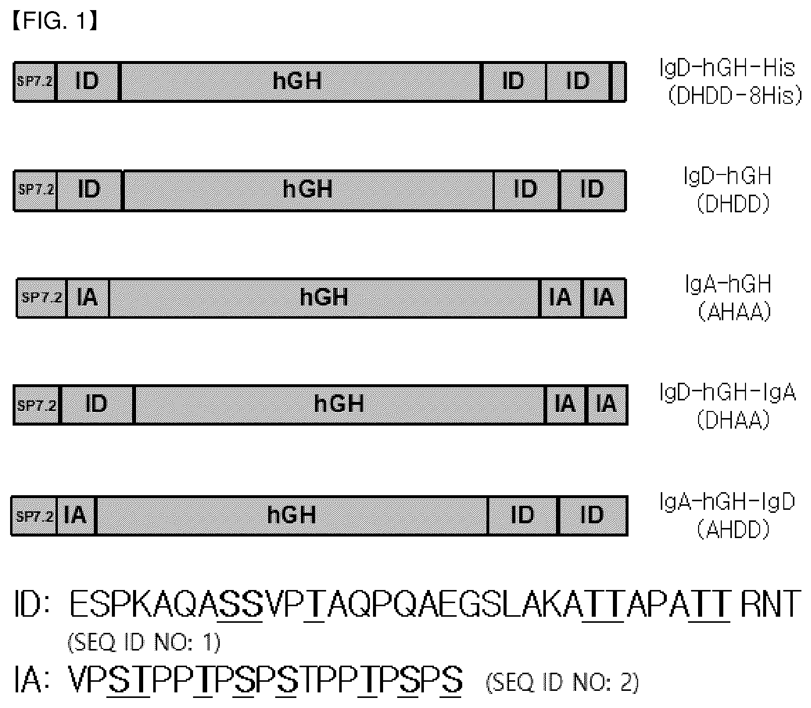
is a diagram schematically showing the structure of the fusion polypeptide IgD-hGH-His (DHDD-8His), IgD-hGH (DHDD), IgA-hGH (AHAA), IgD-hGH-IgA (DHAA), and IgA-hGH-IgD (AHDD) according to one embodiment.
is a graph showing the results of the analysis of the fusion polypeptide IgD-hGH according to one embodiment by Q-TOF Mass Spectrometry.
is a result showing the isomer distribution of the fusion polypeptide IgD-hGH-His analyzed by IEF (Isoelectric focusing).
is a graph showing the results of the analysis of the fusion polypeptide IgD-hGH-His according to one embodiment by Q-TOF Mass Spectrometry.
is a diagram schematically showing the structure of the fusion polypeptide Dulaglutide-ID and Dulaglutide-ID2 according to one embodiment.
is the results showing the isomer distribution of the fusion polypeptide Dulaglutide-ID2 analyzed by IEF (Isoelectric focusing).
is a graph showing the change in blood concentration with time after administration of the fusion polypeptide IgD-hGH compared to when hGH is administered.
is a graph showing the change in blood concentration with time after administration of the fusion polypeptide IgD-hGH-His compared to when hGH is administered.
is a graph showing the change in blood concentration with time after administration of the fusion polypeptides IgA-hGH F3, IgA-hGH F4, and IgA-hGH F5.
is a graph showing the change in blood concentration with time after administration of the fusion polypeptide Dulaglutide-ID2 (pGIgG4DD) compared to when Dulaglutide (Trulicity) is administered.
DETAILED DESCRIPTION OF THE EMBODIMENTS
Hereinafter, the present disclosure will be described in detail with reference to the following examples. However, these examples are for illustration purposes only, and the scope of the disclosure is not limited by these examples.
Example 1: Production of Fusion Polypeptide
1.1. Production of Fusion Polypeptide Containing Human Growth Hormone (hGH) as Target Polypeptide
A fusion polypeptide IgD-hGH-His (DHDD-8His), IgD-hGH (DHDD), IgA-hGH (AHAA), IgD-hGH-IgA (DHAA), and IgA-hGH-IgD (AHDD) (see ; the underlined part of the sequences of IgD and IgA1 is the part capable of performing O-Glycosylation) was produced in which IgD hinge (ESPKAQASSVPTAQPQAEGSLAKATTAPATTRNT; SEQ ID NO: 1), IgA1 hinge (VPSTPPTPSPSTPPTPSPS; SEQ ID NO: 2), or a combination of the hinge of IgD and the hinge of IgA1 was fused with the target polypeptide (human growth hormone: hGH; SEQ ID NO: 3). The amino acid sequences of each part contained in the fusion polypeptide were summarized in Table 2 below.
TABLE 2
SEQ
Amino acid ID
sequence(N-terminus→C-terminus) NO
Signal Peptide MHRPEAMLLL LTLALLGGPT WA 4
(SP7.2)
Target FPTIPLSRLF DNAMLRAHRL HQLAFDTYQE 3
polypeptide FLEAYIPKEQ KYSFLQNPQT SLCFSESIPT
(hGH) PSNREETQQK SNLELLRISL LLIQSWLEPV
QFLRSVFANS LVYGASDSNV YDLLKDLEEG
IQTLMGRLED GSPRTGQIFK QTYSKFDTNS
HNDDALLKNY GLLYCFRKDM DKVETFLRIV
QCRSVEGSCG F
Hinge region ESPKAQASSV PTAQPQAEGS LAKATTAPAT 1
of TRNT
Immunoglobulin
IgD (ID)
Hinge region VPSTPPTPSP STPPTPSPS 2
of
Immunoglobulin
IgA1 (IA)
His-Tag HHHHHHHH 5
1.1.1. IgD-hGH (DHDD)
Plasmid pAF-D1G1 (including the promoter of Korean Patent No. 10-1868139B1), which is a variant of pcDNA3.1(+) (Invitrogen, Cat. No. V790-20), was treated with BamHI (restriction site: GGATCC) and NotI (restriction site: GCGGCCGC), into which the gene encoding the fusion polypeptide of ‘(N-terminus)-[BamHI restriction site-signal peptide (SEQ ID NO: 4)-IgD hinge (IgDH1; SEQ ID NO: 1)-human growth hormone (hGH; SEQ ID NO: 3)-IgD hinge (IgDH1; SEQ ID NO: 1)-IgD hinge (IgDH1; SEQ ID NO: 1)-NotI restriction site]-(C-terminus)’ was inserted to prepare a recombinant vector pDHDD-D1G1 for the production of a fusion polypeptide containing the target polypeptide (human growth hormone) and the hinge region of immunoglobulin (IgD) (293 aa in total (excluding signal peptide); the number of O-Glycosylatable sites: a total of 21); hereinafter, referred to as ‘IgD-hGH’).
The prepared recombinant vector pDHDD-D1G1 was introduced into ExpiCHO-S™ cells (Thermo Fisher Scientific), and cultured in ExpiCHO Expression Medium (Thermo Fisher Scientific; 400 mL) for 12 days (Fed-Batch Culture; Day 1 & Day 5 Feeding) to produce the fusion polypeptide IgD-hGH. The fusion polypeptide IgD-hGH theoretically has a molecular weight of 32.2 kDa (excluding O-Glycans) and 21 O-Glycans.
The fusion polypeptide IgD-hGH produced through the expression of the recombinant vector was purified and O-Glyan site Occupancy was analyzed using Q-TOF Mass Spectrometry.
Specifically, the first purification process was performed by mounting a column made by CaptureSelect™ Human Growth Hormone Affinity Matrix (Life Technologies) having Binding Specificity to hGH on an AKTA™ Purifier (GE Healthcare Life Sciences), and loading a sample. The primary washing was performed with an equilibration buffer, and eluted with 20 mM citric acid pH 3.0 or 0.1M Acetic acid pH 3.0. Immediately after completion of the process, the elution solution was adjusted to pH 7.0 using 2M Tris Buffer and left in a frozen state until before the next purification process.
The second purification process was performed by applying Anion Exchange Chromatography and using TMAE as a resin. After the frozen sample obtained through the first process was dissolved, the conductivity was measured and the sample was diluted with water for injection so as to have a conductivity suitable for loading, and subjected to a pretreatment with a 0.22 um PES Filtration System (Corning, USA). Columns were mounted on AKTA Avant (GE Healthcare Life Sciences) and the sample was loaded. Elution was made in gradient form for isolation according to the conductivity, and fractions were divided and pooled with reference to elution peak.
Concentration or buffer exchange was performed to prepare an analytical sample and an animal experimental sample during the purification process. The sample was placed in Amicon Ultra System (Millipore), centrifuged at low temperature and subjected to concentration or diafiltration. 25 mM Sodium Phosphate pH 7.0 was used as a buffer for analysis, and PBS Buffer was used to prepare animal experimental samples.
The concentration of samples was measured after the purification process, concentration process, or diafiltration, in which the Extinction Coefficient of the substance was calculated using the amino acid sequence, and absorbances at 280 nm and 340 nm were measured with a UV Spectrophotometer (G1103A, Agilent Technologies) and calculated using the following Equation.
( A 280 nm - A 340 nm ) ( Extinction Coefficient ) × ( Dilution Factor )
In the case of animal experimental samples, they were diluted to a predetermined concentration using PBS Buffer, and filtered with 0.22 um Syringe Filter (Millex-GV, 0.22 um, Millipore) in a Biosafety Cabinet before administration, and then stored in a frozen state until subsequent administration.
The results of analyzing IgD-hGH by Q-TOF Mass Spectrometry are shown in (Y-axis: %; X-axis: mass; the numbers 7 to 21 shown above the peak are O-Glycan numbers). As shown in , O-Glycans were distributed from 7 to 21 in IgD-hGH, and the average number of O-Glycans was 13.5.
1.1.2. IgD-hGH-His
Primers in Table 3 were synthesized to add 8His-tag to the C-terminus of IgD-hGH (Example 1.1.1) for convenience of purification.
TABLE 3
Primer Name DNA Sequence (5′→3′) SEQ ID NO
hGH-Pst_F AAGTATTCCTTC CTGCAG AACCCCCAG 14
DD_R CCTGTGCCTTTGGAGACTCTGTGTTACGG 15
G
DD_F GAGTCTCCAAAGGCACAGGCCTCCTCCG 16
TG
DHis_R GTGGTGATGATGGTGTGTGTTACGGGTG 17
GTGGC
His-Not_R GCGGCCGC TTTAGTGATGGTGGTGGTGA 18
TGATGGTG
PCR was performed using each primer, and then overlapping PCR was performed again with an appropriate combination of primers to finally obtain a PCR product of 693 bp (‘(N-terminus)-[PstI restriction site-signal peptide (SEQ ID NO:4)—IgD hinge (IgDH1; SEQ ID NO:1)—human growth hormone (hGH; SEQ ID NO: 3)—IgD hinge (IgDH1; SEQ ID NO: 1)—IgD hinge (IgDH1; SEQ ID NO: 1)—8His-NotI restriction site]-gene encoding (C-terminus)’). Then, the pDHDD-D1G1 and PCR products were treated with PstI and NotI, respectively, and then ligated to finally prepare the recombinant vector pDHDD-8His-D1G1 for the preparation of a fusion polypeptide (total 301 aa (excluding signal peptide); O-Glycosylatable sites—total 21); hereinafter, referred to as ‘IgD-hGH-His’) including the target polypeptide (human growth hormone) and the hinge region of immunoglobulin (IgD) and 8His Tag.
The fusion polypeptide IgD-hGH-His produced through the expression of the recombinant vector was purified and O-Glycan site occupancy was analyzed using IEF (Isoelectric focusing) analysis and Q-TOF Mass Spectrometry.
Specifically, the first column used in the purification process was TMAE which is an anion exchange resin, and IgD-hGH-His was partially isolated from a culture solution and eluted as a first eluate. Then, the first eluate was supplied to a HIS-Tag binding column, a metal affinity resin, which is a second column, and IgD-hGH-His was selectively eluted as a second eluate. Then, the second eluate was supplied to TMAE, an anion exchange resin, which is a third column, to remove a fraction with a low sialic acid content, and eluted as a third eluate. The third eluate was then supplied to a gel filtration column, which is a fourth column, to remove multimers and fragmented proteins, thereby obtaining a fourth eluate.
More specifically, it includes the following steps.
Step 1: equilibrating with a buffer containing TMAE, 0.5×25 cm (4 mL), v=150 cm/hr, 10 mM trolamine (pH 7.0). After loading the culture solution, the column was washed once with an equilibration buffer, and an elution buffer containing 10 mM trolamine and 250 mM sodium chloride (pH 7.0) was eluted in a linear gradient to obtain a first eluate.
Step 2: equilibrating with a buffer containing Ni-NTA His*Bind, 1.0×5 cm (4 mL), v=80 cm/hr, 10 mM sodium phosphate, 1 M sodium chloride, 10 mM imidazole (pH 7.0). After loading the first eluate, the column was washed once with an equilibrium buffer, and an elution buffer containing 10 mM sodium phosphate, 1 M sodium chloride, and 500 mM imidazole (pH 7.0) was eluted in a linear gradient to obtain a second eluate.
Step 3: Diafiltration
Step 4: Equilibrating with a buffer containing TMAE, 0.5×25 cm (4 mL), v=150 cm/hr, 10 mM trolamine (pH 7.0). After loading the second eluate, the column was washed once with an equilibrium buffer, and an elution buffer containing 10 mM trolamine and 100 mM sodium chloride (pH 7.0) was eluted in a linear gradient to obtain a third eluate.
Step 5: Ultrafiltration
Step 6: equilibrating with a buffer containing Sephacryl 5-100, 1.6×30 cm (60 mL), v=30 cm/hr, 20 mM sodium phosphate, 140 mM sodium chloride, pH 7.0. After loading the third eluate, the monomer fraction was eluted with an equilibration buffer to obtain a fourth eluate.
The isomer distribution of the obtained fourth eluate was shown in . In , the theoretical pI value of IgD-hGH-His is 6.65. The value lower than this means that IgD-hGH-His is O-Glycosylation and sialic acid is bound to this O-Glycan to becomes more acidic.
The results of analyzing IgD-hGH-His by Q-TOF Mass Spectrometry are shown in (Y-axis: %; X-axis: mass; values of 7 to 21 shown above the peak are the numbers of O-Glycans). As shown in , in IgD-hGH-His, 0-Glycans were distributed from 8 to 21, and the average number of O-Glycans was 14.7.
1.1.3. IgA-hGH (AHAA)
In the recombinant vector pDHDD-D1G1 constructed in Example 1.1.1, a recombinant vector pAHAA-D1G1 was constructed to have the same configuration, except that the coding genes of three IgD hinges (one on the N-terminus side and two on the C-terminus side of hGH, three in total) were replaced with the coding genes of the IgA1 hinges, respectively., and then expressed in the same manner as in Example 1.1.1 to produce a fusion polypeptide having a configuration of IgA1 hinge (IgA; SEQ ID NO: 2)—human growth hormone (hGH; SEQ ID NO: 3)—IgA1 hinge (IgA; SEQ ID NO: 2)—IgA1 hinge (IgA; SEQ ID NO: 2) (see ; hereinafter referred to as ‘IgA-hGH’). The fusion polypeptide IgA-hGH theoretically has 24 O-Glycans. The fusion polypeptide IgA-hGH produced through expression of the recombinant vector was purified by referring to the method described in Example 1.1.1.
As a result of analyzing the purified IgA-hGH by Q-TOF Mass Spectrometry, the average number of O-Glycans in IgA-hGH was 12.8 in Fraction 3, 14.3 in Fraction 4, and 15.6 in Fraction 5.
1.1.4. IgD-hGH-IgA (DHAA)
7574 bp vector where the recombinant vector pDHDD-D1G1 produced in Example 1.1.1 was cut with BamHI and NotI was ligated with 489 bp of Insert I where pDHDD-DTG1 was cut with BamHI and KasI, and 383 bp of Insert II where the recombinant vector pAHAA-D1G1 used in Example 1.1.3 was cut with KasI and NotI, and thus the recombinant vector pDHAA-D1G1 was constructed so as to have the same configuration except that in the recombinant vector pDHDD-D1G1, 3 IgD hinges (1 on the N-terminus side and 2 on the C-terminus side of hGH, 3 in total), two on the 3′ terminal side were replaced by the coding gene of the IgA1 hinge. The recombinant vector pDHAA-D1G1 was expressed in the same manner as in Example 1.1.1 to produce a fusion polypeptide having the configuration of IgD hinge (IgD; SEQ ID NO: 1)-human growth hormone (hGH; SEQ ID NO: 3)-IgA1 hinge (IgA; SEQ ID NO: 2)-IgA1 hinge (IgA; SEQ ID NO: 2). The fusion polypeptide IgD-hGH-IgA theoretically has 23 O-Glycans.
1.1.5. IgA-hGH-IgD (AHDD)
7574 bp vector where the recombinant vector pDHDD-D1G1 produced in Example 1.1.1 was cut with BamHI and NotI was ligated with 444 bp of Insert I where pAHAA-D1G1 used in Example 1.1.3 was cut with BamHI and KasI, and 473 bp of Insert II where pDHDD-D1G1 used in Example 1.1.1 was cut with KasI and NotI, and thus the recombinant vector pADD-D1G1 was constructed so as to have the same configuration except that one at the 5′ terminal of the three IgD hinge coding genes was replaced by the coding gene for the IgA1 hinge, and then expressed in the same manner as in Example 1.1.1 to produce a fusion polypeptide having the configuration of IgA1 hinge (IgA; SEQ ID NO: 2)—human growth hormone (hGH; SEQ ID NO: 3)—IgD hinge (IgD; SEQ ID NO: 1)—IgD hinge (IgD; SEQ ID NO: 1) (see ; hereinafter referred to as ‘IgA-hGH-IgD’). The fusion polypeptide IgA-hGH-IgD theoretically has 22 O-Glycans.
1.2. Protein of Interest: GLP-1-Fc Fusion Protein
The fusion polypeptides Dulaglutide-ID (including one IgD hinge area) and Dulaglutide-ID2 (including two IgD hinge regions) (see ) were prepared in which IgD hinge (ESPKAQASVPTAQPQAEGSLAKATTAPATTRNT; SEQ ID NO: 1) was fused with the target polypeptide (GLP-1 (Glucagon-like peptide-1)-Fc fusion protein: GLP-1-Fc). The GLP-1-Fc fusion protein exists as a dimer. The encoded amino acid sequence is summarized in Table 4 below.
TABLE 4
Amino acid SEQ
sequence (N- ID
terminus→C-terminus) NO
Signal Peptide (SP7.2) MHRPEAMLLL LTLALLGGPT 4
WA
Target Modified HGEGTFTSDV SSYLEEQAAK 6
polypeptide GLP-1 EFIAWLVKGG G
(GLP-1-Fc) GS Linker GGGGSGGGGS GGGGS
Modified A ESKYGPPCP PCP APEAAGG
IgG4 PSVFLFPPKP KDTLMISRTP
(IgG4 EVTCVVVDVS QEDPEVQFNW
hinge- YVDGVEVHNA KTKPREEQFN
CH2- STYRVVSVLT VLHQDWLNGK
IgG4- EYKCKVSNKG LPSSIEKTIS
CH3_modified) KAK GQPREPQ VYTLPPSQEE
MTKNQVSLTC LVKGFYPSDI
AVEWESNGQP ENNYKTTPPV
LDSDGSFFLY SRLTVDKSRW
QEGNVFSCSV MHEALHNHYT
QKSLSLSLG
Hinge region of ESPKAQASSV PTAQPQAEGS 1
Immunoglobulin IgD LAKATTAPAT TRNT
(ID)
1.2.1. Dulaglutide-ID1
The expression vector pGJg4 (including the promoter of Korean Patent No. 10-1868139B1) expressing GLP-1-Fc, which is a variant of pcDNA3.1(+) (Invitrogen, Cat. No. V790-20), was used as a template, and PCR was performed using Primers JgG4mCH2_F and IgG4JD_R in Table 4 to obtain a PCR product (mIgG4) of 659 bp Modified IgG4. And, the pDHDD-D1G1 prepared in Example 1.1.1 was used as a template, and PCR was performed using Primers gG4D_F and ID_NotR in Table 4 to obtain PCR products of 129 bp (ID1) and 231 bp (ID2). The obtained 659 bp mIgG4 PCR Product and 129 bp ID PCR Product were purified, which was then used as a template. Overlapping PCR was performed using Primers JgG4mCH2_F and ID_NotR in Table 5 below to obtain a PCR product of 770 bp (‘(N-terminus)-[lModified IgG4 Fc part (including BsrGI restriction site))-IgD hinge (IgDH1; SEQ ID NO: 1)-NotI restriction site]—gene encoding (C-terminus)’).
TABLE 5
SEQ ID
Primer Name DNA Sequence (5′→3′) NO
IgG4mCH2_F CACCTGAGGCCGCCGGGGGACCG 19
TCAGTCT
IgG4ID_R CTTTGGAGACTCGCCCAGGGACA 20
GGGACAG
IgG4ID_F CTGGGCGAGTCTCCAAAGGCACA 21
GGCCTCC
ID_NotR TTCTTCTTC GCGGCCGC TTTATGT 22
GTTACG
A 7574 bp vector where pDHDD-D1G1 prepared in Example 1.1.1 was cut with BamHI and NotI was ligated with 607 bp of Insert I where pGIg4 was cut with BamHI and BsrGI and 403 bp of Insert II where the 770 bp PCR product obtained through the overlapping PCR was cut with BsrGI and NotI to prepare a recombinant vector pGIg4D-D1G1 for the production of a fusion polypeptide including the target polypeptide (GLP-1-Fc) and a hinge region of immunoglobulin (IgD) (total 309 aa (excluding signal peptide); O-Glycosylated sites—total 7, exists as dimers, so finally 14); hereinafter, referred to as ‘Dulaglutide-ID1’).
1.2.2. Dulaglutide-ID2
659 bp of mIgG4 PCR product and 231 bp of ID2 PCR product obtained in Example 1.2.1 were used as a template, and overlapping PCR was performed using Primers IgG4mCH2_F and ID_NotR in Table 4 to obtain a PCR product of 882 bp (‘(N-terminus)-[Modified IgG4 Fc part (including BsrGI restriction site)-IgD hinge (IgDH1; SEQ ID NO: 1)-IgD hinge (IgDH1; SEQ ID NO: 1)-NotI restriction site)-gene encoding (C-terminus)’).
A 7574 bp vector where pDHDD-D1G1 prepared in Example 1.1.1 was cut with BamHI and NotI was ligated with 607 bp of Insert I where pGIg4 was cut with BamHI and BsrGI and 505 bp of Insert II where the PCR product of 882 bp obtained through the overlapping PCR was cut with BsrGI and NotI to prepare a recombinant vector pGIg4DD-D1G1 for the production of a fusion polypeptide including the target polypeptide (GLP-1-Fc) and two hinge regions of immunoglobulin (IgD) (total 343 aa (excluding signal peptide); 0-Glycosylated sites-14 in total, 28 as it exists as dimer); hereinafter, referred to as ‘Dulaglutide-ID2’).
The fusion polypeptide Dulaglutide-ID2 produced through the expression of the recombinant vector was purified, and O-Glyan site occupancy was analyzed using isoelectric focusing (IEF) analysis and Q-TOF Mass Spectrometry.
Specifically, proteins were isolated and purified through Protein A affinity chromatography using the Fc region of substance, and then anion exchange chromatography and hydrophobic interaction chromatography were sequentially performed and purified.
The culture solution was filtered using a 0.22 um filtration membrane, injected into Protein A affinity resin equilibrated with an equilibration buffer (10 mM Sodium phosphate, 150 mM Sodium chloride, pH 7.4), and then washed with an equilibrium buffer. After washing, the protein was eluted with an elution buffer (100 mM Sodium citrate pH 3.5), and peaks were collected.
The collected eluate was subjected to a buffer exchange with 20 mM Tris pH 8.0.
The buffer exchanged sample was injected and purified into anion exchange chromatography (Source 15Q, GE Healthcare).
The equilibration buffer and elution buffer used were 20 mM Tris, pH 8.0, 20 mM Tris, 0.5 M NaCl, and pH 8.0, respectively. The equilibration buffer and the elution buffer were used as channels A and B, respectively, to elute the protein under concentration gradient conditions and collect peaks.
The collected protein solution was further purified using hydrophobic interaction chromatography (Butyl sepharose, GE Healthcare).
The equilibration buffer and elution buffer used were 0.1M sodium phosphate pH 6.0, 1.8 ammonium sulfate, pH 8.0, and 0.1M sodium phosphate pH 6.0, respectively. The equilibration buffer and the elution buffer were used as channels A and B, respectively, to elute the protein under concentration gradient conditions and collect peaks.
The isomer distribution of Dulaglutide-ID2 obtained by analyzing the collected peaks by isoelectric focusing (IEF) is shown in . In , the theoretical pI value of Dulaglutide-ID2 is 5.78, but in the case of Fraction #3, the value lower than this means that Dulaglutide-ID2 is O-Glycosylated, and this O-Glycan is attached to sialic acid and becomes more acidic.
As a result of analyzing Dulaglutide-ID2 Fraction #3 by Q-TOF Mass Spectrometry, ID could be performed up to 26, and the average number of O-Glycans was 17.5.
The finally purified protein solution was buffer-exchanged with the same excipients as Trulicity (Sodium citrate hydrate: 2.74 mg/mL, Anhydrous citric acid: 0.14 mg/mL, D-mannitol: 46.4 mg/mL, polysorbate 80: 0.20 mg/mL, pH 6.0-7.0), and concentrated and used as a test material for the animal PK test.
Example 2: Pharmacokinetic Properties (PK Profile) Test of Fusion Polypeptide (In Vivo)
2-1. Target Protein: Human Growth Hormone (hGH)
Fusion polypeptides IgD-hGH, IgD-hGH-His, IgA-hGH F3 (Fraction 3 of Example 1.1.3), IgA-hGH F4 (Fraction 4 of Example 1.1.3), and IgA-hGH F5 (Execution Fraction 5 of Example 1.1.3) prepared in Example 1.1 were subcutaneously administered to SD rats (Orientbio, 7 weeks old, about 300 g; n=3) at a dose of 2 mg/kg, and Pharmacokinetics were tested. Sampling was performed at 0, 0.5, 1, 2, 4, 6, 8, 24, 48 hours, and for comparison, hGH (Eutropin, LG Chem) was administered subcutaneously at a dose of 2 mg/kg in the same manner as above, and tested.
After administration to SD rat as described above, the blood collected by time-point was centrifuged to obtain a serum. ELISA was performed using Human Growth Hormone Quantikine ELISA Kit (R&D Systems), and the concentrations of hGH and fusion polypeptides (IgD-hGH, IgD-hGH-His, IgA-hGH FP3, IgA-hGH FP4 and IgA-hGH FP5) in the blood by time-point were confirmed. Using this data, parameters including AUC (area under the curve) were calculated using software for PK analysis (WinNonlin (Certara L. P.), etc.).
2-1-1. hGH vs. IgD-hGH
PK results of hGH and IgD-hGH are shown in Table 6 and .
TABLE 6
hGH IgD-hGH
Parameter Mean SD Mean SD
Cmax (ng/mL) 1788 50.6 973 233
Tmax (hr) 1 — 4 —
AUCinf (ng*hr/mL) 4703 111 16027 2941
AUClast (ng*hr/mL) 4695 111 15845 2881
T½ (hr) 2.17 1.79 7.11 0.327
AUCextp (%) 0.167 0.059 1.11 0.197
(C max : Maximum blood concentration, T max : Time when peak blood concentration is reached, AUC inf : Area under the blood concentration-time curve calculated by extrapolating from the last measurable blood collection time point to infinite time, AUC last : Area under the blood concentration-time curve until the last measurable blood collection time point, T 1/2 : elimination half-life, AUC Extp (%): [(AUC inf −AUC last )/AUC inf ]*100)
As can be seen in Table 6 and , it can be confirmed that the half-life of the hGH fused with the hinge region (IgD-hGH) increased by about 3.3 times compared to the hGH not fused with the hinge region.
2-1-2. hGH vs. IgD-hGH-His
PK results of hGH and IgD-hGH-His are shown in Table 7 and .
TABLE 7
hGH IgD-hGH-His
Parameter Mean SD Mean SD
Cmax (ng/mL) 502.22 39.88 886.67 195.63
Tmax (hr) 1 — 4 —
AUCinf (ng*hr/mL) 1028.80 120.03 13436.05 2680.39
AUClast (ng*hr/mL) 1003.74 115.76 13332.39 2710.64
T½ (hr) 2.55 0.20 6.61 0.71
AUCextp (%) 2.43 0.23 0.82 0.39
(C max : Maximum blood concentration, T max : Time when peak blood concentration is reached, AUC inf : Area under the blood concentration-time curve calculated by extrapolating from the last measurable blood collection time point to infinite time, AUC last : Area under the blood concentration-time curve until the last measurable blood collection time point, T 1/2 : elimination half-life, AUC Extp (%): [(AUC inf −AUC last )/AUC inf ]*100)
As can be seen in Table 7 and , it can be confirmed that the half-life of hGH (IgD-hGH-His) fused with the hinge region with His-Tag increased by about 2.6 times compared to the hGH not fused with the hinge region.
2-1-3. IgA-hGH (Effect on PK by O-Glycan Number)
In order to see the effect of the number of O-glycans on PK, PK results for each IgA-hGH fraction are shown in Table 8 and .
TABLE 8
IgA-hGH IgA-hGH IgA-hGH
FP3 (Average FP4 (Average FP5 (Average
number of O- number of O- number of O-
glycan: 12.8) glycan: 14.3) glycan: 15.6)
Parameter Mean SD Mean SD Mean SD
Cmax 290 80.5 305 102 195 32.8
(ng/mL)
Tmax (hr) 2 — 4 — 2 —
AUCinf 2510 474 2832 814 2186 89.2
(ng*hr/
mL)
AUClast 2509 475 2826 816 2160 96.8
(ng*hr/
mL)
T½ (hr) 1.98 0.205 2.53 0.287 3.56 0.335
AUCextp 0.050 0.035 0.240 0.182 1.21 0.573
(%)
(C max : Maximum blood concentration, T max : Time when peak blood concentration is reached, AUC inf : Area under the blood concentration-time curve calculated by extrapolating from the last measurable blood collection time point to infinite time, AUC last : Area under the blood concentration-time curve until the last measurable blood collection time point, T 1/2 : elimination half-life, AUC Extp (%): [(AUC inf −AUC last )/AUC inf ]*100) As can be seen in Table 8 and , it can be confirmed that the half-life increases as the number of O-glycan increases.
2-2. Target Protein: GLP-1-Fc Fusion Protein (GLP-1-Fc, Dulaglutide)
Fusion polypeptide Dulaglutide-ID2 prepared in Example 1.2 was subcutaneously administered to SD rats (Orientbio, 7 weeks old, about 300 g; n=3) at a dose of 0.1 mg/kg, and Pharmacokinetics were tested. Sampling was performed at 0, 0.5, 1, 2, 4, 6, 8, 24, 48, 96 and 144 hours, and for comparison, Dulaglutide (Trulicity, Lilly Korea) was administered subcutaneously at a dose of 0.1 mg/kg in the same manner as above, and tested.
After administration to the SD rat as above, the blood collected by time-point was centrifuged to obtain a serum. ELISA was performed using Anti-GLP-1 antibody (NovousBio) and Anti-Human IgG4 Fc Antibody (Sigma-Aldrich), and the concentrations of Dulaglutide and fusion polypeptide Dulaglutide-ID2 in the blood by time-point were confirmed. Using this data, parameters including AUC (area under the curve) were calculated using software for PK analysis (WinNonlin (Certara L. P.), etc.).
The obtained results are shown in Table 9 and .
TABLE 9
Dulaglutide Dulaglutide-ID2
Parameter Mean SD Mean SD
Cmax (ng/mL) 268 24.2 46.8 2.43
Tmax (hr) 24 — 24 —
AUCinf (ng*hr/mL) 11500 1230 3430 535
AUClast (ng*hr/mL) 11700 1200 3870 786
T½ (hr) 26.9 3.06 40.9 7.04
AUCextp (%) 1.97 0.542 10.9 4.09
(C max : Maximum blood concentration, T max : Time when peak blood concentration is reached, AUC inf : Area under the blood concentration-time curve calculated by extrapolating from the last measurable blood collection time point to infinite time, AUC last : Area under the blood concentration-time curve until the last measurable blood collection time point, T 1/2 : elimination half-life, AUC Extp (%): [(AUC inf −AUC last )/AUC inf ]*100) As can be seen in Table 9 and , it can be confirmed that the half-life of GLP-1-Fc (Dulaglutide-ID2) fused with the hinge region increased by about 1.5 times as compared with Dulaglutide, which is not fused with the hinge area. Further, Cmax was about ⅕, and AUClast was about ⅓.
From the above description, those skilled in the art will understand that the present disclosure can be implemented in other specific forms without changing the technical idea or essential features thereof. In this regard, it should be understood that the embodiments described above are illustrative in all respects and non-limiting. The scope of the present disclosure should be construed that all changes or modifications derived from the meaning and scope of the claims to be described later rather than the above detailed description and the equivalent concepts thereof are included in the scope of the present disclosure.
Figures (8)
Citations
This patent cites (24)
- US5416017
- US6165476
- US8304386
- US2003/0044423
- US2005/0069521
- US2006/0051844
- US2008/0300188
- US2012/0094356
- US102439044
- US105198998
- US106256835
- US107286248
- USH01-287041
- USH02-213
- US2010-531134
- US10-0545720
- US10-1038126
- US10-1183262
- US10-2014-0015152
- US10-1380729
- US10-1380732
- US10-2014-0083973
- US10-2015-0125402
- US2017-198435