Anti-sars-cov-2-spike Glycoprotein Antibodies and Antigen-binding Fragments
Abstract
The present disclosure provides antibodies and antigen-binding fragments thereof that bind specifically to a coronavirus spike protein and methods of using such antibodies and fragments for treating or preventing viral infections (e.g., coronavirus infections).
Claims (20)
1 . An isolated recombinant antibody or antigen-binding fragment thereof that specifically binds to a coronavirus spike protein (CoV-S), wherein the antibody or antigen-binding fragment comprises: a heavy chain variable region (HCVR) comprising three heavy chain complementarity determining regions (CDRs) (HCDR1, HCDR2, and HCDR3); and a light chain variable region (LCVR) comprising three light chain CDRs (LCDR1, LCDR2, and LCDR3); wherein the HCDR1, HCDR2, HCDR3, LCDR1, LCDR2, and LCDR3, comprise, respectively, the amino acid sequences of: SEQ ID NO: 4, SEQ ID NO: 6, SEQ ID NO: 8, SEQ ID NO: 12, EVS, and SEQ ID NO: 16; SEQ ID NO: 24, SEQ ID NO: 26, SEQ ID NO: 28, SEQ ID NO: 32, EGN, and SEQ ID NO: 36; SEQ ID NO: 44, SEQ ID NO: 26, SEQ ID NO: 47, SEQ ID NO: 51, EGN, and SEQ ID NO: 36; SEQ ID NO: 60, SEQ ID NO: 62, SEQ ID NO: 64, SEQ ID NO: 51, EGT, and SEQ ID NO: 36; SEQ ID NO: 76, SEQ ID NO: 78, SEQ ID NO: 80, SEQ ID NO: 84, EDS, and SEQ ID NO: 88; SEQ ID NO: 96, SEQ ID NO: 98, SEQ ID NO: 100, SEQ ID NO: 104, AAS, and SEQ ID NO: 108; SEQ ID NO: 116, SEQ ID NO: 118, SEQ ID NO: 120, SEQ ID NO: 124, GAS, and SEQ ID NO: 128; SEQ ID NO: 136, SEQ ID NO: 138, SEQ ID NO: 140, SEQ ID NO: 144, GAS, and SEQ ID NO: 146; SEQ ID NO: 154, SEQ ID NO: 156, SEQ ID NO: 158, SEQ ID NO: 162, GNS, and SEQ ID NO: 166; SEQ ID NO: 174, SEQ ID NO: 176, SEQ ID NO: 178, SEQ ID NO: 182, SND, and SEQ ID NO: 186; SEQ ID NO: 194, SEQ ID NO: 196, SEQ ID NO: 198, SEQ ID NO: 202, DND, and SEQ ID NO: 206; SEQ ID NO: 214, SEQ ID NO: 216, SEQ ID NO: 218, SEQ ID NO: 222, GAS, and SEQ ID NO: 224; SEQ ID NO: 232, SEQ ID NO: 234, SEQ ID NO: 236, SEQ ID NO: 240, DAS, and SEQ ID NO: 244; SEQ ID NO: 252, SEQ ID NO: 254, SEQ ID NO: 256, SEQ ID NO: 260, GAT, and SEQ ID NO: 264; SEQ ID NO: 272, SEQ ID NO: 274, SEQ ID NO: 276, SEQ ID NO: 280, AAS, and SEQ ID NO: 282; SEQ ID NO: 290, SEQ ID NO: 292, SEQ ID NO: 294, SEQ ID NO: 298, SDN, and SEQ ID NO: 302; SEQ ID NO: 310, SEQ ID NO: 312, SEQ ID NO: 314, SEQ ID NO: 318, VNN, and SEQ ID NO: 322; SEQ ID NO: 330, SEQ ID NO: 332, SEQ ID NO: 334, SEQ ID NO: 338, AAS, and SEQ ID NO: 340; SEQ ID NO: 96, SEQ ID NO: 98, SEQ ID NO: 350, SEQ ID NO: 354, AAS, and SEQ ID NO: 356; SEQ ID NO: 364, SEQ ID NO: 366, SEQ ID NO: 368, SEQ ID NO: 372, AAS, and SEQ ID NO: 374; SEQ ID NO: 382, SEQ ID NO: 384, SEQ ID NO: 386, SEQ ID NO: 390, KAS, and SEQ ID NO: 394; SEQ ID NO: 402, SEQ ID NO: 98, SEQ ID NO: 405, SEQ ID NO: 409, DAS, and SEQ ID NO: 411; SEQ ID NO: 419, SEQ ID NO: 421, SEQ ID NO: 423, SEQ ID NO: 427, EVS, and SEQ ID NO: 429; SEQ ID NO: 437, SEQ ID NO: 138, SEQ ID NO: 440, SEQ ID NO: 444, DKN, and SEQ ID NO: 448; SEQ ID NO: 456, SEQ ID NO: 458, SEQ ID NO: 460, SEQ ID NO: 84, ELT, and SEQ ID NO: 467; SEQ ID NO: 475, SEQ ID NO: 477, SEQ ID NO: 479, SEQ ID NO: 483, DVT, and SEQ ID NO: 487; SEQ ID NO: 495, SEQ ID NO: 497, SEQ ID NO: 499, SEQ ID NO: 503, EVT, and SEQ ID NO: 507; SEQ ID NO: 515, SEQ ID NO: 517, SEQ ID NO: 519, SEQ ID NO: 523, EGS, and SEQ ID NO: 36; SEQ ID NO: 533, SEQ ID NO: 535, SEQ ID NO: 537, SEQ ID NO: 483, DVS, and SEQ ID NO: 544; SEQ ID NO: 552, SEQ ID NO: 554, SEQ ID NO: 556, SEQ ID NO: 84, EVS, and SEQ ID NO: 560; SEQ ID NO: 568, SEQ ID NO: 570, SEQ ID NO: 572, SEQ ID NO: 576, ENN, and SEQ ID NO: 580; SEQ ID NO: 588, SEQ ID NO: 590, SEQ ID NO: 592, SEQ ID NO: 596, GAS, and SEQ ID NO: 598; SEQ ID NO: 606, SEQ ID NO: 608, SEQ ID NO: 610, SEQ ID NO: 614, GAS, and SEQ ID NO: 616; SEQ ID NO: 624, SEQ ID NO: 626, SEQ ID NO: 628, SEQ ID NO: 632, LGS, and SEQ ID NO: 636; SEQ ID NO: 644, SEQ ID NO: 646, SEQ ID NO: 648, SEQ ID NO: 652, LGS, and SEQ ID NO: 655; SEQ ID NO: 663, SEQ ID NO: 665, SEQ ID NO: 667, SEQ ID NO: 124, GAS, and SEQ ID NO: 671; SEQ ID NO: 679, SEQ ID NO: 78, SEQ ID NO: 682, SEQ ID NO: 686, GAS, and SEQ ID NO: 688; SEQ ID NO: 696, SEQ ID NO: 698, SEQ ID NO: 700, SEQ ID NO: 704, SAS, and SEQ ID NO: 708; SEQ ID NO: 696, SEQ ID NO: 716, SEQ ID NO: 718, SEQ ID NO: 722, GAS, and SEQ ID NO: 724; SEQ ID NO: 732, SEQ ID NO: 734, SEQ ID NO: 736, SEQ ID NO: 740, AAS, and SEQ ID NO: 742; SEQ ID NO: 750, SEQ ID NO: 497, SEQ ID NO: 752, SEQ ID NO: 756, EVT, and SEQ ID NO: 758; SEQ ID NO: 766, SEQ ID NO: 768, SEQ ID NO: 770, SEQ ID NO: 774, KDS, and SEQ ID NO: 778; SEQ ID NO: 786, SEQ ID NO: 788, SEQ ID NO: 790, SEQ ID NO: 794, GNT, and SEQ ID NO: 798; SEQ ID NO: 806, SEQ ID NO: 497, SEQ ID NO: 808, SEQ ID NO: 812, EVT, and SEQ ID NO: 814; SEQ ID NO: 822, SEQ ID NO: 497, SEQ ID NO: 825, SEQ ID NO: 756, EVS, and SEQ ID NO: 829; SEQ ID NO: 837, SEQ ID NO: 839, SEQ ID NO: 841, SEQ ID NO: 845, KIS, and SEQ ID NO: 849; SEQ ID NO: 857, SEQ ID NO: 859, SEQ ID NO: 861, SEQ ID NO: 865, AAS, and SEQ ID NO: 867; SEQ ID NO: 76, SEQ ID NO: 876, SEQ ID NO: 878, SEQ ID NO: 84, EDS, and SEQ ID NO: 36; SEQ ID NO: 889, SEQ ID NO: 891, SEQ ID NO: 893, SEQ ID NO: 897, GNS, and SEQ ID NO: 899; SEQ ID NO: 154, SEQ ID NO: 908, SEQ ID NO: 910, SEQ ID NO: 914, GHT, and SEQ ID NO: 166; SEQ ID NO: 924, SEQ ID NO: 926, SEQ ID NO: 928, SEQ ID NO: 576, RNN, and SEQ ID NO: 935; SEQ ID NO: 943, SEQ ID NO: 945, SEQ ID NO: 947, SEQ ID NO: 951, LGS, and SEQ ID NO: 655; SEQ ID NO: 959, SEQ ID NO: 961, SEQ ID NO: 963, SEQ ID NO: 967, WAS, and SEQ ID NO: 971; SEQ ID NO: 696, SEQ ID NO: 698, SEQ ID NO: 700, SEQ ID NO: 704, SAS, and SEQ ID NO: 979; SEQ ID NO: 696, SEQ ID NO: 698, SEQ ID NO: 700, SEQ ID NO: 704, SAS, and SEQ ID NO: 985; or SEQ ID NO: 991, SEQ ID NO: 993, SEQ ID NO: 995, SEQ ID NO: 999, GAS, and SEQ ID NO: 1003.
10 . A pair of polynucleotides, wherein: (a) the first polynucleotide encodes a heavy chain variable region (HCVR) comprising three heavy chain complementarity determining regions (CDRs) (HCDR1, HCDR2, and HCDR3), and the second polynucleotide encodes a light chain variable region (LCVR) comprising three light chain CDRs (LCDR1, LCDR2, and LCDR3), wherein the HCDR1, HCDR2, HCDR3, LCDR1, LCDR2, and LCDR3, comprise, respectively, the amino acid sequences of: SEQ ID NO: 4, SEQ ID NO: 6, SEQ ID NO: 8, SEQ ID NO: 12, EVS, and SEQ ID NO: 16; SEQ ID NO: 24, SEQ ID NO: 26, SEQ ID NO: 28, SEQ ID NO: 32, EGN, and SEQ ID NO: 36; SEQ ID NO: 44, SEQ ID NO: 26, SEQ ID NO: 47, SEQ ID NO: 51, EGN, and SEQ ID NO: 36; SEQ ID NO: 60, SEQ ID NO: 62, SEQ ID NO: 64, SEQ ID NO: 51, EGT, and SEQ ID NO: 36; SEQ ID NO: 76, SEQ ID NO: 78, SEQ ID NO: 80, SEQ ID NO: 84, EDS, and SEQ ID NO: 88; SEQ ID NO: 96, SEQ ID NO: 98, SEQ ID NO: 100, SEQ ID NO: 104, AAS, and SEQ ID NO: 108; SEQ ID NO: 116, SEQ ID NO: 118, SEQ ID NO: 120, SEQ ID NO: 124, GAS, and SEQ ID NO: 128; SEQ ID NO: 136, SEQ ID NO: 138, SEQ ID NO: 140, SEQ ID NO: 144, GAS, and SEQ ID NO: 146; SEQ ID NO: 154, SEQ ID NO: 156, SEQ ID NO: 158, SEQ ID NO: 162, GNS, and SEQ ID NO: 166; SEQ ID NO: 174, SEQ ID NO: 176, SEQ ID NO: 178, SEQ ID NO: 182, SND, and SEQ ID NO: 186; SEQ ID NO: 194, SEQ ID NO: 196, SEQ ID NO: 198, SEQ ID NO: 202, DND, and SEQ ID NO: 206; SEQ ID NO: 214, SEQ ID NO: 216, SEQ ID NO: 218, SEQ ID NO: 222, GAS, and SEQ ID NO: 224; SEQ ID NO: 232, SEQ ID NO: 234, SEQ ID NO: 236, SEQ ID NO: 240, DAS, and SEQ ID NO: 244; SEQ ID NO: 252, SEQ ID NO: 254, SEQ ID NO: 256, SEQ ID NO: 260, GAT, and SEQ ID NO: 264; SEQ ID NO: 272, SEQ ID NO: 274, SEQ ID NO: 276, SEQ ID NO: 280, AAS, and SEQ ID NO: 282; SEQ ID NO: 290, SEQ ID NO: 292, SEQ ID NO: 294, SEQ ID NO: 298, SDN, and SEQ ID NO: 302; SEQ ID NO: 310, SEQ ID NO: 312, SEQ ID NO: 314, SEQ ID NO: 318, VNN, and SEQ ID NO: 322; SEQ ID NO: 330, SEQ ID NO: 332, SEQ ID NO: 334, SEQ ID NO: 338, AAS, and SEQ ID NO: 340; SEQ ID NO: 96, SEQ ID NO: 98, SEQ ID NO: 350, SEQ ID NO: 354, AAS, and SEQ ID NO: 356; SEQ ID NO: 364, SEQ ID NO: 366, SEQ ID NO: 368, SEQ ID NO: 372, AAS, and SEQ ID NO: 374; SEQ ID NO: 382, SEQ ID NO: 384, SEQ ID NO: 386, SEQ ID NO: 390, KAS, and SEQ ID NO: 394; SEQ ID NO: 402, SEQ ID NO: 98, SEQ ID NO: 405, SEQ ID NO: 409, DAS, and SEQ ID NO: 411; SEQ ID NO: 419, SEQ ID NO: 421, SEQ ID NO: 423, SEQ ID NO: 427, EVS, and SEQ ID NO: 429; SEQ ID NO: 437, SEQ ID NO: 138, SEQ ID NO: 440, SEQ ID NO: 444, DKN, and SEQ ID NO: 448; SEQ ID NO: 456, SEQ ID NO: 458, SEQ ID NO: 460, SEQ ID NO: 84, ELT, and SEQ ID NO: 467; SEQ ID NO: 475, SEQ ID NO: 477, SEQ ID NO: 479, SEQ ID NO: 483, DVT, and SEQ ID NO: 487; SEQ ID NO: 495, SEQ ID NO: 497, SEQ ID NO: 499, SEQ ID NO: 503, EVT, and SEQ ID NO: 507; SEQ ID NO: 515, SEQ ID NO: 517, SEQ ID NO: 519, SEQ ID NO: 523, EGS, and SEQ ID NO: 36; SEQ ID NO: 533, SEQ ID NO: 535, SEQ ID NO: 537, SEQ ID NO: 483, DVS, and SEQ ID NO: 544; SEQ ID NO: 552, SEQ ID NO: 554, SEQ ID NO: 556, SEQ ID NO: 84, EVS, and SEQ ID NO: 560; SEQ ID NO: 568, SEQ ID NO: 570, SEQ ID NO: 572, SEQ ID NO: 576, ENN, and SEQ ID NO: 580; SEQ ID NO: 588, SEQ ID NO: 590, SEQ ID NO: 592, SEQ ID NO: 596, GAS, and SEQ ID NO: 598; SEQ ID NO: 606, SEQ ID NO: 608, SEQ ID NO: 610, SEQ ID NO: 614, GAS, and SEQ ID NO: 616; SEQ ID NO: 624, SEQ ID NO: 626, SEQ ID NO: 628, SEQ ID NO: 632, LGS, and SEQ ID NO: 636; SEQ ID NO: 644, SEQ ID NO: 646, SEQ ID NO: 648, SEQ ID NO: 652, LGS, and SEQ ID NO: 655; SEQ ID NO: 663, SEQ ID NO: 665, SEQ ID NO: 667, SEQ ID NO: 124, GAS, and SEQ ID NO: 671; SEQ ID NO: 679, SEQ ID NO: 78, SEQ ID NO: 682, SEQ ID NO: 686, GAS, and SEQ ID NO: 688; SEQ ID NO: 696, SEQ ID NO: 698, SEQ ID NO: 700, SEQ ID NO: 704, SAS, and SEQ ID NO: 708; SEQ ID NO: 696, SEQ ID NO: 716, SEQ ID NO: 718, SEQ ID NO: 722, GAS, and SEQ ID NO: 724; SEQ ID NO: 732, SEQ ID NO: 734, SEQ ID NO: 736, SEQ ID NO: 740, AAS, and SEQ ID NO: 742; SEQ ID NO: 750, SEQ ID NO: 497, SEQ ID NO: 752, SEQ ID NO: 756, EVT, and SEQ ID NO: 758; SEQ ID NO: 766, SEQ ID NO: 768, SEQ ID NO: 770, SEQ ID NO: 774, KDS, and SEQ ID NO: 778; SEQ ID NO: 786, SEQ ID NO: 788, SEQ ID NO: 790, SEQ ID NO: 794, GNT, and SEQ ID NO: 798; SEQ ID NO: 806, SEQ ID NO: 497, SEQ ID NO: 808, SEQ ID NO: 812, EVT, and SEQ ID NO: 814; SEQ ID NO: 822, SEQ ID NO: 497, SEQ ID NO: 825, SEQ ID NO: 756, EVS, and SEQ ID NO: 829; SEQ ID NO: 837, SEQ ID NO: 839, SEQ ID NO: 841, SEQ ID NO: 845, KIS, and SEQ ID NO: 849; SEQ ID NO: 857, SEQ ID NO: 859, SEQ ID NO: 861, SEQ ID NO: 865, AAS, and SEQ ID NO: 867; SEQ ID NO: 76, SEQ ID NO: 876, SEQ ID NO: 878, SEQ ID NO: 84, EDS, and SEQ ID NO: 36; SEQ ID NO: 889, SEQ ID NO: 891, SEQ ID NO: 893, SEQ ID NO: 897, GNS, and SEQ ID NO: 899; SEQ ID NO: 154, SEQ ID NO: 908, SEQ ID NO: 910, SEQ ID NO: 914, GHT, and SEQ ID NO: 166; SEQ ID NO: 924, SEQ ID NO: 926, SEQ ID NO: 928, SEQ ID NO: 576, RNN, and SEQ ID NO: 935; SEQ ID NO: 943, SEQ ID NO: 945, SEQ ID NO: 947, SEQ ID NO: 951, LGS, and SEQ ID NO: 655; SEQ ID NO: 959, SEQ ID NO: 961, SEQ ID NO: 963, SEQ ID NO: 967, WAS, and SEQ ID NO: 971; SEQ ID NO: 696, SEQ ID NO: 698, SEQ ID NO: 700, SEQ ID NO: 704, SAS, and SEQ ID NO: 979; SEQ ID NO: 696, SEQ ID NO: 698, SEQ ID NO: 700, SEQ ID NO: 704, SAS, and SEQ ID NO: 985; or SEQ ID NO: 991, SEQ ID NO: 993, SEQ ID NO: 995, SEQ ID NO: 999, GAS, and SEQ ID NO: 1003; (b) the first polynucleotide encodes a HCVR, and the second polynucleotide encodes a LCVR, wherein the HCVR and the LCVR comprise, respectively, the amino acid sequences of SEQ ID NOs: 2 and 10; 22 and 30; 42 and 49; 58 and 66; 74 and 82; 94 and 102; 114 and 122; 134 and 142; 152 and 160; 172 and 180; 192 and 200; 212 and 220; 230 and 238; 250 and 258; 270 and 278; 288 and 296; 308 and 316; 328 and 336; 346 and 352; 362 and 370; 380 and 388; 400 and 407; 417 and 425; 435 and 442; 454 and 462; 473 and 481; 493 and 501; 513 and 521; 531 and 539; 550 and 558; 566 and 574; 586 and 594; 604 and 612; 622 and 630; 642 and 650; 661 and 669; 677 and 684; 694 and 702; 714 and 720; 730 and 738; 748 and 754; 764 and 772; 784 and 792; 804 and 810; 820 and 827; 835 and 843; 855 and 863; 873 and 880; 887 and 895; 905 and 912; 922 and 930; 941 and 949; 957 and 965; 694 and 977; 694 and 983; or 989 and 997; or (c) the first polynucleotide a heavy chain, and the second polynucleotide encodes a light chain, wherein the heavy chain and the light chain comprise, respectively, the amino acid sequences of SEQ ID NOs: 18 and 20; 38 and 40; 54 and 56; 70 and 72; 90 and 92; 110 and 112; 130 and 132; 148 and 150; 168 and 170; 188 and 190; 208 and 210; 226 and 228; 246 and 248; 266 and 268; 284 and 286; 304 and 306; 324 and 326; 342 and 344; 358 and 360; 376 and 378; 396 and 398; 413 and 415; 431 and 433; 450 and 452; 469 and 471; 489 and 491; 509 and 511; 527 and 529; 546 and 548; 562 and 564; 582 and 584; 600 and 602; 618 and 620; 638 and 640; 657 and 659; 673 and 675; 690 and 692; 710 and 712; 726 and 728; 744 and 746; 760 and 762; 780 and 782; 800 and 802; 816 and 818; 831 and 833; 851 and 853; 869 and 871; 883 and 885; 901 and 903; 918 and 920; 937 and 939; 953 and 955; 973 and 975; 710 and 981; 710 and 987; 1005 and 1007; 1075 and 511; or 1077 and 378.
Show 18 dependent claims
2 . The antibody or antigen-binding fragment of claim 1 , wherein the antibody or antigen-binding fragment has one or more of the following characteristics: (a) binds to CoV-S with an EC 50 of less than about 10 −8 M; and/or (b) demonstrates an increase in survival in a coronavirus-infected animal after administration to said coronavirus-infected animal, as compared to a comparable coronavirus-infected animal without said administration.
3 . The antibody or antigen-binding fragment of claim 1 , wherein the HCVR and the LCVR comprise, respectively, the amino acid sequences selected from the group consisting of SEQ ID NOs: 2 and 10; 22 and 30; 42 and 49; 58 and 66; 74 and 82; 94 and 102; 114 and 122; 134 and 142; 152 and 160; 172 and 180; 192 and 200; 212 and 220; 230 and 238; 250 and 258; 270 and 278; 288 and 296; 308 and 316; 328 and 336; 346 and 352; 362 and 370; 380 and 388; 400 and 407; 417 and 425; 435 and 442; 454 and 462; 473 and 481; 493 and 501; 513 and 521; 531 and 539; 550 and 558; 566 and 574; 586 and 594; 604 and 612; 622 and 630; 642 and 650; 661 and 669; 677 and 684; 694 and 702; 714 and 720; 730 and 738; 748 and 754; 764 and 772; 784 and 792; 804 and 810; 820 and 827; 835 and 843; 855 and 863; 873 and 880; 887 and 895; 905 and 912; 922 and 930; 941 and 949; 957 and 965; 694 and 977; 694 and 983; and 989 and 997.
4 . The antibody or antigen-binding fragment of claim 1 , which is an antibody comprising a heavy chain and a light chain, wherein the heavy chain (HC) and the light chain (LC) comprise, respectively, the amino acid sequences selected from the group consisting of SEQ ID NOs: 18 and 20; 38 and 40; 54 and 56; 70 and 72; 90 and 92; 110 and 112; 130 and 132; 148 and 150; 168 and 170; 188 and 190; 208 and 210; 226 and 228; 246 and 248; 266 and 268; 284 and 286; 304 and 306; 324 and 326; 342 and 344; 358 and 360; 376 and 378; 396 and 398; 413 and 415; 431 and 433; 450 and 452; 469 and 471; 489 and 491; 509 and 511; 527 and 529; 546 and 548; 562 and 564; 582 and 584; 600 and 602; 618 and 620; 638 and 640; 657 and 659; 673 and 675; 690 and 692; 710 and 712; 726 and 728; 744 and 746; 760 and 762; 780 and 782; 800 and 802; 816 and 818; 831 and 833; 851 and 853; 869 and 871; 883 and 885; 901 and 903; 918 and 920; 937 and 939; 953 and 955; 973 and 975; 710 and 981; 710 and 987; 1005 and 1007; 1075 and 511; and 1077 and 378.
5 . An antibody or antigen-binding fragment thereof that competes with the antibody or antigen-binding fragment of claim 1 for binding to CoV-S, or that binds to the same epitope as, or to an overlapping epitope on, CoV-S as the antibody or antigen-binding fragment of claim 1 .
6 . The antibody or antigen-binding fragment of claim 1 , which: (i) is multispecific; (ii) comprises one or more of the following properties: (a) inhibits growth of coronavirus; (b) binds to the surface of a coronavirus; (c) limits spread of coronavirus infection of cells in vitro; and (d) protects mice engineered to express the human ACE2 or TMPRSS2 protein from death and/or weight loss caused by coronavirus infection; and/or (iii) is an antibody or antigen-binding fragment wherein said CoV-S is SARS-COV-2-S.
7 . A polynucleotide encoding: (a) an antibody or antigen-binding fragment thereof of claim 1 ; (b) a HCVR and a LCVR comprising the HCVR and LCVR sequences, respectively, selected from the group consisting of SEQ ID NOs: 2 and 10; 22 and 30; 42 and 49; 58 and 66; 74 and 82; 94 and 102; 114 and 122; 134 and 142; 152 and 160; 172 and 180; 192 and 200; 212 and 220; 230 and 238; 250 and 258; 270 and 278; 288 and 296; 308 and 316; 328 and 336; 346 and 352; 362 and 370; 380 and 388; 400 and 407; 417 and 425; 435 and 442; 454 and 462; 473 and 481; 493 and 501; 513 and 521; 531 and 539; 550 and 558; 566 and 574; 586 and 594; 604 and 612; 622 and 630; 642 and 650; 661 and 669; 677 and 684; 694 and 702; 714 and 720; 730 and 738; 748 and 754; 764 and 772; 784 and 792; 804 and 810; 820 and 827; 835 and 843; 855 and 863; 873 and 880; 887 and 895; 905 and 912; 922 and 930; 941 and 949; 957 and 965; 694 and 977; 694 and 983; and 989 and 997; or (c) a heavy chain (HC) and a light chain (LC) comprising the HC and LC sequences, respectively, selected from the group consisting of SEQ ID NOs: 18 and 20; 38 and 40; 54 and 56; 70 and 72; 90 and 92; 110 and 112; 130 and 132; 148 and 150; 168 and 170; 188 and 190; 208 and 210; 226 and 228; 246 and 248; 266 and 268; 284 and 286; 304 and 306; 324 and 326; 342 and 344; 358 and 360; 376 and 378; 396 and 398; 413 and 415; 431 and 433; 450 and 452; 469 and 471; 489 and 491; 509 and 511; 527 and 529; 546 and 548; 562 and 564; 582 and 584; 600 and 602; 618 and 620; 638 and 640; 657 and 659; 673 and 675; 690 and 692; 710 and 712; 726 and 728; 744 and 746; 760 and 762; 780 and 782; 800 and 802; 816 and 818; 831 and 833; 851 and 853; 869 and 871; 883 and 885; 901 and 903; 918 and 920; 937 and 939; 953 and 955; 973 and 975; 710 and 981; 710 and 987; 1005 and 1007; 1075 and 511; and 1077 and 378.
8 . A vector comprising the polynucleotide of claim 7 .
9 . An isolated host cell comprising the antibody or antigen-binding fragment of claim 1 .
11 . A composition or kit comprising the antibody or antigen-binding fragment of claim 1 and a further therapeutic agent, or a pharmaceutical composition comprising the antibody or antigen-binding fragment of claim 1 and pharmaceutically acceptable carrier and, optionally, a further therapeutic agent.
12 . A vessel or injection device comprising the antibody or antigen-binding fragment of claim 1 .
13 . The antibody or antigen-binding fragment thereof of claim 1 that (a) neutralizes an omicron variant of SARS-COV-2, or (b) neutralizes an omicron variant selected from BA.1, BA.1.1, BA.2, BA.2.12.1, BA.3, or BA.4/BA.5.
14 . The antibody or antigen-binding fragment of claim 1 comprising a heavy chain constant region with 252Y, 254T and 256E modifications.
15 . A pair of vectors comprising, respectively, the first polynucleotide and the second polynucleotide of claim 10 .
16 . An isolated host cell comprising the pair of polynucleotides of claim 10 .
17 . An isolated host cell comprising the pair of vectors of claim 15 .
18 . An isolated host cell comprising the polynucleotide of claim 7 .
19 . An isolated host cell comprising the vector of claim 8 .
20 . An isolated host cell comprising the antibody or antigen-binding fragment of claim 3 .
Full Description
Show full text →
CROSS-REFERENCE TO RELATED APPLICATIONS
This application claims the benefit under 35 USC § 119(e) of U.S. Provisional Application Nos. 63/221,846, filed Jul. 14, 2021; 63/245,020, filed Sep. 16, 2021; 63/286,514, filed Dec. 6, 2021; 63/289,126, filed Dec. 13, 2021; 63/289,419, filed Dec. 14, 2021; 63/291,328, filed Dec. 17, 2021; 63/301,002, filed Jan. 19, 2022; 63/306,909, filed Feb. 4, 2022; and 63/354,632 filed Jul. 22, 2022, each of which is incorporated herein by reference in its entirety for all purposes.
SEQUENCE LISTING
This application incorporates by reference a computer readable Sequence Listing in ST.26 XML format, titled 11007US01-Sequence, created on Jul. 12, 2022, and containing 1,488,419 bytes
FIELD OF THE INVENTION
The present invention relates to antibodies and antigen-binding fragments that bind specifically to coronavirus spike proteins and methods for treating or preventing coronavirus infections with said antibodies and fragments.
BACKGROUND OF THE INVENTION
Newly identified viruses, such as coronaviruses, can be difficult to treat because they are not sufficiently characterized. The emergence of these newly identified viruses highlights the need for the development of novel antiviral strategies. Severe acute respiratory syndrome coronavirus 2 (SARS-CoV-2) is a newly-emergent coronavirus which causes a severe acute respiratory disease, COVID-19. SARS-CoV-2 was first identified from an outbreak in Wuhan, China and as of Jul. 8, 2022, the World Health Organization has reported 551,296,228 confirmed cases, resulting in 6,345,595 deaths. Clinical features of COVID-19 include fever, dry cough, and fatigue, and the disease can cause respiratory failure resulting in death.
In view of the continuing threat to human health, and in particular the emergence of new variants of the SARS-CoV-2 virus, there is still an urgent need for preventive and therapeutic antiviral therapies for SARS-CoV-2 control. Because this virus uses its spike glycoprotein for interaction with the cellular receptor ACE2 and the serine protease TMPRSS2 for entry into a target cell, this spike protein represents an attractive target for antibody therapeutics. In particular, fully human antibodies that specifically bind to the SARS-CoV-2-Spike protein (SARS-CoV-2-S) with high affinity and that inhibit virus infectivity could be important in the prevention and treatment of COVID-19.
SUMMARY OF THE INVENTION
There is a need for neutralizing therapeutic anti-SARS-CoV-2-Spike protein (SARS-CoV-2-S) antibodies and their use for treating or preventing viral infection. The present disclosure addresses this need, in part, by providing human anti-SARS-CoV-2-S antibodies, such as those of Table 4, and combinations thereof including, for example, combinations with other therapeutics (e.g., anti-inflammatory agents, antimalarial agents, antiviral agents, or other antibodies or antigen-binding fragments), and methods of use thereof for treating viral infections.
The present disclosure provides neutralizing human antigen-binding proteins that specifically bind to SARS-CoV-2-S, for example, antibodies or antigen-binding fragments thereof.
In one aspect, the present disclosure provides an isolated recombinant antibody or antigen-binding fragment thereof that specifically binds to a coronavirus spike protein (CoV-S), wherein the antibody has one or more of the following characteristics: (a) binds to CoV-S with an EC 50 of less than about 10 −8 M; (b) demonstrates an increase in survival in a coronavirus-infected animal after administration to said coronavirus-infected animal, as compared to a comparable coronavirus-infected animal without said administration; and/or (c) comprises three heavy chain complementarity determining regions (CDRs) (HCDR1, HCDR2, and HCDR3) contained within a heavy chain variable region (HCVR) comprising an amino acid sequence having at least about 90% sequence identity to an HCVR of Table 4; and three light chain CDRs (LCDR1, LCDR2, and LCDR3) contained within a light chain variable region (LCVR) comprising an amino acid sequence having at least about 90% sequence identity to an LCVR Table 4.
In some cases, the antibody or antigen-binding fragment comprises: (a) a heavy chain variable region (e.g., an immunoglobulin HCVR) comprising the HCDR1, HCDR2, and HCDR3 of an antibody of Table 4; and/or (b) a light chain variable region (e.g., an immunoglobulin LCVR) comprising the LCDR1, LCDR2, and LCDR3 of an antibody of Table 4.
In some cases, the antibody or antigen-binding fragment comprises: (a) a heavy chain immunoglobulin variable region comprising an amino acid sequence having at least 90% amino acid sequence identity to an HCVR sequence of Table 4; and/or (b) a light chain immunoglobulin variable region comprising an amino acid sequence having at least 90% amino acid sequence identity to an LCVR sequence of Table 4.
In some embodiments, the antibody or antigen-binding fragment comprises the HCDR1, HCDR2, HCDR3, LCDR1, LCDR2, and LCDR3 of a single antibody of Table 4. In some embodiments, the antibody or antigen-binding fragment comprises an immunoglobulin that comprises the HCVR and the LCVR of a single antibody of Table 4.
In some embodiments, the antibody or antigen-binding fragment comprises: (a) a heavy chain variable region (HCVR) comprising three complementarity determining regions (CDRs) contained within the amino acid sequence of SEQ ID NO: 212; (b) a HCVR comprising HCDR1, HCDR2 and HCDR3 comprising the amino acid sequences of SEQ ID NOs: 214, 216 and 218, respectively; (c) a HCVR comprising the amino acid sequence of SEQ ID NO: 212; (d) a light chain variable region (LCVR) comprising three CDRs contained within the amino acid sequence of SEQ ID NO: 220; (e) a LCVR comprising LCDR1, LCDR2 and LCDR3 comprising the amino acid sequences of SEQ ID NOs: 222, 126 and 224, respectively; (f) a LCVR comprising the amino acid sequence of SEQ ID NO: 220; (g) a heavy chain (HC) comprising the amino acid sequence of SEQ ID NO: 226; (h) a light chain (LC) comprising the amino acid sequence of SEQ ID NO: 228; (i) a HCVR/LCVR pair comprising the CDRs contained within the amino acid sequences of SEQ ID NOs: 212/222, respectively; (j) a HCVR comprising HCDR1, HCDR2 and HCDR3 comprising the amino acid sequences of SEQ ID NOs: 214, 216 and 218, respectively, and a LCVR comprising LCDR1, LCDR2 and LCDR3 comprising the amino acid sequences of SEQ ID NOs: 222, 126 and 224, respectively; (k) a HCVR comprising the amino acid sequence of SEQ ID NO: 212 and a LCVR comprising the amino acid sequence of SEQ ID NO: 220; or (l) a HC comprising the amino acid sequence of SEQ ID NO: 226 and a LC comprising the amino acid sequence of SEQ ID NO: 228.
In some embodiments, the antibody or antigen-binding fragment comprises: (a) a heavy chain variable region (HCVR) comprising three complementarity determining regions (CDRs) contained within the amino acid sequence of SEQ ID NO: 362; (b) a HCVR comprising HCDR1, HCDR2 and HCDR3 comprising the amino acid sequences of SEQ ID NOs: 364, 366 and 368, respectively; (c) a HCVR comprising the amino acid sequence of SEQ ID NO: 362; (d) a light chain variable region (LCVR) comprising three CDRs contained within the amino acid sequence of SEQ ID NO: 370; (e) a LCVR comprising LCDR1, LCDR2 and LCDR3 comprising the amino acid sequences of SEQ ID NOs: 372, 106 and 374, respectively; (f) a LCVR comprising the amino acid sequence of SEQ ID NO: 370; (g) a heavy chain (HC) comprising the amino acid sequence of SEQ ID NO: 376; (h) a HC comprising the amino acid sequence of SEQ ID NO: 1077; (i) a light chain (LC) comprising the amino acid sequence of SEQ ID NO: 378; (j) a HCVR/LCVR pair comprising the CDRs contained within the amino acid sequences of SEQ ID NOs: 362/370, respectively; (k) a HCVR comprising HCDR1, HCDR2 and HCDR3 comprising the amino acid sequences of SEQ ID NOs: 364, 366 and 368, respectively, and a LCVR comprising LCDR1, LCDR2 and LCDR3 comprising the amino acid sequences of SEQ ID NOs: 372, 106 and 374 respectively; (l) a HCVR comprising the amino acid sequence of SEQ ID NO: 362 and a LCVR comprising the amino acid sequence of SEQ ID NO: 370; (m) a HC comprising the amino acid sequence of SEQ ID NO: 376 and a LC comprising the amino acid sequence of SEQ ID NO: 378; or (n) a HC comprising the amino acid sequence of SEQ ID NO: 1077 and a LC comprising the amino acid sequence of SEQ ID NO: 378.
In some embodiments, the antibody or antigen-binding fragment comprises: (a) a heavy chain variable region (HCVR) comprising three complementarity determining regions (CDRs) contained within the amino acid sequence of SEQ ID NO: 493; (b) a HCVR comprising HCDR1, HCDR2 and HCDR3 comprising the amino acid sequences of SEQ ID NOs: 495, 497 and 499, respectively; (c) a HCVR comprising the amino acid sequence of SEQ ID NO: 493; (d) a light chain variable region (LCVR) comprising three CDRs contained within the amino acid sequence of SEQ ID NO: 501; (e) a LCVR comprising LCDR1, LCDR2 and LCDR3 comprising the amino acid sequences of SEQ ID NOs: 503, 505 and 507, respectively; (f) a LCVR comprising the amino acid sequence of SEQ ID NO: 501; (g) a heavy chain (HC) comprising the amino acid sequence of SEQ ID NO: 509; (h) a HC comprising the amino acid sequence of SEQ ID NO: 1075; (i) a light chain (LC) comprising the amino acid sequence of SEQ ID NO: 511; (j) a HCVR/LCVR pair comprising the CDRs contained within the amino acid sequences of SEQ ID NOs: 493/501, respectively; (k) a HCVR comprising HCDR1, HCDR2 and HCDR3 comprising the amino acid sequences of SEQ ID NOs: 495, 497 and 3499 respectively, and a LCVR comprising LCDR1, LCDR2 and LCDR3 comprising the amino acid sequences of SEQ ID NOs: 503, 505 and 507 respectively; (l) a HCVR comprising the amino acid sequence of SEQ ID NO: 493 and a LCVR comprising the amino acid sequence of SEQ ID NO: 501; (m) a HC comprising the amino acid sequence of SEQ ID NO: 509 and a LC comprising the amino acid sequence of SEQ ID NO: 511; or (n) a HC comprising the amino acid sequence of SEQ ID NO: 1075 and a LC comprising the amino acid sequence of SEQ ID NO: 511.
In some embodiments, the antibody or antigen-binding fragment comprises: (a) a heavy chain variable region (HCVR) comprising three complementarity determining regions (CDRs) contained within the amino acid sequence of SEQ ID NO: 887; (b) a HCVR comprising HCDR1, HCDR2 and HCDR3 comprising the amino acid sequences of SEQ ID NOs: 889, 891 and 893, respectively; (c) a HCVR comprising the amino acid sequence of SEQ ID NO: 887; (d) a light chain variable region (LCVR) comprising three CDRs contained within the amino acid sequence of SEQ ID NO: 895; (e) a LCVR comprising LCDR1, LCDR2 and LCDR3 comprising the amino acid sequences of SEQ ID NOs: 897, 164 and 899, respectively; (f) a LCVR comprising the amino acid sequence of SEQ ID NO: 895; (g) a heavy chain (HC) comprising the amino acid sequence of SEQ ID NO: 901; (h) a light chain (LC) comprising the amino acid sequence of SEQ ID NO: 903; (i) a HCVR/LCVR pair comprising the CDRs contained within the amino acid sequences of SEQ ID NOs: 887/895, respectively; (j) a HCVR comprising HCDR1, HCDR2 and HCDR3 comprising the amino acid sequences of SEQ ID NOs: 889, 891 and 893, respectively, and a LCVR comprising LCDR1, LCDR2 and LCDR3 comprising the amino acid sequences of SEQ ID NOs: 897, 164 and 899, respectively; (k) a HCVR comprising the amino acid sequence of SEQ ID NO: 887 and a LCVR comprising the amino acid sequence of SEQ ID NO: 895; or (l) a HC comprising the amino acid sequence of SEQ ID NO: 901 and a LC comprising the amino acid sequence of SEQ ID NO: 903.
In one aspect, the present disclosure provides an antigen-binding protein that competes with the antibody or antigen-binding fragment as discussed above or herein for binding to CoV-S.
In one aspect, the present disclosure provides an antigen-binding protein that binds to the same epitope as, or to an overlapping epitope on, CoV-S as the antibody or antigen-binding fragment discussed above or herein.
In some embodiments, the antibody or antigen-binding fragment discussed above or herein is multispecific.
In some embodiments, the antibody or antigen-binding fragment comprises one or more of the following properties: (a) inhibits growth of coronavirus; (b) binds to the surface of a coronavirus; (c) limits spread of coronavirus infection of cells in vitro; and (d) protects mice engineered to express the human ACE2 or TMPRSS2 protein from death and/or weight loss caused by coronavirus infection.
In any of the various embodiments discussed above or herein, said CoV-S may be SARS-CoV-2-S.
In one aspect, the present disclosure provides a complex comprising an antibody or antigen-binding fragment as discussed above or herein bound to a CoV-S polypeptide. In some cases, the CoV-S is SARS-CoV-2-S.
In one aspect, the present disclosure provides a method for making an antibody or antigen-binding fragment as discussed above or herein, comprising: (a) introducing into a host cell one or more polynucleotides (e.g., a polynucleotide or pair of polynucleotides, as discussed below) encoding said antibody or antigen-binding fragment; (b) culturing the host cell under conditions favorable to expression of the one or more polynucleotides; and (c) optionally, isolating the antibody or antigen-binding fragment from the host cell and/or a medium in which the host cell is grown. In some cases, the host cell is a Chinese hamster ovary cell.
In one aspect, the present disclosure provides an antibody or antigen-binding fragment which is a product of the method discussed above.
In one aspect, the present disclosure provides a polypeptide comprising: (a) HCDR1, HCDR2, and HCDR3 of an HCVR domain of an antibody or antigen-binding fragment that comprises an HCVR amino acid sequence set forth in Table 4; or (b) LCDR1, LCDR2, and LCDR3 of an LCVR domain of an immunoglobulin chain that comprises an LCVR amino acid sequence set forth in Table 4.
In various embodiments, the present disclosure provides a polynucleotide encoding the polypeptide discussed above, a vector comprising the polynucleotide, and/or a host cell comprising the antibody or antigen-binding fragment or polypeptide or polynucleotide or vector. In various embodiments, the present disclosure provides a polynucleotide that encodes a HCVR, a LCVR, or both a HCVR and a LCVR of an antibody or antigen-binding fragment thereof as discussed above or herein. The HCVR and/or LCVR may be defined by the CDRs contained within the HCVR sequence, the LCVR sequence, or both the HCVR sequence and the LCVR sequence, respectively, as set forth in Table 4. The HCVR and/or LCVR may also be defined by the heavy chain CDR sequences, the light chain CDR sequences, or both the heavy and light chain CDR sequences, respectively, as set forth in Table 4. The HCVR and/or LCVR may also be defined by the HCVR sequence, the LCVR sequence, or both the HCVR and LCVR sequences, respectively, as set forth in Table 4. In various embodiments, the present disclosure provides a polynucleotide that encodes a heavy chain, a light chain, or both a heavy chain and a light chain of an antibody as discussed above or herein. The heavy chain and/or light chain may be defined by the heavy and light chain sequences, respectively, as set forth in Table 4. In various embodiments, the polynucleotide comprises a nucleic acid sequence as set forth in Table 5. In various embodiments, the present disclosure provides a vector or vectors comprising the polynucleotides discussed above, and/or a host cell comprising the polynucleotides or vectors, or HCVR or LCVR, or an assembled antibody or antigen-binding fragment thereof as discussed above or herein. In various embodiments, the present disclosure provides a pair of polynucleotides, wherein (a) the first polynucleotide encodes: (i) a HCVR comprising the CDR sequences contained in a HCVR of an antibody of Table 4, (ii) a HCVR comprising the HCDR1, HCDR2 and HCDR3 sequences as set forth for an antibody in Table 4, (iii) a HCVR comprising the HCVR sequence of an antibody of Table 4, or (iv) a heavy chain (HC) comprising the HC sequence of an antibody of Table 4; and (b) the second polynucleotide encodes: (i) a LCVR comprising the CDR sequences contained in a LCVR of an antibody of Table 4, (ii) a LCVR comprising the LCDR1, LCDR2 and LCDR3 sequences as set forth for an antibody in Table 4, (iii) a LCVR comprising the LCVR sequence of an antibody of Table 4, or (iv) a light chain (LC) comprising the LC sequence of an antibody of Table 4. In various embodiments, the present disclosure provides a pair of vectors comprising, respectively, the first and second polynucleotides discussed above, and/or a host cell comprising the pair of vectors.
In some embodiments, the pair of polynucleotides encodes components of an antibody or antigen-binding fragment thereof, such as: (a) the first polynucleotide encodes a HCVR comprising the CDRs contained in a HCVR comprising the amino acid sequence of SEQ ID NO: 212, and the second polynucleotide encodes a LCVR comprising the CDRs contained in a LCVR comprising the amino acid sequence of SEQ ID NO: 220; (b) the first polynucleotide encodes a HCVR comprising the HCDR1, HCDR2 and HCDR2 amino acid sequences of SEQ ID NOs: 214, 216 and 218, respectively, and the second polynucleotide encodes a LCVR comprising the LCDR1, LCDR2 and LCDR3 amino acid sequences of SEQ ID NOs: 222, 126 and 224, respectively; (c) the first polynucleotide encodes a HCVR comprising the amino acid sequence of SEQ ID NO: 212, and the second polynucleotide encodes a LCVR comprising the amino acid sequence of SEQ ID NO: 220; or (d) the first polynucleotide encodes a HC comprising the amino acid sequence of SEQ ID NO: 226, and the second polynucleotide encodes a LC comprising the amino acid sequence of SEQ ID NO: 228.
In some embodiments, the pair of polynucleotides encodes components of an antibody or antigen-binding fragment thereof, such as: (a) the first polynucleotide encodes a HCVR comprising the CDRs contained in a HCVR comprising the amino acid sequence of SEQ ID NO: 362, and the second polynucleotide encodes a LCVR comprising the CDRs contained in a LCVR comprising the amino acid sequence of SEQ ID NO: 370; (b) the first polynucleotide encodes a HCVR comprising the HCDR1, HCDR2 and HCDR2 amino acid sequences of SEQ ID NOs: 364, 366 and 368, respectively, and the second polynucleotide encodes a LCVR comprising the LCDR1, LCDR2 and LCDR3 amino acid sequences of SEQ ID NOs: 372, 106 and 374, respectively; (c) the first polynucleotide encodes a HCVR comprising the amino acid sequence of SEQ ID NO: 362, and the second polynucleotide encodes a LCVR comprising the amino acid sequence of SEQ ID NO: 370; (d) the first polynucleotide encodes a HC comprising the amino acid sequence of SEQ ID NO: 376, and the second polynucleotide encodes a LC comprising the amino acid sequence of SEQ ID NO: 378; or (e) the first polynucleotide encodes a HC comprising the amino acid sequence of SEQ ID NO: 1077, and the second polynucleotide encodes a LC comprising the amino acid sequence of SEQ ID NO: 378.
In some embodiments, the pair of polynucleotides encodes components of an antibody or antigen-binding fragment thereof, such as: (a) the first polynucleotide encodes a HCVR comprising the CDRs contained in a HCVR comprising the amino acid sequence of SEQ ID NO: 493, and the second polynucleotide encodes a LCVR comprising the CDRs contained in a LCVR comprising the amino acid sequence of SEQ ID NO: 501; (b) the first polynucleotide encodes a HCVR comprising the HCDR1, HCDR2 and HCDR2 amino acid sequences of SEQ ID NOs: 495, 497 and 499, respectively, and the second polynucleotide encodes a LCVR comprising the LCDR1, LCDR2 and LCDR3 amino acid sequences of SEQ ID NOs: 503, 505 and 507, respectively; (c) the first polynucleotide encodes a HCVR comprising the amino acid sequence of SEQ ID NO: 493, and the second polynucleotide encodes a LCVR comprising the amino acid sequence of SEQ ID NO: 501; (d) the first polynucleotide encodes a HC comprising the amino acid sequence of SEQ ID NO: 509, and the second polynucleotide encodes a LC comprising the amino acid sequence of SEQ ID NO: 511; or (e) the first polynucleotide encodes a HC comprising the amino acid sequence of SEQ ID NO: 1075, and the second polynucleotide encodes a LC comprising the amino acid sequence of SEQ ID NO: 511.
In some embodiments, the pair of polynucleotides encodes components of an antibody or antigen-binding fragment thereof, such as: (a) the first polynucleotide encodes a HCVR comprising the CDRs contained in a HCVR comprising the amino acid sequence of SEQ ID NO: 887, and the second polynucleotide encodes a LCVR comprising the CDRs contained in a LCVR comprising the amino acid sequence of SEQ ID NO: 895; (b) the first polynucleotide encodes a HCVR comprising the HCDR1, HCDR2 and HCDR2 amino acid sequences of SEQ ID NOs: 889, 891 and 893, respectively, and the second polynucleotide encodes a LCVR comprising the LCDR1, LCDR2 and LCDR3 amino acid sequences of SEQ ID NOs: 897, 164 and 899, respectively; (c) the first polynucleotide encodes a HCVR comprising the amino acid sequence of SEQ ID NO: 887, and the second polynucleotide encodes a LCVR comprising the amino acid sequence of SEQ ID NO: 895; or (d) the first polynucleotide encodes a HC comprising the amino acid sequence of SEQ ID NO: 901, and the second polynucleotide encodes a LC comprising the amino acid sequence of SEQ ID NO: 903.
In one aspect, the present disclosure provides a composition or kit comprising the antibody or antigen-binding fragment discussed above or herein in association with a further therapeutic agent.
In one aspect, the present disclosure provides a pharmaceutical composition comprising the antigen-binding protein discussed above or herein and pharmaceutically acceptable carrier and, optionally, a further therapeutic agent. In some cases, the composition or kit is in association with a further therapeutic agent which is an anti-viral drug or a vaccine. In some embodiments, the further therapeutic agent is selected from the group consisting of: an anti-inflammatory agent, an antimalarial agent, an antibody or antigen-binding fragment thereof that specifically binds TMPRSS2, and an antibody or antigen-binding fragment thereof that specifically binds to CoV-S. In some embodiments, the antimalarial agent is chloroquine or hydroxychloroquine. In some embodiments, the anti-inflammatory agent is an antibody. In some cases, this antibody is sarilumab, tocilizumab, or gimsilumab. In some cases, the composition or kit comprises a second antibody or antigen-binding fragment comprising the HCDR1, HCDR2, HCDR3, LCDR1, LCDR2, and LCDR3 sequences of Table 4. In some cases, the antibody that binds to CoV-S is casirivimab or imdevimab,
In one aspect, the present disclosure provides a vessel or injection device comprising the antigen-binding protein or composition discussed above or herein.
In one aspect, the present disclosure provides a method for treating or preventing infection with a coronavirus, in a subject in need thereof, via administration of a therapeutically effective amount of antigen-binding protein as discussed above or herein. In some cases, the coronavirus is selected from the group consisting of SARS-CoV-2, SARS-CoV, and MERS-CoV.
In some embodiments, the subject is administered one or more further therapeutic agents. In some cases, the one or more further therapeutic agents is an anti-viral drug or a vaccine. In some cases, the one or more further therapeutic agents is selected from the group consisting of: an anti-inflammatory agent, an antimalarial agent, an antibody or antigen-binding fragment thereof that specifically binds TMPRSS2, and an antibody or antigen-binding fragment thereof that specifically binds to CoV-S. In some embodiments, the antimalarial agent is chloroquine or hydroxychloroquine. In some embodiments, the anti-inflammatory agent is an antibody. In some cases, this antibody is sarilumab, tocilizumab, or gimsilumab. In some cases, the subject is administered a second antibody or antigen-binding fragment comprising HCDR1, HCDR2, HCDR3, LCDR1, LCDR2, and LCDR3 sequences of Table 4. In some cases, the subject is administered a second antibody or antigen-binding fragment comprising HCDR1, HCDR2, HCDR3, LCDR1, LCDR2, and LCDR3 sequences of an antibody described in U.S. Pat. No. 10,787,501, e.g., mAb10933, mAb10987, or mAb10985. In some cases, the antibody that binds to CoV-S is casirivimab or imdevimab,
In one aspect, the present disclosure provides a method for administering an antibody or antigen-binding fragment as discussed above or herein into the body of a subject comprising injecting the antibody or antigen-binding fragment into the body of the subject. In some cases, the antibody or antigen-binding fragment is injected into the body of the subject subcutaneously, intravenously or intramuscularly.
In any of the various embodiments of the antibody or antigen-binding fragment, composition, kit, complex, polypeptide, polynucleotide, vector, cell, or methods discussed above or herein, the antibody or antigen-binding binding fragment may comprise a VH3-66 or Vk1-33 variable domain sequence.
In one aspect, the present disclosure provides an isolated antibody or antigen-binding fragment thereof that binds a SARS-CoV-2 spike protein comprising the amino acid sequence set forth in SEQ ID NO: 1008, wherein said isolated antibody or antigen-binding fragment comprises three heavy chain complementarity determining regions (CDRs) (HCDR1, HCDR2 and HCDR3) contained within a heavy chain variable region (HCVR) comprising the amino acid sequence set forth in SEQ ID NO: 887, and three light chain complementarity determining regions (CDRs) (LCDR1, LCDR2 and LCDR3) contained within a light chain variable region (LCVR) comprising the amino acid sequence set forth in SEQ ID NO: 895.
In some cases, the HCDR1 comprises the amino acid sequence set forth in SEQ ID NO: 889, HCDR2 comprises the amino acid sequence set forth in SEQ ID NO: 891, HCDR3 comprises the amino acid sequence set forth in SEQ ID NO: 893, LCDR1 comprises the amino acid sequence set forth in SEQ ID NO: 897, LCDR2 comprises the amino acid sequence set forth in SEQ ID NO: 164, and LCDR3 comprises the amino acid sequence set forth in SEQ ID NO: 899. In some cases, the isolated antibody or antigen-binding fragment thereof comprises an HCVR that comprises the amino acid sequence set forth in SEQ ID NO: 887. In some cases, the isolated antibody or antigen-binding fragment thereof comprises an LCVR that comprises the amino acid sequence set forth in SEQ ID NO: 895. In some cases, the isolated antibody or antigen-binding fragment thereof comprises an HCVR that comprises the amino acid sequence set forth in SEQ ID NO: 887 and an LCVR that comprises the amino acid sequence set forth in SEQ ID NO: 895.
In one aspect, the present disclosure provides an isolated antibody that binds a SARS-CoV-2 spike protein comprising the amino acid sequence set forth in SEQ ID NO: 1008, wherein said isolated antibody comprises an immunoglobulin constant region, three heavy chain complementarity determining regions (CDRs) (HCDR1, HCDR2 and HCDR3) contained within a heavy chain variable region (HCVR) comprising the amino acid sequence set forth in SEQ ID NO: 887, and three light chain complementarity determining regions (CDRs) (LCDR1, LCDR2 and LCDR3) contained within a light chain variable region (LCVR) comprising the amino acid sequence set forth in SEQ ID NO: 895.
In some cases, the HCDR1 comprises the amino acid sequence set forth in SEQ ID NO: 889, HCDR2 comprises the amino acid sequence set forth in SEQ ID NO: 891, HCDR3 comprises the amino acid sequence set forth in SEQ ID NO: 893, LCDR1 comprises the amino acid sequence set forth in SEQ ID NO: 897, LCDR2 comprises the amino acid sequence set forth in SEQ ID NO: 164, and LCDR3 comprises the amino acid sequence set forth in SEQ ID NO: 899. In some cases, the isolated antibody comprises an HCVR that comprises the amino acid sequence set forth in SEQ ID NO: 887 and an LCVR that comprises the amino acid sequence set forth in SEQ ID NO: 895. In some cases, the isolated antibody comprises a heavy chain comprising the amino acid sequence set forth in SEQ ID NO: 901 and a light chain comprising the amino acid sequence set forth in SEQ ID NO: 903. In some embodiments, the isolated antibody comprises an immunoglobulin constant region that is an IgG1 constant region. In some embodiments, the isolated antibody is a recombinant antibody. In some cases, the isolated antibody is multispecific.
In one aspect., the present disclosure provides a pharmaceutical composition comprising the isolated antibody discussed above and a pharmaceutically acceptable carrier or diluent. In some cases, the pharmaceutical composition further comprises a second therapeutic agent. In some cases, the second therapeutic agent is selected from the group consisting of: a second antibody, or an antigen-binding fragment thereof, that binds a SARS-CoV-2 spike protein comprising the amino acid sequence set forth in SEQ ID NO: 1008, an anti-inflammatory agent, an antimalarial agent, and an antibody or antigen-binding fragment thereof that binds TMPRSS2. In some cases, the second therapeutic agent is a second antibody, or an antigen-binding fragment thereof, that binds a SARS-CoV-2 spike protein comprising the amino acid sequence set forth in SEQ ID NO: 1008. In some embodiments, the second antibody or antigen-binding fragment thereof comprises three heavy chain CDRs (HCDR1, HCDR2 and HCDR3) contained within an HCVR comprising the amino acid sequence set forth in SEQ ID NO: 212, and three light chain CDRs (LCDR1, LCDR2 and LCDR3) contained within an LCVR comprising the amino acid sequence set forth in SEQ ID NO: 220. In some embodiments, the second antibody or antigen-binding fragment thereof comprises: HCDR1, comprising the amino acid sequence set forth in SEQ ID NO: 214; HCDR2, comprising the amino acid sequence set forth in SEQ ID NO: 216; HCDR3, comprising the amino acid sequence set forth in SEQ ID NO: 218; LCDR1, comprising the amino acid sequence set forth in SEQ ID NO: 222; LCDR2, comprising the amino acid sequence set forth in SEQ ID NO: 126; and LCDR3, comprising the amino acid sequence set forth in SEQ ID NO: 224. In some embodiments, the second antibody or antigen-binding fragment thereof comprises an HCVR comprising the amino acid sequence set forth in SEQ ID NO: 212 and an LCVR comprising the amino acid sequence set forth in SEQ ID NO: 220. In some embodiments, the second antibody or antigen-binding fragment thereof comprises a heavy chain comprising the amino acid sequence set forth in SEQ ID NO: 226 and a light chain comprising the amino acid sequence set forth in SEQ ID NO: 228.
In one aspect, the present disclosure provides an isolated antibody or antigen-binding fragment thereof that binds a SARS-CoV-2 spike protein comprising the amino acid sequence set forth in SEQ ID NO: 1008, wherein said isolated antibody or antigen-binding fragment comprises three heavy chain complementarity determining regions (CDRs) (HCDR1, HCDR2 and HCDR3) contained within a heavy chain variable region (HCVR) comprising the amino acid sequence set forth in SEQ ID NO: 212, and three light chain complementarity determining regions (CDRs) (LCDR1, LCDR2 and LCDR3) contained within a light chain variable region (LCVR) comprising the amino acid sequence set forth in SEQ ID NO: 220.
In some cases, the HCDR1 comprises the amino acid sequence set forth in SEQ ID NO: 214, HCDR2 comprises the amino acid sequence set forth in SEQ ID NO: 216, HCDR3 comprises the amino acid sequence set forth in SEQ ID NO: 218, LCDR1 comprises the amino acid sequence set forth in SEQ ID NO: 222, LCDR2 comprises the amino acid sequence set forth in SEQ ID NO: 126, and LCDR3 comprises the amino acid sequence set forth in SEQ ID NO: 224. In some cases, the isolated antibody or antigen-binding fragment thereof comprises an HCVR that comprises an amino acid sequence set forth in SEQ ID NO: 212. In some cases, the isolated antibody or antigen-binding fragment thereof comprises an LCVR that comprises an amino acid sequence set forth in SEQ ID NO: 220. In some cases, the isolated antibody or antigen-binding fragment thereof comprises an HCVR that comprises an amino acid sequence set forth in SEQ ID NO: 212 and an LCVR that comprises an amino acid sequence set forth in SEQ ID NO: 220.
In one aspect, the present disclosure provides an isolated antibody that binds a SARS-CoV-2 spike protein comprising the amino acid sequence set forth in SEQ ID NO: 1008, wherein said isolated antibody comprises an immunoglobulin constant region, three heavy chain complementarity determining regions (CDRs) (HCDR1, HCDR2 and HCDR3) contained within a heavy chain variable region (HCVR) comprising the amino acid sequence set forth in SEQ ID NO: 212, and three light chain complementarity determining regions (CDRs) (LCDR1, LCDR2 and LCDR3) contained within a light chain variable region (LCVR) comprising the amino acid sequence set forth in SEQ ID NO: 220.
In some cases, the HCDR1 comprises the amino acid sequence set forth in SEQ ID NO: 214, HCDR2 comprises the amino acid sequence set forth in SEQ ID NO: 216, HCDR3 comprises the amino acid sequence set forth in SEQ ID NO: 218, LCDR1 comprises the amino acid sequence set forth in SEQ ID NO: 222, LCDR2 comprises the amino acid sequence set forth in SEQ ID NO: 126, and LCDR3 comprises the amino acid sequence set forth in SEQ ID NO: 224. In some cases, the isolated antibody comprises an HCVR that comprises the amino acid sequence set forth in SEQ ID NO: 212 and an LCVR that comprises the amino acid sequence set forth in SEQ ID NO: 220. In some cases, the isolated antibody comprises a heavy chain comprising the amino acid sequence set forth in SEQ ID NO: 226 and a light chain comprising the amino acid sequence set forth in SEQ ID NO: 228. In some embodiments, the isolated antibody comprises an immunoglobulin constant region that is an IgG1 constant region. In some embodiments, the isolated antibody is a recombinant antibody. In some embodiments, the isolated antibody is multispecific.
In one aspect, the present disclosure provides a pharmaceutical composition comprising the isolated antibody discussed above and a pharmaceutically acceptable carrier or diluent. In some cases, the pharmaceutical composition further comprises a second therapeutic agent. In some cases, the second therapeutic agent is selected from the group consisting of: a second antibody, or an antigen-binding fragment thereof, that binds a SARS-CoV-2 spike protein comprising the amino acid sequence set forth in SEQ ID NO: 1008, an anti-inflammatory agent, an antimalarial agent, and an antibody or antigen-binding fragment thereof that binds TMPRSS2. In some cases, the second therapeutic agent is a second antibody, or an antigen-binding fragment thereof, that binds a SARS-CoV-2 spike protein comprising the amino acid sequence set forth in SEQ ID NO: 1008. In some cases, the second antibody or antigen-binding fragment thereof comprises three heavy chain CDRs (HCDR1, HCDR2 and HCDR3) contained within an HCVR comprising the amino acid sequence set forth in SEQ ID NO: 887, and three light chain CDRs (LCDR1, LCDR2 and LCDR3) contained within an LCVR comprising the amino acid sequence set forth in SEQ ID NO: 895. In some cases, the second antibody or antigen-binding fragment thereof comprises: HCDR1, comprising the amino acid sequence set forth in SEQ ID NO: 889; HCDR2, comprising the amino acid sequence set forth in SEQ ID NO: 891; HCDR3, comprising the amino acid sequence set forth in SEQ ID NO: 893; LCDR1, comprising the amino acid sequence set forth in SEQ ID NO: 897; LCDR2, comprising the amino acid sequence set forth in SEQ ID NO: 164; and LCDR3, comprising the amino acid sequence set forth in SEQ ID NO: 899. In some cases, the second antibody or antigen-binding fragment thereof comprises an HCVR comprising the amino acid sequence set forth in SEQ ID NO: 887 and an LCVR comprising the amino acid sequence set forth in SEQ ID NO: 895. In some cases, the second antibody or antigen-binding fragment thereof comprises a heavy chain comprising the amino acid sequence set forth in SEQ ID NO: 901 and a light chain comprising the amino acid sequence set forth in SEQ ID NO: 903.
In one aspect, the present disclosure provides an isolated antibody that binds a SARS-CoV-2 spike protein comprising the amino acid sequence set forth in SEQ ID NO: 1008, wherein said isolated antibody comprises an immunoglobulin constant region, three heavy chain complementarity determining regions (CDRs) (HCDR1, HCDR2 and HCDR3) contained within a heavy chain variable region (HCVR) comprising the amino acid sequence set forth in SEQ ID NO: 270, and three light chain complementarity determining regions (CDRs) (LCDR1, LCDR2 and LCDR3) contained within a light chain variable region (LCVR) comprising the amino acid sequence set forth in SEQ ID NO: 278.
In some embodiments, HCDR1 comprises the amino acid sequence set forth in SEQ ID NO: 272, HCDR2 comprises the amino acid sequence set forth in SEQ ID NO: 274, HCDR3 comprises the amino acid sequence set forth in SEQ ID NO: 276, LCDR1 comprises the amino acid sequence set forth in SEQ ID NO: 280, LCDR2 comprises the amino acid sequence set forth in SEQ ID NO: 106, and LCDR3 comprises the amino acid sequence set forth in SEQ ID NO: 282. In some cases, the antibody comprises an HCVR that comprises the amino acid sequence set forth in SEQ ID NO: 270 and an LCVR that comprises the amino acid sequence set forth in SEQ ID NO: 278. In some cases, the antibody comprises a heavy chain comprising the amino acid sequence set forth in SEQ ID NO: 284 and a light chain comprising the amino acid sequence set forth in SEQ ID NO: 286. In some embodiments, the immunoglobulin constant region is an IgG1 constant region. In some cases, the antibody is a recombinant antibody. In some cases, the antibody is multispecific.
In one aspect, the present disclosure provides, the present disclosure provides a pharmaceutical composition comprising the isolated antibody discussed above and a pharmaceutically acceptable carrier or diluent.
In some cases, the pharmaceutical composition further comprises a second therapeutic agent. In some cases, the second therapeutic agent is selected from the group consisting of: a second antibody, or an antigen-binding fragment thereof, that binds a SARS-CoV-2 spike protein comprising the amino acid sequence set forth in SEQ ID NO: 1008, an anti-inflammatory agent, an antimalarial agent, and an antibody or antigen-binding fragment thereof that binds TMPRSS2.
In some embodiments, the second therapeutic agent is a second antibody, or an antigen-binding fragment thereof, that binds a SARS-CoV-2 spike protein comprising the amino acid sequence set forth in SEQ ID NO: 1008. In some cases, the second antibody or antigen-binding fragment thereof comprises three heavy chain CDRs (HCDR1, HCDR2 and HCDR3) contained within an HCVR comprising the amino acid sequence set forth in SEQ ID NO: 212, and three light chain CDRs (LCDR1, LCDR2 and LCDR3) contained within an LCVR comprising the amino acid sequence set forth in SEQ ID NO: 220. In some cases, the second antibody or antigen-binding fragment thereof comprises: HCDR1, comprising the amino acid sequence set forth in SEQ ID NO: 214; HCDR2, comprising the amino acid sequence set forth in SEQ ID NO: 216; HCDR3, comprising the amino acid sequence set forth in SEQ ID NO: 218; LCDR1, comprising the amino acid sequence set forth in SEQ ID NO: 222; LCDR2, comprising the amino acid sequence set forth in SEQ ID NO: 126; and LCDR3, comprising the amino acid sequence set forth in SEQ ID NO: 224. In some cases, the second antibody or antigen-binding fragment thereof comprises an HCVR comprising the amino acid sequence set forth in SEQ ID NO: 212 and an LCVR comprising the amino acid sequence set forth in SEQ ID NO: 220. In some cases, the second antibody or antigen-binding fragment thereof comprises a heavy chain comprising the amino acid sequence set forth in SEQ ID NO: 226 and a light chain comprising the amino acid sequence set forth in SEQ ID NO: 228.
In one aspect, the present disclosure provides a pharmaceutical composition comprising: a) a first isolated antibody that binds a SARS-CoV-2 spike protein comprising the amino acid sequence set forth in SEQ ID NO: 1008, wherein said first isolated antibody comprises an immunoglobulin constant region, three heavy chain complementarity determining regions (CDRs) (HCDR1, HCDR2 and HCDR3) contained within a heavy chain variable region (HCVR) comprising the amino acid sequence set forth in SEQ ID NO: 887, and three light chain complementarity determining regions (CDRs) (LCDR1, LCDR2 and LCDR3) contained within a light chain variable region (LCVR) comprising the amino acid sequence set forth in SEQ ID NO: 895; and b) a second isolated antibody that binds a SARS-CoV-2 spike protein comprising the amino acid sequence set forth in SEQ ID NO: 1008, wherein said second isolated antibody comprises an immunoglobulin constant region, three heavy chain complementarity determining regions (CDRs) (HCDR1, HCDR2 and HCDR3) contained within a heavy chain variable region (HCVR) comprising the amino acid sequence set forth in SEQ ID NO: 1030, and three light chain complementarity determining regions (CDRs) (LCDR1, LCDR2 and LCDR3) contained within a light chain variable region (LCVR) comprising the amino acid sequence set forth in SEQ ID NO: 1038.
In some embodiments, the first isolated antibody comprises: HCDR1 comprising the amino acid sequence set forth in SEQ ID NO: 889, HCDR2 comprising the amino acid sequence set forth in SEQ ID NO: 891, HCDR3 comprising the amino acid sequence set forth in SEQ ID NO: 893, LCDR1 comprising the amino acid sequence set forth in SEQ ID NO: 897, LCDR2 comprising the amino acid sequence set forth in SEQ ID NO: 164, and LCDR3 comprising the amino acid sequence set forth in SEQ ID NO: 899. In some cases, the first isolated antibody comprises: an HCVR that comprises the amino acid sequence set forth in SEQ ID NO: 887 and an LCVR that comprises the amino acid sequence set forth in SEQ ID NO: 895. In some cases, the first isolated antibody comprises: a heavy chain comprising the amino acid sequence set forth in SEQ ID NO: 901 and a light chain comprising the amino acid sequence set forth in SEQ ID NO: 903. In some embodiments, the first isolated antibody comprises: an immunoglobulin constant region that is an IgG1 constant region. In some cases, the first isolated antibody is a recombinant antibody. In some cases, the first isolated antibody is multispecific.
In some embodiments, the pharmaceutical composition further comprises a pharmaceutically acceptable carrier or diluent.
In some embodiments, the second isolated antibody comprises: HCDR1 comprising the amino acid sequence set forth in SEQ ID NO: 1032, HCDR2 comprising the amino acid sequence set forth in SEQ ID NO: 1034, HCDR3 comprising the amino acid sequence set forth in SEQ ID NO: 1036, LCDR1 comprising the amino acid sequence set forth in SEQ ID NO: 1040, LCDR2 comprising the amino acid sequence set forth in SEQ ID NO: 1042, and LCDR3 comprising the amino acid sequence set forth in SEQ ID NO: 1044. In some cases, the second isolated antibody comprises: an HCVR that comprises the amino acid sequence set forth in SEQ ID NO: 1030 and an LCVR that comprises the amino acid sequence set forth in SEQ ID NO: 1038. In some cases, the second isolated antibody comprises: a heavy chain comprising the amino acid sequence set forth in SEQ ID NO: 1048 and a light chain comprising the amino acid sequence set forth in SEQ ID NO: 1048.
In some embodiments, the pharmaceutical composition further comprises a third isolated antibody.
In some embodiments, the third isolated antibody binds a SARS-CoV-2 spike protein comprising the amino acid sequence set forth in SEQ ID NO: 1008, wherein said third isolated antibody comprises an immunoglobulin constant region, three heavy chain complementarity determining regions (CDRs) (HCDR1, HCDR2 and HCDR3) contained within a heavy chain variable region (HCVR) comprising the amino acid sequence set forth in SEQ ID NO: 1010, and three light chain complementarity determining regions (CDRs) (LCDR1, LCDR2 and LCDR3) contained within a light chain variable region (LCVR) comprising the amino acid sequence set forth in SEQ ID NO: 1018. In some cases, the third isolated antibody comprises: HCDR1 comprising the amino acid sequence set forth in SEQ ID NO: 1012, HCDR2 comprising the amino acid sequence set forth in SEQ ID NO: 1014, HCDR3 comprising the amino acid sequence set forth in SEQ ID NO: 1016, LCDR1 comprising the amino acid sequence set forth in SEQ ID NO: 1020, LCDR2 comprising the amino acid sequence set forth in SEQ ID NO: 1022, and LCDR3 comprising the amino acid sequence set forth in SEQ ID NO: 1024. In some cases, the third isolated antibody comprises: an HCVR that comprises the amino acid sequence set forth in SEQ ID NO: 1010 and an LCVR that comprises the amino acid sequence set forth in SEQ ID NO: 1018. In some cases, the third isolated antibody comprises: a heavy chain comprising the amino acid sequence set forth in SEQ ID NO: 1026 and a light chain comprising the amino acid sequence set forth in SEQ ID NO: 1028.
In one aspect, the present disclosure provides a method for treating or preventing infection with an Omicron variant SARS-CoV-2, in a subject in need thereof, comprising administering a therapeutically effective amount of an antibody or antigen binding fragment thereof comprising three heavy chain complementarity determining regions (CDRs) (HCDR1, HCDR2, and HCDR3) contained within a heavy chain variable region (HCVR) comprising an amino acid sequence having at least about 90% sequence identity to an HCVR of Table 4; and three light chain CDRs (LCDR1, LCDR2, and LCDR3) contained within a light chain variable region (LCVR) comprising an amino acid sequence having at least about 90% sequence identity to an LCVR Table 4.
In one aspect, the present disclosure provides a method for preventing one or more COVID-19 symptoms resulting from infection with an Omicron variant SARS-CoV-2, in a subject in need thereof, comprising administering a therapeutically effective amount of an antibody or antigen binding fragment thereof comprising three heavy chain complementarity determining regions (CDRs) (HCDR1, HCDR2, and HCDR3) contained within a heavy chain variable region (HCVR) comprising an amino acid sequence having at least about 90% sequence identity to an HCVR of Table 4; and three light chain CDRs (LCDR1, LCDR2, and LCDR3) contained within a light chain variable region (LCVR) comprising an amino acid sequence having at least about 90% sequence identity to an LCVR Table 4.
In some embodiments of the methods, preventing comprises pre-exposure prophylaxis. In some embodiments of the methods, preventing comprises post-exposure prophylaxis.
In some cases, the antibody or antigen-binding fragment comprises: (a) an immunoglobulin heavy chain variable region comprising the HCDR1, HCDR2, and HCDR3 of an antibody of Table 4; and/or (b) an immunoglobulin light chain variable region comprising the LCDR1, LCDR2, and LCDR3 of an antibody of Table 4.
In some cases, the antibody or antigen-binding fragment comprises the HCDR1, HCDR2, and HCDR3 of an antibody of Table 4 and the LCDR1, LCDR2, and LCDR3 of said antibody of Table 4.
In some embodiments of the methods, the subject is administered one or more further therapeutic agents. In some cases, the one or more further therapeutic agents is an anti-viral drug or a vaccine. In some cases, the one or more further therapeutic agents is selected from the group consisting of: an anti-inflammatory agent, an antimalarial agent, an antibody or antigen-binding fragment thereof that specifically binds TMPRSS2, and an antibody or antigen-binding fragment thereof that specifically binds to CoV-S. In some embodiments, the anti-inflammatory agent is an antibody, such as sarilumab, tocilizumab, or gimsilumab. In some cases, the antibody that binds to CoV-S is casirivimab or imdevimab,
in some embodiments of the methods, the antibody or antigen-binding fragment comprises the HCDR1, HCDR2, HCDR3, LCDR1, LCDR2, and LCDR3 sequences of a second antibody of Table 4.
In some embodiments of the methods, administering comprises injecting the antibody or antigen-binding fragment into the body of the subject. In some cases, the antibody or antigen-binding fragment is injected into the body of the subject subcutaneously, intravenously or intramuscularly.
In one aspect, the present disclosure provides a method for treating or preventing one or more COVID-19 symptoms resulting from infection with SARS-CoV-2, in a subject in need thereof, comprising administering a therapeutically effective amount of an antibody or antigen binding fragment thereof comprising three heavy chain complementarity determining regions (CDRs) (HCDR1, HCDR2, and HCDR3) contained within a heavy chain variable region (HCVR) comprising the amino acid sequence set forth in SEQ ID NO: 362; and three light chain CDRs (LCDR1, LCDR2, and LCDR3) contained within a light chain variable region (LCVR) comprising the amino acid sequence set forth in SEQ ID NO: 370.
In some embodiments, HCDR1 comprises the amino acid sequence set forth in SEQ ID NO: 364, HCDR2 comprises the amino acid sequence set forth in SEQ ID NO: 366, HCDR3 comprises the amino acid sequence set forth in SEQ ID NO: 368, LCDR1 comprises the amino acid sequence set forth in SEQ ID NO: 372, LCDR2 comprises the amino acid sequence set forth in SEQ ID NO: 106, and LCDR3 comprises the amino acid sequence set forth in SEQ ID NO: 374. In some cases, the antibody or antigen-binding fragment comprises an HCVR comprising the amino acid sequence set forth in SEQ ID NO: 362. In some cases, the antibody or antigen-binding fragment comprises an LCVR comprising the amino acid sequence set forth in SEQ ID NO: 370. In some cases, the antibody or antigen-binding fragment thereof comprises a heavy chain comprising the amino acid sequence set forth in SEQ ID NO: 376. In some cases, the antibody or antigen-binding fragment thereof comprises a light chain comprising the amino acid sequence set forth in SEQ ID NO: 378.
In some embodiments, administering comprises administering the antibody or antigen-binding fragment via injection. In some cases, injection is intravenous. In some cases, injection is subcutaneous.
In some embodiments, administering comprises administering 300 mg of said antibody or antigen-binding fragment thereof. In some embodiments, administering comprises administering 600 mg of said antibody or antigen-binding fragment thereof. In some embodiments, administering comprises administering 1200 mg of said antibody or antigen-binding fragment thereof.
In some embodiments, administering comprises administering two doses of said antibody or antigen-binding fragment thereof. In some cases, each of the two doses comprises 300 mg of the antibody or antigen-binding fragment thereof. In some cases, the two doses are administered 4 weeks apart, 5 weeks apart, 6 weeks apart, 7 weeks apart, 8 weeks apart, 9 weeks apart, 10 weeks apart, 11 weeks apart, 12 weeks apart, 13 weeks apart, 14 weeks apart, 15 weeks apart, or 16 weeks apart. In some cases, the two doses are administered 8-16 weeks apart. In some cases, the two doses are administered 12 weeks apart. In some cases, the two doses are administered 8 weeks apart.
In some embodiments, administering reduces the viral load of SARS-CoV-2 in said subject.
In some embodiments, administration of the antibody or antigen-binding fragment thereof occurs prior to SARS-CoV-2 infection.
In one aspect, the present disclosure provides a method for treating or preventing one or more COVID-19 symptoms resulting from infection with SARS-CoV-2, in a subject in need thereof, comprising administering a therapeutically effective amount of an antibody or antigen binding fragment thereof comprising three heavy chain complementarity determining regions (CDRs) (HCDR1, HCDR2, and HCDR3) contained within a heavy chain variable region (HCVR) comprising the amino acid sequence set forth in SEQ ID NO: 362; and three light chain CDRs (LCDR1, LCDR2, and LCDR3) contained within a light chain variable region (LCVR) comprising the amino acid sequence set forth in SEQ ID NO: 370, wherein said administering comprises administering to said subject two doses of said antibody or antigen-binding fragment thereof, each does comprising 300 mg of said antibody or antigen-binding fragment thereof.
In some embodiments, administering is subcutaneous.
In some embodiments, HCDR1 comprises the amino acid sequence set forth in SEQ ID NO: 364, HCDR2 comprises the amino acid sequence set forth in SEQ ID NO: 366, HCDR3 comprises the amino acid sequence set forth in SEQ ID NO: 368, LCDR1 comprises the amino acid sequence set forth in SEQ ID NO: 372, LCDR2 comprises the amino acid sequence set forth in SEQ ID NO: 106, and LCDR3 comprises the amino acid sequence set forth in SEQ ID NO: 374. In some cases, the antibody or antigen-binding fragment comprises an HCVR comprising the amino acid sequence set forth in SEQ ID NO: 362. In some cases, the antibody or antigen-binding fragment comprises an LCVR comprising the amino acid sequence set forth in SEQ ID NO: 370. In some cases, the antibody or antigen-binding fragment thereof comprises a heavy chain comprising the amino acid sequence set forth in SEQ ID NO: 376. In some cases, the antibody or antigen-binding fragment thereof comprises a light chain comprising the amino acid sequence set forth in SEQ ID NO: 378.
In one aspect, the present disclosure provides a polynucleotide encoding an antibody or antigen-binding fragment thereof; or a HCVR and/or a LCVR; or a heavy chain and/or a light chain of an antibody or antigen-binding fragment thereof, as discussed above or herein. The present disclosure further provides a vector or vectors comprising the polynucleotide discussed above or herein, and a host cell comprising the antibody or antigen-binding fragment, the polynucleotide or polynucleotides, and/or the vector or vectors discussed above or herein.
In some embodiments, administering comprises administering 600 mg of the antibody or antigen-binding fragment thereof. In some embodiments, administering comprises administering 300 mg of the antibody or antigen-binding fragment thereof.
In any of the various embodiments of the antibodies or antigen-binding fragments thereof discussed above or herein, the antibody or antigen-binding fragment may neutralize an omicron variant of SARS-CoV-2. In various embodiments, the omicron variant is selected from BA.1, BA.1.1, BA.2, BA.2.12.1, BA.3, or BA.4/ BA.5.
In various embodiments, any of the features or components of embodiments discussed above or herein may be combined, and such combinations are encompassed within the scope of the present disclosure. Any specific value discussed above or herein may be combined with another related value discussed above or herein to recite a range with the values representing the upper and lower ends of the range, and such ranges are encompassed within the scope of the present disclosure.
BRIEF DESCRIPTION OF THE FIGURES
shows cross-competition between 41 anti-SARS-CoV-2-S monoclonal antibodies upon binding to immobilized SARS-Cov-2 RBD-MMH (SEQ ID NO: 1069). Red: Pre-bound mAb-1 reduced binding of mAb-2 to SARS-CoV-2 RBD-MMH by greater than 50%, and binding to SARS-CoV-2 RBD-MMH was also reduced by greater than 50% when the binding order of mAb-1 and mAb-2 was reversed. Yellow: Pre-bound mAb-1 reduced binding of mAb-2 to SARS-CoV-2 RBD-MMH by greater than 50%, but binding to SARS-CoV-2 RBD-MMH was reduced by less than 50% when the binding order of mAb-1 and mAb-2 was reversed.
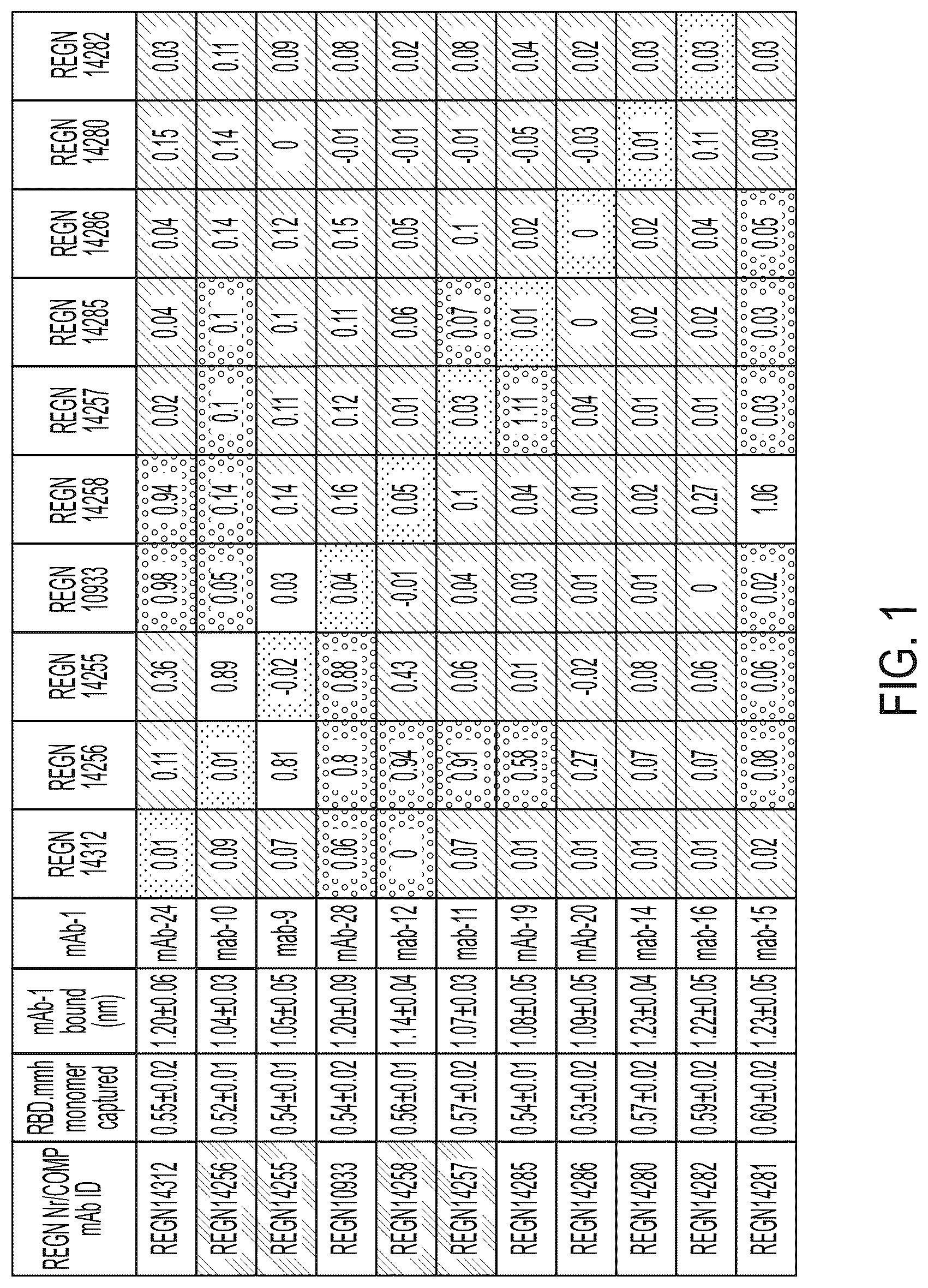
shows cross-competition between 15 anti-SARS-CoV-2-S monoclonal antibodies upon binding to immobilized SARS-Cov-2 RBD-MMH. Red: Pre-bound mAb-1 reduced binding of mAb-2 to SARS-CoV-2 RBD-MMH by greater than 50%, and binding to SARS-CoV-2 RBD-MMH was also reduced by greater than 50% when the binding order of mAb-1 and mAb-2 was reversed. Yellow: Pre-bound mAb-1 reduced binding of mAb-2 to SARS-CoV-2 RBD-MMH by greater than 50%, but binding to SARS-CoV-2 RBD-MMH was reduced by less than 50% when the binding order of mAb-1 and mAb-2 was reversed.
depicts the cryo-EM structure of mAb14256, mAb10987, and the receptor binding domain (RBD) of the SARS-CoV-2 spike glycoprotein, in complex, at 3.9 Å resolution.
shows that mAb14256 binds at the top of the RBD, thereby blocking ACE2 binding. mAb14256 competes with mAb10933 (middle structure) and mAb10985 (not depicted).
depicts the cryo-EM structure of mAb15160, mAb14315, and the receptor binding domain (RBD) of the SARS-CoV-2 spike glycoprotein, in complex, at 3.18 Å resolution.
depicts the cryo-EM structure of antigen-binding fragments of mAb1428 and mAb15160 with the receptor binding domain (RBD) from the Wuhan-Hu-1 strain and BA.1 lineage, in complex, at 3.3 and 3.4 Å resolution, respectively.
DETAILED DESCRIPTION OF THE INVENTION
Before the present methods are described, it is to be understood that this invention is not limited to particular methods, and experimental conditions described, as such methods and conditions may vary. It is also to be understood that the terminology used herein is for the purpose of describing particular embodiments only, and is not intended to be limiting, since the scope of the present invention will be limited only by the appended claims.
Unless defined otherwise, all technical and scientific terms used herein have the same meaning as commonly understood by one of ordinary skill in the art to which this invention belongs. Although any methods and materials similar or equivalent to those described herein can be used in the practice or testing of the present invention, preferred methods and materials are now described. All publications mentioned herein are incorporated herein by reference in their entirety.
The term “coronavirus” or “CoV” refers to any virus of the coronavirus family, including but not limited to SARS-CoV-2, MERS-CoV, and SARS-CoV. SARS-CoV-2 has also been known as 2019-nCoV and Wuhan coronavirus. It binds via the viral spike protein to human host cell receptor angiotensin-converting enzyme 2 (ACE2). The spike protein also binds to and is cleaved by TMPRSS2, which activates the spike protein for membrane fusion of the virus.
The term “CoV-S”, also called “S” or “S protein” refers to the spike protein of a coronavirus, and can refer to specific S proteins such as SARS-CoV-2-S, MERS-CoV S, and SARS-CoV S. The SARS-CoV-2-Spike protein is a 1273 amino acid type I membrane glycoprotein which assembles into trimers that constitute the spikes or peplomers on the surface of the enveloped coronavirus particle. The protein has two essential functions, host receptor binding and membrane fusion, which are attributed to the N-terminal (S1) and C-terminal (S2) halves of the S protein. CoV-S binds to its cognate receptor via a receptor binding domain (RBD) present in the S1 subunit. The amino acid sequence of full-length SARS-CoV-2 spike protein is exemplified by the amino acid sequence provided in SEQ ID NO: 1008. The term “CoV-S” includes protein variants of CoV spike protein isolated from different CoV isolates as well as recombinant CoV spike protein or a fragment thereof. The term also encompasses CoV spike protein or a fragment thereof coupled to, for example, a histidine tag, mouse or human Fc, or a signal sequence such as ROR1.
The term “coronavirus infection” or “CoV infection,” as used herein, refers to infection with a coronavirus such as SARS-CoV-2, MERS-CoV, or SARS-CoV. The term includes coronavirus respiratory tract infections, often in the lower respiratory tract. Symptoms can include high fever, dry cough, shortness of breath, pneumonia, gastro-intestinal symptoms such as diarrhea, organ failure (kidney failure and renal dysfunction), septic shock, and death in severe cases.
Viruses
The present invention includes methods for treating or preventing a viral infection in a subject. The term “virus” includes any virus whose infection in the body of a subject is treatable or preventable by administration of an anti-CoV-S antibody or antigen-binding fragment thereof (e.g., wherein infectivity of the virus is at least partially dependent on CoV-S). In an embodiment of the invention, a “virus” is any virus that expresses spike protein (e.g., CoV-S). The term “virus” also includes a CoV-S-dependent respiratory virus which is a virus that infects the respiratory tissue of a subject (e.g., upper and/or lower respiratory tract, trachea, bronchi, lungs) and is treatable or preventable by administration of an anti-CoV-S antibody or antigen-binding fragment thereof. For example, in an embodiment of the invention, virus includes coronavirus, SARS-CoV-2 (severe acute respiratory syndrome coronavirus 2), SARS-CoV (severe acute respiratory syndrome coronavirus), and MERS-CoV (Middle East respiratory syndrome (MERS) coronavirus). Coronaviruses can include the genera of alphacoronaviruses, betacoronaviruses, gammacoronaviruses, and deltacoronaviruses. In some embodiments, the antibodies or antigen-binding fragments provided herein can bind to and/or neutralize an alphacoronavirus, a betacoronavirus, a gammacoronavirus, and/or a deltacoronavirus. In certain embodiments, this binding and/or neutralization can be specific for a particular genus of coronavirus or for a particular subgroup of a genus. “Viral infection” refers to the invasion and multiplication of a virus in the body of a subject.
Coronavirus virions are spherical with diameters of approximately 125 nm. The most prominent feature of coronaviruses is the club-shape spike projections emanating from the surface of the virion. These spikes are a defining feature of the virion and give them the appearance of a solar corona, prompting the name, coronaviruses. Within the envelope of the virion is the nucleocapsid. Coronaviruses have helically symmetrical nucleocapsids, which is uncommon among positive-sense RNA viruses, but far more common for negative-sense RNA viruses. SARS-CoV-2, MERS-CoV, and SARS-CoV belong to the coronavirus family. The initial attachment of the virion to the host cell is initiated by interactions between the S protein and its receptor. The sites of receptor binding domains (RBD) within the S1 region of a coronavirus S protein vary depending on the virus, with some having the RBD at the C-terminus of S1. The S-protein/receptor interaction is the primary determinant for a coronavirus to infect a host species and also governs the tissue tropism of the virus. Many coronaviruses utilize peptidases as their cellular receptor. Following receptor binding, the virus must next gain access to the host cell cytosol. This is generally accomplished by acid-dependent proteolytic cleavage of S protein by a cathepsin, TMPRRS2 or another protease, followed by fusion of the viral and cellular membranes.
The coronaviruses described herein also include variant coronaviruses, which in some embodiments are classified as “variants of interest” or “variants of concern” by the World Health Organization (WHO). Variant coronaviruses can have mutations in their spike glycoprotein, which may change the virus's properties such as severity of COVID-19 or transmissibility. WHO defines a variant of concern as one that is associated with one or more of the following changes from wild-type, to such a degree that it is a matter of global public health significance: 1) increased transmissibility or detrimental change in COVID-19 epidemiology; 2) increased virulence or changed clinical disease presentation; or 3) decreased effectiveness of public health measures and social measures, including available therapeutics, vaccines, and diagnostics. Variants of interest are defined by WHO as SARS-CoV-2 viruses 1) with genetic changes that are predicted or have been demonstrated to affect virus characteristics including immune escape, therapeutic escape, diagnostic escape, transmissibility, and disease severity; and 2) that have been identified as causing significant community transmission or multiple COVID-19 clusters, in multiple countries and with increasing relative prevalence alongside an increasing number of cases over time, or with other epidemiological impacts that suggest an emerging risk to global public health. As of Dec. 13, 2021, there are five variants of concern as classified by WHO (alpha, beta, gamma, delta, and omicron), and two variants of interest as classified by WHO (lambda and mu). Each variant can be defined by the mutations in its spike glycoprotein as compared to the wild-type spike glycoprotein. For example, the Omicron variant (also classified as B.1.1.529) comprises the following mutations in its spike glycoprotein: A67V, Δ69-70, T95I, G142D/Δ143-145, Δ211/L212I, ins214EPE, G339D, S371L, S373P, S375F, K417N, N440K, G446S, S477N, T478K, E484A, Q493R, G4965, Q498R, N501Y, Y505H, T547K, D614G, H655Y, N679K, P681H, N764K, D796Y, N856K, Q954H, N969K, and L981F (full length omicron spike glycoprotein: SEQ ID NO: 1072). Moreover, these variants may have further lineages that encompass a group of related viruses. For example, as of Jul. 8, 2022, the Omicron variant has BA.1, BA.1.1, BA.2, BA.2.12.1, BA.3, and BA.4/ BA.5 lineages. In some embodiments, the antibodies and antigen-binding fragments thereof described herein can bind to any of these variants and/or lineages. In further embodiments, the antibodies and antigen-binding fragments thereof can neutralize these variants and/or lineages.
Anti-CoV-S Antibodies and Antigen-Binding Fragments
The present invention provides antigen-binding proteins, such as antibodies and antigen-binding fragments thereof, that specifically bind to CoV spike protein or an antigenic fragment thereof.
The term “antibody”, as used herein, refers to immunoglobulin molecules comprising four polypeptide chains, two heavy chains (HCs) and two light chains (LCs) inter-connected by disulfide bonds (i.e., “full antibody molecules”), as well as multimers thereof (e.g. IgM). Exemplary antibodies include, for example, those listed in Table 4. Each heavy chain comprises a heavy chain variable region (“HCVR” or “V H ”) and a heavy chain constant region (comprised of domains C H 1, C H 2 and C H 3). Each light chain is comprised of a light chain variable region (“LCVR or “V L ”) and a light chain constant region (C L ). The V H and V L regions can be further subdivided into regions of hypervariability, termed complementarity determining regions (CDR), interspersed with regions that are more conserved, termed framework regions (FR). Each V H and V L comprises three CDRs and four FRs, arranged from amino-terminus to carboxy-terminus in the following order: FR1, CDR1, FR2, CDR2, FR3, CDR3, FR4. Heavy chain CDRs can also be referred to as HCDRs or CDR-Hs, and numbered as described above (e.g., HCDR1, HCDR2, and HCDR3 or HCDR1, HCDR2, and HCDR3). Likewise, light chain CDRs can be referred to as LCDRs or CDR-Ls, and numbered LCDR1, LCDR2, and LCDR3, or LCDR1, LCDR2, and LCDR3. In certain embodiments of the invention, the FRs of the antibody (or antigen binding fragment thereof) are identical to the human germline sequences, or are naturally or artificially modified. Exemplary human germline sequences include, but are not limited to, VH3-66 and Vk1-33. Thus, the present disclosure provides anti-CoV-S antibodies or antigen-binding fragments thereof (e.g., anti-SARS-CoV-2-S antibodies or antigen-binding fragments thereof) comprising HCDR and LCDR sequences of Table 4 within a VH3-66 or Vk1-33 variable heavy chain or light chain region. The present disclosure further provides anti-CoV-S antibodies or antigen-binding fragments thereof (e.g., anti-SARS-CoV-2-S antibodies or antigen-binding fragments thereof) comprising HCDR and LCDR sequences of Table 4 within a combination of a light chain selected from IgKV4-1, IgKV1-5, IgKV1-9, IgKV1-12, IgKV3-15, IgKV1-16, IgKV1-17, IgKV3-20, IgLV3-21, IgKV2-24, IgKV1-33, IgKV1-39, IgLV1-40, IgLV1-44, IgLV1-51, IgLV3-1, IgKV1-6, IgLV2-8, IgKV3-11, IgLV2-11, IgLV2-14, IgLV2-23, or IgLV6-57, and a heavy chain selected from IgHV1-69, IgHV3-64, IgHV4-59, IgHV3-53, IgHV3-48, IgHV4-34, IgHV3-33, IgHV3-30, IgHV3-23, IgHV3-20, IgHV1-18, IgHV3-15, IgHV3-11, IgHV3-9, IgHV1-8, IgHV3-7, IgHV2-5, IgHV1-2, IgHV2-70, IgHV3-66, IgHV5-51, IgHV1-46, IgHV4-39, IgHV4-31, IgHV3-30-3, IgHV2-26, or IgHV7-4-1. The present disclosure further provides anti-CoV-S antibodies or antigen-binding fragments thereof (e.g., anti-SARS-CoV-2-S antibodies or antigen-binding fragments thereof) comprising HCVR and LCVR sequences of Table 4 within a combination of a light chain selected from IgKV4-1, IgKV1-5, IgKV1-9, IgKV1-12, IgKV3-15, IgKV1-16, IgKV1-17, IgKV3-20, IgLV3-21, IgKV2-24, IgKV1-33, IgKV1-39, IgLV1-40, IgLV1-44, IgLV1-51, IgLV3-1, IgKV1-6, IgLV2-8, IgKV3-11, IgLV2-11, IgLV2-14, IgLV2-23, or IgLV6-57, and a heavy chain selected from IgHV1-69, IgHV3-64, IgHV4-59, IgHV3-53, IgHV3-48, IgHV4-34, IgHV3-33, IgHV3-30, IgHV3-23, IgHV3-20, IgHV1-18, IgHV3-15, IgHV3-11, IgHV3-9, IgHV1-8, IgHV3-7, IgHV2-5, IgHV1-2, IgHV2-70, IgHV3-66, IgHV5-51, IgHV1-46, IgHV4-39, IgHV4-31, IgHV3-30-3, IgHV2-26, or IgHV7-4-1.
Typically, the variable domains of both the heavy and light immunoglobulin chains comprise three hypervariable regions, also called complementarity determining regions (CDRs), located within relatively conserved framework regions (FR). In general, from N-terminal to C-terminal, both light and heavy chains variable domains comprise FR1, CDR1, FR2, CDR2, FR3, CDR3 and FR4. In an embodiment of the invention, the assignment of amino acids to each domain is in accordance with the definitions of Sequences of Proteins of Immunological Interest, Kabat, et al.; National Institutes of Health, Bethesda, Md.; 5 th ed.; NIH Publ. No. 91-3242 (1991); Kabat (1978) Adv. Prot. Chem. 32:1-75; Kabat, et al., (1977) J. Biol. Chem. 252:6609-6616; Chothia, et al., (1987) J Mol. Biol. 196:901-917 or Chothia, et al., (1989) Nature 342:878-883.
The present invention includes monoclonal anti-CoV-S antigen-binding proteins, e.g., antibodies and antigen-binding fragments thereof, as well as monoclonal compositions comprising a plurality of isolated monoclonal antigen-binding proteins. The term “monoclonal antibody”, as used herein, refers to a population of substantially homogeneous antibodies, i.e., the antibody molecules comprising the population are identical in amino acid sequence except for possible naturally occurring mutations that may be present in minor amounts. A “plurality” of such monoclonal antibodies and fragments in a composition refers to a concentration of identical (i.e., as discussed above, in amino acid sequence except for possible naturally occurring mutations that may be present in minor amounts) antibodies and fragments which is above that which would normally occur in nature, e.g., in the blood of a host organism such as a mouse or a human.
In an embodiment of the invention, an anti-CoV-S antigen-binding protein, e.g., antibody or antigen-binding fragment comprises a heavy chain constant domain, e.g., of the type IgA (e.g., IgA1 or IgA2), IgD, IgE, IgG (e.g., IgG1, IgG2, IgG3 and IgG4) or IgM. In an embodiment of the invention, an antigen-binding protein, e.g., antibody or antigen-binding fragment comprises a light chain constant domain, e.g., of the type kappa or lambda.
The term “human” antigen-binding protein, such as an antibody, as used herein, includes antibodies having variable and constant regions derived from human germline immunoglobulin sequences whether in a human cell or grafted into a non-human cell, e.g., a mouse cell. See e.g., U.S. Pat. Nos. 8,502,018, 6,596,541 or 5,789,215. The human mAbs of the invention may include amino acid residues not encoded by human germline immunoglobulin sequences (e.g., mutations introduced by random or site-specific mutagenesis in vitro or by somatic mutation in vivo), for example in the CDRs and, in particular, CDR3. However, the term “human antibody”, as used herein, is not intended to include mAbs in which CDR sequences derived from the germline of another mammalian species (e.g., mouse) have been grafted onto human FR sequences. The term includes antibodies recombinantly produced in a non-human mammal or in cells of a non-human mammal. The term is not intended to include antibodies isolated from or generated in a human subject. See below.
The present invention includes anti-CoV-S chimeric antigen-binding proteins, e.g., antibodies and antigen-binding fragments thereof, and methods of use thereof. As used herein, a “chimeric antibody” is an antibody having the variable domain from a first antibody and the constant domain from a second antibody, where the first and second antibodies are from different species. (U.S. Pat. No. 4,816,567; and Morrison et al., (1984) Proc. Natl. Acad. Sci. USA 81: 6851-6855).
The present invention includes anti-CoV-S hybrid antigen-binding proteins, e.g., antibodies and antigen-binding fragments thereof, and methods of use thereof. As used herein, a “hybrid antibody” is an antibody having the variable domain from a first antibody and the constant domain from a second antibody, wherein the first and second antibodies are from different animals, or wherein the variable domain, but not the constant region, is from a first animal. For example, a variable domain can be taken from an antibody isolated from a human and expressed with a fixed constant region not isolated from that antibody. Exemplary hybrid antibodies are described in Example 1, which refers to antibody heavy chain variable region and light chain variable region derived PCR products that were cloned into expression vectors containing a heavy constant region and a light constant region, respectively. Hybrid antibodies are synthetic and non-naturally occurring because the variable and constant regions they contain are not isolated from a single natural source.
The term “recombinant” antigen-binding proteins, such as antibodies or antigen-binding fragments thereof, refers to such molecules created, expressed, isolated or obtained by technologies or methods known in the art as recombinant DNA technology which include, e.g., DNA splicing and transgenic expression. The term includes antibodies expressed in a non-human mammal (including transgenic non-human mammals, e.g., transgenic mice), or a cell (e.g., CHO cells) expression system, or a non-human cell expression system, or isolated from a recombinant combinatorial human antibody library. In some embodiments, a recombinant antibody shares a sequence with an antibody isolated from an organism (e.g., a mouse or a human), but has been expressed via recombinant DNA technology. Such antibodies may have post-translational modifications (e.g., glycosylation) that differ from the antibody as isolated from the organism.
In some embodiments, the antibodies disclosed herein lack fucose in its constant region glycosylation. Methods of measuring fucose in an antibody composition have been described in the art, e.g., U.S. Pat. No. 8,409,838 (Regeneron Pharmaceuticals), incorporated herein by reference. In some embodiments, fucose is undetectable in a composition comprising a population of antibody molecules. In some embodiments, an antibody lacking fucose has enhanced ADCC activity.
In some embodiments, antibodies that lack fucose can be produced using cell lines that are deficient in their ability to fucosylate proteins, i.e., the ability to fucosylate proteins is reduced or eliminated. Fucosylation of glycans requires synthesis of GDP-fucose via the de novo pathway or the salvage pathway, both of which involve sequential function of several enzymes, leading to addition of a fucose molecule to the first N-acetylglucosamine (GlcNAc) moiety of the reducing end of a glycan. The two key enzymes of the de novo pathway responsible for production of GDP-fucose are GDP-D-mannose-4,6-dehydratase (GMD) and GDP-keto-6-deoxymannose-3,5-epimerase,4-reductase (FX). In the absence of fucose, these two de novo pathway enzymes (GMD and FX) convert mannose and/or glucose to GDP-fucose which is then transported into the Golgi complex where nine fucosyl-transferases (FUT1-9) act in concert to fucosylate the first GlcNAc molecule of a glycan. In the presence of fucose, however, the salvage pathway enzymes, fucose-kinase and GDP-fucose pyrophosphorylase, convert fucose into GDP-fucose.
Cell lines that are deficient in their ability to fucosylate proteins have been described in the art. In some embodiments, a cell line deficient in its ability to fucosylate proteins is a mammalian cell line (e.g., CHO cell lines, such as CHO K1, DXB-11 CHO, Veggie-CHO) comprising a mutation or genetic modification in one or more of endogenous FUT1 to 9 genes resulting in a lack of one or more functional fucosyl-transferases. In some embodiments, the mammalian cell line comprises a mutation in an endogenous FUT8 gene (e.g., a FUT8 knock-out cell line in which the FUT8 gene has been disrupted resulting in a lack of a functional □1,6-fucosyltransferase in the cell line, as described in U.S. Pat. No. 7,214,775 (Kyowa Hakko Kogyo Co., Ltd.) and U.S. Pat. No. 7,737,725 (Kyowa Hakko Kirin Co., Ltd), incorporated herein by reference. In some embodiments, the mammalian cell line comprises a mutation or genetic modification in an endogenous GMD gene resulting in a lack of a functional GMD in the cell line, e.g., a GMD knock-out cell line in which the GMD gene has been disrupted, described in e.g., U.S. Pat. No. 7,737,725 (Kyowa Hakko Kirin Co., Ltd), incorporated herein by reference. In some embodiments, the mammalian cell line comprises a mutation or genetic modification in an endogenous Fx gene resulting in a lack of a functional Fx protein. In some embodiments, the mammalian cell line is an Fx knock-out cell line in which the endogenous Fx gene has been disrupted (see, e.g., U.S. Pat. No. 7,737,725 (Kyowa Hakko Kirin Co., Ltd), incorporated herein by reference). In some embodiments, the mammalian cell line comprises a mutation in an endogenous Fx mutation that confers temperature sensitive phenotypes (as described in, e.g., U.S. Pat. No. 8,409,838 (Regeneron Pharmaceuticals), incorporated herein by reference). In some embodiments, the mammalian cell line deficient in its ability to fucosylate proteins is a cell line that has been selected based on resistance to certain lectins, e.g., the Lens culinaris lectin. See, e.g., U.S. Pat. No. 8,409,838 (Regeneron Pharmaceuticals), incorporated herein by reference.
Recombinant anti-CoV-S antigen-binding proteins, e.g., antibodies and antigen-binding fragments, disclosed herein may also be produced in an E. coli / T7 expression system. In this embodiment, nucleic acids encoding the anti-CoV-S antibody immunoglobulin molecules of the invention (e.g., as found in Table 4) may be inserted into a pET-based plasmid and expressed in the E. coli /T7 system. For example, the present invention includes methods for expressing an antibody or antigen-binding fragment thereof or immunoglobulin chain thereof in a host cell (e.g., bacterial host cell such as E. coli such as BL21 or BL21DE3) comprising expressing T7 RNA polymerase in the cell which also includes a polynucleotide encoding an immunoglobulin chain that is operably linked to a T7 promoter. For example, in an embodiment of the invention, a bacterial host cell, such as an E. coli , includes a polynucleotide encoding the T7 RNA polymerase gene operably linked to a lac promoter and expression of the polymerase and the chain is induced by incubation of the host cell with IPTG (isopropyl-beta-D-thiogalactopyranoside). See U.S. Pat. Nos. 4,952,496 and 5,693,489 or Studier & Moffatt, Use of bacteriophage T7 RNA polymerase to direct selective high-level expression of cloned genes, J. Mol. Biol. 1986 May 5; 189(1): 113-30.
There are several methods by which to produce recombinant antibodies which are known in the art. One example of a method for recombinant production of antibodies is disclosed in U.S. Pat. No. 4,816,567.
Transformation can be by any known method for introducing polynucleotides (e.g., DNA or RNA, including mRNA) into a host cell. Methods for introduction of heterologous polynucleotides into mammalian cells are well known in the art and include dextran-mediated transfection, calcium phosphate precipitation, polybrene-mediated transfection, protoplast fusion, electroporation, encapsulation of the polynucleotide(s) in liposomes, lipid nanoparticle technology, biolistic injection and direct microinjection of the DNA into nuclei. In addition, nucleic acid molecules may be introduced into mammalian cells by viral vectors such as lentivirus or adeno-associated virus. Methods of transforming cells are well known in the art. See, for example, U.S. Pat. Nos. 4,399,216; 4,912,040; 4,740,461 and 4,959,455. In some embodiments, an antibody or antigen-binding fragment thereof of the present disclosure can be introduced to a subject in nucleic acid form (e.g., DNA or RNA, including mRNA), such that the subject's own cells produce the antibody. The present disclosure further provides modifications to nucleotide sequences encoding the anti-CoV-S antibodies described herein that result in increased antibody expression, increased antibody stability, increased nucleic acid (e.g., mRNA) stability, or improved affinity or specificity of the antibodies for the CoV spike protein.
Thus, the present invention includes recombinant methods for making an anti-CoV-S antigen-binding protein, such as an antibody or antigen-binding fragment thereof of the present invention, or an immunoglobulin chain thereof, comprising (i) introducing one or more polynucleotides (e.g., including the nucleotide sequence of any one or more of the sequences of Table 5) encoding light and/or heavy immunoglobulin chains, or CDRs, of the antigen-binding protein, e.g., of Table 4, for example, wherein the polynucleotide is in a vector; and/or integrated into a host cell chromosome and/or is operably linked to a promoter; (ii) culturing the host cell (e.g., CHO or Pichia or Pichia pastoris ) under condition favorable to expression of the polynucleotide and, (iii) optionally, isolating the antigen-binding protein, (e.g., antibody or fragment) or chain from the host cell and/or medium in which the host cell is grown. For example, a polynucleotide can be integrated into a host cell chromosome through targeted insertion with a vector such as adeno-associated virus (AAV), e.g., after cleavage of the chromosome using a gene editing system (e.g., CRISPR (for example, CRISPR-Cas9), TALEN, megaTAL, zinc finger, or Argonaute). Targeted insertions can take place, for example, at host cell loci such as an albumin or immunoglobulin genomic locus. Alternatively, insertion can be at a random locus, e.g., using a vector such as lentivirus. When making an antigen-binding protein (e.g., antibody or antigen-binding fragment) comprising more than one immunoglobulin chain, e.g., an antibody that comprises two heavy immunoglobulin chains and two light immunoglobulin chains, co-expression of the chains in a single host cell leads to association of the chains, e.g., in the cell or on the cell surface or outside the cell if such chains are secreted, so as to form the antigen-binding protein (e.g., antibody or antigen-binding fragment). The methods include those wherein only a heavy immunoglobulin chain or only a light immunoglobulin chain (e.g., any of those discussed herein including mature fragments and/or variable domains thereof) is expressed. Such chains are useful, for example, as intermediates in the expression of an antibody or antigen-binding fragment that includes such a chain. For example, the present invention also includes anti-CoV-S antigen-binding proteins, such as antibodies and antigen-binding fragments thereof, comprising a heavy chain immunoglobulin (or variable domain thereof or comprising the CDRs thereof) encoded by a polynucleotide comprising a nucleotide sequence set forth in Table 5 and a light chain immunoglobulin (or variable domain thereof or comprising the CDRs thereof) encoded by a nucleotide sequence set forth in Table 5 which are the product of such production methods, and, optionally, the purification methods set forth herein. For example, in some embodiments, the product of the method is an anti-CoV-S antigen-binding protein which is an antibody or fragment comprising an HCVR comprising an amino acid sequence set forth in Table 4 and an LCVR comprising an amino acid sequence set forth in Table 4, wherein the HCVR and LCVR sequences are selected from a single antibody listed in Table 4. In some embodiments, the product of the method is an anti-CoV-S antigen-binding protein which is an antibody or fragment comprising HCDR1, HCDR2, and HCDR3 comprising amino acid sequences set forth in Table 4 and LCDR1, LCDR2, and LCDR3 comprising amino acid sequences set forth in Table 4, wherein the six CDR sequences are selected from a single antibody listed in Table 4. In some embodiments, the product of the method is an anti-CoV-S antigen-binding protein which is an antibody or fragment comprising a heavy chain comprising an HC amino acid sequence set forth in Table 4 and a light chain comprising an LC amino acid sequence set forth in Table 4.
Eukaryotic and prokaryotic host cells, including mammalian cells, may be used as hosts for expression of an anti-CoV-S antigen-binding protein. Such host cells are well known in the art and many are available from the American Type Culture Collection (ATCC). These host cells include, inter alia, Chinese hamster ovary (CHO) cells, NS0, SP2 cells, HeLa cells, baby hamster kidney (BHK) cells, monkey kidney cells (COS), human hepatocellular carcinoma cells (e.g., Hep G2), A549 cells, 3T3 cells, HEK-293 cells and a number of other cell lines. Mammalian host cells include human, mouse, rat, dog, monkey, pig, goat, bovine, horse and hamster cells. Other cell lines that may be used are insect cell lines (e.g., Spodoptera frugiperda or Trichoplusia ni ), amphibian cells, bacterial cells, plant cells and fungal cells. Fungal cells include yeast and filamentous fungus cells including, for example, Pichia pastoris, Pichia finlandica, Pichia trehalophila, Pichia koclamae, Pichia membranaefaciens, Pichia minuta ( Ogataea minuta, Pichia lindneri ), Pichia opuntiae, Pichia thermotolerans, Pichia salictaria, Pichia guercuum, Pichia pijperi, Pichia stiptis, Pichia methanolica, Pichia sp., Saccharomyces cerevisiae, Saccharomyces sp., Hansenula polymorpha, Kluyveromyces sp., Kluyveromyces lactis, Candida albicans, Aspergillus nidulans, Aspergillus niger, Aspergillus oryzae, Trichoderma reesei, Chrysosporium lucknowense, Fusarium sp., Fusarium gramineum, Fusarium venenatum, Physcomitrella patens and Neurospora crassa . The present invention includes an isolated host cell (e.g., a CHO cell) comprising an antigen-binding protein, such as those of Table 4; or a polynucleotide encoding such a polypeptide thereof.
Polynucleotides, as discussed herein, may encode all or a portion of an antibody or antigen-binding fragment as discussed throughout the present disclosure. In some cases, a single polynucleotide may encode both a HCVR and a LCVR (e.g., defined with reference to the CDRs contained within the respective amino acid sequence-defined HCVR and LCVR, defined with reference to the amino acid sequences of the CDRs of the HCVR and LCVR. respectively, or defined with reference to the amino acid sequences of the HCVR and LCVR, respectively) of an antibody or antigen-binding fragment, or the HCVR and LCVR may be encoded by separate polynucleotides (i.e., a pair of polynucleotides). In the latter case, in which the HCVR and LCVR are encoded by separate polynucleotides, the polynucleotides may be combined in a single vector or may be contained in separate vectors (i.e., a pair of vectors). In any case, a host cell used to express the polynucleotide(s) or vector(s) may contain the full complement of component parts to generate the antibody or antigen-binding fragment thereof. For example, a host cell may comprise separate vectors, each encoding a HCVR and a LCVR, respectively, of an antibody or antigen-binding fragment thereof as discussed above or herein. Similarly, the polynucleotide or polynucleotides, and the vector or vectors, may be used to express the full-length heavy chain and full-length light chain of an antibody as discussed above or herein. For example, a host cell may comprise a single vector with polynucleotides encoding both a heavy chain and a light chain of an antibody, or the host cell may comprise separate vectors with polynucleotides encoding, respectively, a heavy chain and a light chain of an antibody as discussed above or herein.
In an exemplary embodiment, the present disclosure provides a polynucleotide (or a vector comprising the polynucleotide, or a host cell comprising the polynucleotide or the vector) encoding a HCVR comprising the HCDRs of SEQ ID NOs: 214, 216 and 218 (e.g., in an embodiment, the polynucleotide comprises SEQ ID NOs: 213, 215 and 217). In an exemplary embodiment, the present disclosure provides a polynucleotide (or a vector comprising the polynucleotide, or a host cell comprising the polynucleotide or the vector) encoding a LCVR comprising the LCDRs of SEQ ID NOs: 222, 126 and 224 (e.g., in an embodiment, the polynucleotide comprises SEQ ID NOs: 221, 125 and 223). In an exemplary embodiment, the present disclosure provides a polynucleotide (or a vector comprising the polynucleotide, or a host cell comprising the polynucleotide or the vector) encoding a HCVR comprising the amino acid sequence of SEQ ID NO: 212 (e.g., in an embodiment, the polynucleotide comprises SEQ ID NO: 211). In an exemplary embodiment, the present disclosure provides a polynucleotide (or a vector comprising the polynucleotide, or a host cell comprising the polynucleotide or the vector) encoding a LCVR comprising the amino acid sequence of SEQ ID NO: 220 (e.g., in an embodiment, the polynucleotide comprises SEQ ID NO: 219). In an exemplary embodiment, the present disclosure provides a polynucleotide (or a vector comprising the polynucleotide, or a host cell comprising the polynucleotide or the vector) encoding a heavy chain (HC) comprising the amino acid sequence of SEQ ID NO: 226 (e.g., in an embodiment, the polynucleotide comprises SEQ ID NO: 225). In an exemplary embodiment, the present disclosure provides a polynucleotide (or a vector comprising the polynucleotide, or a host cell comprising the polynucleotide or the vector) encoding a light chain (LC) comprising the amino acid sequence of SEQ ID NO: 228 (e.g., in an embodiment, the polynucleotide comprises SEQ ID NO: 227). In an embodiment, the present disclosure provides a pair of polynucleotides or a pair of vectors encoding, respectively, a HCVR and a LCVR, or a HC and a LC, as discussed in this paragraph, as well as a host cell containing the pair or polynucleotides and/or the pair of vectors to encode the antibody designated mAb14315 or an antigen-binding fragment thereof.
In an exemplary embodiment, the present disclosure provides a polynucleotide (or a vector comprising the polynucleotide, or a host cell comprising the polynucleotide or the vector) encoding a HCVR comprising the HCDRs of SEQ ID NOs: 364, 366 and 368 (e.g., in an embodiment, the polynucleotide comprises SEQ ID NOs: 363, 365 and 367). In an exemplary embodiment, the present disclosure provides a polynucleotide (or a vector comprising the polynucleotide, or a host cell comprising the polynucleotide or the vector) encoding a LCVR comprising the LCDRs of SEQ ID NOs: 372, 106 and 374 (e.g., in an embodiment, the polynucleotide comprises SEQ ID NOs: 371, 105 and 373). In an exemplary embodiment, the present disclosure provides a polynucleotide (or a vector comprising the polynucleotide, or a host cell comprising the polynucleotide or the vector) encoding a HCVR comprising the amino acid sequence of SEQ ID NO: 362 (e.g., in an embodiment, the polynucleotide comprises SEQ ID NO: 361). In an exemplary embodiment, the present disclosure provides a polynucleotide (or a vector comprising the polynucleotide, or a host cell comprising the polynucleotide or the vector) encoding a LCVR comprising the amino acid sequence of SEQ ID NO: 370 (e.g., in an embodiment, the polynucleotide comprises SEQ ID NO: 369). In an exemplary embodiment, the present disclosure provides a polynucleotide (or a vector comprising the polynucleotide, or a host cell comprising the polynucleotide or the vector) encoding a heavy chain (HC) comprising the amino acid sequence of SEQ ID NO: 376 (e.g., in an embodiment, the polynucleotide comprises SEQ ID NO: 375). In an exemplary embodiment, the present disclosure provides a polynucleotide (or a vector comprising the polynucleotide, or a host cell comprising the polynucleotide or the vector) encoding a light chain (LC) comprising the amino acid sequence of SEQ ID NO: 378 (e.g., in an embodiment, the polynucleotide comprises SEQ ID NO: 377). In an embodiment, the present disclosure provides a pair of polynucleotides or a pair of vectors encoding, respectively, a HCVR and a LCVR, or a HC and a LC, as discussed in this paragraph, as well as a host cell containing the pair or polynucleotides and/or the pair of vectors to encode the antibody designated mAb15160 or an antigen-binding fragment thereof.
In an exemplary embodiment, the present disclosure provides a polynucleotide (or a vector comprising the polynucleotide, or a host cell comprising the polynucleotide or the vector) encoding a HCVR comprising the HCDRs of SEQ ID NOs: 495, 497 and 499 (e.g., in an embodiment, the polynucleotide comprises SEQ ID NOs: 494, 496 and 498). In an exemplary embodiment, the present disclosure provides a polynucleotide (or a vector comprising the polynucleotide, or a host cell comprising the polynucleotide or the vector) encoding a LCVR comprising the LCDRs of SEQ ID NOs: 503, 505 and 507 (e.g., in an embodiment, the polynucleotide comprises SEQ ID NOs: 502, 504 and 506). In an exemplary embodiment, the present disclosure provides a polynucleotide (or a vector comprising the polynucleotide, or a host cell comprising the polynucleotide or the vector) encoding a HCVR comprising the amino acid sequence of SEQ ID NO: 493 (e.g., in an embodiment, the polynucleotide comprises SEQ ID NO: 492). In an exemplary embodiment, the present disclosure provides a polynucleotide (or a vector comprising the polynucleotide, or a host cell comprising the polynucleotide or the vector) encoding a LCVR comprising the amino acid sequence of SEQ ID NO: 501 (e.g., in an embodiment, the polynucleotide comprises SEQ ID NO: 500). In an exemplary embodiment, the present disclosure provides a polynucleotide (or a vector comprising the polynucleotide, or a host cell comprising the polynucleotide or the vector) encoding a heavy chain (HC) comprising the amino acid sequence of SEQ ID NO: 509 (e.g., in an embodiment, the polynucleotide comprises SEQ ID NO: 508). In an exemplary embodiment, the present disclosure provides a polynucleotide (or a vector comprising the polynucleotide, or a host cell comprising the polynucleotide or the vector) encoding a light chain (LC) comprising the amino acid sequence of SEQ ID NO: 511 (e.g., in an embodiment, the polynucleotide comprises SEQ ID NO: 510). In an embodiment, the present disclosure provides a pair of polynucleotides or a pair of vectors encoding, respectively, a HCVR and a LCVR, or a HC and a LC, as discussed in this paragraph, as well as a host cell containing the pair or polynucleotides and/or the pair of vectors to encode the antibody designated mAb14284 or an antigen-binding fragment thereof.
In an exemplary embodiment, the present disclosure provides a polynucleotide (or a vector comprising the polynucleotide, or a host cell comprising the polynucleotide or the vector) encoding a HCVR comprising the HCDRs of SEQ ID NOs: 889, 891 and 893 (e.g., in an embodiment, the polynucleotide comprises SEQ ID NOs: 888, 890 and 892). In an exemplary embodiment, the present disclosure provides a polynucleotide (or a vector comprising the polynucleotide, or a host cell comprising the polynucleotide or the vector) encoding a LCVR comprising the LCDRs of SEQ ID NOs: 897, 164 and 899 (e.g., in an embodiment, the polynucleotide comprises SEQ ID NOs: 896, 163 and 898). In an exemplary embodiment, the present disclosure provides a polynucleotide (or a vector comprising the polynucleotide, or a host cell comprising the polynucleotide or the vector) encoding a HCVR comprising the amino acid sequence of SEQ ID NO: 887 (e.g., in an embodiment, the polynucleotide comprises SEQ ID NO: 886). In an exemplary embodiment, the present disclosure provides a polynucleotide (or a vector comprising the polynucleotide, or a host cell comprising the polynucleotide or the vector) encoding a LCVR comprising the amino acid sequence of SEQ ID NO: 895 (e.g., in an embodiment, the polynucleotide comprises SEQ ID NO: 894). In an exemplary embodiment, the present disclosure provides a polynucleotide (or a vector comprising the polynucleotide, or a host cell comprising the polynucleotide or the vector) encoding a heavy chain (HC) comprising the amino acid sequence of SEQ ID NO: 901 (e.g., in an embodiment, the polynucleotide comprises SEQ ID NO: 900). In an exemplary embodiment, the present disclosure provides a polynucleotide (or a vector comprising the polynucleotide, or a host cell comprising the polynucleotide or the vector) encoding a light chain (LC) comprising the amino acid sequence of SEQ ID NO: 903 (e.g., in an embodiment, the polynucleotide comprises SEQ ID NO: 902). In an embodiment, the present disclosure provides a pair of polynucleotides or a pair of vectors encoding, respectively, a HCVR and a LCVR, or a HC and a LC, as discussed in this paragraph, as well as a host cell containing the pair or polynucleotides and/or the pair of vectors to encode the antibody designated mAb14256 or an antigen-binding fragment thereof.
In an exemplary embodiment, the present disclosure provides a polynucleotide (or a vector comprising the polynucleotide, or a host cell comprising the polynucleotide or the vector) encoding a HCVR comprising the HCDRs of SEQ ID NOs: 495, 497 and 499 (e.g., in an embodiment, the polynucleotide comprises SEQ ID NOs: 494, 496 and 498). In an exemplary embodiment, the present disclosure provides a polynucleotide (or a vector comprising the polynucleotide, or a host cell comprising the polynucleotide or the vector) encoding a LCVR comprising the LCDRs of SEQ ID NOs: 503, 505 and 507 (e.g., in an embodiment, the polynucleotide comprises SEQ ID NOs: 502, 504 and 506). In an exemplary embodiment, the present disclosure provides a polynucleotide (or a vector comprising the polynucleotide, or a host cell comprising the polynucleotide or the vector) encoding a HCVR comprising the amino acid sequence of SEQ ID NO: 493 (e.g., in an embodiment, the polynucleotide comprises SEQ ID NO: 492). In an exemplary embodiment, the present disclosure provides a polynucleotide (or a vector comprising the polynucleotide, or a host cell comprising the polynucleotide or the vector) encoding a LCVR comprising the amino acid sequence of SEQ ID NO: 501 (e.g., in an embodiment, the polynucleotide comprises SEQ ID NO: 500). In an exemplary embodiment, the present disclosure provides a polynucleotide (or a vector comprising the polynucleotide, or a host cell comprising the polynucleotide or the vector) encoding a heavy chain (HC) comprising the amino acid sequence of SEQ ID NO: 1075 (e.g., in an embodiment, the polynucleotide comprises SEQ ID NO: 1074). In an exemplary embodiment, the present disclosure provides a polynucleotide (or a vector comprising the polynucleotide, or a host cell comprising the polynucleotide or the vector) encoding a light chain (LC) comprising the amino acid sequence of SEQ ID NO: 511 (e.g., in an embodiment, the polynucleotide comprises SEQ ID NO: 510). In an embodiment, the present disclosure provides a pair of polynucleotides or a pair of vectors encoding, respectively, a HCVR and a LCVR, or a HC and a LC, as discussed in this paragraph, as well as a host cell containing the pair or polynucleotides and/or the pair of vectors to encode the antibody designated mAb17090 or an antigen-binding fragment thereof.
In an exemplary embodiment, the present disclosure provides a polynucleotide (or a vector comprising the polynucleotide, or a host cell comprising the polynucleotide or the vector) encoding a HCVR comprising the HCDRs of SEQ ID NOs: 364, 366 and 368 (e.g., in an embodiment, the polynucleotide comprises SEQ ID NOs: 363, 365 and 367). In an exemplary embodiment, the present disclosure provides a polynucleotide (or a vector comprising the polynucleotide, or a host cell comprising the polynucleotide or the vector) encoding a LCVR comprising the LCDRs of SEQ ID NOs: 372, 106 and 374 (e.g., in an embodiment, the polynucleotide comprises SEQ ID NOs: 371, 105 and 373). In an exemplary embodiment, the present disclosure provides a polynucleotide (or a vector comprising the polynucleotide, or a host cell comprising the polynucleotide or the vector) encoding a HCVR comprising the amino acid sequence of SEQ ID NO: 362 (e.g., in an embodiment, the polynucleotide comprises SEQ ID NO: 361). In an exemplary embodiment, the present disclosure provides a polynucleotide (or a vector comprising the polynucleotide, or a host cell comprising the polynucleotide or the vector) encoding a LCVR comprising the amino acid sequence of SEQ ID NO: 370 (e.g., in an embodiment, the polynucleotide comprises SEQ ID NO: 369). In an exemplary embodiment, the present disclosure provides a polynucleotide (or a vector comprising the polynucleotide, or a host cell comprising the polynucleotide or the vector) encoding a heavy chain (HC) comprising the amino acid sequence of SEQ ID NO: 1077 (e.g., in an embodiment, the polynucleotide comprises SEQ ID NO: 1076). In an exemplary embodiment, the present disclosure provides a polynucleotide (or a vector comprising the polynucleotide, or a host cell comprising the polynucleotide or the vector) encoding a light chain (LC) comprising the amino acid sequence of SEQ ID NO: 378 (e.g., in an embodiment, the polynucleotide comprises SEQ ID NO: 377). In an embodiment, the present disclosure provides a pair of polynucleotides or a pair of vectors encoding, respectively, a HCVR and a LCVR, or a HC and a LC, as discussed in this paragraph, as well as a host cell containing the pair or polynucleotides and/or the pair of vectors to encode the antibody designated mAb15160_2 or an antigen-binding fragment thereof.
The term “specifically binds” refers to those antigen-binding proteins (e.g., mAbs) having a binding affinity to an antigen, such as a CoV-S protein (e.g., SARS-CoV-2-S), expressed as K D , of at least about 10 −8 M, as measured by real-time, label free bio-layer interferometry assay, for example, at 25° C. or 37° C., e.g., an Octet® HTX biosensor, or by surface plasmon resonance, e.g., BIACORE™, or by solution-affinity ELISA. The present invention includes antigen-binding proteins that specifically bind to a CoV-S protein.
The terms “antigen-binding portion” or “antigen-binding fragment” of an antibody or antigen-binding protein, and the like, as used herein, include any naturally occurring, enzymatically obtainable, synthetic, or genetically engineered polypeptide or glycoprotein that specifically binds an antigen to form a complex. Non-limiting examples of antigen-binding fragments include: (i) Fab fragments; (ii) F(ab) 2 fragments; (iii) Fd fragments; (iv) Fv fragments; (v) single-chain Fv (scFv) molecules; (vi) dAb fragments; and (vii) minimal recognition units consisting of the amino acid residues that mimic the hypervariable region of an antibody (e.g., an isolated complementarity determining region (CDR) such as a CDR3 peptide), or a constrained FR3-CDR3-FR4 peptide. Other engineered molecules, such as domain-specific antibodies, single domain antibodies, domain-deleted antibodies, chimeric antibodies, CDR-grafted antibodies, diabodies, triabodies, tetrabodies, minibodies, nanobodies (e.g., as defined in WO08/020079 or WO09/138519) (e.g., monovalent nanobodies, bivalent nanobodies, etc.), small modular immunopharmaceuticals (SMIPs), and shark variable IgNAR domains, are also encompassed within the expression “antigen-binding fragment,” as used herein. In an embodiment of the invention, the antigen-binding fragment comprises three or more CDRs of an antibody of Table 4 (e.g., HCDR1, HCDR2 and HCDR3; or LCDR1, LCDR2 and LCDR3).
An antigen-binding fragment of an antibody will, in an embodiment of the invention, comprise at least one variable domain. The variable domain may be of any size or amino acid composition and will generally comprise at least one CDR, which is adjacent to or in frame with one or more framework sequences. In antigen-binding fragments having a V H domain associated with a V L domain, the V H and V L domains may be situated relative to one another in any suitable arrangement. For example, the variable region may be dimeric and contain V H -V H , V H -V L or V L -V L dimers. Alternatively, the antigen-binding fragment of an antibody may contain a monomeric V H or V L domain.
In certain embodiments, an antigen-binding fragment of an antibody may contain at least one variable domain covalently linked to at least one constant domain. Non-limiting, exemplary configurations of variable and constant domains that may be found within an antigen-binding fragment of an antibody of the present invention include: (i) V H -C H 1; (ii) V H -C H 2; (iii) V H -C H 3; (iv) V H -C H 1-C H 2; (v) V H -C H 1-C H 2-C H 3; (vi) V H -C H 2-C H 3; (vii) V H -C L ; (viii) V L -C H 1; (ix) V L -C H 2; (x) V L -C H 3; (xi) V L -C H 1-C H 2; (xii) V L -C H 1-C H 2-C H 3; (xiii) V L -C H 2-C H 3; and (xiv) V L -C L . In any configuration of variable and constant domains, including any of the exemplary configurations listed above, the variable and constant domains may be either directly linked to one another or may be linked by a full or partial hinge or linker region. A hinge region may consist of at least 2 (e.g., 5, 10, 15, 20, 40, 60 or more) amino acids, which result in a flexible or semi-flexible linkage between adjacent variable and/or constant domains in a single polypeptide molecule. Moreover, an antigen-binding fragment of an antibody of the present invention may comprise a homo-dimer or hetero-dimer (or other multimer) of any of the variable and constant domain configurations listed above in non-covalent association with one another and/or with one or more monomeric V H or V L domain (e.g., by disulfide bond(s)).
Antigen-binding proteins (e.g., antibodies and antigen-binding fragments) may be mono-specific or multi-specific (e.g., bi-specific). Multispecific antigen-binding proteins are discussed further herein.
In specific embodiments, antibody or antibody fragments of the invention may be conjugated to a moiety such a ligand or a therapeutic moiety (“immunoconjugate”), such as an anti-viral drug, a second anti-influenza antibody, or any other therapeutic moiety useful for treating a viral infection, e.g., influenza viral infection. See below.
The present invention also provides a complex comprising an anti-CoV-S antigen-binding protein, e.g., antibody or antigen-binding fragment, discussed herein complexed with CoV-S polypeptide or an antigenic fragment thereof and/or with a secondary antibody or antigen-binding fragment thereof (e.g., detectably labeled secondary antibody) that binds specifically to the anti-CoV-S antibody or fragment. In an embodiment of the invention, the antibody or fragment is in vitro (e.g., is immobilized to a solid substrate) or is in the body of a subject. In an embodiment of the invention, the CoV-S is in vitro (e.g., is immobilized to a solid substrate) or is on the surface of a virus or is in the body of a subject. Immobilized anti-CoV-S antibodies and antigen-binding fragments thereof which are covalently linked to an insoluble matrix material (e.g., glass or polysaccharide such as agarose or sepharose, e.g., a bead or other particle thereof) are also part of the present invention; optionally, wherein the immobilized antibody is complexed with CoV-S or antigenic fragment thereof or a secondary antibody or fragment thereof.
“Isolated” antigen-binding proteins, antibodies or antigen-binding fragments thereof, polypeptides, polynucleotides and vectors, are at least partially free of other biological molecules from the cells or cell culture from which they are produced. Such biological molecules include nucleic acids, proteins, other antibodies or antigen-binding fragments, lipids, carbohydrates, or other material such as cellular debris and growth medium. An isolated antibody or antigen-binding fragment may further be at least partially free of expression system components such as biological molecules from a host cell or of the growth medium thereof. Generally, the term “isolated” is not intended to refer to a complete absence of such biological molecules or to an absence of water, buffers, or salts or to components of a pharmaceutical formulation that includes the antibodies or fragments.
The term “epitope” refers to an antigenic determinant (e.g., a CoV-S polypeptide) that interacts with a specific antigen-binding site of an antigen-binding protein, e.g., a variable region of an antibody molecule, known as a paratope. A single antigen may have more than one epitope. Thus, different antibodies may bind to different areas on an antigen and may have different biological effects. The term “epitope” also refers to a site on an antigen to which B and/or T cells respond. It also refers to a region of an antigen that is bound by an antibody. Epitopes may be defined as structural or functional. Functional epitopes are generally a subset of the structural epitopes and have those residues that directly contribute to the affinity of the interaction. Epitopes may be linear or conformational, that is, composed of non-linear amino acids. In certain embodiments, epitopes may include determinants that are chemically active surface groupings of molecules such as amino acids, sugar side chains, phosphoryl groups, or sulfonyl groups, and, in certain embodiments, may have specific three-dimensional structural characteristics, and/or specific charge characteristics.
Methods for determining the epitope of an antigen-binding protein, e.g., antibody or fragment or polypeptide, include alanine scanning mutational analysis, peptide blot analysis (Reineke (2004) Methods Mol. Biol. 248: 443-63), peptide cleavage analysis, crystallographic studies and NMR analysis. In addition, methods such as epitope excision, epitope extraction and chemical modification of antigens can be employed (Tomer (2000) Prot. Sci. 9: 487-496). Another method that can be used to identify the amino acids within a polypeptide with which an antigen-binding protein (e.g., antibody or fragment or polypeptide) (e.g., coversin) interacts is hydrogen/deuterium exchange detected by mass spectrometry. In general terms, the hydrogen/deuterium exchange method involves deuterium-labeling the protein of interest, followed by binding the antigen-binding protein, e.g., antibody or fragment or polypeptide, to the deuterium-labeled protein. Next, the CoV-S protein/antigen-binding protein complex is transferred to water and exchangeable protons within amino acids that are protected by the antibody complex undergo deuterium-to-hydrogen back-exchange at a slower rate than exchangeable protons within amino acids that are not part of the interface. As a result, amino acids that form part of the protein/antigen-binding protein interface may retain deuterium and therefore exhibit relatively higher mass compared to amino acids not included in the interface. After dissociation of the antigen-binding protein (e.g., antibody or fragment or polypeptide), the target protein is subjected to protease cleavage and mass spectrometry analysis, thereby revealing the deuterium-labeled residues which correspond to the specific amino acids with which the antigen-binding protein interacts. See, e.g., Ehring (1999) Analytical Biochemistry 267: 252-259; Engen and Smith (2001) Anal. Chem. 73: 256A-265A.
The term “competes” as used herein, refers to an antigen-binding protein (e.g., antibody or antigen-binding fragment thereof) that binds to an antigen (e.g., CoV-S) and inhibits or blocks the binding of another antigen-binding protein (e.g., antibody or antigen-binding fragment thereof) to the antigen. The term also includes competition between two antigen-binding proteins e.g., antibodies, in both orientations, i.e., a first antibody that binds and blocks binding of second antibody and vice versa. In certain embodiments, the first antigen-binding protein (e.g., antibody) and second antigen-binding protein (e.g., antibody) may bind to the same epitope. Alternatively, the first and second antigen-binding proteins (e.g., antibodies) may bind to different, but, for example, overlapping epitopes, wherein binding of one inhibits or blocks the binding of the second antibody, e.g., via steric hindrance. Competition between antigen-binding proteins (e.g., antibodies) may be measured by methods known in the art, for example, by a real-time, label-free bio-layer interferometry assay. Epitope mapping (e.g., via alanine scanning or hydrogen-deuterium exchange (HDX)) can be used to determine whether two or more antibodies are non-competing (e.g., on a spike protein receptor binding domain (RBD) monomer), competing for the same epitope, or competing but with diverse micro-epitopes (e.g., identified through HDX). In an embodiment of the invention, competition between a first and second anti-CoV-S antigen-binding protein (e.g., antibody) is determined by measuring the ability of an immobilized first anti-CoV-S antigen-binding protein (e.g., antibody) (not initially complexed with CoV-S protein) to bind to soluble CoV-S protein complexed with a second anti-CoV-S antigen-binding protein (e.g., antibody). A reduction in the ability of the first anti-CoV-S antigen-binding protein (e.g., antibody) to bind to the complexed CoV-S protein, relative to uncomplexed CoV-S protein, indicates that the first and second anti-CoV-S antigen-binding proteins (e.g., antibodies) compete. The degree of competition can be expressed as a percentage of the reduction in binding. Such competition can be measured using a real time, label-free bio-layer interferometry assay, e.g., on an Octet RED384 biosensor (Pall ForteBio Corp.), ELISA (enzyme-linked immunosorbent assays) or SPR (surface plasmon resonance).
Binding competition between anti-CoV-S antigen-binding proteins (e.g., monoclonal antibodies (mAbs)) can be determined using a real time, label-free bio-layer interferometry assay on an Octet RED384 biosensor (Pall ForteBio Corp.). For example, to determine competition between two anti-CoV-S monoclonal antibodies, the anti-CoV-S mAb can be first captured onto anti-hFc antibody coated Octet biosensor tips (Pall ForteBio Corp., #18-5060) by submerging the tips into a solution of anti-CoV-S mAb (subsequently referred to as “mAb1”). As a positive-control for blocking, the antibody captured biosensor tips can then be saturated with a known blocking isotype control mAb (subsequently referred to as “blocking mAb”) by dipping into a solution of blocking mAb. To determine if mAb2 competes with mAb1, the biosensor tips can then be subsequently dipped into a co-complexed solution of CoV-S polypeptide and a second anti-CoV-S mAb (subsequently referred to as “mAb2”), that had been pre-incubated for a period of time and binding of mAb1 to the CoV-S polypeptide can be determined. The biosensor tips can be washed in buffer in between every step of the experiment. The real-time binding response can be monitored during the course of the experiment and the binding response at the end of every step can be recorded.
For example, in an embodiment of the invention, the competition assay is conducted at 25° C. and pH about 7, e.g., 7.4, e.g., in the presence of buffer, salt, surfactant and a non-specific protein (e.g., bovine serum albumin).
Typically, an antibody or antigen-binding fragment of the invention which is modified in some way retains the ability to specifically bind to CoV-S, e.g., retains at least 10% of its CoV-S binding activity (when compared to the parental antibody) when that activity is expressed on a molar basis. Preferably, an antibody or antigen-binding fragment of the invention retains at least 20%, 50%, 70%, 80%, 90%, 95% or 100% or more of the CoV-S binding affinity as the parental antibody. It is also intended that an antibody or antigen-binding fragment of the invention can include conservative or non-conservative amino acid substitutions (referred to as “conservative variants” or “function conserved variants” of the antibody) that do not substantially alter its biologic activity.
A “variant” of a polypeptide, such as an immunoglobulin chain (e.g., mAb8021 V H , V L , HC, or LC, mAb8028 V H , V L , HC, or LC, or mAb8029 V H , V L , HC, or LC), refers to a polypeptide comprising an amino acid sequence that is at least about 70-99.9% (e.g., 70, 72, 74, 75, 76, 79, 80, 81, 82, 83, 84, 85, 86, 87, 88, 89, 90, 91, 92, 93, 94, 95, 96, 97, 98, 99, 99.5, 99.9%) identical or similar to a referenced amino acid sequence that is set forth herein (e.g., SEQ ID NO: 2, 10, 18, 20, 22, 30, 38, 40, 42, 50, 58, or 60); when the comparison is performed by a BLAST algorithm wherein the parameters of the algorithm are selected to give the largest match between the respective sequences over the entire length of the respective reference sequences (e.g., expect threshold: 10; word size: 3; max matches in a query range: 0; BLOSUM 62 matrix; gap costs: existence 11, extension 1; conditional compositional score matrix adjustment).
A “variant” of a polynucleotide refers to a polynucleotide comprising a nucleotide sequence that is at least about 70-99.9% (e.g., at least about 70, 72, 74, 75, 76, 79, 80, 81, 82, 83, 84, 85, 86, 87, 88, 89, 90, 91, 92, 93, 94, 95, 96, 97, 98, 99, 99.5, or 99.9%) identical to a referenced nucleotide sequence that is set forth herein (e.g., SEQ ID NO: 1, 9, 17, 19, 21, 29, 37, 39, 41, 49, 57, or 59); when the comparison is performed by a BLAST algorithm wherein the parameters of the algorithm are selected to give the largest match between the respective sequences over the entire length of the respective reference sequences (e.g., expect threshold: 10; word size: 28; max matches in a query range: 0; match/mismatch scores: 1, -2; gap costs: linear).
Anti-CoV-S antigen-binding proteins, e.g., antibodies and antigen-binding fragments thereof of the present invention, in an embodiment of the invention, include a heavy chain immunoglobulin variable region having at least 70% (e.g., 80%, 85%, 90%, 91%, 92%, 93%, 94%, 95%, 96%, 97%, 98%, 99%, or greater) amino acid sequence identity to the HCVR amino acid sequences set forth in Table 4; and/or a light chain immunoglobulin variable region having at least 70% (e.g., 80%, 85%, 90%, 91%, 92%, 93%, 94%, 95%, 96%, 97%, 98%, 99%, or greater) amino acid sequence identity to the LCVR amino acid sequences set forth in Table 4.
In addition, a variant anti-CoV-S antigen-binding protein may include a polypeptide comprising an amino acid sequence that is set forth herein except for one or more (e.g., 1, 2, 3, 4, 5, 6, 7, 8, 9 or 10) mutations such as, for example, missense mutations (e.g., conservative substitutions), non-sense mutations, deletions, or insertions. For example, the present invention includes antigen-binding proteins which include an immunoglobulin light chain variant comprising an LCVR amino acid sequence set forth in Table 4 but having one or more of such mutations and/or an immunoglobulin heavy chain variant comprising an HCVR amino acid sequence set forth in Table 4 but having one or more of such mutations. In an embodiment of the invention, a variant anti-CoV-S antigen-binding protein includes an immunoglobulin light chain variant comprising LCDR1, LCDR2 and LCDR3 wherein one or more (e.g., 1 or 2 or 3) of such CDRs has one or more of such mutations (e.g., conservative substitutions) and/or an immunoglobulin heavy chain variant comprising HCDR1, HCDR2 and HCDR3 wherein one or more (e.g., 1 or 2 or 3) of such CDRs has one or more of such mutations (e.g., conservative substitutions). Substitutions can be in a CDR, framework, or constant region.
The invention further provides variant anti-CoV-S antigen-binding proteins, e.g., antibodies or antigen-binding fragments thereof, comprising one or more variant CDRs (e.g., any one or more of LCDR1, LCDR2, LCDR3, HCDR1, HCDR2 and/or HCDR3) that are set forth herein with at least 70%, 75%, 80%, 85%, 90%, 91%, 92%, 93%, 94%, 95%, 96%, 97%, 98%, 99%, or 99.9% sequence identity or similarity to, e.g., the heavy chain and light chain CDRs of Table 4.
Embodiments of the present invention also include variant antigen-binding proteins, e.g., anti-CoV-S antibodies and antigen-binding fragments thereof, that comprise immunoglobulin V HS and V LS ; or HCs and LCs, which comprise an amino acid sequence having 70% or more (e.g., 80%, 85%, 90%, 91%, 92%, 93%, 94%, 95%, 96%, 97%, 98%, 99%, or greater) overall amino acid sequence identity or similarity to the amino acid sequences of the corresponding V HS , V LS , HCs or LCs specifically set forth herein, but wherein the LCDR1, LCDR2, LCDR3, HCDR1, HCDR2 and HCDR3 of such immunoglobulins are not variants and comprise CDR amino acid sequence set forth in Table 4. Thus, in such embodiments, the CDRs within variant antigen-binding proteins are not, themselves, variants.
Conservatively modified variant anti-CoV-S antibodies and antigen-binding fragments thereof are also part of the present invention. A “conservatively modified variant” or a “conservative substitution” refers to a variant wherein there is one or more substitutions of amino acids in a polypeptide with other amino acids having similar characteristics (e.g. charge, side-chain size, hydrophobicity/hydrophilicity, backbone conformation and rigidity, etc.). Such changes can frequently be made without significantly disrupting the biological activity of the antibody or fragment. Those of skill in this art recognize that, in general, single amino acid substitutions in non-essential regions of a polypeptide do not substantially alter biological activity (see, e.g., Watson et al. (1987) Molecular Biology of the Gene, The Benjamin/Cummings Pub. Co., p. 224 (4 th Ed.)). In addition, substitutions of structurally or functionally similar amino acids are less likely to significantly disrupt biological activity.
Examples of groups of amino acids that have side chains with similar chemical properties include 1) aliphatic side chains: glycine, alanine, valine, leucine and isoleucine; 2) aliphatic-hydroxyl side chains: serine and threonine; 3) amide-containing side chains: asparagine and glutamine; 4) aromatic side chains: phenylalanine, tyrosine, and tryptophan; 5) basic side chains: lysine, arginine, and histidine; 6) acidic side chains: aspartate and glutamate, and 7) sulfur-containing side chains: cysteine and methionine. Preferred conservative amino acids substitution groups are: valine-leucine-isoleucine, phenylalanine-tyrosine, lysine-arginine, alanine-valine, glutamate-aspartate, and asparagine-glutamine. Alternatively, a conservative replacement is any change having a positive value in the PAM250 log-likelihood matrix disclosed in Gonnet et al. (1992) Science 256: 1443 45.
Function-conservative variants of the anti-CoV-S antibodies and antigen-binding fragments thereof are also part of the present invention. Any of the variants of the anti-CoV-S antibodies and antigen-binding fragments thereof (as discussed herein) may be “function-conservative variants”. Such function-conservative variants may, in some cases, also be characterized as conservatively modified variants. “Function-conservative variants,” as used herein, refers to variants of the anti-CoV-S antibodies or antigen-binding fragments thereof in which one or more amino acid residues have been changed without significantly altering one or more functional properties of the antibody or fragment. In an embodiment of the invention, a function-conservative variant anti-CoV-S antibody or antigen-binding fragment thereof of the present invention comprises a variant amino acid sequence and exhibits one or more of the following functional properties:
•
• Inhibits growth of coronavirus (e.g., SARS-CoV-2, SARS-CoV, and/or MERS-CoV) in ACE2- and/or TMPRSS2-expressing cells (e.g., Calu-3 cells); • Does not significantly bind to MDCK/Tet-on cells which do not express ACE2 and/or TMPRSS2; • Limits spread of coronavirus infection (e.g., by SARS-CoV-2, SARS-CoV, and/or MERS-CoV) of cells, e.g., Calu-3, in vitro; and/or • Protects a mouse engineered to express the human TMPRSS2 and/or ACE2 protein from death caused by coronavirus infection (e.g., SARS-CoV-2, SARS-CoV, or MERS-CoV), for example, wherein the mice are infected with an otherwise lethal dose of the virus, optionally when combined with a second therapeutic agent. • Protects a mouse engineered to express the human TMPRSS2 and/or ACE2 protein from weight loss caused by coronavirus infection (e.g., SARS-CoV-2, SARS-CoV, or MERS-CoV), for example, wherein the mice are infected with a dose of the virus that would otherwise cause weight loss, optionally when combined with a second therapeutic agent.
A “neutralizing” or “antagonist” anti-CoV-S antigen-binding protein, e.g., antibody or antigen-binding fragment, refers to a molecule that inhibits an activity of CoV-S to any detectable degree, e.g., inhibits the ability of CoV-S to bind to a receptor such as ACE2, to be cleaved by a protease such as TMPRSS2, or to mediate viral entry into a host cell or viral reproduction in a host cell.
Table 4 refers to antigen-binding proteins, such as antibodies and antigen-binding fragments thereof, that comprise the heavy chain or V H (or a variant thereof) and light chain or V L (or a variant thereof) as set forth below; or that comprise a V H that comprises the CDRs thereof (HCDR1 (or a variant thereof), HCDR2 (or a variant thereof) and HCDR3 (or a variant thereof)) and a V L that comprises the CDRs thereof (LCDR1 (or a variant thereof), LCDR2 (or a variant thereof) and LCDR3 (or a variant thereof)), e.g., wherein the immunoglobulin chains, variable regions and/or CDRs comprise the specific amino acid sequences described below.
The antibodies described herein also include embodiments wherein the V H is fused to a wild-type IgG4 (e.g., wherein residue 108 is S) or to IgG4 variants (e.g., wherein residue 108 is P).
Antibodies and antigen-binding fragments of the present invention comprise immunoglobulin chains including the amino acid sequences set forth herein as well as cellular and in vitro post-translational modifications to the antibody. For example, the present invention includes antibodies and antigen-binding fragments thereof that specifically bind to CoV-S comprising heavy and/or light chain amino acid sequences set forth herein (e.g., HCDR1, HCDR2, HCDR3, LCDR1, LCDR2 and/or LCDR3) as well as antibodies and fragments wherein one or more amino acid residues is glycosylated, one or more Asn residues is deamidated, one or more residues (e.g., Met, Trp and/or His) is oxidized, the N-terminal Gln is pyroglutamate (pyroE) and/or the C-terminal Lysine is missing. The amino acid and nucleotide sequences of exemplary anti-SARS-CoV-2-Spike protein (SARS-CoV-2-S) antibodies are shown in the Table of Exemplary Sequences (Table 1), below.
TABLE 1
Table of Exemplary Sequences
Antibody Component
Designation Part Sequence SEQ ID NO
mAb14256 Amino Acids
HCVR QVQLQESGPGLVKPSETLSLTCTVSGGSISSHYW 887
SWIRQPPGKGLEWIGYIYYSGSSNYNPSLKSRVT
ISVDTSKNQFSLKLNSVTAADTAVYYCARHYDIL
TGFDWFDPWGQGTLVTVSS
HCDR1 GGSISSHY 889
HCDR2 IYYSGSS 891
HCDR3 ARHYDILTGFDWFDP 893
LCVR QSVLTQPPSVSGAPGQRVTISCTGSSSNIGTHYD 895
VHWYQQLPGTAPKLLIYGNSNRPSGVPDRFSGSK
SGTSASLAITGLQAEDEADYYCQSFDNSLTAPYV
FGTGTKVTVL
LCDR1 SSNIGTHYD 897
LCDR2 GNS 164
LCDR3 QSFDNSLTAPYV 899
HC QVQLQESGPGLVKPSETLSLTCTVSGGSISSHYW 901
SWIRQPPGKGLEWIGYIYYSGSSNYNPSLKSRVT
ISVDTSKNQFSLKLNSVTAADTAVYYCARHYDIL
TGFDWFDPWGQGTLVTVSSASTKGPSVFPLAPSS
KSTSGGTAALGCLVKDYFPEPVTVSWNSGALTSG
VHTFPAVLQSSGLYSLSSVVTVPSSSLGTQTYIC
NVNHKPSNTKVDKKVEPKSCDKTHTCPPCPAPEL
LGGPSVFLFPPKPKDTLMISRTPEVTCVVVDVSH
EDPEVKFNWYVDGVEVHNAKTKPREEQYNSTYRV
VSVLTVLHQDWLNGKEYKCKVSNKALPAPIEKTI
SKAKGQPREPQVYTLPPSRDELTKNQVSLTCLVK
GFYPSDIAVEWESNGQPENNYKTTPPVLDSDGSF
FLYSKLTVDKSRWQQGNVFSCSVMHEALHNHYTQ
KSLSLSPGK
LC QSVLTQPPSVSGAPGQRVTISCTGSSSNIGTHYD 903
VHWYQQLPGTAPKLLIYGNSNRPSGVPDRFSGSK
SGTSASLAITGLQAEDEADYYCQSFDNSLTAPYV
FGTGTKVTVLGQPKAAPSVTLFPPSSEELQANKA
TLVCLISDFYPGAVTVAWKADSSPVKAGVETTTP
SKQSNNKYAASSYLSLTPEQWKSHRSYSCQVTHE
GSTVEKTVAPTECS
Nucleic Acids
HCVR caggtgcagctgcaggagtcgggcccaggactgg 886
tgaagccttcggagaccctgtccctcacctgcac
tgtctctggtggctccatcagtagtcactactgg
agctggatccggcagcccccagggaagggactgg
aatggattgggtatatttattacagcgggagctc
caactacaacccctccctcaagagtcgagtcacc
atatcagtagacacgtccaagaaccagttctccc
tgaaactgaattctgtgaccgccgcagacacggc
cgtgtattactgtgcgagacattacgatattttg
actggttttgactggttcgacccctggggccagg
gaaccctggtcaccgtctcctca
HCDR1 ggtggctccatcagtagtcactac 888
HCDR2 atttattacagcgggagctcc 890
HCDR3 gcgagacattacgatattttgactggttttgact 892
ggttcgacccc
LCVR cagtctgtgctgacgcagccgccctcagtgtcag 894
gggccccagggcagagggtcaccatctcctgcac
tgggagcagttccaacatcgggacacattatgat
gtacactggtaccaacaacttccaggaacagccc
ccaaactcctcatctatggtaacagcaatcggcc
ctcaggggtccctgaccgattctctggctccaag
tctggcacctcagcctccctggccatcactgggc
tccaggctgaggatgaggctgattattactgcca
gtcctttgacaacagcctgactgccccttatgtc
ttcggaactgggaccaaggtcaccgtccta
LCDR1 agttccaacatcgggacacattatgat 896
LCDR2 ggtaacagc 163
LCDR3 cagtcctttgacaacagcctgactgccccttatg 898
tc
HC caggtgcagctgcaggagtcgggcccaggactgg 900
tgaagccttcggagaccctgtccctcacctgcac
tgtctctggtggctccatcagtagtcactactgg
agctggatccggcagcccccagggaagggactgg
aatggattgggtatatttattacagcgggagctc
caactacaacccctccctcaagagtcgagtcacc
atatcagtagacacgtccaagaaccagttctccc
tgaaactgaattctgtgaccgccgcagacacggc
cgtgtattactgtgcgagacattacgatattttg
actggttttgactggttcgacccctggggccagg
gaaccctggtcaccgtctcctcagcctccaccaa
gggcccatcggtcttccccctggcaccctcctcc
aagagcacctctgggggcacagcggccctgggct
gcctggtcaaggactacttccccgaaccggtgac
ggtgtcgtggaactcaggcgccctgaccagcggc
gtgcacaccttcccggctgtcctacagtcctcag
gactctactccctcagcagcgtggtgaccgtgcc
ctccagcagcttgggcacccagacctacatctgc
aacgtgaatcacaagcccagcaacaccaaggtgg
acaagaaagttgagcccaaatcttgtgacaaaac
tcacacatgcccaccgtgcccagcacctgaactc
ctggggggaccgtcagtcttcctcttccccccaa
aacccaaggacaccctcatgatctcccggacccc
tgaggtcacatgcgtggtggtggacgtgagccac
gaagaccctgaggtcaagttcaactggtacgtgg
acggcgtggaggtgcataatgccaagacaaagcc
gcgggaggagcagtacaacagcacgtaccgtgtg
gtcagcgtcctcaccgtcctgcaccaggactggc
tgaatggcaaggagtacaagtgcaaggtctccaa
caaagccctcccagcccccatcgagaaaaccatc
tccaaagccaaagggcagccccgagaaccacagg
tgtacaccctgcccccatcccgggatgagctgac
caagaaccaggtcagcctgacctgcctggtcaaa
ggcttctatcccagcgacatcgccgtggagtggg
agagcaatgggcagccggagaacaactacaagac
cacgcctcccgtgctggactccgacggctccttc
ttcctctacagcaagctcaccgtggacaagagca
ggtggcagcaggggaacgtcttctcatgctccgt
gatgcatgaggctctgcacaaccactacacgcag
aagtccctctccctgtctccgggtaaatga
LC cagtctgtgctgacgcagccgccctcagtgtcag 902
gggccccagggcagagggtcaccatctcctgcac
tgggagcagttccaacatcgggacacattatgat
gtacactggtaccaacaacttccaggaacagccc
ccaaactcctcatctatggtaacagcaatcggcc
ctcaggggtccctgaccgattctctggctccaag
tctggcacctcagcctccctggccatcactgggc
tccaggctgaggatgaggctgattattactgcca
gtcctttgacaacagcctgactgccccttatgtc
ttcggaactgggaccaaggtcaccgtcctaggcc
agcccaaggccgccccctccgtgaccctgttccc
cccctcctccgaggagctgcaggccaacaaggcc
accctggtgtgcctgatctccgacttctaccccg
gcgccgtgaccgtggcctggaaggccgactcctc
ccccgtgaaggccggcgtggagaccaccaccccc
tccaagcagtccaacaacaagtacgccgcctcct
cctacctgtccctgacccccgagcagtggaagtc
ccaccggtcctactcctgccaggtgacccacgag
ggctccaccgtggagaagaccgtggcccccaccg
agtgctcctga
mAb14315 Amino Acids
HCVR QVQLVQSGAEVKKPGSSVKVSCKASGDTFSTYAI 212
NWVRQAPGQGLEWMGRFIHIFGTANYAQKFQGRV
TITADESTSTAYMELRSLRSEDTAVYYCARDGVD
YGDYRPDYWGQGTLVTVSS
HCDR1 GDTFSTYA 214
HCDR2 FIHIFGTA 216
HCDR3 ARDGVDYGDYRPDY 218
LCVR EIVLTQSPGTLSLSPGERATLSCRASQSVSSNYL 220
AWYQQKPGQAPRLLIYGASSRATGIPERFSGSGS
GTDFTLTISRLEPEDFAVYYCQQYGSSLYTFGQG
TKLEIK
LCDR1 QSVSSNY 222
LCDR2 GAS 126
LCDR3 QQYGSSLYT 224
HC QVQLVQSGAEVKKPGSSVKVSCKASGDTFSTYAI 226
NWVRQAPGQGLEWMGRFIHIFGTANYAQKFQGRV
TITADESTSTAYMELRSLRSEDTAVYYCARDGVD
YGDYRPDYWGQGTLVTVSSASTKGPSVFPLAPSS
KSTSGGTAALGCLVKDYFPEPVTVSWNSGALTSG
VHTFPAVLQSSGLYSLSSVVTVPSSSLGTQTYIC
NVNHKPSNTKVDKKVEPKSCDKTHTCPPCPAPEL
LGGPSVFLFPPKPKDTLMISRTPEVTCVVVDVSH
EDPEVKFNWYVDGVEVHNAKTKPREEQYNSTYRV
VSVLTVLHQDWLNGKEYKCKVSNKALPAPIEKTI
SKAKGQPREPQVYTLPPSRDELTKNQVSLTCLVK
GFYPSDIAVEWESNGQPENNYKTTPPVLDSDGSF
FLYSKLTVDKSRWQQGNVFSCSVMHEALHNHYTQ
KSLSLSPGK
LC EIVLTQSPGTLSLSPGERATLSCRASQSVSSNYL 228
AWYQQKPGQAPRLLIYGASSRATGIPERFSGSGS
GTDFTLTISRLEPEDFAVYYCQQYGSSLYTFGQG
TKLEIKRTVAAPSVFIFPPSDEQLKSGTASVVCL
LNNFYPREAKVQWKVDNALQSGNSQESVTEQDSK
DSTYSLSSTLTLSKADYEKHKVYACEVTHQGLSS
PVTKSFNRGEC
Nucleic Acids
HCVR caggtgcagctggtgcagtctggggctgaggtga 211
agaagcctgggtcctcggtgaaggtctcctgcaa
ggcttctggagacaccttcagcacctatgctatc
aactgggtgcgacaggcccctggacaagggcttg
agtggatgggaagattcatccatatctttggtac
agcaaactacgcacagaagttccagggcagagtc
accattaccgcggacgaatccacgagcacagcct
acatggagctgcgcagcctgagatctgaggacac
ggccgtttattactgcgcgagagacggagtagac
tacggtgactaccgacctgactactggggccagg
gaaccctggtcaccgtctcctca
HCDR1 ggagacaccttcagcacctatgct 213
HCDR2 ttcatccatatctttggtacagca 215
HCDR3 gcgagagacggagtagactacggtgactaccgac 217
ctgactac
LCVR gaaattgtgttgacgcagtctccaggcaccctgt 219
ctttgtctccaggggaaagagccaccctctcctg
cagggccagtcagagtgttagtagcaactactta
gcctggtaccagcagaaacctggccaggctccca
gactcctcatctatggtgcatccagcagggccac
tggcatcccagagaggttcagtggcagtgggtct
gggacagacttcactctcaccatcagcagactgg
agcctgaagattttgcagtatattactgtcagca
gtatggtagctcgctgtacacttttggccagggg
accaagctggagatcaaa
LCDR1 cagagtgttagtagcaactac 221
LCDR2 ggtgcatcc 125
LCDR3 cagcagtatggtagctcgctgtacact 223
HC caggtgcagctggtgcagtctggggctgaggtga 225
agaagcctgggtcctcggtgaaggtctcctgcaa
ggcttctggagacaccttcagcacctatgctatc
aactgggtgcgacaggcccctggacaagggcttg
agtggatgggaagattcatccatatctttggtac
agcaaactacgcacagaagttccagggcagagtc
accattaccgcggacgaatccacgagcacagcct
acatggagctgcgcagcctgagatctgaggacac
ggccgtttattactgcgcgagagacggagtagac
tacggtgactaccgacctgactactggggccagg
gaaccctggtcaccgtctcctcagcctccaccaa
gggcccatcggtcttccccctggcaccctcctcc
aagagcacctctgggggcacagcggccctgggct
gcctggtcaaggactacttccccgaaccggtgac
ggtgtcgtggaactcaggcgccctgaccagcggc
gtgcacaccttcccggctgtcctacagtcctcag
gactctactccctcagcagcgtggtgaccgtgcc
ctccagcagcttgggcacccagacctacatctgc
aacgtgaatcacaagcccagcaacaccaaggtgg
acaagaaagttgagcccaaatcttgtgacaaaac
tcacacatgcccaccgtgcccagcacctgaactc
ctggggggaccgtcagtcttcctcttccccccaa
aacccaaggacaccctcatgatctcccggacccc
tgaggtcacatgcgtggtggtggacgtgagccac
gaagaccctgaggtcaagttcaactggtacgtgg
acggcgtggaggtgcataatgccaagacaaagcc
gcgggaggagcagtacaacagcacgtaccgtgtg
gtcagcgtcctcaccgtcctgcaccaggactggc
tgaatggcaaggagtacaagtgcaaggtctccaa
caaagccctcccagcccccatcgagaaaaccatc
tccaaagccaaagggcagccccgagaaccacagg
tgtacaccctgcccccatcccgggatgagctgac
caagaaccaggtcagcctgacctgcctggtcaaa
ggcttctatcccagcgacatcgccgtggagtggg
agagcaatgggcagccggagaacaactacaagac
cacgcctcccgtgctggactccgacggctccttc
ttcctctacagcaagctcaccgtggacaagagca
ggtggcagcaggggaacgtcttctcatgctccgt
gatgcatgaggctctgcacaaccactacacgcag
aagtccctctccctgtctccgggtaaatga
LC gaaattgtgttgacgcagtctccaggcaccctgt 227
ctttgtctccaggggaaagagccaccctctcctg
cagggccagtcagagtgttagtagcaactactta
gcctggtaccagcagaaacctggccaggctccca
gactcctcatctatggtgcatccagcagggccac
tggcatcccagagaggttcagtggcagtgggtct
gggacagacttcactctcaccatcagcagactgg
agcctgaagattttgcagtatattactgtcagca
gtatggtagctcgctgtacacttttggccagggg
accaagctggagatcaaacgaactgtggctgcac
catctgtcttcatcttcccgccatctgatgagca
gttgaaatctggaactgcctctgttgtgtgcctg
ctgaataacttctatcccagagaggccaaagtac
agtggaaggtggataacgccctccaatcgggtaa
ctcccaggagagtgtcacagagcaggacagcaag
gacagcacctacagcctcagcagcaccctgacgc
tgagcaaagcagactacgagaaacacaaagtcta
cgcctgcgaagtcacccatcagggcctgagctcg
cccgtcacaaagagcttcaacaggggagagtgtt
ag
mAb15151 Amino Acids
HCVR QVQLVESGGGVVQPGRSLRLSCAASGFVFNSYGM 270
HWVRQAPGKGLEWVAVLWYEGSKNYYADSVKGRF
TISRDNSKNTLYLQMNSLRAEDTAVYYCARHGSG
SFFGYYLDYWGQGTLVTVSS
HCDR1 GFVFNSYG 272
HCDR2 LWYEGSKN 274
HCDR3 ARHGSGSFFGYYLDY 276
LCVR DIQMTQSPSSLSASVGDRVTITCRASHNINDFLN 278
WYQQKPGKAPRLLIYAASSLQSGVPSRFSGSGSG
TDFTLTISSLQPEDFATYYCQESYTTPPTFGQGT
KLEIK
LCDR1 HNINDF 280
LCDR2 AAS 106
LCDR3 QESYTTPPT 282
HC QVQLVESGGGVVQPGRSLRLSCAASGFVFNSYGM 284
HWVRQAPGKGLEWVAVLWYEGSKNYYADSVKGRF
TISRDNSKNTLYLQMNSLRAEDTAVYYCARHGSG
SFFGYYLDYWGQGTLVTVSSASTKGPSVFPLAPS
SKSTSGGTAALGCLVKDYFPEPVTVSWNSGALTS
GVHTFPAVLQSSGLYSLSSVVTVPSSSLGTQTYI
CNVNHKPSNTKVDKKVEPKSCDKTHTCPPCPAPE
LLGGPSVFLFPPKPKDTLMISRTPEVTCVVVDVS
HEDPEVKFNWYVDGVEVHNAKTKPREEQYNSTYR
VVSVLTVLHQDWLNGKEYKCKVSNKALPAPIEKT
ISKAKGQPREPQVYTLPPSRDELTKNQVSLTCLV
KGFYPSDIAVEWESNGQPENNYKTTPPVLDSDGS
FFLYSKLTVDKSRWQQGNVFSCSVMHEALHNHYT
QKSLSLSPGK
LC DIQMTQSPSSLSASVGDRVTITCRASHNINDFLN 286
WYQQKPGKAPRLLIYAASSLQSGVPSRFSGSGSG
TDFTLTISSLQPEDFATYYCQESYTTPPTFGQGT
KLEIKRTVAAPSVFIFPPSDEQLKSGTASVVCLL
NNFYPREAKVQWKVDNALQSGNSQESVTEQDSKD
STYSLSSTLTLSKADYEKHKVYACEVTHQGLSSP
VTKSFNRGEC
Nucleic Acids
HCVR caggtgcagctggtggagtctgggggaggcgtgg 269
tccagcctgggaggtccctgagactctcctgtgc
agcgtctggtttcgtcttcaatagctatggcatg
cactgggtccgccaggctccaggcaaggggctgg
agtgggtggcagttctatggtatgaaggaagtaa
aaattactatgcagactccgtgaagggccgattc
accatctccagagacaattccaagaacacactgt
atctgcaaatgaacagcctgagagccgaggacac
ggctgtgtattactgtgcgaggcatggttcaggg
agtttttttgggtactacttggactactggggcc
agggaaccctggtcaccgtctcctca
HCDR1 ggtttcgtcttcaatagctatggc 271
HCDR2 ctatggtatgaaggaagtaaaaat 273
HCDR3 gcgaggcatggttcagggagtttttttgggtact 275
acttggactac
LCVR gacatccagatgacccagtctccatcctccctgt 277
ctgcatctgtaggagacagagtcaecatcacttg
ccgggcaagtcacaacattaatgactttttaaat
tggtatcagcagaaaccagggaaagcccctaggc
tcctgatctatgctgcatccagtttgcaaagtgg
ggtcccatcaaggttcagtggcagtggatctggg
acagatttcactctcaccatcagcagtctacaac
ctgaagattttgcaacttactactgtcaagagag
ttacactacccctccgacttttggccaggggacc
aagctggagatcaaa
LCDR1 cacaacattaatgacttt 279
LCDR2 gctgcatcc 105
LCDR3 caagagagttacactacccctccgact 281
HC caggtgcagctggtggagtctgggggaggcgtgg 283
tccagcctgggaggtccctgagactctcctgtgc
agcgtctggtttcgtcttcaatagctatggcatg
cactgggtccgccaggctccaggcaaggggctgg
agtgggtggcagttctatggtatgaaggaagtaa
aaattactatgcagactccgtgaagggccgattc
accatctccagagacaattccaagaacacactgt
atctgcaaatgaacagcctgagagccgaggacac
ggctgtgtattactgtgcgaggcatggttcaggg
agtttttttgggtactacttggactactggggcc
agggaaccctggtcaccgtctcctcagcctccac
caagggcccatcggtcttccccctggcaccctcc
tccaagagcacctctgggggcacagcggccctgg
gctgcctggtcaaggactacttccccgaaccggt
gacggtgtcgtggaactcaggcgccctgaccagc
ggcgtgcacaccttcccggctgtcctacagtcct
caggactctactccctcagcagcgtggtgaccgt
gccctccagcagcttgggcacccagacctacatc
tgcaacgtgaatcacaagcccagcaacaccaagg
tggacaagaaagttgagcccaaatcttgtgacaa
aactcacacatgcccaccgtgcccagcacctgaa
ctcctggggggaccgtcagtcttcctcttccccc
caaaacccaaggacaccctcatgatctcccggac
ccctgaggtcacatgcgtggtggtggacgtgagc
cacgaagaccctgaggtcaagttcaactggtacg
tggacggcgtggaggtgcataatgccaagacaaa
gccgcgggaggagcagtacaacagcacgtaccgt
gtggtcagcgtcctcaccgtcctgcaccaggact
ggctgaatggcaaggagtacaagtgcaaggtctc
caacaaagccctcccagcccccatcgagaaaacc
atctccaaagccaaagggcagccccgagaaccac
aggtgtacaccctgcccccatcccgggatgagct
gaccaagaaccaggtcagcctgacctgcctggtc
aaaggcttctatcccagcgacatcgccgtggagt
gggagagcaatgggcagccggagaacaactacaa
gaccacgcctcccgtgctggactccgacggctcc
ttcttcctctacagcaagctcaccgtggacaaga
gcaggtggcagcaggggaacgtcttctcatgctc
cgtgatgcatgaggctctgcacaaccactacacg
cagaagtccctctccctgtctccgggtaaatga
LC gacatccagatgacccagtctccatcctccctgt 285
ctgcatctgtaggagacagagtcaecatcacttg
ccgggcaagtcacaacattaatgactttttaaat
tggtatcagcagaaaccagggaaagcccctaggc
tcctgatctatgctgcatccagtttgcaaagtgg
ggtcccatcaaggttcagtggcagtggatctggg
acagatttcactctcaccatcagcagtctacaac
ctgaagattttgcaacttactactgtcaagagag
ttacactacccctccgacttttggccaggggacc
aagctggagatcaaacgaactgtggctgcaccat
ctgtcttcatcttcccgccatctgatgagcagtt
gaaatctggaactgcctctgttgtgtgcctgctg
aataacttctatcccagagaggccaaagtacagt
ggaaggtggataacgccctccaatcgggtaactc
ccaggagagtgtcacagagcaggacagcaaggac
agcacctacagcctcagcagcaccctgacgctga
gcaaagcagactacgagaaacacaaagtctacgc
ctgcgaagtcacccatcagggcctgagctcgccc
gtcacaaagagcttcaacaggggagagtgttag
mAb14284 Amino Acids
HCVR QITLKESGPTLVKPTQTLTLTCTFSGFSFSTSGV 493
GVGWIRQPPGKTLEWLALIYWDDDKRYSPSLKSR
LTITKDTSKNQVVLTMTNMDPVDTATYFCAHHGI
PT1FGYWGQGALVTVSS
HCDR1 GFSFSTSGVG 495
HCDR2 IYWDDDK 497
HCDR3 AHHGIPTIFGY 499
LCVR QSALTQPASVSGSPGQSITISCTGTSSDLGVFNY 501
VSWYQQHPGKAPKLMIYEVTNRPSGVSNRFSGSK
SGNTASLTISGLQAEDEADYYCSSYTTSSTVFGG
GTKLTVL
LCDR1 SSDLGVFNY 503
LCDR2 EVT 505
LCDR3 SSYTTSSTV 507
HC QITLKESGPTLVKPTQTLTLTCTFSGFSFSTSGV 509
GVGWIRQPPGKTLEWLALIYWDDDKRYSPSLKSR
LTITKDTSKNQVVLTMTNMDPVDTATYFCAHHGI
PTIFGYWGQGALVTVSSASTKGPSVFPLAPSSKS
TSGGTAALGCLVKDYFPEPVTVSWNSGALTSGVH
TFPAVLQSSGLYSLSSVVTVPSSSLGTQTYICNV
NHKPSNTKVDKKVEPKSCDKTHTCPPCPAPELLG
GPSVFLFPPKPKDTLMISRTPEVTCVVVDVSHED
PEVKFNWYVDGVEVHNAKTKPREEQYNSTYRVVS
VLTVLHQDWLNGKEYKCKVSNKALPAPIEKTISK
AKGQPREPQVYTLPPSRDELTKNQVSLTCLVKGF
YPSDIAVEWESNGQPENNYKTTPPVLDSDGSFFL
YSKLTVDKSRWQQGNVFSCSVMHEALHNHYTQKS
LSLSPGK
LC QSALTQPASVSGSPGQSITISCTGTSSDLGVFNY 511
VSWYQQHPGKAPKLMIYEVTNRPSGVSNRFSGSK
SGNTASLTISGLQAEDEADYYCSSYTTSSTVFGG
GTKLTVLGQPKAAPSVTLFPPSSEELQANKATLV
CLISDFYPGAVTVAWKADSSPVKAGVETTTPSKQ
SNNKYAASSYLSLTPEQWKSHRSYSCQVTHEGST
VEKTVAPTECS
Nucleic Acids
HCVR cagatcaccttgaaggagtctggtcctacgctgg 492
tgaaacccacacagaccctcacgctgacctgcac
cttctctgggttctcatttagcacttctggagtg
ggtgtgggctggatccgtcagcccccaggaaaga
ccctggagtggcttgcactcatttattgggatga
tgataagcgctacagcccatctctgaagagcagg
ctcaecattaccaaggacacctccaaaaaccagg
tggtccttacaatgaccaacatggaccctgtgga
cactgccacatatttctgtgcacaccatggaata
cctacgatctttggctactggggccagggagccc
tggtcaccgtctcctca
HCDR1 gggttctcatttagcacttctggagtgggt 494
HCDR2 atttattgggatgatgataag 496
HCDR3 gcacaccatggaatacctacgatctttggctac 498
LCVR cagtctgccctgactcagcctgcctccgtgtctg 500
ggtctcctggacagtcgatcaccatctcctgcac
tggaaccagcagtgaccttggtgtttttaactat
gtctcctggtaccaacagcacccaggcaaagccc
ccaaactcatgatttatgaggtcactaatcggcc
ctcaggggtttctaatcgcttctctggctccaag
tctggcaacacggcctccctgaccatctctgggc
tccaggctgaggacgaggctgattattattgcag
ctcatatacaaccagcagcactgttttcggcgga
gggaccaagctgaccgtccta
LCDR1 agcagtgaccttggtgtttttaactat 502
LCDR2 gaggtcact 504
LCDR3 agctcatatacaaccagcagcactgtt 506
HC cagatcaccttgaaggagtctggtcctacgctgg 508
tgaaacccacacagaccctcacgctgacctgcac
cttctctgggttctcatttagcacttctggagtg
ggtgtgggctggatccgtcagcccccaggaaaga
ccctggagtggcttgcactcatttattgggatga
tgataagcgctacagcccatctctgaagagcagg
ctcaccattaccaaggacacctccaaaaaccagg
tggtccttacaatgaccaacatggaccctgtgga
cactgccacatatttctgtgcacaccatggaata
cctacgatctttggctactggggccagggagccc
tggtcaccgtctcctcagcctccaccaagggccc
atcggtcttccccctggcaccctcctccaagagc
acctctgggggcacagcggccctgggctgcctgg
tcaaggactacttccccgaaccggtgacggtgtc
gtggaactcaggcgccctgaccagcggcgtgcac
accttcccggctgtcctacagtcctcaggactct
actccctcagcagcgtggtgaccgtgccctccag
cagcttgggcacccagacctacatctgcaacgtg
aatcacaagcccagcaacaccaaggtggacaaga
aagttgagcccaaatcttgtgacaaaactcacac
atgcccaccgtgcccagcacctgaactcctgggg
ggaccgtcagtcttcctcttccccccaaaaccca
aggacaccctcatgatctcccggacccctgaggt
cacatgcgtggtggtggacgtgagccacgaagac
cctgaggtcaagttcaactggtacgtggacggcg
tggaggtgcataatgccaagacaaagccgcggga
ggagcagtacaacagcacgtaccgtgtggtcagc
gtcctcaccgtcctgcaccaggactggctgaatg
gcaaggagtacaagtgcaaggtctccaacaaagc
cctcccagcccccatcgagaaaaccatctccaaa
gccaaagggcagccccgagaaccacaggtgtaca
ccctgcccccatcccgggatgagctgaccaagaa
ccaggtcagcctgacctgcctggtcaaaggcttc
tatcccagcgacatcgccgtggagtgggagagca
atgggcagccggagaacaactacaagaccacgcc
tcccgtgctggactccgacggctccttcttcctc
tacagcaagctcaccgtggacaagagcaggtggc
agcaggggaacgtcttctcatgctccgtgatgca
tgaggctctgcacaaccactacacgcagaagtcc
ctctccctgtctccgggtaaatga
LC cagtctgccctgactcagcctgcctccgtgtctg 510
ggtctcctggacagtcgatcaccatctcctgcac
tggaaccagcagtgaccttggtgtttttaactat
gtctcctggtaccaacagcacccaggcaaagccc
ccaaactcatgatttatgaggtcactaatcggcc
ctcaggggtttctaatcgcttctctggctccaag
tctggcaacacggcctccctgaccatctctgggc
tccaggctgaggacgaggctgattattattgcag
ctcatatacaaccagcagcactgttttcggcgga
gggaccaagctgaccgtcctaggccagcccaagg
ccgccccctccgtgaccctgttccccccctcctc
cgaggagctgcaggccaacaaggccaccctggtg
tgcctgatctccgacttctaccccggcgccgtga
ccgtggcctggaaggccgactcctcccccgtgaa
ggccggcgtggagaccaccaccccctccaagcag
tccaacaacaagtacgccgcctcctcctacctgt
ccctgacccccgagcagtggaagtcccaccggtc
ctactcctgccaggtgacccacgagggctccacc
gtggagaagaccgtggcccccaccgagtgctcct
ga
mAb15160 Amino Acids
HCVR EVQLVESGGGLVQPGGSLRLSCSASGFTFSRYAM 362
YWVRQAPGKGLEYVSAISSDGGSTYDADSVKGRF
TISRANSKNTLYLQMSSLRAEDTAVYYCVKGLRE
LLYYYYGMDVWGQGTTVTVSS
HCDR1 GFTFSRYA 364
HCDR2 ISSDGGST 366
HCDR3 VKGLRELLYYYYGMDV 368
LCVR DIQMTQSPSSLSASVGDRVTITCRAGQSISSFLN 370
WYQQKPGKAPKLLIYAASSLQSGVPSRFSGSGSG
TDFTLTISSLQPEDFATYYCQQSYITPFTFGPGT
KVDIK
LCDR1 QSISSF 372
LCDR2 AAS 106
LCDR3 QQSYITPFT 374
HC EVQLVESGGGLVQPGGSLRLSCSASGFTFSRYAM 376
YWVRQAPGKGLEYVSAISSDGGSTYDADSVKGRF
TISRANSKNTLYLQMSSLRAEDTAVYYCVKGLRE
LLYYYYGMDVWGQGTTVTVSSASTKGPSVFPLAP
SSKSTSGGTAALGCLVKDYFPEPVTVSWNSGALT
SGVHTFPAVLQSSGLYSLSSVVTVPSSSLGTQTY
ICNVNHKPSNTKVDKKVEPKSCDKTHTCPPCPAP
ELLGGPSVFLFPPKPKDTLMISRTPEVTCVVVDV
SHEDPEVKFNWYVDGVEVHNAKTKPREEQYNSTY
RVVSVLTVLHQDWLNGKEYKCKVSNKALPAPIEK
TISKAKGQPREPQVYTLPPSRDELTKNQVSLTCL
VKGFYPSDIAVEWESNGQPENNYKTTPPVLDSDG
SFFLYSKLTVDKSRWQQGNVFSCSVMHEALHNHY
TQKSLSLSPGK
LC DIQMTQSPSSLSASVGDRVTITCRAGQSISSFLN 378
WYQQKPGKAPKLLIYAASSLQSGVPSRFSGSGSG
TDFTLTISSLQPEDFATYYCQQSYITPFTFGPGT
KVDIKRTVAAPSVFIFPPSDEQLKSGTASVVCLL
NNFYPREAKVQWKVDNALQSGNSQESVTEQDSKD
STYSLSSTLTLSKADYEKHKVYACEVTHQGLSSP
VTKSFNRGEC
Nucleic Acids
HCVR gaggtgcagctggtggagtctgggggaggcttgg 361
tccagcctggggggtccctgagactctcctgttc
agcctctggattcaccttcagtaggtacgctatg
tactgggtccgccaggctccagggaagggactgg
aatatgtttcagetattagtagtgatgggggtag
cacatacgacgcagactccgtgaagggcagattc
accatctccagagccaattccaagaacacgctgt
accttcaaatgagcagtctgagagctgaggacac
ggctgtgtattattgtgtgaaaggtctgcgggag
ttactctactactattacggaatggacgtctggg
gccaagggactacggtcaccgtctcctca
HCDR1 ggattcaccttcagtaggtacgct 363
HCDR2 attagtagtgatgggggtagcaca 365
HCDR3 gtgaaaggtctgcgggagttactctactactatt 367
acggaatggacgtc
LCVR gacatccagatgacccagtctccatcctccctgt 369
ctgcatctgtaggagacagagtcaecateaettg
ccgggcaggtcagagcattagcagctttttaaat
tggtatcagcagaagccagggaaagcccctaagc
tcctgatctatgctgcatccagtttgcaaagtgg
ggtcccatcaaggttcagtggcagtggatctggg
acagatttcactctcaccatcagcagtctccaac
ctgaagattttgcaacttactactgtcaacagag
ttacattacccccttcactttcggccctgggacc
aaggtggatatcaaa
LCDR1 cagagcattagcagcttt 371
LCDR2 gctgcatcc 105
LCDR3 caacagagttacattacccccttcact 373
HC gaggtgcagctggtggagtctgggggaggcttgg 375
tccagcctggggggtccctgagactctcctgttc
agcctctggattcaccttcagtaggtacgctatg
tactgggtccgccaggctccagggaagggactgg
aatatgtttcagetattagtagtgatgggggtag
cacatacgacgcagactccgtgaagggcagattc
accatctccagagccaattccaagaacacgctgt
accttcaaatgagcagtctgagagctgaggacac
ggctgtgtattattgtgtgaaaggtctgcgggag
ttactctactactattacggaatggacgtctggg
gccaagggactacggtcaccgtctcctcagcctc
caccaagggcccatcggtcttccccctggcaccc
tcctccaagagcacctctgggggcacagcggccc
tgggctgcctggtcaaggactacttccccgaacc
ggtgacggtgtcgtggaactcaggcgccctgacc
agcggcgtgcacaccttcccggctgtcctacagt
cctcaggactctactccctcagcagcgtggtgac
cgtgccctccagcagcttgggcacccagacctac
atctgcaacgtgaatcacaagcccagcaacacca
aggtggacaagaaagttgagcccaaatcttgtga
caaaactcacacatgcccaccgtgcccagcacct
gaactcctggggggaccgtcagtcttcctcttcc
ccccaaaacccaaggacaccctcatgatctcccg
gacccctgaggtcacatgcgtggtggtggacgtg
agccacgaagaccctgaggtcaagttcaactggt
acgtggacggcgtggaggtgcataatgccaagac
aaagccgcgggaggagcagtacaacagcacgtac
cgtgtggtcagcgtcctcaccgtcctgcaccagg
actggctgaatggcaaggagtacaagtgcaaggt
ctccaacaaagccctcccagcccccatcgagaaa
accatctccaaagccaaagggcagccccgagaac
cacaggtgtacaccctgcccccatcccgggatga
gctgaccaagaaccaggtcagcctgacctgcctg
gtcaaaggcttctatcccagcgacatcgccgtgg
agtgggagagcaatgggcagccggagaacaacta
caagaccacgcctcccgtgctggactccgacggc
tccttcttcctctacagcaagctcaccgtggaca
agagcaggtggcagcaggggaacgtcttctcatg
ctccgtgatgcatgaggctctgcacaaccactac
acgcagaagtccctctccctgtctccgggtaaat
ga
LC gacatccagatgacccagtctccatcctccctgt 377
ctgcatctgtaggagacagagtcaecateaettg
ccgggcaggtcagagcattagcagctttttaaat
tggtatcagcagaagccagggaaagcccctaagc
tcctgatctatgctgcatccagtttgcaaagtgg
ggtcccatcaaggttcagtggcagtggatctggg
acagatttcactctcaccatcagcagtctccaac
ctgaagattttgcaacttactactgtcaacagag
ttacattacccccttcactttcggccctgggacc
aaggtggatatcaaacgaactgtggctgcaccat
ctgtcttcatcttcccgccatctgatgagcagtt
gaaatctggaactgcctctgttgtgtgcctgctg
aataacttctatcccagagaggccaaagtacagt
ggaaggtggataacgccctccaatcgggtaactc
ccaggagagtgtcacagagcaggacagcaaggac
agcacctacagcctcagcagcaccctgacgctga
gcaaagcagactacgagaaacacaaagtctacgc
ctgcgaagtcacccatcagggcctgagctcgccc
gtcacaaagagcttcaacaggggagagtgttag
Administration of Antibodies
The present invention provides methods for administering an anti-CoV-S antigen-binding protein of the present invention, e.g., those of Table 4, comprising introducing the antigen-binding protein into the body of a subject (e.g., a human). For example, the method comprises piercing the body of the subject with a needle of a syringe and injecting the antigen-binding protein into the body of the subject, e.g., into the vein, artery, tumor, muscular tissue or subcutis of the subject.
The present invention provides a vessel (e.g., a plastic or glass vial, e.g., with a cap or a chromatography column, hollow bore needle or a syringe cylinder) comprising an anti-CoV-S antigen-binding protein of the present invention, e.g., those of Table 4.
The present invention also provides an injection device comprising one or more antigen-binding proteins (e.g., antibody or antigen-binding fragment) that bind specifically to CoV-S, e.g., those of Table 4, or a pharmaceutical composition thereof. The injection device may be packaged into a kit. An injection device is a device that introduces a substance into the body of a subject via a parenteral route, e.g., intramuscular, subcutaneous or intravenous. For example, an injection device may be a syringe (e.g., pre-filled with the pharmaceutical composition, such as an auto-injector) which, for example, includes a cylinder or barrel for holding fluid to be injected (e.g., comprising the antibody or fragment or a pharmaceutical composition thereof), a needle for piecing skin and/or blood vessels for injection of the fluid; and a plunger for pushing the fluid out of the cylinder and through the needle bore. In an embodiment of the invention, an injection device that comprises an antigen-binding protein, e.g., an antibody or antigen-binding fragment thereof, from a combination of the present invention, or a pharmaceutical composition thereof is an intravenous (IV) injection device. Such a device can include the antigen-binding protein or a pharmaceutical composition thereof in a cannula or trocar/needle which may be attached to a tube which may be attached to a bag or reservoir for holding fluid (e.g., saline) introduced into the body of the subject through the cannula or trocar/needle. The antibody or fragment or a pharmaceutical composition thereof may, in an embodiment of the invention, be introduced into the device once the trocar and cannula are inserted into the vein of a subject and the trocar is removed from the inserted cannula. The IV device may, for example, be inserted into a peripheral vein (e.g., in the hand or arm); the superior vena cava or inferior vena cava, or within the right atrium of the heart (e.g., a central IV); or into a subclavian, internal jugular, or a femoral vein and, for example, advanced toward the heart until it reaches the superior vena cava or right atrium (e.g., a central venous line). In an embodiment of the invention, an injection device is an autoinjector; a jet injector or an external infusion pump. A jet injector uses a high-pressure narrow jet of liquid which penetrate the epidermis to introduce the antibody or fragment or a pharmaceutical composition thereof to a subject's body. External infusion pumps are medical devices that deliver the antibody or fragment or a pharmaceutical composition thereof into a subject's body in controlled amounts. External infusion pumps may be powered electrically or mechanically. Different pumps operate in different ways, for example, a syringe pump holds fluid in the reservoir of a syringe, and a moveable piston controls fluid delivery, an elastomeric pump holds fluid in a stretchable balloon reservoir, and pressure from the elastic walls of the balloon drives fluid delivery. In a peristaltic pump, a set of rollers pinches down on a length of flexible tubing, pushing fluid forward. In a multi-channel pump, fluids can be delivered from multiple reservoirs at multiple rates.
Preparation of Human Antibodies
Methods for generating human antibodies in transgenic mice are known in the art. Any such known methods can be used in the context of the present invention to make human antibodies that specifically bind to CoV-S. An immunogen comprising any one of the following can be used to generate antibodies to CoV-S. In certain embodiments of the invention, the antibodies of the invention are obtained from mice immunized with a full length, native CoV-S, or with a live attenuated or inactivated virus, or with DNA encoding the protein or fragment thereof. Alternatively, the CoV-S protein or a fragment thereof may be produced using standard biochemical techniques and modified and used as immunogen. In one embodiment of the invention, the immunogen is a recombinantly produced CoV-S protein or fragment thereof. In certain embodiments of the invention, the immunogen may be a CoV-S polypeptide vaccine. In certain embodiments, one or more booster injections may be administered. In certain embodiments, the immunogen may be a recombinant CoV-S polypeptide expressed in E. coli or in any other eukaryotic or mammalian cells such as Chinese hamster ovary (CHO) cells.
Using VELOCIMMUNE® technology (see, for example, U.S. Pat. No. 6,596,541, Regeneron Pharmaceuticals, VELOCIMMUNE®) or any other known method for generating monoclonal antibodies, high affinity chimeric antibodies to CoV-S can be initially isolated having a human variable region and a mouse constant region. The VELOCIMMUNE® technology involves generation of a transgenic mouse having a genome comprising human heavy and light chain variable regions operably linked to endogenous mouse constant region loci such that the mouse produces an antibody comprising a human variable region and a mouse constant region in response to antigenic stimulation. The DNA encoding the variable regions of the heavy and light chains of the antibody are isolated and operably linked to DNA encoding the human heavy and light chain constant regions. The DNA is then expressed in a cell capable of expressing the fully human antibody.
Generally, a VELOCIMMUNE® mouse is challenged with the antigen of interest, and lymphatic cells (such as B-cells) are recovered from the mice that express antibodies. The lymphatic cells may be fused with a myeloma cell line to prepare immortal hybridoma cell lines, and such hybridoma cell lines are screened and selected to identify hybridoma cell lines that produce antibodies specific to the antigen of interest. DNA encoding the variable regions of the heavy chain and light chain may be isolated and linked to desirable isotypic constant regions of the heavy chain and light chain. Such an antibody protein may be produced in a cell, such as a CHO cell. Alternatively, DNA encoding the antigen-specific chimeric antibodies or the variable domains of the light and heavy chains may be isolated directly from antigen-specific lymphocytes.
Initially, high affinity chimeric antibodies are isolated having a human variable region and a mouse constant region. As in the experimental section below, the antibodies are characterized and selected for desirable characteristics, including affinity, selectivity, epitope, etc. The mouse constant regions are replaced with a desired human constant region to generate the fully human antibody of the invention, for example wild-type or modified IgG1 or IgG4. While the constant region selected may vary according to specific use, high affinity antigen-binding and target specificity characteristics reside in the variable region.
Anti-Coronavirus Spike Protein Antibodies Comprising Fc Variants
According to certain embodiments of the present invention, anti-CoV-S antigen-binding proteins, e.g., antibodies or antigen-binding fragments, are provided comprising an Fc domain comprising one or more mutations, which, for example, enhance or diminish antibody binding to the FcRn receptor, e.g., at acidic pH as compared to neutral pH. For example, the present invention includes anti-CoV-S antibodies comprising a mutation in the C H 2 or a C H 3 region of the Fc domain, wherein the mutation(s) increases the affinity of the Fc domain to FcRn in an acidic environment (e.g., in an endosome where pH ranges from about 5.5 to about 6.0). Such mutations may result in an increase in serum half-life of the antibody when administered to an animal. Non-limiting examples of such Fc modifications include, e.g., a modification at position 250 (e.g., E or Q); 250 and 428 (e.g., L or F); 252 (e.g., L/Y/F/W or T), 254 (e.g., S or T), and 256 (e.g., S/R/Q/E/D or T); or a modification at position 428 and/or 433 (e.g., H/L/R/S/P/Q or K) and/or 434 (e.g., A, W, H, F or Y [N434A, N434W, N434H, N434F or N434Y]); or a modification at position 250 and/or 428; or a modification at position 307 or 308 (e.g., 308F, V308F), and 434. In one embodiment, the modification comprises a 428L (e.g., M428L) and 434S (e.g., N434S) modification; a 428L, 2591 (e.g., V259I), and 308F (e.g., V308F) modification; a 433K (e.g., H433K) and a 434 (e.g., 434Y) modification; a 252, 254, and 256 (e.g., 252Y, 254T, and 256E) modification; a 250Q and 428L modification (e.g., T250Q and M428L); and a 307 and/or 308 modification (e.g., 308F or 308P). In yet another embodiment, the modification comprises a 265A (e.g., D265A) and/or a 297A (e.g., N297A) modification.
For example, the present invention includes anti-CoV-S antigen-binding proteins, e.g., antibodies or antigen-binding fragments, comprising an Fc domain comprising one or more pairs or groups of mutations selected from the group consisting of: 250Q and 248L (e.g., T250Q and M248L); 252Y, 254T and 256E (e.g., M252Y, S254T and T256E); 428L and 434S (e.g., M428L and N434S); 257I and 311I (e.g., P257I and Q311I); 257I and 434H (e.g., P257I and N434H); 376V and 434H (e.g., D376V and N434H); 307A, 380A and 434A (e.g., T307A, E380A and N434A); and 433K and 434F (e.g., H433K and N434F). In particular, antibodies designated mAb17090 and mAb15160_2 each contain M252Y, S254T and T256E modifications in the heavy chain constant region as compared to mAb14286 and mAb15160, respectively.
Anti-CoV-S antigen-binding proteins, e.g., antibodies and antigen-binding fragments thereof, that comprise a V H and/or V L as set forth herein comprising any possible combinations of the foregoing Fc domain mutations, are contemplated within the scope of the present invention.
The present invention also includes anti-CoV-S antigen-binding proteins, antibodies or antigen-binding fragments, comprising a V H set forth herein and a chimeric heavy chain constant (C H ) region, wherein the chimeric C H region comprises segments derived from the C H regions of more than one immunoglobulin isotype. For example, the antibodies of the invention may comprise a chimeric C H region comprising part or all of a C H 2 domain derived from a human IgG1, human IgG2 or human IgG4 molecule, combined with part or all of a C H 3 domain derived from a human IgG1, human IgG2 or human IgG4 molecule. According to certain embodiments, the antibodies of the invention comprise a chimeric C H region having a chimeric hinge region. For example, a chimeric hinge may comprise an “upper hinge” amino acid sequence (amino acid residues from positions 216 to 227 according to EU numbering) derived from a human IgG1, a human IgG2 or a human IgG4 hinge region, combined with a “lower hinge” sequence (amino acid residues from positions 228 to 236 according to EU numbering) derived from a human IgG1, a human IgG2 or a human IgG4 hinge region. According to certain embodiments, the chimeric hinge region comprises amino acid residues derived from a human IgG1 or a human IgG4 upper hinge and amino acid residues derived from a human IgG2 lower hinge. An antibody comprising a chimeric C H region as described herein may, in certain embodiments, exhibit modified Fc effector functions without adversely affecting the therapeutic or pharmacokinetic properties of the antibody. (See, e.g., WO2014/022540).
Immunoconjugates
The invention encompasses an anti-CoV-S antigen-binding proteins, e.g., antibodies or antigen-binding fragments, conjugated to another moiety, e.g., a therapeutic moiety (an “immunoconjugate”), such as a toxoid or an anti-viral drug to treat influenza virus infection. In an embodiment of the invention, an anti-CoV-S antibody or fragment is conjugated to any of the further therapeutic agents set forth herein. As used herein, the term “immunoconjugate” refers to an antigen-binding protein, e.g., an antibody or antigen-binding fragment, which is chemically or biologically linked to a radioactive agent, a cytokine, an interferon, a target or reporter moiety, an enzyme, a peptide or protein or a therapeutic agent. The antigen-binding protein may be linked to the radioactive agent, cytokine, interferon, target or reporter moiety, enzyme, peptide or therapeutic agent at any location along the molecule so long as it is able to bind its target (CoV-S). Examples of immunoconjugates include antibody-drug conjugates and antibody-toxin fusion proteins. In one embodiment of the invention, the agent may be a second, different antibody that binds specifically to CoV-S. The type of therapeutic moiety that may be conjugated to the anti-CoV-S antigen-binding protein (e.g., antibody or fragment) will take into account the condition to be treated and the desired therapeutic effect to be achieved. See, e.g., Arnon et al., “Monoclonal Antibodies For Immunotargeting Of Drugs In Cancer Therapy”, Monoclonal Antibodies And Cancer Therapy, Reisfeld et al. (eds.), pp. 243-56 (Alan R. Liss, Inc. 1985); Hellstrom et al., “Antibodies For Drug Delivery”, Controlled Drug Delivery (2 nd Ed.), Robinson et al. (eds.), pp. 623-53 (Marcel Dekker, Inc. 1987); Thorpe, “Antibody Carriers Of Cytotoxic Agents In Cancer Therapy: A Review”, Monoclonal Antibodies 1984: Biological And Clinical Applications, Pinchera et al. (eds.), pp. 475-506 (1985); “Analysis, Results, And Future Prospective Of The Therapeutic Use Of Radiolabeled Antibody In Cancer Therapy”, Monoclonal Antibodies For Cancer Detection And Therapy, Baldwin et al. (eds.), pp. 303-16 (Academic Press 1985), and Thorpe et al., “The Preparation And Cytotoxic Properties Of Antibody-Toxin Conjugates”, Immunol. Rev., 62: 119-58 (1982).
Multi-Specific Antibodies
The present invention includes anti-CoV-S antigen-binding proteins, e.g., antibodies and antigen-binding fragments thereof, as well as methods of use thereof and methods of making such antigen-binding proteins. The term “anti-CoV-S” antigen-binding proteins, e.g., antibodies or antigen-binding fragments, includes multispecific (e.g., bispecific or biparatopic) molecules that include at least one first antigen-binding domain that specifically binds to CoV-S (e.g., an antigen-binding domain from an antibody of Table 4) and at least one second antigen-binding domain that binds to a different antigen or to an epitope in CoV-S which is different from that of the first antigen-binding domain. In some embodiments, the first antigen-binding domain and the second antigen-binding domain are both selected from the antigen-binding domains of Table 4. In an embodiment of the invention, the first and second epitopes overlap. In another embodiment of the invention, the first and second epitopes do not overlap. For example, in an embodiment of the invention, a multispecific antibody is a bispecific IgG antibody (e.g., IgG1 or IgG4) that includes a first antigen-binding domain that binds specifically to CoV-S including the heavy and light immunoglobulin chain of an antibody of Table 4, and a second antigen-binding domain that binds specifically to a different epitope of CoV-S. In some embodiments, a bispecific IgG antibody (e.g., IgG1 or IgG4) includes a first antigen-binding domain that binds specifically to CoV-S and a second binding domain that binds to a host cell protein, e.g., ACE2 or TMPRSS2.
The antibodies of Table 4 include multispecific molecules, e.g., antibodies or antigen-binding fragments, that include the CDR-Hs and CDR-Ls, V H and V L , or HC and LC of those antibodies, respectively (including variants thereof as set forth herein).
In an embodiment of the invention, an antigen-binding domain that binds specifically to CoV-S, which may be included in a multispecific molecule, comprises:
•
• (1)
• (i) a heavy chain variable domain sequence that comprises HCDR1, HCDR2, and HCDR3 amino acid sequences set forth in Table 4, and • (ii) a light chain variable domain sequence that comprises LCDR1, LCDR2, and LCDR3 amino acid sequences set forth in Table 4; • or, • (2)
• (i) a heavy chain variable domain sequence comprising an amino acid sequence set forth in Table 4, and • (ii) a light chain variable domain sequence comprising an amino acid sequence set forth in Table 4; • or, • (3)
• (i) a heavy chain immunoglobulin sequence comprising an amino acid sequence set forth in Table 4, and • (ii) a light chain immunoglobulin sequence comprising an amino acid sequence set forth in Table 4.
In an embodiment of the invention, the multispecific antibody or fragment includes more than two different binding specificities (e.g., a trispecific molecule), for example, one or more additional antigen-binding domains which are the same or different from the first and/or second antigen-binding domain.
In one embodiment of the invention, a bispecific antigen-binding fragment comprises a first scFv (e.g., comprising V H and V L sequences of Table 4) having binding specificity for a first epitope (e.g., CoV-S) and a second scFv having binding specificity for a second, different epitope. For example, in an embodiment of the invention, the first and second scFv are tethered with a linker, e.g., a peptide linker (e.g., a GS linker such as (GGGGS) n (SEQ ID NO: 834) wherein n is, for example, 1, 2, 3, 4, 5, 6, 7, 8, 9 or 10). Other bispecific antigen-binding fragments include an F(ab) 2 of a bispecific IgG antibody which comprises the heavy and light chain CDRs of Table 4 and of another antibody that binds to a different epitope.
Therapeutic Methods
The present invention provides methods for treating or preventing viral infection (e.g., coronavirus infection) by administering a therapeutically effective amount of anti-CoV-S antigen-binding protein, e.g., antibody or antigen-binding fragment, (e.g., of Table 4) to a subject (e.g., a human) in need of such treatment or prevention.
Coronavirus infection may be treated or prevented, in a subject, by administering an anti-CoV-S antigen-binding protein of the present invention to a subject. Exemplary coronaviruses include SARS-CoV-2, which may further include variants such as alpha, beta, gamma, delta, and omicron.
An effective or therapeutically effective dose of anti-CoV-S antigen-binding protein, e.g., antibody or antigen-binding fragment (e.g., of Table 4), for treating or preventing a viral infection refers to the amount of the antibody or fragment sufficient to alleviate one or more signs and/or symptoms of the infection in the treated subject, whether by inducing the regression or elimination of such signs and/or symptoms or by inhibiting the progression of such signs and/or symptoms. The dose amount may vary depending upon the age and the size of a subject to be administered, target disease, conditions, route of administration, and the like. In an embodiment of the invention, an effective or therapeutically effective dose of antibody or antigen-binding fragment thereof of the present invention, for treating or preventing viral infection, e.g., in an adult human subject, is about 0.01 to about 200 mg/kg, e.g., up to about 150 mg/kg. In an embodiment of the invention, the dosage is up to about 10.8 or 11 grams (e.g., about 1, 2, 3, 4, 5, 6, 7, 8, 9, 10 or 11 grams). Depending on the severity of the infection, the frequency and the duration of the treatment can be adjusted. In certain embodiments, the antigen-binding protein of the present invention can be administered at an initial dose, followed by one or more secondary doses. In certain embodiments, the initial dose may be followed by administration of a second or a plurality of subsequent doses of antibody or antigen-binding fragment thereof in an amount that can be approximately the same or less than that of the initial dose, wherein the subsequent doses are separated by at least 1 day to 3 days; at least one week, at least 2 weeks; at least 3 weeks; at least 4 weeks; at least 5 weeks; at least 6 weeks; at least 7 weeks; at least 8 weeks; at least 9 weeks; at least 10 weeks; at least 12 weeks; or at least 14 weeks.
In some embodiments, mAb10933 and mAb10987 (casirivimab and imdevimab, respectively) can be administered to a subject in a method of prophylaxis, e.g., pre-exposure prophylaxis, against symptomatic COVID-19. In some embodiments, the subject is immunocompromised, e.g., has an immunodeficiency such as a primary or secondary immunodeficiency. In some embodiments, the subject has not mounted an effective response to COVID-19 vaccination. Exemplary criteria associated with immune compromise include: active or recent treatment for solid tumor and hematologic malignancies; receipt of solid-organ or recent hematopoietic stem cell transplants; severe primary immunodeficiency; advanced or untreated HIV infection; active treatment with high-dose corticosteroids, alkylating agents, antimetabolites, tumor-necrosis (TNF) blockers, and other biologic agents that are immunosuppressive or immunomodulatory; and chronic medical conditions such as asplenia and chronic renal disease, in which some patients with these conditions may be associated with varying degrees of immune deficit. In some embodiments, a subject meets ≥1 of the following criteria:
•
• a. Solid organ transplant (SOT) or hematopoietic blood cell transplant (HSCT) recipients receiving any immunosuppressive medication • b. Active hematologic malignancies or that have completed therapy within 3 months • c. Solid organ malignancies receiving active treatment with T cell or B cell immunosuppressive therapy • d. Moderate or severe primary immunodeficiency (such as hypogammaglobulinemia, Common variable immune deficiency, severe combined immunodeficiency) • e. HIV and CD4 <200 cells/microliter • f. Patients with rheumatologic disease, autoimmune disease, or multiple sclerosis receiving immunosuppressive therapy that modulates Th1, Th17 or B cell responses • g. Receiving any of the following immunosuppressant drugs for more than 3 weeks:
• T cell suppressing agents (eg, ≥5 mg prednisone equivalent/day during >3 weeks), proteasome inhibitors (such as bortezomib, lenalidomide), alemtuzumab, anti-thymocyte globulin, CAR-T therapy, calcineurin inhibitors) • Alkylating agents • Purine analogs, such as fludarabine or cladribine • B cell depleting agents, such as rituximab and ocrelizumab • mTOR inhibitors • Antimetabolites, such as mycophenolate • JAK inhibitors.
Exemplary dosing regimens include:
•
• Co-administration of mAb10933 and mAb10987 combination therapy, 1200 mg (600 mg per mAb) on day 1, then 600 mg (300 mg per mAb) subcutaneous (SC) every four weeks (Q4W) • Co-administration of mAb10933 and mAb10987 combination therapy, 300 mg (150 mg per mAb) SC Q4W; and • Co-administration of mAb10933 and mAb10987 combination therapy, 300 mg (150 mg per mAb) SC Q12W.
As used herein, the term “subject” refers to a mammal (e.g., rat, mouse, cat, dog, cow, pig, sheep, horse, goat, rabbit), preferably a human, for example, in need of prevention and/or treatment of a disease or disorder such as viral infection or cancer. The subject may have a viral infection, e.g., an influenza infection, or be predisposed to developing an infection. Subjects predisposed to developing an infection, or subjects who may be at elevated risk for contracting an infection (e.g., of coronavirus or influenza virus), include subjects with compromised immune systems because of autoimmune disease, subjects receiving immunosuppressive therapy (for example, following organ transplant), subjects afflicted with human immunodeficiency syndrome (HIV) or acquired immune deficiency syndrome (AIDS), subjects with forms of anemia that deplete or destroy white blood cells, subjects receiving radiation or chemotherapy, or subjects afflicted with an inflammatory disorder. Additionally, subjects of very young (e.g., 5 years of age or younger) or old age (e.g., 65 years of age or older) are at increased risk. Moreover, a subject may be at risk of contracting a viral infection due to proximity to an outbreak of the disease, e.g. subject resides in a densely-populated city or in close proximity to subjects having confirmed or suspected infections of a virus, or choice of employment, e.g. hospital worker, pharmaceutical researcher, traveler to infected area, or frequent flier.
“Treat” or “treating” means to administer an anti-CoV-S antigen-binding protein, e.g., antibody or antigen-binding fragment of the present invention (e.g., of Table 4), to a subject having one or more signs or symptoms of a disease or infection, e.g., viral infection, for which the antigen-binding protein is effective when administered to the subject at an effective or therapeutically effective amount or dose (as discussed herein).
The present invention also encompasses prophylactically administering an anti-CoV-S antigen-binding protein, e.g., antibody or antigen-binding fragment thereof of the present invention (e.g., of Table 4), to a subject who is at risk of viral infection so as to prevent such infection. Passive antibody-based immunoprophylaxis has proven an effective strategy for preventing subject from viral infection. See e.g., Berry et al., Passive broad-spectrum influenza immunoprophylaxis. Influenza Res Treat. 2014; 2014:267594. Epub 2014 Sep. 22; and Jianqiang et al., Passive immune neutralization strategies for prevention and control of influenza A infections, Immunotherapy. 2012 February; 4(2): 175-186; Prabhu et al., Antivir Ther. 2009; 14(7):911-21, Prophylactic and therapeutic efficacy of a chimeric monoclonal antibody specific for H5 hemagglutinin against lethal H5N1 influenza. “Prevent” or “preventing” means to administer an anti-CoV-S antigen-binding protein, e.g., antibody or antigen-binding fragment of the present invention (e.g., of Table 4), to a subject to inhibit the manifestation of a disease or infection (e.g., viral infection) in the body of a subject, for which the antigen-binding protein is effective when administered to the subject at an effective or therapeutically effective amount or dose (as discussed herein). As used herein, prophylaxis can be pre-exposure prophylaxis (e.g., administration of an antibody or antigen-binding fragment described herein to an individual prior to exposure to the SARS-CoV-2 virus), or post-exposure prophylaxis (e.g., administration of an antibody or antigen-binding fragment described herein to an individual prior to exposure to the SARS-CoV-2 virus). In some embodiments, post-exposure prophylaxis can prevent one or more symptoms of COVID-19 despite infection with SARS-CoV-2.
In an embodiment of the invention, a sign or symptom of a viral infection in a subject is survival or proliferation of virus in the body of the subject, e.g., as determined by viral titer assay (e.g., coronavirus propagation in embryonated chicken eggs or coronavirus spike protein assay). Other signs and symptoms of viral infection are discussed herein.
As noted above, in some embodiments the subject may be a non-human animal, and the antigen-binding proteins (e.g., antibodies and antigen-binding fragments) discussed herein may be used in a veterinary context to treat and/or prevent disease in the non-human animals (e.g., cats, dogs, pigs, cows, horses, goats, rabbits, sheep, and the like).
The present invention provides a method for treating or preventing viral infection (e.g., coronavirus infection) or for inducing the regression or elimination or inhibiting the progression of at least one sign or symptom of viral infection such as:
•
• fever or feeling feverish/chills; • cough; • sore throat; • runny or stuffy nose; • sneezing; • muscle or body aches; • headaches; • fatigue (tiredness); • vomiting; • diarrhea; • respiratory tract infection; • chest discomfort; • shortness of breath; • bronchitis; and/or • pneumonia, which sign or symptom is secondary to viral infection, in a subject in need thereof (e.g., a human), by administering a therapeutically effective amount of anti-CoV-S antigen-binding protein (e.g., of Table 4) to the subject, for example, by injection of the protein into the body of the subject. Combinations and Pharmaceutical Compositions
To prepare pharmaceutical compositions of the anti-CoV-S antigen-binding proteins, e.g., antibodies and antigen-binding fragments thereof (e.g., of Table 4), antigen-binding protein is admixed with a pharmaceutically acceptable carrier or excipient. See, e.g., Remington's Pharmaceutical Sciences and U.S. Pharmacopeia: National Formulary, Mack Publishing Company, Easton, Pa. (1984); Hardman, et al. (2001) Goodman and Gilman's The Pharmacological Basis of Therapeutics, McGraw-Hill, New York, N.Y.; Gennaro (2000) Remington: The Science and Practice of Pharmacy, Lippincott, Williams, and Wilkins, New York, N.Y.; Avis, et al. (eds.) (1993) Pharmaceutical Dosage Forms: Parenteral Medications, Marcel Dekker, NY; Lieberman, et al. (eds.) (1990) Pharmaceutical Dosage Forms: Tablets, Marcel Dekker, NY; Lieberman, et al. (eds.) (1990) Pharmaceutical Dosage Forms: Disperse Systems, Marcel Dekker, NY; Weiner and Kotkoskie (2000) Excipient Toxicity and Safety, Marcel Dekker, Inc., New York, N.Y. In an embodiment of the invention, the pharmaceutical composition is sterile. Such compositions are part of the present invention.
The scope of the present invention includes desiccated, e.g., freeze-dried, compositions comprising an anti-CoV-S antigen-binding proteins, e.g., antibody or antigen-binding fragment thereof (e.g., of Table 4), or a pharmaceutical composition thereof that includes a pharmaceutically acceptable carrier but substantially lacks water.
In a further embodiment of the invention, a further therapeutic agent that is administered to a subject in association with an anti-CoV-S antigen-binding protein, e.g., antibody or antigen-binding fragment thereof (e.g., of Table 4), disclosed herein is administered to the subject in accordance with the Physicians' Desk Reference 2003 (Thomson Healthcare; 57 th edition (Nov. 1, 2002)).
The mode of administration can vary. Routes of administration include oral, rectal, transmucosal, intestinal, parenteral; intramuscular, subcutaneous, intradermal, intramedullary, intrathecal, direct intraventricular, intravenous, intraperitoneal, intranasal, intraocular, inhalation, insufflation, topical, cutaneous, transdermal or intra-arterial.
The present invention provides methods for administering an anti-CoV-S antigen-binding protein, e.g., antibody or antigen-binding fragment thereof (e.g., of Table 4), comprising introducing the protein into the body of a subject. For example, the method comprises piercing the body of the subject with a needle of a syringe and injecting the antigen-binding protein into the body of the subject, e.g., into the vein, artery, tumor, muscular tissue or subcutis of the subject.
The present invention provides a vessel (e.g., a plastic or glass vial, e.g., with a cap or a chromatography column, hollow bore needle or a syringe cylinder) comprising any of the anti-CoV-S antigen-binding proteins, e.g., antibodies or antigen-binding fragments thereof (e.g., of Table 4), polypeptides (e.g., an HC, LC, V H or V L of Table 4) or polynucleotides (e.g., of Table 5) or vectors set forth herein or a pharmaceutical composition thereof comprising a pharmaceutically acceptable carrier.
In an embodiment of the present disclosure, an anti-CoV-S antigen-binding protein, e.g., antibody or antigen-binding fragment thereof of the present invention (e.g., of Table 4), is administered in association with one or more further therapeutic agents. A further therapeutic agent includes, but is not limited to: an anti-inflammatory agent, an antimalarial agent, a second antibody or antigen-binding fragment thereof that specifically binds TMPRSS2, and a second antibody or antigen-binding fragment thereof that specifically binds to CoV-S (e.g., an antibody described herein, or described in U.S. Pat. No. 10,787,501 which is hereby specifically incorporated by reference in its entirety). In some embodiments, an antimalarial agent is chloroquine or hydroxychloroquine. In some embodiments, an anti-inflammatory agent is an antibody such as sarilumab, tocilizumab, or gimsilumab. In some embodiments, the further therapeutic agent is a second antibody or antigen-binding fragment disclosed herein, e.g, of Table 4. In some cases, the antibody that binds to CoV-S is casirivimab or imdevimab, in certain embodiments, one, two, three, four, or more antibodies, or antigen-binding fragments thereof, of Table 4 can be administered in combination (e.g., concurrently or sequentially). In particular, combinations of antibodies can be selected such that the antibodies do not cross-compete, as described in Example 7. In some embodiments, mAb14256 is administered in combination with mAb14315. In some embodiments, mAb15151 is administered in combination with mAb14315. In some embodiments, an antibody (e.g., one antibody or two antibodies) of the present disclosure can be combined with an antibody (e.g., one antibody or two antibodies) described in U.S. Pat. No. 10,787,501 (“the '501 patent”). In certain embodiments, mAb10987 of the '501 patent is administered in combination with mAb14256 and/or mAb15151 of the present disclosure. In certain embodiments, mAb10933 and mAb10987 of the '501 patent are administered in combination with mAb14256 of the present disclosure. In certain embodiments, mAb10985 and mAb10987 of the '501 patent are administered in combination with mAb15151 of the present disclosure. In certain embodiments, mAb10985 of the '501 patent is administered in combination with mAb14315 and/or mAb15151 of the present disclosure. In certain embodiments, mAb15160 of the present disclosure is administered in combination with mAb10987 of the '501 patent. In certain embodiments, mAb15160 of the present disclosure is administered in combination with mAb10985 of the '501 patent, In certain embodiments, mAb15160 of the present disclosure is administered in combination with mAb10985 and mAb10987 of the '501 patent. In certain embodiments, mAb15160 of the present disclosure is administered in combination with any one, two, or three of mAb14256, mAb14315, and mAb15151, optionally further administered with mAb10987 and/or mAb10985, In certain embodiments, any one of, two of, three of, of four of mAb14256, mAb14315, mAb15151, and mAb15160 is administered in combination with i) mAb10933, ii) mAb10987, or iii) mAb10933 and mAb10987. In certain embodiments, any combination of one, two, three, four, five, six, or seven of mAb10933, mAb10987, mAb10985, mAb14256, mAb1435, mAb15151; and mAb15160 is administered in combination. In certain embodiments, an antibody or antigen-binding fragment thereof selected from mAb10987, mAb14284, mAb14315 and mAb17090 is combined with an antibody or antigen-binding fragment thereof selected from mAb10933, mAb14256, mAb15160 and mAb15160_2. In certain embodiments, the combination comprises mAb10987 and mAb10933. In certain embodiments, the combination comprises mAb10987 and mAb14256. In certain embodiments, the combination comprises mAb10987 and mAb15160. In certain embodiments, the combination comprises mAb10987 and mAb15160_2. In certain embodiments, the combination comprises mAb14284 and mAb10933. In certain embodiments, the combination comprises mAb14284 and mAb14256. In certain embodiments, the combination comprises mAb14284 and mAb15160. In certain embodiments, the combination comprises mAb14284 and mAb15160_2. In certain embodiments, the combination comprises mAb14315 and mAb10933. In certain embodiments, the combination comprises mAb14315 and mAb14256. In certain embodiments, the combination comprises mAb14315 and mAb15160. In certain embodiments, the combination comprises mAb14315 and mAb15160_2. In certain embodiments, the combination comprises mAb17090 and mAb10933. In certain embodiments, the combination comprises mAb17090 and mAb14256. In certain embodiments, the combination comprises mAb17090 and mAb15160. In certain embodiments, the combination comprises mAb17090 and mAb15160_2. In any of the combinations discussed above or herein, the antibody or antigen-binding fragment may be defined by the CDRs contained within the HCVR and LCVR sequences identified in Table 4, by the heavy and light chain CDR sequences identified in Table 4, by the HCVR and LCVR sequences identified in Table 4, or by the full-length heavy and light chain sequences identified in Table 4, and each of these specific combinations is encompassed within the present disclosure. Certain exemplary combinations of two antibodies are shown below. In some embodiments, an antibody that specifically binds TMPRSS2 is H1H7017N, as described in International Patent Pub. No. WO/2019/147831.
TABLE 2
Exemplary Combinations of Two Antibodies
mAb/
Combination mAb mAb mAb mAb mAb mAb mAb mAb mAb mAb
No. 10933 10985 10987 14256 14315 15151 14284 17090 15160 15160_2
mAb10933 X 1 2 3 4 5 6 7 8 9
mAb10985 10 X 11 12 13 14 15 16 17 18
mAb10987 19 20 X 21 22 23 24 25 26 27
mAb14256 28 29 30 X 31 32 33 34 35 36
mAb14315 37 38 39 40 X 41 42 43 44 45
mAb15151 46 47 48 49 50 X 51 52 53 54
mAb14284 55 56 57 58 59 60 X 61 62 63
mAb17090 64 65 66 67 68 69 70 X 71 72
mAb15160 73 74 75 76 77 78 79 80 X 81
mAb15160_2 82 83 84 85 86 87 88 89 90 X
TABLE 3
Exemplary Sequences from U.S. Pat. No. 10,787,501
Antibody Component
Designation Part Sequence SEQ ID NO
mAb10933 Amino Acids
HCVR QVQLVESGGGLVKPGGSLRLSCAASGFTFSDYYM 1010
SWIRQAPGKGLEWVSYITYSGSTIYYADSVKGRF
TISRDNAKSSLYLQMNSLRAEDTAVYYCARDRGT
TMVPFDYWGQGTLVTVSS
HCDR1 GFTFSDYY 1012
HCDR2 ITYSGSTI 1014
HCDR3 ARDRGTTMVPFDY 1016
LCVR DIQMTQSPSSLSASVGDRVTITCQASQDITNYLN 1018
WYQQKPGKAPKLLIYAASNLETGVPSRFSGSGSG
TDFTFTISGLQPEDIATYYCQQYDNLPLTFGGGT
KVEIK
LCDR1 QDITNY 1020
LCDR2 AAS 1022
LCDR3 QQYDNLPLT 1024
HC QVQLVESGGGLVKPGGSLRLSCAASGFTFSDYYM 1026
SWIRQAPGKGLEWVSYITYSGSTIYYADSVKGRF
TISRDNAKSSLYLQMNSLRAEDTAVYYCARDRGT
TMVPFDYWGQGTLVTVSSASTKGPSVFPLAPSSK
STSGGTAALGCLVKDYFPEPVTVSWNSGALTSGV
HTFPAVLQSSGLYSLSSVVTVPSSSLGTQTYICN
VNHKPSNTKVDKKVEPKSCDKTHTCPPCPAPELL
GGPSVFLFPPKPKDTLMISRTPEVTCVVVDVSHE
DPEVKFNWYVDGVEVHNAKTKPREEQYNSTYRVV
SVLTVLHQDWLNGKEYKCKVSNKALPAPIEKTIS
KAKGQPREPQVYTLPPSRDELTKNQVSLTCLVKG
FYPSDIAVEWESNGQPENNYKTTPPVLDSDGSFF
LYSKLTVDKSRWQQGNVFSCSVMHEALHNHYTQK
SLSLSPGK
LC DIQMTQSPSSLSASVGDRVTITCQASQDITNYLN 1028
WYQQKPGKAPKLLIYAASNLETGVPSRFSGSGSG
TDFTFTISGLQPEDIATYYCQQYDNLPLTFGGGT
KVEIKRTVAAPSVFIFPPSDEQLKSGTASVVCLL
NNFYPREAKVQWKVDNALQSGNSQESVTEQDSKD
STYSLSSTLTLSKADYEKHKVYACEVTHQGLSSP
VTKSFNRGEC
Nucleic Acids
HCVR CAGGTGCAGCTGGTGGAGTCTGGGGGAGGCTTGG 1009
TCAAGCCTGGAGGGTCCCTGAGACTCTCCTGTGC
AGCCTCTGGATTCACCTTCAGTGACTACTACATG
AGCTGGATCCGCCAGGCTCCAGGGAAGGGGCTGG
AGTGGGTTTCATACATTACTTATAGTGGTAGTAC
CATATACTACGCAGACTCTGTGAAGGGCCGATTC
ACCATCTCCAGGGACAACGCCAAGAGCTCACTGT
ATCTGCAAATGAACAGCCTGAGAGCCGAGGACAC
GGCCGTGTATTACTGTGCGAGAGATCGCGGTACA
ACTATGGTCCCCTTTGACTACTGGGGCCAGGGAA
CCCTGGTCACCGTCTCCTCA
HCDR1 GGATTCACCTTCAGTGACTACTAC 1011
HCDR2 ATTACTTATAGTGGTAGTACCATA 1013
HCDR3 GCGAGAGATCGCGGTACAACTATGGTCCCCTTTG 1015
ACTAC
LCVR GACATCCAGATGACCCAGTCTCCATCCTCCCTGT 1017
CTGCATCTGTAGGAGACAGAGTCACCATCACTTG
CCAGGCGAGTCAGGACATTACCAACTATTTAAAT
TGGTATCAGCAGAAACCAGGGAAAGCCCCTAAGC
TCCTGATCTACGCTGCATCCAATTTGGAAACAGG
GGTCCCATCAAGGTTCAGTGGAAGTGGATCTGGG
ACAGATTTTACTTTCACCATCAGCGGCCTGCAGC
CTGAAGATATTGCAACATATTACTGTCAACAGTA
TGATAATCTCCCTCTCACTTTCGGCGGAGGGACC
AAGGTGGAGATCAAA
LCDR1 CAGGACATTACCAACTAT 1019
LCDR2 GCTGCATCC 1021
LCDR3 CAACAGTATGATAATCTCCCTCTCACT 1023
HC CAGGTGCAGCTGGTGGAGTCTGGGGGAGGCTTGG 1025
TCAAGCCTGGAGGGTCCCTGAGACTCTCCTGTGC
AGCCTCTGGATTCACCTTCAGTGACTACTACATG
AGCTGGATCCGCCAGGCTCCAGGGAAGGGGCTGG
AGTGGGTTTCATACATTACTTATAGTGGTAGTAC
CATATACTACGCAGACTCTGTGAAGGGCCGATTC
ACCATCTCCAGGGACAACGCCAAGAGCTCACTGT
ATCTGCAAATGAACAGCCTGAGAGCCGAGGACAC
GGCCGTGTATTACTGTGCGAGAGATCGCGGTACA
ACTATGGTCCCCTTTGACTACTGGGGCCAGGGAA
CCCTGGTCACCGTCTCCTCAGCCTCCACCAAGGG
CCCATCGGTCTTCCCCCTGGCACCCTCCTCCAAG
AGCACCTCTGGGGGCACAGCGGCCCTGGGCTGCC
TGGTCAAGGACTACTTCCCCGAACCGGTGACGGT
GTCGTGGAACTCAGGCGCCCTGACCAGCGGCGTG
CACACCTTCCCGGCTGTCCTACAGTCCTCAGGAC
TCTACTCCCTCAGCAGCGTGGTGACCGTGCCCTC
CAGCAGCTTGGGCACCCAGACCTACATCTGCAAC
GTGAATCACAAGCCCAGCAACACCAAGGTGGACA
AGAAAGTTGAGCCCAAATCTTGTGACAAAACTCA
CACATGCCCACCGTGCCCAGCACCTGAACTCCTG
GGGGGACCGTCAGTCTTCCTCTTCCCCCCAAAAC
CCAAGGACACCCTCATGATCTCCCGGACCCCTGA
GGTCACATGCGTGGTGGTGGACGTGAGCCACGAA
GACCCTGAGGTCAAGTTCAACTGGTACGTGGACG
GCGTGGAGGTGCATAATGCCAAGACAAAGCCGCG
GGAGGAGCAGTACAACAGCACGTACCGTGTGGTC
AGCGTCCTCACCGTCCTGCACCAGGACTGGCTGA
ATGGCAAGGAGTACAAGTGCAAGGTCTCCAACAA
AGCCCTCCCAGCCCCCATCGAGAAAACCATCTCC
AAAGCCAAAGGGCAGCCCCGAGAACCACAGGTGT
ACACCCTGCCCCCATCCCGGGATGAGCTGACCAA
GAACCAGGTCAGCCTGACCTGCCTGGTCAAAGGC
TTCTATCCCAGCGACATCGCCGTGGAGTGGGAGA
GCAATGGGCAGCCGGAGAACAACTACAAGACCAC
GCCTCCCGTGCTGGACTCCGACGGCTCCTTCTTC
CTCTACAGCAAGCTCACCGTGGACAAGAGCAGGT
GGCAGCAGGGGAACGTCTTCTCATGCTCCGTGAT
GCATGAGGCTCTGCACAACCACTACACGCAGAAG
TCCCTCTCCCTGTCTCCGGGTAAATGA
LC GACATCCAGATGACCCAGTCTCCATCCTCCCTGT 1027
CTGCATCTGTAGGAGACAGAGTCACCATCACTTG
CCAGGCGAGTCAGGACATTACCAACTATTTAAAT
TGGTATCAGCAGAAACCAGGGAAAGCCCCTAAGC
TCCTGATCTACGCTGCATCCAATTTGGAAACAGG
GGTCCCATCAAGGTTCAGTGGAAGTGGATCTGGG
ACAGATTTTACTTTCACCATCAGCGGCCTGCAGC
CTGAAGATATTGCAACATATTACTGTCAACAGTA
TGATAATCTCCCTCTCACTTTCGGCGGAGGGACC
AAGGTGGAGATCAAACGAACTGTGGCTGCACCAT
CTGTCTTCATCTTCCCGCCATCTGATGAGCAGTT
GAAATCTGGAACTGCCTCTGTTGTGTGCCTGCTG
AATAACTTCTATCCCAGAGAGGCCAAAGTACAGT
GGAAGGTGGATAACGCCCTCCAATCGGGTAACTC
CCAGGAGAGTGTCACAGAGCAGGACAGCAAGGAC
AGCACCTACAGCCTCAGCAGCACCCTGACGCTGA
GCAAAGCAGACTACGAGAAACACAAAGTCTACGC
CTGCGAAGTCACCCATCAGGGCCTGAGCTCGCCC
GTCACAAAGAGCTTCAACAGGGGAGAGTGTTAG
mAb10987 Amino Acids
HCVR QVQLVESGGGVVQPGRSLRLSCAASGFTFSNYAM 1030
YWVRQAPGKGLEWVAVISYDGSNKYYADSVKGRF
TISRDNSKNTLYLQMNSLRTEDTAVYYCASGSDY
GDYLLVYWGQGTLVTVSS
HCDR1 GFTFSNYA 1032
HCDR2 ISYDGSNK 1034
HCDR3 ASGSDYGDYLLVY 1036
LCVR QSALTQPASVSGSPGQSITISCTGTSSDVGGYNY 1038
VSWYQQHPGKAPKLMIYDVSKRPSGVSNRFSGSK
SGNTASLTISGLQSEDEADYYCNSLTSISTWVFG
GGTKLTVL
LCDR1 SSDVGGYNY 1040
LCDR2 DVS 1042
LCDR3 NSLTSISTWV 1044
HC QVQLVESGGGVVQPGRSLRLSCAASGFTFSNYAM 1046
YWVRQAPGKGLEWVAVISYDGSNKYYADSVKGRF
TISRDNSKNTLYLQMNSLRTEDTAVYYCASGSDY
GDYLLVYWGQGTLVTVSSASTKGPSVFPLAPSSK
STSGGTAALGCLVKDYFPEPVTVSWNSGALTSGV
HTFPAVLQSSGLYSLSSVVTVPSSSLGTQTYICN
VNHKPSNTKVDKKVEPKSCDKTHTCPPCPAPELL
GGPSVFLFPPKPKDTLMISRTPEVTCVVVDVSHE
DPEVKFNWYVDGVEVHNAKTKPREEQYNSTYRVV
SVLTVLHQDWLNGKEYKCKVSNKALPAPIEKTIS
KAKGQPREPQVYTLPPSRDELTKNQVSLTCLVKG
FYPSDIAVEWESNGQPENNYKTTPPVLDSDGSFF
LYSKLTVDKSRWQQGNVFSCSVMHEALHNHYTQK
SLSLSPGK
LC QSALTQPASVSGSPGQSITISCTGTSSDVGGYNY 1048
VSWYQQHPGKAPKLMIYDVSKRPSGVSNRFSGSK
SGNTASLTISGLQSEDEADYYCNSLTSISTWVFG
GGTKLTVLGQPKAAPSVTLFPPSSEELQANKATL
VCLISDFYPGAVTVAWKADSSPVKAGVETTTPSK
QSNNKYAASSYLSLTPEQWKSHRSYSCQVTHEGS
TVEKTVAPTECS
Nucleic Acids
HCVR CAGGTGCAGCTGGTGGAGTCTGGGGGAGGCGTGG 1029
TCCAGCCTGGGAGGTCCCTGAGACTCTCCTGTGC
AGCCTCTGGATTCACCTTCAGTAACTATGCTATG
TACTGGGTCCGCCAGGCTCCAGGCAAGGGGCTGG
AGTGGGTGGCAGTTATATCATATGATGGAAGTAA
TAAATACTATGCAGACTCCGTGAAGGGCCGATTC
ACCATCTCCAGAGACAATTCCAAGAACACGCTGT
ATCTGCAAATGAACAGCCTGAGAACTGAGGACAC
GGCTGTGTATTACTGTGCGAGTGGCTCCGACTAC
GGTGACTACTTATTGGTTTACTGGGGCCAGGGAA
CCCTGGTCACCGTCTCCTCA
HCDR1 GGATTCACCTTCAGTAACTATGCT 1031
HCDR2 ATATCATATGATGGAAGTAATAAA 1033
HCDR3 GCGAGTGGCTCCGACTACGGTGACTACTTATTGG 1035
TTTAC
LCVR CAGTCTGCCCTGACTCAGCCTGCCTCCGTGTCTG 1037
GGTCTCCTGGACAGTCGATCACCATCTCCTGCAC
TGGAACCAGCAGTGACGTTGGTGGTTAT7XACTAT
GTCTCCTGGTACCAACAACACCCAGGCAAAGCCC
CCAAACTCATGATTTATGATGTCAGTAAGCGGCC
CTCAGGGGTTTCTAATCGCTTCTCTGGCTCCAAG
TCTGGCAACACGGCCTCCCTGACCATCTCTGGGC
TCCAGTCTGAGGACGAGGCTGATTATTACTGCAA
CTCTTTGACAAGCATCAGCACTTGGGTGTTCGGC
GGAGGGACCAAGCTGACCGTCCTA
LCDR1 AGCAGTGACGTTGGTGGTTATAACTAT 1039
LCDR2 GATGTCAGT 1041
LCDR3 AACTCTTTGACAAGCATCAGCACTTGGGTG 1043
HC CAGGTGCAGCTGGTGGAGTCTGGGGGAGGCGTGG 1045
TCCAGCCTGGGAGGTCCCTGAGACTCTCCTGTGC
AGCCTCTGGATTCACCTTCAGTAACTATGCTATG
TACTGGGTCCGCCAGGCTCCAGGCAAGGGGCTGG
AGTGGGTGGCAGTTATATCATATGATGGAAGTAA
TAAATACTATGCAGACTCCGTGAAGGGCCGATTC
ACCATCTCCAGAGACAATTCCAAGAACACGCTGT
ATCTGCAAATGAACAGCCTGAGAACTGAGGACAC
GGCTGTGTATTACTGTGCGAGTGGCTCCGACTAC
GGTGACTACTTATTGGTTTACTGGGGCCAGGGAA
CCCTGGTCACCGTCTCCTCAGCCTCCACCAAGGG
CCCATCGGTCTTCCCCCTGGCACCCTCCTCCAAG
AGCACCTCTGGGGGCACAGCGGCCCTGGGCTGCC
TGGTCAAGGACTACTTCCCCGAACCGGTGACGGT
GTCGTGGAACTCAGGCGCCCTGACCAGCGGCGTG
CACACCTTCCCGGCTGTCCTACAGTCCTCAGGAC
TCTACTCCCTCAGCAGCGTGGTGACCGTGCCCTC
CAGCAGCTTGGGCACCCAGACCTACATCTGCAAC
GTGAATCACAAGCCCAGCAACACCAAGGTGGACA
AGAAAGTTGAGCCCAAATCTTGTGACAAAACTCA
CACATGCCCACCGTGCCCAGCACCTGAACTCCTG
GGGGGACCGTCAGTCTTCCTCTTCCCCCCAAAAC
CCAAGGACACCCTCATGATCTCCCGGACCCCTGA
GGTCACATGCGTGGTGGTGGACGTGAGCCACGAA
GACCCTGAGGTCAAGTTCAACTGGTACGTGGACG
GCGTGGAGGTGCATAATGCCAAGACAAAGCCGCG
GGAGGAGCAGTACAACAGCACGTACCGTGTGGTC
AGCGTCCTCACCGTCCTGCACCAGGACTGGCTGA
ATGGCAAGGAGTACAAGTGCAAGGTCTCCAACAA
AGCCCTCCCAGCCCCCATCGAGAAAACCATCTCC
AAAGCCAAAGGGCAGCCCCGAGAACCACAGGTGT
ACACCCTGCCCCCATCCCGGGATGAGCTGACCAA
GAACCAGGTCAGCCTGACCTGCCTGGTCTAAGGC
TTCTATCCCAGCGACATCGCCGTGGAGTGGGAGA
GCAATGGGCAGCCGGAGAACAACTACAAGACCAC
GCCTCCCGTGCTGGACTCCGACGGCTCCTTCTTC
CTCTACAGCAAGCTCACCGTGGACAAGAGCAGGT
GGCAGCAGGGGAACGTCTTCTCATGCTCCGTGAT
GCATGAGGCTCTGCACAACCACTACACGCAGAAG
TCCCTCTCCCTGTCTCCGGGTAAATGA
LC CAGTCTGCCCTGACTCAGCCTGCCTCCGTGTCTG 1047
GGTCTCCTGGACAGTCGATCACCATCTCCTGCAC
TGGAACCAGCAGTGACGTTGGTGGTTATAACTAT
GTCTCCTGGTACCAACAACACCCAGGCAAAGCCC
CCAAACTCATGATTTATGATGTCAGTAAGCGGCC
CTCAGGGGTTTCTAATCGCTTCTCTGGCTCCAAG
TCTGGCAACACGGCCTCCCTGACCATCTCTGGGC
TCCAGTCTGAGGACGAGGCTGATTATTACTGCAA
CTCTTTGACAAGCATCAGCACTTGGGTGTTCGGC
GGAGGGACCAAGCTGACCGTCCTAGGCCAGCCCA
AGGCCGCCCCCTCCGTGACCCTGTTCCCCCCCTC
CTCCGAGGAGCTGCAGGCCAACAAGGCCACCCTG
GTGTGCCTGATCTCCGACTTCTACCCCGGCGCCG
TGACCGTGGCCTGGAAGGCCGACTCCTCCCCCGT
GAAGGCCGGCGTGGAGACCACCACCCCCTCCAAG
CAGTCCAACAACAAGTACGCCGCCTCCTCCTACC
TGTCCCTGACCCCCGAGCAGTGGAAGTCCCACCG
GTCCTACTCCTGCCAGGTGACCCACGAGGGCTCC
ACCGTGGAGAAGACCGTGGCCCCCACCGAGTGCT
CCTGA
mAb10985 Amino Acids
HCVR EVQLVESGGGLVQPGRSLRLSCAASGFTFDDYAM 1050
HWVRQAPGKGLEWVSGISWNRGSIGYADSVKGRF
TISRDNAKNSLYLQMSSLRAEDTALYYCAKDGER
WDSWVPSARNGMDVWGQGTTVTVSS
HCDR1 GFTFDDYA 1052
HCDR2 ISWNRGSI 1054
HCDR3 AKDGERWDSVVVPSARNGMDV 1056
LCVR QSVLTQPPSVSGAPGQRVTISCTGSSSNIGAGYD 1058
VHWYQQLPGTAPKLLIYGNSNRPSGVPDRFSGSK
SGTSASLAITGLQAEDEADYYCQSYDSSLSGSYV
FGTGTKVTVL
LCDR1 SSNIGAGYD 1060
LCDR2 GNS 1062
LCDR3 QSYDSSLSGSYV 1064
HC EVQLVESGGGLVQPGRSLRLSCAASGFTFDDYAM 1066
HWVRQAPGKGLEWVSGISWNRGSIGYADSVKGRF
TISRDNAKNSLYLQMSSLRAEDTALYYCAKDGER
WDSVVVPSARNGMDVWGQGTTVTVSSASTKGPSV
FPLAPSSKSTSGGTAALGCLVKDYFPEPVTVSWN
SGALTSGVHTFPAVLQSSGLYSLSSVVTVPSSSL
GTQTYICNVNHKPSNTKVDKKVEPKSCDKTHTCP
PCPAPELLGGPSVFLFPPKPKDTLMISRTPEVTC
VVVDVSHEDPEVKFNWYVDGVEVHNAKTKPREEQ
YNSTYRVVSVLTVLHQDWLNGKEYKCKVSNKALP
APIEKTISKAKGQPREPQVYTLPPSRDELTKNQV
SLTCLVKGFYPSDIAVEWESNGQPENNYKTTPPV
LDSDGSFFLYSKLTVDKSRWQQGNVFSCSVMHEA
LHNHYTQKSLSLSPGK
LC QSVLTQPPSVSGAPGQRVTISCTGSSSNIGAGYD 1068
VHWYQQLPGTAPKLLIYGNSNRPSGVPDRFSGSK
SGTSASLAITGLQAEDEADYYCQSYDSSLSGSYV
FGTGTKVTVLGQPKAAPSVTLFPPSSEELQANKA
TLVCLISDFYPGAVTVAWKADSSPVKAGVETTTP
SKQSNNKYAASSYLSLTPEQWKSHRSYSCQVTHE
GSTVEKTVAPTECS
Nucleic Acids
HCVR GAAGTGCAGCTGGTGGAGTCTGGGGGAGGCTTGG 1049
TACAGCCTGGCAGGTCCCTGAGACTCTCCTGTGC
AGCCTCTGGATTCACCTTTGATGATTATGCCATG
CACTGGGTCCGGCAAGCTCCAGGGAAGGGCCTGG
AGTGGGTCTCAGGTATTAGTTGGAATAGGGGTAG
CATAGGCTATGCGGACTCTGTGAAGGGCCGATTC
ACCATCTCCAGAGACAACGCCAAGAACTCCCTGT
ATCTGCAAATGAGCAGTCTGAGAGCTGAGGACAC
GGCCTTGTATTACTGCGCAAAAGATGGCGAGAGA
TGGGATAGTGTAGTAGTACCATCTGCTAGGAACG
GTATGGACGTCTGGGGCCAAGGGACCACGGTCAC
CGTCTCCTCA
HCDR1 GGATTCACCTTTGATGATTATGCC 1051
HCDR2 ATTAGTTGGAATAGGGGTAGCATA 1053
HCDR3 GCAAAAGATGGCGAGAGATGGGATAGTGTAGTAG 1055
TACCATCTGCTAGGAACGGTATGGACGTC
LCVR CAGTCTGTGCTGACGCAGCCGCCCTCAGTGTCTG 1057
GGGCCCCAGGGCAGAGGGTCACCATCTCCTGCAC
TGGGAGCAGCTCCAACATCGGGGCAGGTTATGAT
GTACATTGGTACCAGCAGCTTCCAGGAACAGCCC
CCAAACTCCTCATCTATGGTAACAGCAATCGGCC
CTCAGGGGTCCCTGACCGATTCTCTGGCTCCAAG
TCTGGCACCTCAGCCTCCCTGGCCATCACTGGGC
TCCAGGCTGAGGATGAGGCTGATTATTACTGCCA
GTCCTATGACAGCAGCCTGAGTGGCTCTTATGTC
TTCGGAACTGGGACCAAGGTCACCGTCCTA
LCDR1 AGCTCCAACATCGGGGCAGGTTATGAT 1059
LCDR2 GGTAACAGC 1061
LCDR3 CAGTCCTATGACAGCAGCCTGAGTGGCTCTTATG 1063
TC
HC GAAGTGCAGCTGGTGGAGTCTGGGGGAGGCTTGG 1065
TACAGCCTGGCAGGTCCCTGAGACTCTCCTGTGC
AGCCTCTGGATTCACCTTTGATGATTATGCCATG
CACTGGGTCCGGCAAGCTCCAGGGAAGGGCCTGG
AGTGGGTCTCAGGTATTAGTTGGAATAGGGGTAG
CATAGGCTATGCGGACTCTGTGAAGGGCCGATTC
ACCATCTCCAGAGACAACGCCAAGAACTCCCTGT
ATCTGCAAATGAGCAGTCTGAGAGCTGAGGACAC
GGCCTTGTATTACTGCGCAAAAGATGGCGAGAGA
TGGGATAGTGTAGTAGTACCATCTGCTAGGAACG
GTATGGACGTCTGGGGCCAAGGGACCACGGTCAC
CGTCTCCTCAGCCTCCACCAAGGGCCCATCGGTC
TTCCCCCTGGCACCCTCCTCCAAGAGCACCTCTG
GGGGCACAGCGGCCCTGGGCTGCCTGGTCAAGGA
CTACTTCCCCGAACCGGTGACGGTGTCGTGGAAC
TCAGGCGCCCTGACCAGCGGCGTGCACACCTTCC
CGGCTGTCCTACAGTCCTCAGGACTCTACTCCCT
CAGCAGCGTGGTGACCGTGCCCTCCAGCAGCTTG
GGCACCCAGACCTACATCTGCAACGTGAATCACA
AGCCCAGCAACACCAAGGTGGACAAGAAAGTTGA
GCCCAAATCTTGTGACAAAACTCACACATGCCCA
CCGTGCCCAGCACCTGAACTCCTGGGGGGACCGT
CAGTCTTCCTCTTCCCCCCAAAACCCAAGGACAC
CCTCATGATCTCCCGGACCCCTGAGGTCACATGC
GTGGTGGTGGACGTGAGCCACGAAGACCCTGAGG
TCAAGTTCAACTGGTACGTGGACGGCGTGGAGGT
GCATAATGCCAAGACAAAGCCGCGGGAGGAGCAG
TACAACAGCACGTACCGTGTGGTCAGCGTCCTCA
CCGTCCTGCACCAGGACTGGCTGAATGGCAAGGA
GTACAAGTGCAAGGTCTCCAACAAAGCCCTCCCA
GCCCCCATCGAGAAAACCATCTCCAAAGCCAAAG
GGCAGCCCCGAGAACCACAGGTGTACACCCTGCC
CCCATCCCGGGATGAGCTGACCAAGAACCAGGTC
AGCCTGACCTGCCTGGTCAAAGGCTTCTATCCCA
GCGACATCGCCGTGGAGTGGGAGAGCAATGGGCA
GCCGGAGAACAACTACAAGACCACGCCTCCCGTG
CTGGACTCCGACGGCTCCTTCTTCCTCTACAGCA
AGCTCACCGTGGACAAGAGCAGGTGGCAGCAGGG
GAACGTCTTCTCATGCTCCGTGATGCATGAGGCT
CTGCACAACCACTACACGCAGAAGTCCCTCTCCC
TGTCTCCGGGTAAATGA
LC CAGTCTGTGCTGACGCAGCCGCCCTCAGTGTCTG 1067
GGGCCCCAGGGCAGAGGGTCACCATCTCCTGCAC
TGGGAGCAGCTCCAACATCGGGGCAGGTTATGAT
GTACATTGGTACCAGCAGCTTCCAGGAACAGCCC
CCAAACTCCTCATCTATGGTAACAGCAATCGGCC
CTCAGGGGTCCCTGACCGATTCTCTGGCTCCAAG
TCTGGCACCTCAGCCTCCCTGGCCATCACTGGGC
TCCAGGCTGAGGATGAGGCTGATTATTACTGCCA
GTCCTATGACAGCAGCCTGAGTGGCTCTTATGTC
TTCGGAACTGGGACCAAGGTCACCGTCCTAGGCC
AGCCCAAGGCCGCCCCCTCCGTGACCCTGTTCCC
CCCCTCCTCCGAGGAGCTGCAGGCCAACAAGGCC
ACCCTGGTGTGCCTGATCTCCGACTTCTACCCCG
GCGCCGTGACCGTGGCCTGGAAGGCCGACTCCTC
CCCCGTGAAGGCCGGCGTGGAGACCACCACCCCC
TCCAAGCAGTCCAACAACAAGTACGCCGCCTCCT
CCTACCTGTCCCTGACCCCCGAGCAGTGGAAGTC
CCACCGGTCCTACTCCTGCCAGGTGACCCACGAG
GGCTCCACCGTGGAGAAGACCGTGGCCCCCACCG
AGTGCTCCTGA
In some embodiments, anti-CoV-S antigen-binding proteins (e.g., anti-SARS-CoV-2-S antibodies or antigen-binding fragments thereof) from different human donors may be combined. The present invention includes a composition comprising two (or more) anti-SARS-CoV-2-S antibodies or antigen-binding fragments comprising variable domains from human subjects, wherein the two (or more) antibodies or antigen-binding fragments are derived from different subjects (e.g., two different human subjects). Antibody variable regions derived from human B cells are discussed, e.g., in Examples 1 and 2 (Table 6), which describes that variable domains cloned from such B cells are combined with a constant region not from those B cells to produce hybrid antibodies.
In some embodiments, the further therapeutic agent is an anti-viral drug and/or a vaccine. As used herein, the term “anti-viral drug” refers to any anti-infective drug or therapy used to treat, prevent, or ameliorate a viral infection in a subject. The term “anti-viral drug” includes, but is not limited to a cationic steroid antimicrobial, leupeptin, aprotinin, ribavirin, or interferon-alpha2b. Methods for treating or preventing virus (e.g., coronavirus) infection in a subject in need of said treatment or prevention by administering an antibody or antigen-binding fragment of Table 4 in association with a further therapeutic agent are part of the present invention.
For example, in an embodiment of the invention, the further therapeutic agent is a vaccine, e.g., a coronavirus vaccine. In an embodiment of the invention, a vaccine is an inactivated/killed virus vaccine, a live attenuated virus vaccine or a virus subunit vaccine.
For example, in an embodiment of the invention, the further therapeutic agent is:
See Shen et al. Biochimie 142: 1-10 (2017).
In an embodiment of the invention, the anti-viral drug is an antibody or antigen-binding fragment that binds specifically to coronavirus, e.g., SARS-CoV-2, SARS-CoV, or MERS-CoV. Exemplary anti-CoV-S antibodies include, but are not limited to: H4sH15188P; H1H15188P; H1H15211P; H1H15177P; H4sH15211P; H1H15260P2; H1H15259P2; H1H15203P; H4sH15260P2; H4sH15231P2; H1H15237P2; H1H15208P; H1H15228P2; H1H15233P2; H1H15264P2; H1H15231P2; H1H15253P2; H1H15215P; and H1H15249P2, as set forth in International patent application publication no. WO/2015/179535, or an antigen-binding fragment thereof, e.g., wherein the antibody or fragment comprises a light chain immunoglobulin that includes LCDR1, LCDR2 and LCDR3 (e.g., the V L or light chain thereof); and a heavy chain that includes HCDR1, HCDR2 and HCDR3 (e.g., the V H or heavy chain thereof) of any of the foregoing anti-CoV-S antibodies.
In a certain embodiment of the invention, the further therapeutic agent is not aprotinin, leupeptin, a cationic steroid antimicrobial, an influenza vaccine (e.g., killed, live, attenuated whole virus or subunit vaccine), or an antibody against influenza virus (e.g., an anti-hemagglutinin antibody).
The term “in association with” indicates that the components, an anti-CoV-S antigen-binding protein, e.g., antibody or antigen-binding fragment thereof of the present invention, along with another agent, can be formulated into a single composition, e.g., for simultaneous delivery, or formulated separately into two or more compositions (e.g., a kit). Each component can be administered to a subject at a different time than when the other component is administered; for example, each administration may be given non-simultaneously (e.g., separately or sequentially) at intervals over a given period of time. Moreover, the separate components may be administered to a subject by the same or by a different route (e.g., wherein an anti-CoV-S antibody or antigen-binding fragment thereof.
Kits
Further provided are kits comprising one or more components that include, but are not limited to, an anti-CoV-S antigen-binding protein, e.g., an antibody or antigen-binding fragment as discussed herein (e.g., of Table 4), in association with one or more additional components including, but not limited to, a further therapeutic agent, as discussed herein. The antigen-binding protein and/or the further therapeutic agent can be formulated as a single composition or separately in two or more compositions, e.g., with a pharmaceutically acceptable carrier, in a pharmaceutical composition.
In one embodiment of the invention, the kit includes an anti-CoV-S antigen-binding protein, e.g., an antibody or antigen-binding fragment thereof of the invention (e.g., of Table 4), or a pharmaceutical composition thereof in one container (e.g., in a sterile glass or plastic vial) and a further therapeutic agent in another container (e.g., in a sterile glass or plastic vial).
In another embodiment, the kit comprises a combination of the invention, including an anti-CoV-S antigen-binding protein, e.g., antibody or antigen-binding fragment thereof of the invention (e.g., of Table 4), or pharmaceutical composition thereof in combination with one or more further therapeutic agents formulated together, optionally, in a pharmaceutical composition, in a single, common container.
If the kit includes a pharmaceutical composition for parenteral administration to a subject, the kit can include a device (e.g., an injection device) for performing such administration. For example, the kit can include one or more hypodermic needles or other injection devices as discussed above containing the anti-CoV-S antigen-binding protein, e.g., antibody or antigen-binding fragment thereof of the present invention (e.g., of Table 4).
The kit can include a package insert including information concerning the pharmaceutical compositions and dosage forms in the kit. Generally, such information aids patients and physicians in using the enclosed pharmaceutical compositions and dosage forms effectively and safely. For example, the following information regarding a combination of the invention may be supplied in the insert: pharmacokinetics, pharmacodynamics, clinical studies, efficacy parameters, indications and usage, contraindications, warnings, precautions, adverse reactions, overdosage, proper dosage and administration, how supplied, proper storage conditions, references, manufacturer/distributor information and patent information.
Diagnostic Uses of the Antibodies
The anti-CoV-S antigen-binding proteins, e.g., antibodies or antigen-binding fragments thereof of the present invention (e.g., of Table 4), may be used to detect and/or measure CoV-S in a sample. Exemplary assays for CoV-S may include, e.g., contacting a sample with an anti-CoV-S antigen-binding protein of the invention, wherein the anti-CoV-S antigen-binding protein is labeled with a detectable label or reporter molecule or used as a capture ligand to selectively isolate CoV-S from samples. The presence of an anti-CoV-S antigen-binding protein complexed with CoV-S indicates the presence of CoV-S in the sample. Alternatively, an unlabeled anti-CoV-S antibody can be used in combination with a secondary antibody which is itself detectably labeled. The detectable label or reporter molecule can be a radioisotope, such as 3 H, 14 C, 32 P, 35 S, or 125 I; a fluorescent or chemiluminescent moiety such as fluorescein isothiocyanate, or rhodamine; or an enzyme such as alkaline phosphatase, β-galactosidase, horseradish peroxidase, or luciferase. Specific exemplary assays that can be used to detect or measure CoV-S in a sample include neutralization assays, enzyme-linked immunosorbent assay (ELISA), radioimmunoassay (RIA), and fluorescence-activated cell sorting (FACS). Thus, the present invention includes a method for detecting the presence of spike protein polypeptide in a sample comprising contacting the sample with an anti-CoV-S antigen-binding protein and detecting the presence of a CoV-S/anti-CoV-S antigen-binding protein wherein the presence of the complex indicates the presence of CoV-S.
An anti-CoV-S antigen-binding protein of the invention (e.g., of Table 4) may be used in a Western blot or immune-protein blot procedure for detecting the presence of CoV-S or a fragment thereof in a sample. Such a procedure forms part of the present invention and includes the steps of e.g.:
•
• providing a membrane or other solid substrate comprising a sample to be tested for the presence of CoV-S, e.g., optionally including the step of transferring proteins from a sample to be tested for the presence of CoV-S (e.g., from a PAGE or SDS-PAGE electrophoretic separation of the proteins in the sample) onto a membrane or other solid substrate using a method known in the art (e.g., semi-dry blotting or tank blotting); and contacting the membrane or other solid substrate to be tested for the presence of CoV-S or a fragment thereof with an anti-CoV-S antigen-binding protein of the invention.
Such a membrane may take the form, for example, of a nitrocellulose or vinyl-based (e.g., polyvinylidene fluoride (PVDF)) membrane to which the proteins to be tested for the presence of CoV-S in a non-denaturing PAGE (polyacrylamide gel electrophoresis) gel or SDS-PAGE (sodium dodecyl sulfate polyacrylamide gel electrophoresis) gel have been transferred (e.g., following electrophoretic separation in the gel). Before contacting the membrane with the anti-CoV-S antigen-binding protein, the membrane is optionally blocked, e.g., with non-fat dry milk or the like so as to bind non-specific protein binding sites on the membrane.
•
• (2) washing the membrane one or more times to remove unbound anti-CoV-S antigen-binding protein and other unbound substances; and • (3) detecting the bound anti-CoV-S antigen-binding protein.
Detection of the bound antigen-binding protein indicates that the CoV-S protein is present on the membrane or substrate and in the sample. Detection of the bound antigen-binding protein may be by binding the antigen-binding protein with a secondary antibody (an anti-immunoglobulin antibody) which is detectably labeled and, then, detecting the presence of the secondary antibody label.
The anti-CoV-S antigen-binding proteins (e.g., antibodies and antigen-binding fragments (e.g., of Table 4)) disclosed herein may also be used for immunohistochemistry. Such a method forms part of the present invention and comprises, e.g.,
•
• contacting tissue to be tested for the presence of CoV-S protein with an anti-CoV-S antigen-binding protein of the invention; and • detecting the antigen-binding protein on or in the tissue.
If the antigen-binding protein itself is detectably labeled, it can be detected directly. Alternatively, the antigen-binding protein may be bound by a detectably labeled secondary antibody wherein the label is then detected.
EXAMPLES
The following examples are put forth so as to provide those of ordinary skill in the art with a complete disclosure and description of how to make and use the methods and compositions of the invention and are not intended to limit the scope of what the inventors regard as their invention. Efforts have been made to ensure accuracy with respect to numbers used (e.g., amounts, temperature, etc.) but some experimental errors and deviations should be accounted for. Unless indicated otherwise, parts are parts by weight, molecular weight is average molecular weight, temperature is in degrees Centigrade, room temperature is about 25° C., and pressure is at or near atmospheric.
Example 1: Generation of Human Antibodies to SARS-CoV-2 Spike Protein (SARS-CoV-2-S)
Human antibodies to SARS-CoV-2-Spike protein (SARS-CoV-2-S) were generated in a VELOCIMMUNE® mouse comprising DNA encoding human immunoglobulin heavy and kappa light chain variable regions or human immunoglobulin heavy and lambda light chain variable regions. Each mouse was immunized with a vector expressing the SARS-CoV-2-S receptor binding domain (RBD) (amino acids 1-1273 of NCBI accession number (MN908947.3), SEQ ID NO: 1008), followed by a booster with a SARS-CoV-2-S vector or a SARS-CoV-2-S protein. The antibody immune response was monitored by a SARS-CoV-2-S-specific immunoassay. Anti-SARS-CoV-2-S antibodies were isolated directly from antigen-positive mouse B cells without fusion to myeloma cells, as described in U.S. Pat. No. 7,582,298, herein specifically incorporated by reference in its entirety. Using this method, fully human anti-SARS-CoV-2-S antibodies (i.e., antibodies possessing human variable domains and human constant domains) were obtained.
Antibody variable regions were also isolated from human blood samples. Whole blood was received from patients 6-8 weeks after a laboratory-confirmed PCR positive test for SARS-CoV-2 and symptomatic COVID-19 disease. Red blood cells were lysed using an ammonium chloride based lysis buffer (Life Technologies) and B cells were enriched by negative selection. Single B cells that bound the SARS-CoV-2 spike protein were isolated by fluorescent-activated cell sorting (FACS). Isolated B cells were single-well plated and mixed with antibody light and heavy variable region-specific PCR primers. cDNAs for each single B cell were synthesized via a reverse transcriptase (RT) reaction. Each resulting RT product was then split and transferred into two corresponding wells for subsequent antibody heavy and light chain PCRs. One set of the resulting RT products was first amplified by PCR using a 5′ degenerate primer specific for antibody heavy variable region leader sequence or a 5′ degenerate primer specific for antibody light chain variable region leader sequence and a 3′ primer specific for antibody constant region, to form an amplicon. The amplicons were then amplified again by PCR using a 5′ degenerate primer specific for antibody heavy variable region framework 1 or a 5′ degenerate primer specific for antibody light chain variable region framework 1 and a 3′ primer specific for antibody constant region, to generate amplicons for cloning. The antibody heavy chain and light chain derived PCR products were cloned into expression vectors containing heavy constant region and light constant region, respectively, thereby producing expression vectors for hybrid antibodies. The expression vectors expressing full-length heavy and light chain pairs were transfected into CHO cells to produce antibody proteins for testing.
The biological properties of exemplary antibodies generated in accordance with the methods of this Example are described in detail in the Examples set forth below.
Example 2: Heavy and Light Chain Variable Region Amino Acid and Nucleotide Sequences
Table 4 sets forth the amino acid sequence identifiers of the heavy and light chain variable regions and CDRs, as well as the heavy chain and light chain sequences, of exemplary anti-SARS-CoV-2-S antibodies. The corresponding nucleic acid sequence identifiers are set forth in Table 5.
TABLE 4
Amino Acid Sequence Identifiers
SEQ ID NOs
mAb HCVR HCDR1 HCDR2 HCDR3 LCVR LCDR1 LCDR2 LCDR3 HC LC
mAb15163 2 4 6 8 10 12 14 16 18 20
mAb15164 22 24 26 28 30 32 34 36 38 40
mAb15165 42 44 26 47 49 51 34 36 54 56
mAb15166 58 60 62 64 66 51 68 36 70 72
mAb15167 74 76 78 80 82 84 86 88 90 92
mAb15170 94 96 98 100 102 104 106 108 110 112
mAb14296 114 116 118 120 122 124 126 128 130 132
mAb14297 134 136 138 140 142 144 126 146 148 150
mAb14312 152 154 156 158 160 162 164 166 168 170
mAb14313 172 174 176 178 180 182 184 186 188 190
mAb14314 192 194 196 198 200 202 204 206 208 210
mAb14315 212 214 216 218 220 222 126 224 226 228
mAb14316 230 232 234 236 238 240 242 244 246 248
mAb15150 250 252 254 256 258 260 262 264 266 268
mAb15151 270 272 274 276 278 280 106 282 284 286
mAb15156 288 290 292 294 296 298 300 302 304 306
mAb15157 308 310 312 314 316 318 320 322 324 326
mAb15158 328 330 332 334 336 338 106 340 342 344
mAb15159 346 96 98 350 352 354 106 356 358 360
mAb15160 362 364 366 368 370 372 106 374 376 378
mAb15161 380 382 384 386 388 390 392 394 396 398
mAb15162 400 402 98 405 407 409 242 411 413 415
mAb14280 417 419 421 423 425 427 14 429 431 433
mAb14281 435 437 138 440 442 444 446 448 450 452
mAb14282 454 456 458 460 462 84 465 467 469 471
mAb14283 473 475 477 479 481 483 485 487 489 491
mAb14284 493 495 497 499 501 503 505 507 509 511
mAb14285 513 515 517 519 521 523 525 36 527 529
mAb14286 531 533 535 537 539 483 542 544 546 548
mAb14287 550 552 554 556 558 84 14 560 562 564
mAb14288 566 568 570 572 574 576 578 580 582 584
mAb14289 586 588 590 592 594 596 126 598 600 602
mAb14290 604 606 608 610 612 614 126 616 618 620
mAb14291 622 624 626 628 630 632 634 636 638 640
mAb14292 642 644 646 648 650 652 634 655 657 659
mAb14293 661 663 665 667 669 124 126 671 673 675
mAb14295 677 679 78 682 684 686 126 688 690 692
mAb13459 694 696 698 700 702 704 706 708 710 712
mAb14230 714 696 716 718 720 722 126 724 726 728
mAb14231 730 732 734 736 738 740 106 742 744 746
mAb14232 748 750 497 752 754 756 505 758 760 762
mAb14233 764 766 768 770 772 774 776 778 780 782
mAb14234 784 786 788 790 792 794 796 798 800 802
mAb14235 804 806 497 808 810 812 505 814 816 818
mAb14247 820 822 497 825 827 756 14 829 831 833
mAb14248 835 837 839 841 843 845 847 849 851 853
mAb14249 855 857 859 861 863 865 106 867 869 871
mAb14255 873 76 876 878 880 84 86 36 883 885
mAb14256 887 889 891 893 895 897 164 899 901 903
mAb14257 905 154 908 910 912 914 916 166 918 920
mAb14258 922 924 926 928 930 576 933 935 937 939
mAb14259 941 943 945 947 949 951 634 655 953 955
mAb14260 957 959 961 963 965 967 969 971 973 975
mAb13457 694 696 698 700 977 704 706 979 710 981
mAb13458 694 696 698 700 983 704 706 985 710 987
mAb14294 989 991 993 995 997 999 1001 1003 1005 1007
mAb17090 493 495 497 499 501 503 505 507 1075 511
mAb15160_2 362 364 366 368 370 372 106 374 1077 378
TABLE 5
Nucleic Acid Sequence Identifiers
SEQ ID NOs
mAb HCVR HCDR1 HCDR2 HCDR3 LCVR LCDR1 LCDR2 LCDR3 HC LC
mAb15163 1 3 5 7 9 11 13 15 17 19
mAb15164 21 23 25 27 29 31 33 35 37 39
mAb15165 41 43 45 46 48 50 33 52 53 55
mAb15166 57 59 61 63 65 50 67 52 69 71
mAb15167 73 75 77 79 81 83 85 87 89 91
mAb15170 93 95 97 99 101 103 105 107 109 111
mAb14296 113 115 117 119 121 123 125 127 129 131
mAb14297 133 135 137 139 141 143 125 145 147 149
mAb14312 151 153 155 157 159 161 163 165 167 169
mAb14313 171 173 175 177 179 181 183 185 187 189
mAb14314 191 193 195 197 199 201 203 205 207 209
mAb14315 211 213 215 217 219 221 125 223 225 227
mAb14316 229 231 233 235 237 239 241 243 245 247
mAb15150 249 251 253 255 257 259 261 263 265 267
mAb15151 269 271 273 275 277 279 105 281 283 285
mAb15156 287 289 291 293 295 297 299 301 303 305
mAb15157 307 309 311 313 315 317 319 321 323 325
mAb15158 327 329 331 333 335 337 105 339 341 343
mAb15159 345 347 348 349 351 353 105 355 357 359
mAb15160 361 363 365 367 369 371 105 373 375 377
mAb15161 379 381 383 385 387 389 391 393 395 397
mAb15162 399 401 403 404 406 408 241 410 412 414
mAb14280 416 418 420 422 424 426 13 428 430 432
mAb14281 434 436 438 439 441 443 445 447 449 451
mAb14282 453 455 457 459 461 463 464 466 468 470
mAb14283 472 474 476 478 480 482 484 486 488 490
mAb14284 492 494 496 498 500 502 504 506 508 510
mAb14285 512 514 516 518 520 522 524 52 526 528
mAb14286 530 532 534 536 538 540 541 543 545 547
mAb14287 549 551 553 555 557 83 13 559 561 563
mAb14288 565 567 569 571 573 575 577 579 581 583
mAb14289 585 587 589 591 593 595 125 597 599 601
mAb14290 603 605 607 609 611 613 125 615 617 619
mAb14291 621 623 625 627 629 631 633 635 637 639
mAb14292 641 643 645 647 649 651 653 654 656 658
mAb14293 660 662 664 666 668 123 125 670 672 674
mAb14295 676 678 680 681 683 685 125 687 689 691
mAb13459 693 695 697 699 701 703 705 707 709 711
mAb14230 713 695 715 717 719 721 125 723 725 727
mAb14231 729 731 733 735 737 739 105 741 743 745
mAb14232 747 749 496 751 753 755 504 757 759 761
mAb14233 763 765 767 769 771 773 775 777 779 781
mAb14234 783 785 787 789 791 793 795 797 799 801
mAb14235 803 805 496 807 809 811 504 813 815 817
mAb14247 819 821 823 824 826 755 13 828 830 832
mAb14248 834 836 838 840 842 844 846 848 850 852
mAb14249 854 856 858 860 862 864 105 866 868 870
mAb14255 872 874 875 877 879 83 85 881 882 884
mAb14256 886 888 890 892 894 896 163 898 900 902
mAb14257 904 906 907 909 911 913 915 165 917 919
mAb14258 921 923 925 927 929 931 932 934 936 938
mAb14259 940 942 944 946 948 950 633 654 952 954
mAb14260 956 958 960 962 964 966 968 970 972 974
mAb13457 693 695 697 699 976 703 705 978 709 980
mAb13458 693 695 697 699 982 703 705 984 709 986
mAb14294 988 990 992 994 996 998 1000 1002 1004 1006
mAb17090 492 494 496 498 500 502 504 506 1074 510
mAb15160_2 361 363 365 367 369 371 105 373 1076 377
Antibodies disclosed herein have fully human variable regions but can have mouse constant regions (e.g., a mouse IgG1 Fc or a mouse IgG2 Fc (a or b isotype)) or human constant regions (e.g., a human IgG1 Fc or a human IgG4 Fc). As will be appreciated by a person of ordinary skill in the art, an antibody having a particular Fc isotype can be converted to an antibody with a different Fc isotype (e.g., an antibody with a mouse IgG1 Fc can be converted to an antibody with a human IgG4, etc.), but in any event, the variable domains (including the CDRs)—which are indicated by the numerical identifiers shown in Tables 4 and 5 will remain the same, and the binding properties to antigen are expected to be identical or substantially similar regardless of the nature of the constant domain.
As described above, the antibodies were obtained by direct isolation from antigen-positive VELOCIMMUNE® mouse B cells or derived from variable regions cloned from antigen-positive human B cells. A summary of these sources is shown in Table 6.
TABLE 6
Antibody/Variable Region sources
mAb Source
mAb13457 mouse B cells
mAb13458 mouse B cells
mAb13459 mouse B cells
mAb14230 human B cells
mAb14231 human B cells
mAb14232 human B cells
mAb14233 human B cells
mAb14234 human B cells
mAb14235 human B cells
mAb14247 human B cells
mAb14248 human B cells
mAb14249 human B cells
mAb14255 mouse B cells
mAb14256 mouse B cells
mAb14257 mouse B cells
mAb14258 mouse B cells
mAb14259 human B cells
mAb14260 mouse B cells
mAb14280 human B cells
mAb14281 human B cells
mAb14282 human B cells
mAb14283 human B cells
mAb14284 human B cells
mAb14285 human B cells
mAb14286 human B cells
mAb14287 human B cells
mAb14288 human B cells
mAb14290 human B cells
mAb14289 human B cells
mAb14291 human B cells
mAb14292 human B cells
mAb14293 human B cells
mAb14294 human B cells
mAb14295 human B cells
mAb14296 human B cells
mAb14297 human B cells
mAb14312 mouse B cells
mAb14313 mouse B cells
mAb14314 mouse B cells
mAb14315 human B cells
mAb14316 human B cells
mAb15156 mouse B cells
mAb15157 mouse B cells
mAb15158 human B cells
mAb15159 human B cells
mAb15160 human B cells
mAb15161 human B cells
mAb15162 human B cells
mAb15163 human B cells
mAb15164 human B cells
mAb15165 human B cells
mAb15166 human B cells
mAb15167 human B cells
mAb15170 human B cells
mAb15150 human B cells
mAb15151 human B cells
Example 3: Biacore Binding Kinetics of Purified Anti-SARS-CoV-2-S Monoclonal Antibodies
Equilibrium dissociation constant (K D ) for different SARS-COV-2 RBD reagents binding to purified CHOt anti-SARS-COV-2 monoclonal antibodies (mAbs) were determined using a real-time surface plasmon resonance based Biacore T200/ Biacore 8K biosensor. All binding studies were performed in 10 mM HEPES, 150 mM NaCl, 3 mM EDTA, and 0.05% v/v Surfactant Tween-20, pH 7.4 (HBS-ET) running buffer at 25° C. and 37° C. The Biacore CM5 sensor chip surface was first derivatized by amine coupling with either mouse anti-human Fc specific mAb (Regeneron, mAb2567) to capture anti-SARS-COV-2 mAbs. Binding studies were performed on human SARS-COV-2 RBD extracellular domain expressed with a C-terminal myc-myc-hexahistidine tag (SARS-COV-2 RBD-mmH) (monomeric RBD) (SEQ ID NO: 1069), SARS-COV-2 RBD extracellular domain expressed with a C-terminal mouse IgG2a Fc (SARS-COV-2 RBD mFc) (dimeric RBD) (SEQ ID NO: 1070), and SARS-CoV2 Spike ecto foldon Trimer expressed with a C-terminal myc-myc-hexahistidine (SARS-CoV2 Spike ECD foldon) (trimeric RBD). Use of these reagents allowed for the testing of the antibodies' ability to bind monomeric, dimeric, and trimeric RBD peptides, respectively.
Three concentrations, 50 nM, 12.5 nM and 3.12 nM, of hSARS-COV-2 RBD-mmH, SARS-COV-2 RBD mFc and SARS-CoV2 Spike ECD foldon prepared in HBS-ET running buffer, were injected for 1.5-3 minutes at a flow rate of 50 μL/min while the dissociation of mAb bound different SARS-COV-2 RBD reagents was monitored for 5-8 minutes in HBS-ET running buffer. At the end of each cycle, the SARS-COV-2 RBD mAb capture surface was regenerated using either 12 sec injection of 20 mM phosphoric acid for mouse anti-human Fc specific mAb surface. The association rate (k a ) and dissociation rate (k d ) were determined by fitting the real-time binding sensorgrams to a 1:1 binding model with mass transport limitation using BiaEvaluation software v3.1 or Biacore Insight Evaluation software v2.0. or curve-fitting software. Binding dissociation equilibrium constant (K D ) and dissociative half-life (t½) were calculated from the kinetic rates as:
K D ( M ) = kd ka , and t 1 / 2 ( min ) = ln ( 2 ) 60 * kd
Binding kinetics parameters for different anti-SARS-COV-2 mAbs binding to monomeric, dimeric, and trimeric SARS-COV-2 RBD reagents of the invention at 25° C. and 37° C. are shown in Tables 7 through 12, respectively. An isotype control, mAb1932, was also used as a control.
TABLE 7
Kinetic Binding Parameters of the Interaction of Anti-SARS-COV-2 Monoclonal
Antibodies to Monomeric SARS-COV-2 RBD-mmH (SEQ ID NO: 1069) at 25° C.
RBD
mAb monomer
Capture bound at
Level 50 nM k a k d KD t½
mAb Captured (RU) (RU) (1/Ms) (1/s) (M) (min)
mAb14312 468.6 ± 4.6 190.8 1.53E+06 9.29E−04 6.06E−10 12.4
mAb14260 415.3 ± 1.6 173.5 1.87E+06 1.38E−03 7.38E−10 8.4
mAb14284 731.1 ± 1.2 236.5 6.66E+05 5.49E−04 8.24E−10 21.0
mAb14258 423.9 ± 0.4 180.2 1.89E+06 1.61E−03 8.54E−10 7.2
mAb14294 408.7 ± 2.0 138.3 6.00E+05 5.48E−04 9.14E−10 21.1
mAb14283 410.1 ± 8.9 171.9 1.46E+06 1.58E−03 1.08E−09 7.3
mAb14235 340.0 ± 4.0 117.1 7.89E+05 1.05E−03 1.33E−09 11.0
mAb14289 395.1 ± 1.1 151 1.16E+06 1.58E−03 1.36E−09 7.3
mAb13458 435.7 ± 4.4 158.4 9.09E+05 1.39E−03 1.53E−09 8.3
mAb14313 532.2 ± 1.9 218.2 1.42E+06 2.17E−03 1.53E−09 5.3
mAb14247 179.0 ± 0.5 63.3 8.38E+05 1.63E−03 1.94E−09 7.1
mAb14315 465.8 ± 0.9 127.1 4.07E+05 1.02E−03 2.49E−09 11.4
mAb13459 665.7 ± 4.5 219.3 6.93E+05 1.74E−03 2.51E−09 6.6
mAb13457 412.5 ± 7.4 148.4 7.74E+05 2.05E−03 2.65E−09 5.6
mAb14257 518.6 ± 1.0 215.1 3.40E+06 9.52E−03 2.80E−09 1.2
mAb14255 432.4 ± 4.8 180.4 1.75E+06 4.99E−03 2.84E−09 2.3
mAb14286 394.6 ± 5.2 158.5 3.23E+06 1.04E−02 3.22E−09 1.1
mAb14282 414.4 ± 4.7 170.1 1.93E+06 9.57E−03 4.97E−09 1.2
mAb14281 423.6 ± 0.4 157.8 1.10E+06 5.54E−03 5.02E−09 2.1
mAb14230 315.0 ± 0.3 113.6 1.13E+06 8.52E−03 7.51E−09 1.4
mAb14256 440.5 ± 3.6 175.4 1.11E+06 9.27E−03 8.32E−09 1.2
mAb14285 443.1 ± 0.6 157.9 1.80E+06 2.11E−02 1.17E−08 0.5
mAb14280 474.3 ± 0.5 177.3 1.66E+06 2.55E−02 1.54E−08 0.5
mAb14234 458.8 ± 0.9 152.8 5.50E+05 1.13E−02 2.05E−08 1.0
mAb14232 71.6 ± 1.4 18.8 1.12E+06 3.50E−02 3.12E−08 0.3
mAb14292 431.3 ± 0.7 11.1 4.17E+05 1.71E−02 4.09E−08 0.7
mAb14314 597.8 ± 0.8 11.6 IC IC IC IC
mAb14248 400.8 ± 1.6 269.5 7.06E+05 2.05E−05 2.90E−11 564.0
mAb14295 441.5 ± 3.7 59.8 3.01E+05 <1.00E−05 3.32E−11 21155.0
mAb14233 492.8 ± 3.4 85.7 4.94E+05 3.6E−05 7.41E−11 315.5
mAb14287 419.8 ± 0.3 91.9 3.10E+05 4.59E−05 1.48E−10 251.4
mAb14293 432.5 ± 0.6 119.8 2.43E+05 8.10E−05 3.33E−10 142.6
mAb14291 391.3 ± 3.3 47.8 IC IC IC IC
mAb14249 407.5 ± 1.0 42.3 IC IC IC IC
mAb14316 264.7 ± 1.6 40 IC IC IC IC
mAb14296 529.6 ± 0.7 32.3 IC IC IC IC
mAb14231 407.0 ± 2.2 27.3 IC IC IC IC
mAb14288 458.5 ± 2.3 24.7 IC IC IC IC
mAb14297 444.0 ± 1.4 13.7 IC IC IC IC
mAb14290 378.1 ± 1.2 12 IC IC IC IC
mAb14259 335.8 ± 2.8 7.8 IC IC IC IC
mAb15156 427.3 ± 17.1 91 2.89E+05 1.21E−03 4.18E−09 9.6
mAb15157 291.6 ± 11.5 20.3 2.78E+05 1.25E−03 4.49E−09 9.3
mAb15158 317.3 ± 6.6 26.3 1.04E+05 8.12E−05 7.79E−10 142.2
mAb15159 286.3 ± 16.0 62 4.89E+04 1.30E−04 2.66E−09 88.8
mAb15160 304.2 ± 8.3 9.3 IC IC IC IC
mAb15161 250.6 ± 15.3 40.3 1.73E+05 2.34E−04 1.35E−09 49.3
mAb15162 287.1 ± 12.2 49.6 1.16E+05 8.50E−04 7.30E−09 13.6
mAb15163 358.2 ± 12.8 51 2.18E+05 1.67E−05 7.79E−11 693.3
mAb15164 315.8 ± 11.1 41.2 2.28E+05 3.49E−04 1.53E−09 33.1
mAb15165 271.1 ± 13.8 33.9 5.39E+05 2.12E−04 3.93E−10 54.6
mAb15166 249.2 ± 11.0 13.3 2.46E+05 5.35E−03 2.17E−08 2.2
mAb15167 329.5 ± 10.9 21.8 2.65E+05 4.14E−03 1.56E−08 2.8
mAb15170 278.8 ± 9.9 21.8 2.00E+05 1.53E−05 1.00E−10 756.4
mAb15150 276.5 ± 9.6 33.3 7.74E+05 4.61E−05 5.94E−11 250.8
mAb15151 354.8 ± 13.2 63.4 2.00E+05 7.78E−04 3.89E−09 14.8
mAb1932 790.6 ± 9.6 0.6 NB NB NB NB
NB: No binding (NB) was observed under the current experimental conditions.
IC: Observed binding did not fit to the binding simulation model and no binding kinetic parameters were determined under the current experimental conditions.
≤1.00E−05 indicates that no dissociation was observed under the current experimental conditions and the k d value was manually fixed at 1.00E−05 s −1 while fitting the real time binding sensorgrams.
TABLE 8
Kinetic Binding Parameters of the Interaction of Anti-SARS-COV-2 Monoclonal
Antibodies to Monomeric SARS-COV-2 RBD-mmH (SEQ ID NO: 1069) at 37° C.
RBD
mAb monomer
Capture bound at
mAb Level 50 nM k a k d KD t½
Captured (RU) (RU) (1/Ms) (1/s) (M) (min)
mAb14312 543 221.2 1.57E+06 9.88E−04 6.31E−10 11.7
mAb14260 525.5 219.6 1.95E+06 1.44E−03 7.41E−10 8.0
mAb14284 867.3 345.3 6.44E+05 6.05E−04 9.40E−10 19.1
mAb14258 521 215.5 2.14E+06 1.70E−03 7.96E−10 6.8
mAb14294 465.5 185.9 6.00E+05 5.90E−04 9.84E−10 19.6
mAb14283 531.3 217.1 1.54E+06 1.79E−03 1.17E−09 6.4
mAb14235 388.4 153.8 7.93E+05 1.09E−03 1.37E−09 10.6
mAb14289 461.9 185.8 1.20E+06 1.76E−03 1.46E−09 6.5
mAb13458 550.6 212.8 9.07E+05 1.39E−03 1.54E−09 8.3
mAb14313 676 272.8 2.81E+06 2.59E−03 9.21E−10 4.5
mAb14247 222.1 91.2 8.32E+05 1.63E−03 1.95E−09 7.1
mAb14315 581.1 193 4.35E+05 9.85E−04 2.26E−09 11.7
mAb13459 822.4 299 7.86E+05 2.04E−03 2.60E−09 5.7
mAb13457 472.1 180.3 8.01E+05 2.34E−03 2.92E−09 4.9
mAb14257 600.6 242.3 3.26E+06 7.62E−03 2.34E−09 1.5
mAb14255 518.4 206 1.94E+06 5.35E−03 2.76E−09 2.2
mAb14286 436.6 169.1 3.38E+06 1.04E−02 3.07E−09 1.1
mAb14282 492.7 186.2 1.98E+06 1.06E−02 5.37E−09 1.1
mAb14281 486.7 182 1.12E+06 5.82E−03 5.20E−09 2.0
mAb14230 369.8 133.2 1.15E+06 8.76E−03 7.61E−09 1.3
mAb14256 555.8 193.4 1.40E+06 1.15E−02 8.24E−09 1.0
mAb14285 543.5 165.6 2.09E+06 2.74E−02 1.31E−08 0.4
mAb14280 586 176.2 1.53E+06 2.48E−02 1.62E−08 0.5
mAb14234 565.9 162 5.34E+05 1.19E−02 2.23E−08 1.0
mAb14232 98.1 21.1 4.69E+06 3.72E−02 7.92E−09 0.3
mAb14292 507.6 10.4 3.59E+05 2.18E−02 6.07E−08 0.5
mAb14314 693.8 12.5 4.79E+05 1.15E−02 2.39E−08 1.0
mAb14248 509.5 491.7 7.02E+05 1.90E−05 2.71E−11 607.3
mAb14295 558.8 196.2 5.78E+05 <1.00E−05 1.73E−11 21155.0
mAb14233 561.8 276.8 3.20E+05 3.38E−05 1.06E−10 341.7
mAb14287 528.8 154.9 7.04E+05 4.63E−05 6.58E−11 249.4
mAb14293 531 166.7 9.62E+05 8.69E−05 9.03E−11 132.9
mAb14291 516.4 224.5 3.08E+05 4.46E−05 1.45E−10 258.9
mAb14249 482.5 143 2.38E+05 5.34E−05 2.25E−10 216.5
mAb14316 304.6 132.1 8.05E+05 <1.00E−05 1.24E−11 21155.0
mAb14296 672.2 91.3 1.33E+05 <1.00E−05 7.55E−11 21155.0
mAb14231 490.1 54.7 3.18E+05 1.57E−04 4.94E−10 73.5
mAb14288 576.6 72.9 1.06E+05 5.85E−05 5.54E−10 197.5
mAb14297 529.3 32.1 3.01E+05 2.09E−04 6.94E−10 55.3
mAb14290 457.9 32.9 2.34E+05 1.09E−05 4.66E−11 1057.7
mAb14259 395.2 9 NB NB NB NB
mAb15156 463.2 ± 0.7 106.9 3.73E+05 5.35E−03 1.44E−08 2.2
mAb15157 309.1 ± 1 65.7 3.44E+05 3.99E−03 1.16E−08 2.9
mAb15158 298 ± 0.5 44.3 1.26E+05 2.07E−04 1.65E−09 55.7
mAb15159 293.3 ± 1.9 42 1.46E+05 1.76E−04 1.21E−09 65.6
mAb15160 400.6 ± 1.2 79.3 2.50E+05 1.94E−03 7.79E−09 5.9
mAb15161 283.9 ± 0.9 61.1 2.63E+05 8.66E−04 3.29E−09 13.3
mAb15162 322.4 ± 2.9 38.9 1.03E+05 2.99E−03 2.92E−08 3.9
mAb15163 343.6 ± 1.7 72.7 2.18E+05 3.87E−04 1.78E−09 29.9
mAb15164 385 ± 1.1 93.4 2.83E+05 1.79E−03 6.33E−09 6.5
mAb15165 276.1 ± 2.3 82.5 4.75E+05 1.47E−03 3.10E−09 7.9
mAb15166 266.8 ± 0.3 19.7 3.50E+05 3.34E−02 9.53E−08 0.3
mAb15167 405.3 ± 0.6 46.8 3.52E+05 1.95E−02 5.55E−08 0.6
mAb15170 299.4 ± 1.4 56.3 2.83E+05 8.36E−05 2.97E−10 138.1
mAb15150 248.5 ± 1.2 64.9 4.93E+05 7.84E−05 1.58E−10 147.4
mAb15151 595.8 ± 1.6 141.4 1.80E+05 4.42E−03 2.46E−08 2.6
mAb1932 921.8 ± 2 1.3 NB NB NB NB
NB: No binding (NB) was observed under the current experimental conditions.
IC: Observed binding did not fit to the binding simulation model and no binding kinetic parameters were determined under the current experimental conditions.
≤1.00E−05 indicates that no dissociation was observed under the current experimental conditions and the k d value was manually fixed at 1.00E−05 s −1 while fitting the real time binding sensorgrams.
TABLE 9
Kinetic Binding Parameters of the Interaction of Anti-SARS-COV-2 Monoclonal
Antibodies to Dimeric SARS-COV-2 RBD mFc (SEQ ID NO: 1070) at 25° C.
RBD
mFc
mAb dimer
Capture bound
Level at k a k d KD t½
mAb Captured (RU) 50 nM (1/Ms) (1/s) (M) (min)
mAb14312 465.2 ± 3.8 349.2 1.68E+06 5.60E−05 3.34E−11 206.4
mAb14260 416.5 ± 2.2 327.4 1.96E+06 8.70E−05 4.43E−11 132.8
mAb14284 732.9 ± 0.5 482.2 8.31E+05 2.12E−05 2.55E−11 544.6
mAb14258 420.7 ± 1 332.8 3.56E+04 1.09E−04 3.04E−09 106.5
mAb14294 404.3 ± 5.6 291.4 9.11E+05 2.68E−05 2.94E−11 430.3
mAb14283 417.5 ± 0.6 319.6 1.55E+06 8.14E−05 5.24E−11 141.9
mAb14235 338.6 ± 2.2 242.7 1.10E+06 6.89E−05 6.27E−11 167.6
mAb14289 393.3 ± 2.8 295.3 1.43E+06 8.95E−05 6.28E−11 129.1
mAb13458 439.8 ± 2.5 326.7 1.18E+06 7.55E−05 6.40E−11 153.1
mAb14313 528.5 ± 10.9 407.4 2.36E+06 9.97E−05 4.22E−11 115.9
mAb14247 177.9 ± 0.7 135.5 1.19E+06 1.56E−04 1.31E−10 73.9
mAb14315 455.9 ± 5.0 303.7 7.99E+05 6.26E−05 7.84E−11 184.4
mAb13457 410.1 ± 7 308.2 1.33E+06 8.59E−05 6.45E−11 134.5
mAb14257 519.6 ± 2.1 403.1 2.81E+06 2.54E−04 9.07E−11 45.4
mAb14255 437.3 ± 0.4 340.7 2.19E+06 1.59E−04 7.26E−11 72.6
mAb14286 393.5 ± 1.5 296.8 2.95E+06 6.55E−04 2.22E−10 17.6
mAb14282 418.3 ± 0.9 326.3 2.17E+06 4.03E−04 1.86E−10 28.7
mAb14281 424.2 ± 0.6 305.1 1.46E+06 2.13E−04 1.46E−10 54.3
mAb14230 315.5 ± 0.6 240.6 2.00E+06 2.86E−04 1.42E−10 40.5
mAb14256 440.9 ± 5.5 331.9 2.84E+06 1.81E−04 6.38E−11 63.7
mAb14285 435.9 ± 1 318.7 5.78E+06 5.02E−04 8.68E−11 23
mAb14280 476.4 ± 0.9 358.2 3.02E+06 3.70E−04 1.23E−10 31.2
mAb14234 458.2 ± 5 328.7 1.02E+06 1.21E−04 1.19E−10 95.2
mAb14232 71.7 ± 1.4 52.4 1.97E+06 1.73E−03 8.78E−10 6.7
mAb14292 431.8 ± 1.4 76.8 1.04E+06 3.51E−02 3.36E−08 0.3
mAb14314 601.8 ± 1.1 173.6 2.22E+06 5.60E−02 2.53E−08 0.2
mAb14248 404.4 ± 1.8 4.9 NB NB NB NB
mAb14295 441.3 ± 4.7 8.4 NB NB NB NB
mAb14233 491.0 ± 2.3 2.3 NB NB NB NB
mAb14287 424.9 ± 3.8 9.4 NB NB NB NB
mAb14293 440.5 ± 0.3 5.8 NB NB NB NB
mAb14291 391.0 ± 4.3 3.3 NB NB NB NB
mAb14249 412.7 ± 1.1 4.6 NB NB NB NB
mAb14316 266.4 ± 1.7 11.4 IC IC IC IC
mAb14296 529.8 ± 1.7 4.6 NB NB NB NB
mAb14231 407.8 ± 1 4.8 NB NB NB NB
mAb14288 462.8 ± 1.2 4 NB NB NB NB
mAb14297 443.5 ± 1.2 6.8 NB NB NB NB
mAb14290 382.1 ± 3.6 7.7 NB NB NB NB
mAb14259 339.7 ± 0.5 8.3 NB NB NB NB
mAb15156 914.2 ± 2.1 493.4 1.41E+06 5.22E−05 3.69E−11 221.2
mAb15157 543.7 ± 0.5 294.3 3.57E+05 6.67E−05 1.87E−10 173.2
mAb15158 573.9 ± 1.8 175.4 4.53E+05 1.00E−05 2.21E−11 1,155.0
mAb15159 519.6 ± 0.8 224.5 6.91E+05 1.19E−05 1.72E−11 971.4
mAb15160 610.1 ± 1.6 268.3 2.58E+05 1.05E−05 4.06E−11 1,101.0
mAb15161 547.4 ± 0.5 328.4 1.06E+06 1.26E−05 1.19E−11 913.8
mAb15162 771.1 ± 0.3 354.1 1.29E+06 2.30E−04 1.79E−10 50.2
mAb15163 529.5 ± 1.1 164.2 9.30E+05 1.00E−05 1.08E−11 1,155.0
mAb15164 719.6 ± 0.5 308.8 3.72E+05 2.12E−05 5.69E−11 545.6
mAb15165 644.5 ± 6.2 222.6 3.01E+05 3.88E+05 1.29E−10 297.5
mAb15166 715.6 ± 4.0 334.4 1.02E+06 1.62E−05 1.60E−11 713.0
mAb15167 501.7 ± 2.5 241.5 4.81E+05 1.05E−04 2.17E−10 110.3
mAb15170 608.7 ± 2.3 237.5 3.39E+05 1.00E−05 2.95E−11 1,155.0
mAb15151 1143 ± 2.6 491.7 9.24E+06 3.12E−05 3.38E−12 369.7
mAb1932 1175.9 ± 2.3 6.8 NB NB NB NB
NB: No binding (NB) was observed under the current experimental conditions.
IC: Observed binding did not fit to the binding simulation model and no binding kinetic parameters were determined under the current experimental conditions.
TABLE 10
Kinetic Binding Parameters of the Interaction of Anti-SARS-COV-2 Monoclonal
Antibodies to dimeric SARS-COV-2 RBD mFc (SEQ ID NO: 1070) at 37° C..
RBD
mFc
mAb dimer
Capture bound
mAb Level at k a k d KD t½
Captured (RU) 50 nM (1/Ms) (1/s) (M) (min)
mAb14312 550.3 422.3 1.67E+06 5.80E−05 3.47E−11 199.1
mAb14260 521.7 408.3 1.93E+06 1.04E−04 5.40E−11 110.6
mAb14284 872 659.3 8.30E+05 2.24E−05 2.70E−11 516.1
mAb14258 271.5 392.5 IC IC IC IC
mAb14294 467.7 370.4 9.06E+05 3.50E−05 3.86E−11 330.1
mAb14283 534.6 414.9 1.52E+06 9.59E−05 6.31E−11 120.5
mAb14235 393.8 308.2 1.09E+06 6.68E−05 6.12E−11 173
mAb14289 467.8 372.3 1.41E+06 1.28E−04 9.07E−11 90.1
mAb13458 548.4 425.4 1.16E+06 9.12E−05 7.84E−11 126.7
mAb14313 703.3 538 2.52E+06 1.46E−04 5.82E−11 78.9
mAb14247 223 191.8 1.18E+06 1.50E−04 1.27E−10 77
mAb14315 577.7 416.2 8.77E+05 7.91E−05 9.03E−11 145.9
mAb13459 830 613.1 9.85E+05 9.24E−05 9.39E−11 125
mAb13457 467.7 357.9 1.26E+06 1.17E−04 9.35E−11 98.5
mAb14257 597.6 482 2.81E+06 3.39E−04 1.21E−10 34.1
mAb14255 523 407.4 2.12E+06 2.50E−04 1.18E−10 46.2
mAb14286 433.2 336.7 2.94E+06 5.28E−04 1.80E−10 21.9
mAb14282 493.7 393.8 2.21E+06 5.46E−04 2.47E−10 21.1
mAb14281 481.8 370.1 1.34E+06 3.65E−04 2.71E−10 31.7
mAb14230 374 294.6 1.82E+06 4.90E−04 2.68E−10 23.6
mAb14256 573.9 430.4 1.78E+06 4.73E−04 2.67E−10 24.4
mAb14285 537 386.8 4.45E+06 1.03E−03 2.31E−10 11.2
mAb14280 589.2 444.9 2.92E+06 7.72E−04 2.64E−10 15
mAb14234 571.3 414.8 9.80E+05 4.02E−04 4.10E−10 28.8
mAb14232 103.7 69 2.10E+06 1.88E−03 8.93E−10 6.1
mAb14292 503.4 97.9 1.86E+06 6.44E−02 3.46E−08 0.2
mAb14314 700.2 167.3 1.48E+06 3.57E−02 2.41E−08 0.3
mAb14248 506.9 8.1 NB NB NB NB
mAb14295 555.3 13.8 NB NB NB NB
mAb14233 559.6 3.9 NB NB NB NB
mAb14287 528.2 19.8 NB NB NB NB
mAb14293 532.7 9.1 NB NB NB NB
mAb14291 529.1 6.1 NB NB NB NB
mAb14249 481 6.6 NB NB NB NB
mAb14316 303.8 14.5 NB NB NB NB
mAb14296 667.7 7.6 NB NB NB NB
mAb14231 494.9 8 NB NB NB NB
mAb14288 572.7 8.4 NB NB NB NB
mAb14297 525.8 12.4 NB NB NB NB
mAb14290 450.6 10.3 NB NB NB NB
mAb14259 395.4 9.4 NB NB NB NB
mAb15156 1206.3 ± 2.9 672.7 1.78E+05 1.12E−04 6.30E−10 103.2
mAb15157 689.3 ± 1.8 411.9 9.22E+05 1.07E−04 1.16E−10 107.9
mAb15158 699.6 ± 1.7 279.4 1.71E+05 2.90E−05 1.70E−10 398.3
mAb15159 756.1 ± 1.1 314.8 2.20E+05 2.99E−05 1.35E−10 386.3
mAb15160 913.9 ± 1.3 440.3 4.36E+05 7.90E−05 1.81E−10 146.2
mAb15161 674.7 ± 0.6 352.5 1.05E+06 4.90E−05 4.69E−11 235.7
mAb15162 835.4 ± 2.9 322.7 2.00E+05 1.01E−04 5.00E−10 114.8
mAb15163 778.3 ± 3.3 407 4.16E+05 2.50E−05 6.01E−11 462.0
mAb15164 937.9 ± 2.2 466.3 1.82E+06 9.40E−05 5.18E−11 122.9
mAb15165 716.1 ± 0 191.1 1.00E+05 4.55E−05 4.50E−10 254.0
mAb15166 616.1 ± 2.1 350.6 7.81E+05 9.80E−05 1.26E−10 117.9
mAb15167 993.3 ± 3.2 499.8 1.40E+06 1.24E−04 8.90E−11 93.1
mAb15170 779.1 ± 2.9 374.2 2.60E+05 2.41E−05 9.10E−11 480.0
mAb15150 554 ± 1.8 338.7 9.46E+05 2.80E−05 2.96E−11 412.5
mAb15151 1452 ± 4.1 653.6 3.70E+06 3.10E−03 8.50E−10 3.7
mAb1932 2129.7 ± 4 20.6 NB NB NB NB
NB: No binding (NB) was observed under the current experimental conditions.
IC: Observed binding did not fit to the binding simulation model and no binding kinetic parameters were determined under the current experimental conditions.
TABLE 11
Kinetic Binding Parameters of the Interaction of Anti-SARS-COV-2 Monoclonal
Antibodies to Trimeric SARS-CoV2 Spike ECD foldon Fusion Protein at 25° C..
mAb Spike
Capture trimer.his
Level bound at k a k d KD t½
mAb Captured (RU) 50 nM (1/Ms) (1/s) (M) (min)
mAb14312 463.3 ± 4.0 393.4 1.93E+06 5.24E−05 2.72E−11 220.5
mAb14260 411.6 ± 6.6 378.1 1.52E+06 5.87E−05 3.86E−11 196.6
mAb14284 729.9 ± 1.8 469.4 8.12E+05 1.31E−05 1.61E−11 883
mAb1425 8 420 ± 1.7 406.3 2.12E+06 4.91E−05 2.32E−11 235.3
mAb14294 400.7 ± 3.8 341.7 6.97E+05 2.29E−05 3.29E−11 503.7
mAb14283 417.4 ± 5.5 352.5 1.61E+06 8.66E−05 5.39E−11 133.3
mAb1423 5 336.9 ± 1.2 297.5 7.79E+05 3.69E−05 4.74E−11 312.9
mAb14289 387.0 ± 3.9 345.3 9.12E+05 6.35E−05 6.96E−11 182
mAb13458 436.2 ± 3.7 340.8 1.21E+06 4.26E−05 3.51E−11 271.3
mAb14313 528.7 ± 2.2 462 2.00E+06 1.13E−04 5.65E−11 102.1
mAb14247 172 ± 4.9 143.3 9.44E+05 4.91E−05 5.21E−11 235
mAb14315 458.4 ± 1.1 335.4 6.52E+05 3.15E−05 4.83E−11 366.2
mAb13459 668.9 ± 8.1 483.2 1.41E+06 5.46E−05 3.89E−11 211.5
mAb13457 405.4 ± 4.6 358.2 7.68E+05 5.52E−05 7.18E−11 209.2
mAb14257 518.4 ± 1.3 535.3 2.45E+06 1.07E−04 4.34E−11 108.5
mAb14255 431.9 ± 3.2 454.1 1.96E+06 1.38E−04 7.06E−11 83.5
mAb14286 389.5 ± 2.0 425 2.63E+06 8.71E−05 3.31E−11 132.6
mAb14282 413.2 ± 3.4 421.7 2.14E+06 1.33E−04 6.24E−11 86.6
mAb14281 415 ± 5.6 310.7 8.34E+05 8.09E−05 9.70E−11 142.8
mAb14230 313.1 ± 2.6 340 1.51E+06 6.09E−05 4.04E−11 189.8
mAb14256 441.9 ± 6.3 356.7 1.47E+06 1.89E−04 1.29E−10 61
mAb14285 435.7 ± 1.6 486.2 2.34E+06 3.98E−05 1.70E−11 290.4
mAb14280 472.8 ± 1.3 458.7 1.99E+06 2.69E−05 1.36E−11 428.7
mAb14234 455.7 ± 5.8 365.5 7.07E+05 5.48E−05 7.75E−11 210.7
mAb14232 69.7 ± 1 72.8 7.63E+05 1.49E−04 1.95E−10 77.7
mAb14292 429.2 ± 0.5 138.4 5.84E+05 4.21E−03 7.21E−09 2.7
mAb14314 600.4 ± 1.7 184 4.92E+05 4.21E−03 8.56E−09 2.7
mAb14248 403.3 ± 0.4 1.4 NB NB NB NB
mAb14295 446.7 ± 2.3 2.4 NB NB NB NB
mAb14233 488.5 ± 2.0 0.3 NB NB NB NB
mAb14287 421.9 ± 3.0 0.9 NB NB NB NB
mAb14293 448.8 ± 0.4 2.4 NB NB NB NB
mAb14291 396.1 ± 4.2 0.3 NB NB NB NB
mAb14249 413.5 ± 1.6 −0.1 NB NB NB NB
mAb14316 269.1 ± 1.4 3.9 NB NB NB NB
mAb14296 528.3 ± 1.5 1.1 NB NB NB NB
mAb14231 410.9 ± 1.7 0.2 NB NB NB NB
mAb14288 459.0 ± 0.8 1.9 NB NB NB NB
mAb14297 451.0 ± 0.8 1.4 NB NB NB NB
mAb14290 379.4 ± 6.1 3.8 NB NB NB NB
mAb14259 340.6 ± 0.2 1.9 NB NB NB NB
mAb15156 447.5 ± 29.5 204.7 3.42E+05 6.61E−05 1.93E−10 174.6
mAb15157 303.7 ± 13.3 42.5 3.02E+05 9.28E−05 3.08E−10 124.5
mAb15158 328.2 ± 11.9 55.1 9.15E+04 1.40E−05 1.53E−10 825.0
mAb15159 307.7 ± 25.7 147.1 8.75E+04 2.02E−05 2.31E−10 571.5
mAb15160 318.2 ± 10.2 55.8 3.04E+05 3.10E−05 1.02E−10 372.6
mAb15161 272 ± 25.5 127 2.80E+05 1.38E−05 4.90E−11 840.0
mAb15162 303.6 ± 18 115.1 1.05E+05 6.79E−05 6.48E−10 170.0
mAb15163 373.8 ± 15.3 97.6 2.14E+05 1.15E−05 5.62E−11 1,002.6
mAb15164 331.2 ± 15.7 107.4 2.98E+05 2.58E−05 8.65E−11 448.0
mAb15165 286.2 ± 19.5 103.9 3.91E+05 1.09E−05 2.79E−11 1,058.7
mAb15166 264.4 ± 15.1 87 2.76E+05 1.74E−04 6.30E−10 66.3
mAb15167 348.2 ± 20.2 114.1 3.36E+05 1.59E−04 4.72E−10 72.8
mAb15170 290 ± 10.6 48.4 1.20E+05 1.29E−05 1.10E−10 893.3
mAb15150 290.2 ± 16 87.6 3.17E+05 1.00E−05 3.15E−11 1,155.0
mAb15151 375.4 ± 21.7 144.9 3.35E+05 6.26E−05 1.87E−10 184.5
mAb1932 806.9 ± 10.9 2.6 NB NB NB NB
NB: No binding (NB) was observed under the current experimental conditions.
IC: Observed binding did not fit to the binding simulation model and no binding kinetic parameters were determined under the current experimental conditions.
TABLE 12
Kinetic Binding Parameters of the Interaction of Anti-SARS-COV-2 Monoclonal
Antibodies to Trimeric SARS-CoV2 Spike ECD foldon Fusion Protein at 37° C.
mAb Spike
Capture trimer.his
Mab Level bound at k a k d KD t½
Captured (RU) 50 nM (1/Ms) (1/s) (M) (min)
mAb14312 541.7 500 9.66E+05 5.44E−05 5.63E−11 212.4
mAb14260 519.4 495.3 9.49E+05 5.73E−05 6.04E−11 201.5
mAb14284 865.5 631.1 8.14E+05 2.05E−05 2.51E−11 564.8
mAb14258 518 523.9 1.02E+06 7.93E−05 7.76E−11 145.7
mAb14294 463.8 440.6 6.94E+05 2.29E−05 3.30E−11 503.9
mAb14283 535.5 470 8.65E+05 7.32E−05 8.47E−11 157.8
mAb14235 386.4 396.9 7.35E+05 3.80E−05 5.17E−11 304
mAb14289 463.7 455.7 9.11E+05 6.63E−05 7.28E−11 174.3
mAb13458 548.9 466 8.15E+05 4.08E−05 5.00E−11 283.2
mAb14313 687.8 610.7 1.01E+06 9.94E−05 9.84E−11 116.2
mAb14247 221.2 247 6.74E+05 4.72E−05 7.01E−11 244.6
mAb14315 576.2 457.2 7.43E+05 3.25E−05 4.37E−11 355.7
mAb13459 826.8 632.1 7.94E+05 4.76E−05 5.99E−11 242.9
mAb13457 468.9 452.4 7.67E+05 5.98E−05 7.79E−11 193.3
mAb14257 602.9 684.6 1.17E+06 1.65E−04 1.41E−10 70.1
mAb14255 516.8 575.4 1.03E+06 1.61E−04 1.55E−10 71.9
mAb14286 437.5 539.2 2.59E+06 1.41E−04 5.44E−11 82
mAb14282 497 566.5 1.05E+06 1.71E−04 1.62E−10 67.7
mAb14281 488.8 429.8 8.34E+05 1.16E−04 1.39E−10 99.7
mAb14230 372.3 435.6 9.06E+05 8.97E−05 9.91E−11 128.7
mAb14256 570.7 509.1 7.96E+05 2.42E−04 3.04E−10 47.7
mAb14285 533.8 647.3 1.16E+06 4.18E−05 3.59E−11 276.3
mAb14280 594.3 650.1 9.85E+05 2.86E−05 2.90E−11 403.7
mAb14234 564.5 473.8 6.99E+05 7.16E−05 1.02E−10 161.4
mAb14232 104.1 119.6 7.51E+05 1.33E−04 1.77E−10 87.1
mAb14292 504.7 236.7 5.92E+05 6.82E−03 1.15E−08 1.7
mAb14314 697.7 259.4 4.59E+05 6.48E−03 1.41E−08 1.8
mAb14248 505.9 2 NB NB NB NB
mAb14295 558.8 6.1 NB NB NB NB
mAb14233 558.6 1.5 NB NB NB NB
mAb14287 523.6 5.2 NB NB NB NB
mAb14293 537.9 3.5 NB NB NB NB
mAb14291 511.8 1.1 NB NB NB NB
mAb14249 478.6 2.7 NB NB NB NB
mAb14316 306.5 4.8 NB NB NB NB
mAb14296 660.5 2.5 NB NB NB NB
mAb14231 489.4 2 NB NB NB NB
mAb14288 571.6 2.9 NB NB NB NB
mAb14297 530.7 2.5 NB NB NB NB
mAb14290 457.4 4.3 NB NB NB NB
mAb14259 395.1 3 NB NB NB NB
mAb15156 463.7 ± 4.1 474.1 1.89E+06 2.12E−04 1.12E−10 54.5
mAb15157 307.2 ± 1.0 298.2 1.70E+06 2.40E−04 1.41E−10 48.2
mAb15158 295.7 ±1.3 224.5 7.58E+05 4.07E−05 5.41E−11 283.6
mAb15159 293.3 ± 2.0 196.2 4.67E+05 3.22E−05 6.86E−11 359.1
mAb15160 400.3 ± 1.4 301.9 1.26E+06 1.67E−04 1.32E−10 69.2
mAb15161 283.7 ± 0.9 281.6 1.48E+06 9.14E−05 6.16E−11 126.4
mAb15162 323.0 ± 1.5 205.5 4.40E+05 1.87E−04 4.25E−10 61.7
mAb15163 343.6 ± 1.3 323.9 1.35E+06 4.85E−05 3.64E−11 238.1
mAb15164 387.4 ± 1.5 379.2 1.78E+06 1.38E−04 7.77E−11 84.0
mAb15165 279.2 ± 1.5 318.7 2.12E+06 1.15E−04 5.42E−11 100.2
mAb15166 265.2 ± 0.5 282.8 1.54E+06 8.32E−05 5.40E−11 138.8
mAb15167 406.6 ± 1.0 446.1 1.76E+06 9.78E−05 5.57E−11 118.1
mAb15170 303.3 ± 1.4 221.3 8.46E+05 1.00E−05 1.18E−11 1,155.0
mAb15150 249.8 ± 1.8 246.8 1.68E+06 3.42E−05 2.02E−11 337.6
mAb15151 595.2 ± 0.8 372.7 1.92E+06 2.48E−04 1.29E−10 46.6
mAb1932 926.9 ± 3.8 11.4 NB NB NB NB
hBST: human B-cell Sorting Technology
NB: No binding (NB) was observed under the current experimental conditions.
IC: Observed binding did not fit to the binding simulation model and no binding kinetic parameters were determined under the current experimental conditions.
Example 4: Neutralization of SARS-CoV-2 Wild-Type and Variant Spike Proteins
To test whether anti-SARS-CoV-2 spike protein antibodies can neutralize SARS-CoV-2 variants, these antibodies were screened against a panel of VSV pseudotype viruses expressing wild-type and variant spike proteins.
Generation of Recombinant VSV
Non-replicative pseudoparticles were generated using a VSV genome encoding the firefly luciferase and GFP genes instead of the native viral glycoprotein (VSV-G). Infectious particles complemented with VSV-G (VSV-ΔG-Fluc-2A-GFP/VSV-G) were recovered and produced using standard techniques with minor modifications. HEK293T cells (ATCC CRL-3216) were plated on poly-lysine treated plates and incubated overnight in DMEM without glutamine (Life Technologies), 10% fetal bovine serum (Life Technologies) and 1% Penicillin/Streptomycin/L-glutamine (Life Technologies). The following day, the cells were transfected with the VSV genomic clone driven by a T7 promoter and helper plasmids expressing the VSV-N, VSV-P, VSV-G, VSV-L, and T7 RNA polymerase with Lipofectamine LTX reagent (Life Technologies). After 48 hours, the transfected cells were co-cultured with BHK-21 cells (ATCC CCL-10) transfected with VSV-G using the SE cell Line 4D-Nucleofector X Kit L (Lonza) in DMEM without glutamine (Life Technologies), 3% fetal bovine serum (Life Technologies) and 1% Penicillin/Streptomycin/L-glutamine (Life Technologies). Cells were monitored for GFP expression or cytopathic effect (CPE) indicative of virus replication. Virus was then plaque purified, expanded, and titered in BHK-21 cells transiently expressing VSV-G. Fully replicative VSV-SARS-CoV-2-S virus was generated by replacing the VSV glycoprotein with the native SARS-CoV-2 sequences encoding residues 1-1255 of the spike protein (NCBI Accession No. MN908947.3). VSV-SARS-CoV-2-Spike virus was recovered as described above but the HEK293T cells were instead cocultured with BHK-21 cells transfected with both VSV-G and hACE2. VSV-SARS-CoV-2-S virus was plaque-purified and titered in Vero cells (ATCC CCL-81) and expanded in Vero E6 cells (ATCC CRL-1586). After collection, stocks of both viruses were centrifuged at 3000×g for 5 minutes to clarify, sucrose cushioned to concentrate 10-fold, aliquoted, and frozen at −80 C.
Pseudotyping of VSV
Non-replicative pseudoparticles were generated as previously described (Baum et al., Science 2020). Human codon-optimized SARS-CoV-2 spike (NCBI Accession No. MN908947.3) was cloned into an expression plasmid. A total of 1.2×10⁷ HEK293T cells (ATCC CRL-3216) were seeded overnight in 15-cm dishes in DMEM without glutamine (Life Technologies) containing 10% heat-inactivated fetal bovine serum (Life Technologies), and Penicillin-Streptomycin-L-Glutamine (Life Technologies). The following day, the cells were transfected with 15 μg spike expression plasmid with Lipofectamine LTX (Life Technologies) following the manufacturer's protocol. At 24 hours post transfection, the cells were washed with phosphate buffered saline (PBS) and infected at a MOI of 1 with the VSV-ΔG-Fluc-2A-GFP/VSV-G virus diluted in 10 mL Opti-MEM (Life Technologies). The cells were incubated 1 hour at 37 C with 5% CO2. Cells were washed three times with PBS to remove residual input virus and overlaid with DMEM with glutamine (Life Technologies) with 0.7% IgG-free BSA (Sigma), sodium pyruvate (Life Technologies), and Gentamicin (Life Technologies). After 24 hours at 37° C. with 5% CO 2 , the supernatant containing pseudoparticles was collected, centrifuged at 3000×g for 5 minutes to clarify, aliquoted, and frozen at −80° C. Variants were cloned into the spike expression plasmid using site-directed mutagenesis and pseudoparticles were produced as described above.
Neutralization Assays with VSV Based Pseudoparticles.
Vero cells (ATCC: CCL-81) were seeded in 96-well black, clear bottom tissue culture treated plated (Corning: 3904) at 20,000 cells/well in DMEM media without glutamine (Life Technologies) containing 10% heat-inactivated fetal bovine serum (Life Technologies), and 1× Penicillin/Streptomycin/L-Glutamine (Life Technologies) 24 hours prior to assay. Cells were allowed to reach approximately 85% confluence before use in assay. Antibodies were diluted in Infection Media containing DMEM with glutamine (Life Technologies), 0.7% Low IgG BSA (Sigma), 1× Sodium Pyruvate (Life Technologies), and 0.5% Gentamicin (Life Technologies) to 2× assay concentration and diluted 3-fold down in Infection media, for an 11-point dilution curve in the assay beginning at 3 μg/mL (20 nM). Antibody dilutions were mixed 1:1 with pseudoparticles for 30 minutes at room temperature prior to addition onto Vero cells. Cells were incubated at 37° C., 5% CO 2 for 24 hours. Supernatant was removed from cells prior to lysis with 100 μL Glo Lysis Buffer (Promega). 100 μL resuspended Bright Glo substrate (Promega) was then added and luminescence was read on a Spectramax i3x (Molecular Devices). Exported values were analyzed using GraphPad Prism (v8.4.1).
Individual monoclonal antibody half maximal inhibitory concentration (IC50) against VSV-SARS-CoV-2 spike protein (S)-expressing pseudovirus encoding the Wuhan-Hu-1 (NCBI Accession Number MN908947.3) sequence of spike protein (S-wt) or the D614G spike protein variant (SEQ ID NO: 1071) were determined in Vero cells (Table 13). The majority of antibodies displayed neutralization potency in the picomolar range (pM), with some exhibiting neutralization potency in nanomolar (nM) range, and some non-neutralizing. In addition, IC50s were measured for individual monoclonal antibodies tested against certain variant of concern/interest VSV-SARS-CoV-2 spike protein (S)-expressing pseudoviruses (Table 14), including the Omicron variant (individual mutations contained in Omicron in Table 15; complete set of Omicron mutations in Table 16, and comparison to and combination with mAb10933 and mAb10987 in Table 17). In the Omicron neutralization assay, mAb15160 was unaffected, while mAb14315 had a marginally decreased neutralization. Based on these results, a mAb14315/ mAb15160 combination and a mAb15160/ mAb14256 combination maintain effective neutralization of the Omicron variant. In addition, mAb14284, mAb14235, and mAb14287 were potent neutralizers and were unaffected by the Omicron variants, while mAb15151 had a half-log decrease in neutralization of the Omicron variant but maintained potency nonetheless based on its strong neutralization properties. As shown in the tables, the lower the fold change from wt or D614G, the less impact the Omicron mutations have on neutralization. Additional mAb15160 and mAb14284 neutralization data is presented in Tables 18 and 19, showing that these antibodies are potent neutralizers of different variants, including Omicron lineages.
TABLE 13
mAb neutralization potency (IC50 (M)) against the wild-type or
D614G strain of VSV-SARS-CoV-2-S pseudoparticles in Vero cells
mAb IC50 (M)
mAb13457 2.48E−11
mAb13458 2.61E−11
mAb13459 3.66E−11
mAb14230 2.41E−11
mAb14231 1.02E−09
mAb14232 6.35E−11
mAb14233 5.42E−11
mAb14234 3.98E−11
mAb14235 3.38E−11
mAb14247 5.80E−11
mAb14248 6.20E−11
mAb14249 1.62E−10
mAb14255 3.43E−11
mAb14256 5.47E−11
mAb14257 2.91E−11
mAb14258 5.57E−11
mAb14259 3.72E−11
mAb14260 9.28E−11
mAb14280 2.86E−11
mAb14281 3.35E−11
mAb14282 3.27E−11
mAb14283 5.49E−11
mAb14284 3.14E−11
mAb14285 3.25E−11
mAb14286 ~2.616E11
mAb14287 1.99E−10
mAb14288 1.38E−09
mAb14289 4.02E−11
mAb14290 1.20E−09
mAb14291 1.23E−10
mAb14292 5.77E−11
mAb14293 1.10E−09
mAb14294 5.67E−11
mAb14295 1.95E−10
mAb14296 5.26E−10
mAb14297 3.85E−10
mAb14312 4.66E−11
mAb14313 2.72E−10
mAb14314 no activity
mAb14315 4.76E−11
mAb14316 1.29E−10
mAb15156 2.09E−11
mAb15157 3.72E−11
mAb15158 1.24E−10
mAb15159 5.78E−11
mAb15160 4.58E−11
mAb15161 4.91E−11
mAb15162 1.11E−10
mAb15163 6.28E−11
mAb15164 2.48E−11
mAb15165 2.96E−11
mAb15166 1.61E−11
mAb15167 2.95E−11
mAb15170 1.22E−10
mAb15150 6.21E−11
mAb15151 7.09E−12
Underlined IC50 values determined using D614G spike protein.
TABLE 14
Pseudoparticle neutralization IC50 (M) of variants of concern/interest
(VOCs/VOIs) variants in Vero cells
B.1.1.7 B.1.351 P.1 E484K L452R
mAb IC50 FC IC50 FC IC50 FC IC50 FC IC50 FC
mAb 1.8E−11 1.2 3.9E−11 1.9 4.1E−11 1.7 4.5E−11 2.8 2.9E−11 1.4
14255
mAb 4.3E−11 1.0 1.5E−11 0.4 1.3E−11 0.3 1.6E−11 0.8 2.8E−11 0.7
14256
mAb 1.1E−11 1.6 1.7E−08 >48988 ND ND NA >2640 ND ND
14257
mAb 1.0E−10 1.5 NA >1754 ND ND NA >794 ND ND
14258
mAb ND ND 2.9E−10 9.3 ND ND ND ND 7E−11 2.2
14312
mAb 8.0E−11 2.7 9.8E−12 0.3 4.1E−11 0.6 1.5E−11 0.5 4.2E−11 1.0
14315
mAb 3.1E−11 1.5 4.8E−12 0.2 ND ND ND ND 3.7E−11 1.4
15156
mAb 5.7E−11 1.5 1.4E−11 0.4 ND ND ND ND ND ND
15157
mAb 1.1E−10 0.9 2.1E−08 172 ND ND ND ND ND ND
15158
mAb 7.7E−11 1.3 2.6E−11 0.5 ND ND ND ND ND ND
15159
mAb 5.3E−11 1.2 1.1E−11 0.2 ND ND ND ND 5.8E−11 1.6
15160
mAb 7.2E−11 1.5 1.5E−11 0.3 ND ND ND ND 5.9E−11 1.1
15161
mAb 7.5E−11 0.7 4.2E−11 0.4 ND ND ND ND ND ND
15162
mAb 6.5E−11 1.0 2.5E−10 4.0 ND ND ND ND ND ND
15163
mAb 2.7E−11 1.1 2.6E−11 1.0 ND ND ND ND 3.0E−11 2.0
15164
mAb 2.9E−11 1.0 1.6E−11 0.6 ND ND ND ND 2.9E−11 2.2
15165
mAb 2.4E−11 1.5 1.9E−10 12.0 ND ND ND ND ND ND
15166
mAb 2.9E−11 1.0 3.1E−10 10.4 ND ND ND ND ND ND
15167
mAb 9.5E−11 0.8 8.9E−11 0.7 ND ND ND ND ND ND
15170
mAb 8.3E−11 1.3 5.7E−12 0.1 ND ND ND ND ND ND
15150
mAb 1.1E−11 1.0 5.4E−12 0.8 ND ND 2.3E−11 2.1 2E−11 0.9
15151
Full B.1.1.7 (H69del, V70del, Y145del, N501Y, A570D, D614G, P681H, T716I, S982A, D1118H), B. 1.351 (D80Y, D215Y, L241del, L242del, A243del, L242del, K417N, E484K, N501Y, D614G, A701V), and P.l (L18F, T20N, P26S, D138Y, R190S, K417T, E484K, N501Y, D614G, H655Y, T1027I, VI176F) variants were assessed. Key RBD residues from B.1.429 (L452R) and B. 1.526 (E484K) lineages were assessed.
Fold-decrease in potency (FC) was calculated relative to wt or D614G control from same assay
TABLE 15
Pseudoparticle neutralization IC50 (M) and
fold-change of variants in Vero cells
Spike protein mAb14256 mAb14315 mAb15160 mAb10985
WT/D614G 2.606E−11 2.95E−11 4.58E−11 2.17E−10
( IC50 )
K417N 1.46 0.66 0.86 1.38
(FC)
N440K 2.51 0.94 ND 1.41
(FC)
S477N 1.01 0.60 1.01 1.32
(FC)
T478K 0.81 0.96 1.98 0.98
(FC)
Q493R 0.86 0.45 ND 0.38
(FC)
N501Y 1.06 ND 1.81 1.28
(FC)
Italics: IC50 values for each antibody against wt or D614G control spike protein
Fold-decrease in potency (FC) was calculated relative to wt or D614G control from the same assay
ND: Not determined
TABLE 16
Pseudoparticle neutralization IC50 (M), IC90 (M), and IC50 fold-change
of Omicron variant SARS-CoV-2 in Vero cells
SARS-CoV-2 spike mAb15160 + mAb14256 +
glycoprotein mAb14256 mAb14315 mAb15160 mAb14315 mAb15160
D614G (control) IC50 5.33E−11 4.97E−11 2.95E−11 4.76E−11 4.84E−11
(IC90, where provided) (4.08E−10)
Omicron IC50 NC (NC) 1.45E−10 1.71E−11 6.47E−11 1.02E−10
(IC90, where provided)
D614G (control) IC50 1.00 (0.00) 1.00 (0.00) 1.00 (0.00) 1.00 (0.00) 1.00 (0.00)
Omicron IC50 FC 375.38 (2.57) 2.91 (0.46) 0.58 (−0.24) 1.36 (0.13) 2.10 (0.32)
(log10 FC)
SARS-CoV-2 spike
glycoprotein mAb14284 mAb14235 mAb14294 mAb14234 mAb14312
D614G (control) 8.25E−12 3.79E−12 5.00E−12 2.38E−12 1.00E−12
Omicron 1.03E−11 6.84E−12 6.67E−12 2.62E−12 7.94E−13
D614G (control) 1.00 (0.00) 1.00 (0.00) 1.00 (0.00) 1.00 (0.00) 1.00 (0.00)
IC50 FC (log10 FC)
Omicron IC50 FC 1.25 (0.00) 1.81 (0.26) 1418.80 (3.15) 21.82 (1.34) 5287.59 (3.72)
(log10 FC)
SARS-CoV-2 spike
glycoprotein mAb15170 mAb15162 mAb15150 mAb15151 mAb15159 mAb15161
D614G (control) 6.41E−12 4.31E−12 8.52E−13 7.78E−13 4.34E−12 1.92E−12
Omicron 6.11E−12 7.25E−12 1.23E−12 2.44E−12 5.05E−12 1.08E−12
D614G (control) 1.00 (0.00) 1.00 (0.00) 1.00 (0.00) 1.00 (0.00) 1.00 (0.00) 1.00 (0.00)
IC50 FC (log10 FC)
Omicron IC50 FC 37.20 (1.57) 27.88 (1.45) 392.47 (2.59) 3.14 (0.50) 214.77 (2.33) 160.43 (2.21)
(log10 FC)
SARS-CoV-2 spike
glycoprotein mAb15157 mAb15156 mAb14287 mAb14297
D614G (control) 1.01E−12 4.62E−13 2.18E−12 9.72E−12
Omicron 1.15E−12 4.96E−13 4.42E−12 NC
D614G (control) 1.00 (0.00) 1.00 (0.00) 1.00 (0.00) 1.00 (0.00)
IC50 FC (log10 FC)
Omicron IC50 FC 19841.27 (4.30) 74.72 (1.87) 0.20 (−0.69) 2058.67 (3.31)
(log10 FC)
Omicron spike protein used comprises SEQ ID NO: 1073 the following mutations: A67V, Δ69-70, T95I, G142D/Δ143-145, Δ211/L212I, ins214EPE, G339D, S371L, S373P, S375F, K417N, N440K, G446S, S477N, T478K, E484A, Q493R, N501Y, Y505H, T547K, D614G, H655Y, N679K, P681H, N764K, D796Y, N856K, Q954H, N969K, L981F
NC: No IC50 calculated due to no activity
FC: Fold change calculated relative to D614G control
Values in bold and underlined: Fold change is at least this value, calculated as highest assay concentration/D614G IC50; highest concentration in assay was 2.00E−08
TABLE 17
Pseudoparticle neutralization IC50 (M) and IC50 fold-change of Omicron
variant (B.1.1.529/BA.1) SARS-CoV-2 in Vero cells
mAb15160 + mAb10933 +
mAb10933 + mAb10987 2:1:1
mAb10987 mAb15160 (inferred values)
Fold Decrease Fold Decrease Fold Decrease
in IC 50 over in IC 50 over in IC 50 over
Variant a IC 50 (M) Ref Virus b IC 50 (M) Ref Virus b IC 50 (M) Ref Virus b
D614G control 9.91E−12 1.0 1.92E−11 1.0 3.84E−11 1.0
B.1.1.529/BA.1 NC >2018.98 c 3.18E−11 1.65 6.36E−11 3.3
(Omicron)
a Amino acid substitutions versus Wuhan lineage S protein: B.1.1.529/BA.1 (A67V, del69-70, T95I, G142D/del143-145, del211/L212I, ins214EPE, G339D, S371L, S373P, S375F, K417N, N440K, G446S, S477N, T478K, E484A, Q493R, G496S, Q498R, N501Y, Y505H, T547K, D614G, H655Y, N679K, P681H, N764K, D796Y, N856K, Q954H, N969K, L981F)
b Fold decrease relative to reference (ref) D614G virus was calculated by dividing the IC 50 value generated by the antibody(ies) in the presence of a particular variant by the IC 50 value generated by the antibody(ies) in the presence of reference virus from the same assay.
c Where an IC 50 value could not be accurately calculated, the change in mAb potency in the presence of each mutant was calculated by dividing the maximum antibody concentration tested (20 nM) by the IC 50 value calculated for the reference D614G VLP. The actual fold change is at least that of the indicated value.
Abbreviations: NC, not calculated due to poor or lack of neutralization
TABLE 18
IC 50 and IC 90 Values for mAb15160-Mediated Neutralization of Entry of pVSV-Luc-
SARS-CoV-2-S Pseudotyped with S Protein Variants into Vero Cells
mAb15160 mAb10933 (Casirivimab) mAb10987 (Imdevimab) Casirivimab + Imdevimab
Fold Fold Fold Fold
Change Change Change Change
in IC 50 in IC 50 in IC 50 in IC 50
SARS-CoV-2 over over over over
S Protein IC 50 Ref IC 50 IC 90 Ref IC 50 IC 90 Ref IC 50 IC 90 Ref
Variant [M] IC 90 [M] Virus [M] [M] Virus [M] [M] Virus [M] [M] Virus
Full Sequences or Key Residues of Variants under Surveillance a and Other Multi-Variant Lineages
B.1.1.7 5.28E− 2.21E−10 1.15 2.26E− 2.23E− 0.69 1.71E−11 9.31E− 0.41 1.11E− 9.09E− 0.46
(Alpha) 11 11 10 11 11 11
B.1.351 (Beta) 1.07E− 1.70E−10 0.23 2.87E− 2.68E− 87.97 2.12E−11 4.43E− 0.51 2.67E− 2.54E− 1.11
11 09 08 11 11 10
P.1 (Gamma) 1.02E− 6.55E−11 0.48 7.52E− 3.16E− 167.54 4.58E−12 6.46E− 0.18 1.01E− 7.63E− 1.16
11 09 07 11 11 11
B.1.617.2 2.80E− 1.55E−10 3.07 4.32E− 4.23E− 0.55 1.04E−11 2.11E− 1.26 7.45E− 5.02E− 1.06
(Delta) 11 12 11 10 12 11
K417N + 5.61E− 1.04E−10 0.16 1.81E− 5.01E− 1.23 8.08E−12 1.46E− 0.58 7.50E− 5.53E− 0.55
L452R + 12 11 10 10 12 11
T478K
(AY.1 [Delta])
L145H + 2.32E− 1.82E−10 1.73 1.15E− 8.27E− 0.70 3.60E−11 6.57E− 1.94 1.55E− 7.38E− 1.51
A222V + 11 11 11 10 11 11
L452R +
T478K
(AY4.2
[Delta])
L452R 5.83E− 2.24E−10 1.55 5.60E− 3.17E− 0.76 7.83E−11 7.26E− 1.03 4.96E− 1.64E− 1.15
(B.1.427/B.1. 11 11 10 10 11 10
429 [Epsilon])
E484K (B.1.526 [Iota]) 5.97E− 4.83E−10 2.84 2.49E− 3.53E− 5.55 2.62E−11 4.30E− 1.01 3.20E− 1.60E− 3.67
11 10 09 10 11 10
B.1.617.1 2.98E− 1.76E−10 2.98 1.14E− 1.06E− 22.12 1.51E−11 1.63E− 1.68 2.45E− 9.08E− 3.07
(Kappa) 11 10 09 10 11 11
F490S + L452Q 2.64E− 2.35E−10 0.73 1.37E− 1.73E− 0.93 4.23E−11 5.72E− 3.05 1.53E− 1.23E− 1.12
(C.37 11 11 10 10 11 10
[Lambda])
R346K + 2.52E− 2.95E−10 1.71 2.10E− 1.74E− 19.22 9.03E−12 1.00E− 0.60 1.44E− 1.54E− 1.29
E484K + 11 10 09 10 11 10
N501Y
(B.1.621
[Mu])
BA.1 1.84E− 1.80E−10 1.05 NC NC >1731.60 b NC NC >754.43 b NC NC >1012.66 b
(Omicron) 11
BA.1.1 2.26E− 1.57E−10 0.96 NC NC >1336.01 b NC NC >1108.65 b NC NC >1460.92 b
(Omicron) 11
BA.2 1.46E− 1.53E−10 2.08 6.80E− 8.04E− 461.82 2.19E−09 1.99E− 158.12 2.60E− 1.82E− 190.26
(Omicron) 11 09 08 08 09 08
BA.2.12.1 5.63E− 4.67E−10 2.22 NC NC >702.26 3.18E−09 1.79E− 137.46 4.77E− 1.71E− 275.13
(Omicron) 11 08 09 08
BA.3 6.06E− 1.16E−09 3.34 NC NC 1258.65 NC NC 899.69 NC NC 1456.66
(Omicron) 11
BA.4/BA.5 NC NC >701.48 NC NC 652.76 1.38E−09 1.10E− 54.10 3.67E− 1.91E− 200.81
(Omicron) 08 09 08
N439K + E484K 3.57E− 4.00E−10 3.92 1.22E− 1.98E− 15.58 NC NC >2417.79 b 4.23E− 2.99E− 60.04
(AV.1) 11 10 09 10 09
N440K + E484K 3.25E− 4.37E−10 3.57 2.03E− 2.41E− 25.89 1.85E−09 1.07E− 223.53 2.76E− 1.60E− 39.20
(B.1.619, 11 10 09 07 10 09
B.1.619.1,
B.1.625)
C.1.2 3.75E− 2.03E−10 2.79 1.76E− 1.22E− 10.69 8.62E−12 8.51E− 0.46 1.57E− 8.03E− 1.53
11 10 09 11 11 11
All Single-Mutation Variants
Y145H 1.88E− 1.03E−10 1.40 1.61E− 7.83E− 0.98 2.13E−11 1.51E− 1.15 1.46E− 4.36E− 1.43
11 11 11 10 11 11
W152C 1.24E− 1.60E−10 0.34 4.99E− 1.37E− 0.34 9.60E−12 1.05E− 0.69 8.86E− 1.01E− 0.65
11 12 10 10 12 10
V308L 2.86E− 2.58E−10 0.79 1.39E− 1.46E− 0.95 1.62E−11 1.60E− 1.17 1.23E− 1.05E− 0.90
11 11 10 10 11 10
G339D 5.47E− 2.42E−10 1.46 2.54E− 2.26E− 0.94 2.52E−11 1.98E− 1.00 2.22E− 9.66E− 1.43
11 11 10 10 11 11
E340A 2.16E− 2.02E−10 0.74 2.36E− 1.10E− 0.78 2.13E−11 2.26E− 0.59 1.48E− 1.02E− 1.22
11 11 10 10 11 10
E340K 2.54E− 1.81E−10 0.86 1.98E− 1.95E− 0.65 1.63E−11 4.20E− 0.45 1.13E− 1.36E− 0.93
11 11 10 10 11 10
R346K 3.95E− 2.56E−10 2.99 2.45E− 1.48E− 1.44 2.90E−11 1.85E− 2.51 2.10E− 8.42E− 2.39
11 11 10 10 11 11
R346S 3.21E− 1.65E−10 1.36 2.31E− 1.34E− 1.54 2.76E−11 2.28E− 1.53 1.90E− 8.63E− 1.39
11 11 10 10 11 11
S371L 2.62E− 1.01E−09 7.00 1.37E− 3.99E− 5.07 1.78E−10 6.48E− 7.07 9.21E− 2.52E− 5.96
10 10 10 10 11 10
A372T 3.OOE− 1.57E−10 0.16 2.01E− 7.46E− 0.05 4.09E−12 8.55E− 0.35 3.46E− 4.35E− 0.35
12 13 12 11 12 11
S373P 3.58E− 1.66E−10 0.96 1.90E− 1.07E− 0.71 1.88E−11 1.16E− 0.74 1.26E− 7.11E− 0.82
11 11 10 10 11 11
S375F 1.72E− 7.65E−11 0.46 8.68E− 1.13E− 0.32 7.45E−12 2.75E− 0.30 5.17E− 4.04E− 0.33
11 12 10 11 12 11
D389Y 6.90E− 6.82E−11 0.17 2.24E− 3.73E− 0.12 5.74E−12 8.36E− 0.41 4.69E− 3.96E− 0.24
12 12 11 11 12 11
D405N 3.26E− 2.10E−10 1.38 3.06E− 2.69E− 2.04 1.29E−11 1.78E− 0.71 1.73E− 7.98E− 1.27
11 11 10 10 11 11
E406D 1.91E− 2.51E−10 1.91 5.69E− 5.05E− 110.30 2.08E−11 4.32E− 2.31 3.67E− 4.50E− 4.60
11 10 09 10 11 10
E406Q 2.05E− 1.72E−10 2.07 1.07E− 6.74E− 255.89 1.12E−11 1.63E− 2.32 2.10E− 2.99E− 11.37
11 09 09 10 11 10
K417N 2.47E− 2.29E−10 1.27 8.96E− 1.15E− 8.74 6.22E−12 7.65E− 1.35 1.35E− 5.16E− 1.54
11 11 09 11 11 11
K417R c 5.04E− 1.96E−11 2.37 4.10E− 2.96E− 61.23 2.92E−11 2.92E− 1.70 1.24E− 6.59E− 2.45
12 09 08 10 10 10
N439K 2.13E− 1.06E−10 0.57 4.47E− 4.31E− 0.60 1.77E−08 1.12E− 231.51 7.25E− 2.50E− 1.69
11 11 10 07 11 10
N440D 5.41E− 3.94E−10 2.28 1.69E− 2.09E− 1.04 1.92E−09 1.42E− 119.13 6.71E− 4.36E− 4.90
11 11 10 08 11 10
N440K 2.44E− 2.06E−10 1.26 1.02E− 1.22E− 0.99 1.34E−09 1.57E− 291.40 1.88E− 3.28E− 2.15
11 11 10 08 11 10
V445T 2.23E− 1.85E−10 0.94 2.27E− 1.82E− 1.52 7.25E−09 6. HE− 401.72 4.86E− 4.00E− 3.55
11 11 10 OS 11 10
G446D 1.58E− 2.80E−10 0.44 1.75E− 1.79E− 1.19 7.66E−09 3.38E− 553.07 3.52E− 2.71E− 2.58
11 11 10 08 11 10
G446R 2.59E− 1.72E−10 1.09 1.09E− 1.05E− 0.68 4.41E−09 4.26E− 273.91 4.36E− 2.74E− 3.18
11 11 10 08 11 10
G446S 1.13E− 1.74E−10 0.58 8.70E− 7.95E− 0.85 4.24E−09 1.61E− 920.25 1.95E− 2.24E− 2.23
11 12 11 08 11 10
Y449F 2.03E− 2.25E−10 0.64 8.48E− 6.34E− 0.59 1.07E−11 9.56E− 0.96 6.23E− 4.90E− 0.82
11 12 11 11 12 11
Y449H 3.68E− 2.12E−10 1.26 2.74E− 1.56E− 0.91 3.11E−12 6.68E− 0.09 1.42E− 1.43E− 1.17
11 11 10 11 11 10
Y449N 2.68E− 2.64E−10 1.13 1.23E− 1.16E− 0.82 5.14E−12 3.84E− 0.28 7.83E− 3.38E− 0.57
11 11 10 11 12 11
N450K 4.75E− 2.88E−10 0.83 4.40E− 1.97E− 0.91 2.95E−11 1.60E− 0.84 3.18E− 1.07E− 0.91
11 11 10 10 11 10
L452R 5.83E− 2.24E−10 1.55 5.60E− 3.17E− 0.76 7.83E−11 7.26E− 1.03 4.96E− 1.64E− 1.15
11 11 10 10 11 10
L455F 3.15E− 3.00E−10 0.78 7.69E− 9.21E− 426.00 1.93E−11 1.05E− 1.39 2.73E− 2.94E− 1.39
11 09 08 10 11 10
K458N 4.50E− 2.76E−10 1.86 1.76E− 1.89E− 1.14 1.92E−11 1.38E− 1.60 1.39E− 7.82E− 2.58
11 11 10 10 11 11
K458R 2.20E− 1.94E−10 2.77 7.59E− 1.09E− 1.36 7.71E−12 1.57E− 0.96 6.58E− 9.08E− 1.28
11 12 10 10 12 11
A475V 1.47E− 1.09E−10 0.61 1.48E− 1.49E− 0.96 1.05E−11 8.96E− 0.88 8.43E− 5.26E− 1.56
11 11 10 11 12 11
S477N 3.78E− 1.47E−10 1.01 9.78E− 4.22E− 1.32 6.54E−11 4.44E− 0.86 4.15E− 1.68E− 0.97
11 11 10 10 11 10
S477R 3.87E− 2.16E−10 1.60 1.29E− 1.14E− 0.84 3.19E−11 1.70E− 2.67 1.19E− 6.78E− 2.19
11 11 10 10 11 11
T478I 4.19E− 2.76E−10 1.73 2.44E− 1.84E− 1.58 2.42E−11 1.73E− 2.02 1.58E− 9.96E− 2.93
11 11 10 10 11 11
T478K 2.45E− 1.87E−10 1.26 7.11E− 9.04E− 0.69 1.23E−11 2.96E− 2.66 6.28E− 6.43E− 0.72
11 12 11 10 12 11
P479S 2.63E− 1.88E−10 1.09 2.31E− 1.76E− 1.50 2.12E−11 1.56E− 1.77 1.03E− 6.23E− 1.90
11 11 10 10 11 11
E484A 5.09E− 2.73E−10 1.36 1.03E− 7.12E− 3.83 1.10E−11 1.04E− 0.43 1.87E− 1.04E− 1.21
11 10 10 10 11 10
E484K 5.97E− 4.83E−10 2.84 2.49E− 3.53E− 5.55 2.62E−11 4.30E− 1.01 3.20E− 1.60E− 3.67
11 10 09 10 11 10
G485S 2.30E− 1.31E−10 0.97 4.24E− 3.46E− 2.83 1.23E−11 1.10E− 0.68 1.93E− 6.01E− 1.41
11 11 10 10 11 11
F486V NC NC >531.21 b NC NC >269.61 b 5.75E−11 2.40E− 0.75 9.85E− 3.68E− 2.29
10 11 10
Y489H 2.53E− 6.61E−09 121.48 NC NC >2311.60 b 3.55E−12 7.16E− 0.35 2.15E− 2.42E− 2.57
09 11 11 10
F490L 5.68E− 2.08E−10 1.94 2.96E− 1.69E− 0.98 2.37E−11 1.51E− 0.65 1.43E− 1.53E− 1.18
11 11 10 10 11 10
F490S 1.76E− 2.69E−10 0.60 2.53E− 1.45E− 0.84 7.24E−12 4.51E− 0.20 1.65E− 1.81E− 1.36
11 11 10 10 11 10
F490Y 1.86E− 1.67E−10 0.79 9.70E− 1.47E− 0.65 1.33E−11 1.51E− 0.74 1.62E− 7.39E− 1.18
11 12 10 10 11 11
Q493E 8.85E− 6.10E−11 1.11 NC NC >3588.73 b 1.34E−11 8.69E− 1.67 3.05E− 2.69E− 5.95
12 11 11 10
Q493K 4.57E− 6.68E−10 3.40 6.25E− 1.31E− 378.67 1.77E−11 1.53E− 0.95 4.22E− 3.40E− 4.11
11 09 07 10 11 10
Q493R 9.47E− 5.79E−10 4.88 8.68E− 8.94E− 84.71 1.76E−11 1.12E− 3.82 2.79E− 2.56E− 3.18
11 10 09 10 11 10
G496S 4.53E− 1.72E−10 1.21 2.25E− 1.07E− 0.83 1.28E−10 9.49E− 5.08 2.66E− 9.64E− 1.72
11 11 10 10 11 11
Q498R 4.72E− 2.55E−10 1.26 1.89E− 2.02E− 0.70 1.93E−11 2.00E− 0.77 1.53E− 7.85E− 0.99
11 11 10 10 11 11
T500A 1.18E− 6.09E−10 0.12 3.74E− 3.45E− 0.90 4.91E−11 4.77E− 10.21 9.85E− 1.05E− 5.32
12 12 10 10 12 10
T500F 8.35E− 1.97E−10 0.84 2.76E− 8.39E− 0.66 1.70E−12 2.56E− 0.35 2.54E− 1.90E− 1.37
12 12 11 11 12 11
N501Y 5.40E− 2.79E−10 2.78 2.13E− 2.08E− 2.08 6.85E−12 8.63E− 1.49 1.03E− 5.18E− 1.17
11 11 10 11 11 11
Y505F 4.34E− 3.08E−10 1.07 2.06E− 2.27E− 1.14 1.84E−11 1.50E− 1.32 1.62E− 1.10E− 0.82
11 11 10 10 11 10
Y505H 2.84E− 1.64E−10 0.76 1.40E− 8.52E− 0.52 9.54E−12 5.75E− 0.38 1.40E− 5.52E− 0.91
11 11 11 11 11 11
a Full sequences and/or key residues of the S protein from variants under surveillance; full S protein sequences: B.1.1.7 (H69del, V70del, Y145del, N501Y, A570D, D614G, P681H, T716I, S982A, D1118H), B.1.351 (D80Y, D215G, L241del, L242del, A243del, K417N, E484K, N501Y, D614G, A701V), P.1 (L18F, T20N, P26S, D138Y, R190S, K417T, E484K, N501Y, D614G, H655Y, T1027I, V1176F), B.1.617.2 (T19R, G142D, E156G, F157del, R158del, L452R, T478K, D614G, P681R, D950N), B.1.617.1 (T95I, G142D, E154K, L452R, E484Q, D614G, P681R, Q1071H), BA.1 (A67V, del69-70, T95I, G142D/del143-145, del211/L212I, ins214EPE, G339D, S371L, S373P, S375F, K417N, N440K, G446S, S477N, T478K, E484A, Q493R, G496S, Q498R, N501Y, Y505H, T547K, D614G, H655Y, N679K, P681H, N764K, D796Y, N856K, Q954H, N969K, L981F), BA.1.1 (BA.1 + R346K), BA.2 (T19I, del24-26, A27S, G142D, V213G, G339D, S371F, S373P, S375F, T376A, D405N, R408S, K417N, N440K, S477N, T478K, E484A, Q493R, Q498R, N501Y, Y505H, D614G, H655Y, N679K, P681H, N764K, D796Y, Q954H, N969K); key S protein residues: AY.1 (K417N + L452R + T478K), AY4.2 (Y145H + A222V + L452R + T478K), B.1.427/B.1.429 (L452R), B.1.526 (E484K), B.1.621 (R346K + E484K + N501Y), AV.1 (N439K + E484K), B.1.619/B.1.619.1/B.1.625 (N440K + E484K), C.1.2 (Y449H + E484K + N501Y).
b Where an IC 50 value could not be accurately calculated, the change in mAb potency in the presence of each mutant was calculated by dividing the maximum antibody concentration tested (20 nM) by the IC 50 value calculated for casirivimab, imdevimab, or casirivimab + timdevimab in the presence of reference pVSV-SARS-CoV-2-S pseudoparticles.
c The IC 50 and IC 90 values for neutralization of this variant by mAb15160 and imdevimab were compared to different reference virus datasets than casirivimab and casirivimab + imdevimab.
del, deletion; ins, insertion; NA, not applicable; NC, not calculated due to poor or lack of neutralization; NT, not tested
Fold change calculated relative to D614G control
TABLE 19
Summary of IC 50 and IC 90 Values for REGN14284-Mediated Neutralization of Entry
of pVSV-Luc-SARS-CoV-2-S Pseudotyped with S Protein Variants into Vero Cells
Fold Change in
IC 50
SARS-CoV-2 S IC 50 IC 90 over Ref
Protein Variant [M] [M] Virus
Full Sequences or Key Residues of Variants under
Surveillance a and Other Multi-Variant Lineages
Alpha B.1.1.7 a 1.14E−11 7.58E−11 1.14
Beta B.1.351 a 9.73E−12 6.15E−11 0.98
Gamma P.1 a 8.34E−12 7.52E−11 0.84
Delta B.1.617.2 a 6.09E−12 6.07E−11 0.69
K417N + L452R + T478K (AY.1) * 3.42E−12 3.53E−11 0.50
Y145H + A222V + L452R + T478K 2.53E−11 2.34E−10 0.75
(AY4.2)
Epsilon W152C + L452R (B.1.427/B.1.429) 1.28E−11 9.23E−11 1.18
Iota E484K (B.1.526) 5.40E−11 5.30E−10 1.61
Kappa B.1.617.1 a 4.24E−11 3.84E−10 1.26
Lambda F490S + L452Q (C.37) * 1.20E−11 1.58E−10 1.17
Mu R346K + E484K + N501Y (B.1.621) 9.89E−12 8.66E−11 0.99
Omicron BA.1 a * 8.86E−12 1.09E−10 1.63
BA.1.1 a 1.00E−11 1.03E−10 0.58
BA.2 a * 1.13E−11 1.54E−10 0.98
BA.2.12.1 a * 2.55E−11 2.12E−10 1.40
BA.3 a 2.22E−11 4.41E−10 2.52
BA.4/BA.5 a * 1.85E−11 1.89E−10 0.79
NA C.1.2 3.16E−11 3.20E−10 0.94
All Single-Mutation Variants
W152C 9.40E−12 6.46E−11 0.81
S247R 3.70E−12 3.53E−11 0.90
V308L 1.38E−11 1.30E−10 1.19
N334D 6.69E−12 1.75E−10 1.01
G339D 4.01E−11 2.86E−10 1.68
E340A 1.43E−11 1.28E−10 1.86
E340K 1.16E−11 8.07E−11 1.51
R346K 1.04E−11 9.43E−11 1.41
R346S 2.73E−11 1.05E−10 1.59
S371F 4.66E−11 2.80E−10 12.75
S371Y 4.00E−11 3.21E−10 7.78
A372T 1.16E−11 1.14E−10 1.56
T376A 2.64E−12 4.44E−11 0.72
T376S 1.54E−11 1.59E−10 0.65
D405N 8.15E−12 7.93E−11 0.47
E406W 1.67E−11 4.82E−10 1.91
R408S 9.12E−12 6.38E−11 2.49
K417T 6.45E−12 7.44E−11 1.76
Y421F 4.47E−12 8.04E−11 0.51
N439K 4.13E−11 8.23E−10 1.98
N439V 1.59E−11 1.97E−10 0.76
N440D 5.16E−11 5.34E−10 2.47
N440K 3.09E−11 7.31E−10 1.48
L441F 1.80E−11 1.36E−10 0.86
L441Q 2.78E−11 2.69E−10 1.33
K444L NC NC >957.40 b
K444M 5.23E−10 5.92E−09 25.03
K444N NC NC >957.40 b
K444Q 4.93E−09 5.90E−08 236.00
K444R 4.33E−12 4.92E−11 1.54
K444T NC NC >5599.10 b
V445A 1.88E−10 2.81E−09 57.18
V445T 9.18E−10 8.56E−09 53.44
G446D 2.34E−10 1.94E−09 20.28
G446R 3.37E−11 2.51E−10 10.23
G446S 8.96E−12 6.40E−11 2.72
G446V 3.38E−11 7.30E−10 9.46
G447V 2.35E−11 3.12E−10 8.34
N448S 2.62E−12 4.64E−11 0.30
Y449D 8.48E−12 1.01E−10 2.57
Y449F 7.32E−12 4.74E−11 0.85
Y449H 1.04E−11 8.45E−11 1.36
Y449N 7.92E−12 9.05E−11 0.46
Y449S 8.70E−12 8.60E−11 2.64
N450K 8.36E−12 1.19E−10 0.72
L452M 8.87E−12 6.13E−11 2.16
L452Q 2.61E−11 1.79E−10 1.10
L452R 5.49E−11 7.32E−10 1.63
F456L 5.87E−12 1.05E−10 0.67
Y473F 6.01E−12 8.57E−11 0.68
A475V 7.47E−12 8.37E−11 0.65
S477N 1.37E−11 2.02E−10 1.70
T478K 1.34E−11 1.69E−10 1.66
E484A 1.38E−11 1.74E−10 0.58
E484K 5.40E−11 5.30E−10 1.61
E484Q 3.04E−12 4.51E−11 0.35
G485S 8.52E−12 9.47E−11 0.50
G485V 9.62E−12 9.33E−11 1.10
F486S 1.79E−11 5.36E−10 2.03
F486V 6.23E−11 3.18E−10 7.72
Y489H 8.93E−13 4.43E−11 0.15
F490L 2.12E−11 1.41E−10 2.76
F490S 2.04E−11 1.19E−10 2.65
F490Y 1.20E−11 7.26E−11 0.70
Q498H 1.10E−11 1.41E−10 3.09
Q498R 2.24E−11 1.35E−10 0.94
T500A 1.46E−11 1.02E−10 4.07
T500F 8.56E−12 3.85E−11 2.40
T500N 2.28E−11 1.52E−10 3.45
N501T 5.57E−12 3.47E−11 1.69
N501Y 1.22E−11 1.42E−10 1.51
Y505H 2.46E−11 2.00E−10 1.03
*The values shown for the indicated variant/reference virus represent the geometric mean from at least 3 replicate assays.
a Full S protein sequences and/or key residues of variants under surveillance were assessed. Key residues assessed and the lineages they represent are shown in the table. Full sequences are indicated by footnote ‘a’ within the table and contain the following substitutions in the Wuhan-Hu-1 S protein reference sequence: B.1.1.7 (H69del, V70del, Y145del, N501Y, A570D, D614G, P681H, T716I, S982A, D1118H), B.1.351 (D80Y, D215G, L241del, L242del, A243del, K417N, E484K, N501Y, D614G, A701V), P.1 (L18F, T20N, P26S, D138Y, R190S, K417T, E484K, N501Y, D614G, H655Y, T1027I, V1176F), B.1.617.2 (T19R, G142D, E156G, F157del, R158del, L452R, T478K, D614G, P681R, D950N), B.1.617.1 (T95I, G142D, E154K, L452R, E484Q, D614G, P681R, Q1071H), BA.1 (A67V, del69-70, T95I, G142D/del143-145, del211/L212I, ins214EPE, G339D, S371L, S373P, S375F, K417N, N440K, G446S, S477N, T478K, E484A, Q493R, G496S, Q498R, N501Y, Y505H, T547K, D614G, H655Y, N679K, P681H, N764K, D796Y, N856K, Q954H, N969K, L981F). BA.1.1 (BA.1 + R346K), BA.2 (T19I, del24-26, A27S, G142D, V213G, G339D, S371F, S373P, S375F, T376A, D405N, R408S, K417N, N440K, S477N, T478K, E484A, Q493R, Q498R, N501Y, Y505H, D614G, H655Y, N679K, P681H, N764K, D796Y, Q954H, N969K), BA.2.12.1 (BA.2 + L452Q), BA.3 (G142D, G339D, S371F, S373P, S375F, D405N, K417N D614G, H655Y, N679K, P681H, D796Y, Q954H, N969K), BA.4/BA.5 (both lineages have an identical S protein sequence [T19I, L24del, P25del, P26del, A27S, H69del, V70del, G142D, V213G, G339D, S371F, S373P, S375F, T376A, D405N, R408S, K417N, N440K, L452R, S477N, T478K, E484A, F486V, Q498R, N501Y, Y505H, D614G, H655Y, N679K, P681H, N764K, D796Y, Q954H, N969K])
b Where an IC 50 value could not be accurately calculated, the change in mAb potency in the presence of each mutant was calculated by dividing the maximum antibody concentration tested (20 nM) by the IC 50 value calculated for the antibody in the presence of reference pVSV-SARS-CoV-2-S pseudoparticles.
del, deletion; ins, insertion; NC, not calculated due to poor or lack of neutralization
mAb14284 was tested at concentrations ranging from approximately 300 fM to 20 nM. Fold change over reference (ref) virus was calculated by dividing the IC 50 value determined for the antibody in the presence of a particular variant by the IC 50 value determined for the antibody in the presence of reference pseudoparticles (pVSV-SARS-CoV-2 pseudotyped with the D614G mutation) from the same assay.
Example 5: Anti-SARS-CoV-2 Antibodies Block RBD Binding to hACE2 as Determined by ELISA
An ELISA-based blocking assay was used to determine the ability of anti-SARS-CoV-2 antibodies to block the binding of the SARS-COV-2 Spike protein receptor binding domain (RBD) to its receptor, human angiotensin converting enzyme 2 (hACE2).
The SARS-CoV-2 protein used in this assay was comprised of the receptor binding domain (RBD) portion of the SARS-CoV-2 Spike protein (amino acids Arg319-Phe541) expressed with a C-terminal myc-myc-6-Histidine tag (SARS-CoV-2 RBD.mmh). The human ACE-2 protein used in the experiments was purchased from R&D Systems, comprising a portion of the human ACE-2 extracellular domain (amino acids Leu18-Ser740) and a C-terminal 10-Histidine tag (hACE-2-10His; ACE2 NCBI Accession No. Q9BYF1).
Experiments were carried out using the following procedure. The hACE-2-10His receptor was coated at 2 μg/ml in PBS on a 96-well microtiter plate overnight at 4° C. Nonspecific binding sites were subsequently blocked using a 0.5% (w/v) solution of BSA in PBS. In other microtiter plates, a constant amount of 300 pM of SARS-CoV-2 RBD.mmh protein was bound for one hour with anti-SARS-COV-2-S antibodies or an irrelevant human IgG1 antibody control at dilutions from 1.7 pM to 100 nM in PBS+0.5% BSA. The fixed concentration of SARS-CoV-2 RBD was selected to be near the concentration which generated 50% of the maximal binding (EC50 value) to the plate-adhered hACE-2. The antibody-protein complexes were transferred to the microtiter plate coated hACE-2-10His. After 1 hour of incubation at room temperature, the wells were washed, and plate-bound SARS-COV-2 RBD.mmh was detected with a polyclonal goat anti-myc antibody conjugated with horseradish peroxidase (HRP) (NovusBio). The plates were then developed using TMB substrate solution (BD Biosciences, #51-2606KC and #51-2607KC) according to the manufacturer's recommended procedure and absorbance at 450 nm was measured on a Victor X5 plate reader (PerkinElmer, Shelton CT).
Binding data were analyzed using a sigmoidal dose-response model within Prism™ software (GraphPad). The calculated IC50 value, defined as the concentration of antibody required to block 50% of SARS-CoV-2 RBD binding to plate-coated hACE2, was used as an indicator of blocking potency. Percent blocking of anti-SARS-CoV-2-S antibody at a given concentration was calculated based on the following formula:
% Blocking = 100 - ( [ Experimental Signal ( highest Ab conc ) - Background Signal ( buffer ) ] [ Maximum Signal ( hEGF . mFc alone ) - Background Signal ( buffer ) ] × 100 )
Antibodies that blocked binding less than or equal to 50% at the highest concentration tested were classified as non-blockers and IC50 values were not reported for those antibodies.
The ability of anti-SARS-CoV-2 antibodies to block SARS-CoV-2 RBD binding to human ACE2 was assessed using a blocking ELISA. In this assay 300 pM SARS-COV-2 RBD.mmh was titrated with a wide concentration range of the anti-SARS-COV-2-S antibody. The inhibition of the binding of the SARS-COV-2 RBD to hACE-2 in the presence of the SARS-COV-2 antibody was evaluated. The plate-bound SARS-COV-2 RBD.mmh was detected with a HRP-conjugated goat anti-myc antibody. The IC50 values and maximum blocking at the highest tested concentrations of the SARS-COV-2 antibodies are summarized in Table 20.
Of the fifty-six antibodies tested, forty-two displayed antibody concentration-dependent blocking of SARS-COV-2 RBD binding to hACE-2 with maximum blocking ranging from 61% to about 100% at the highest antibody concentration tested (100 nM). The IC50 values for the identified blocking antibodies ranged from 102 pM to 19.7 nM. The remaining fourteen antibodies displayed less than 50% blocking activity at the highest concentration tested and were classified as non-blockers. An isotype control, mAb193281, was also used as a control.
TABLE 20
Blocking potency of Anti-SAR-COV-2-S Antibodies on
Spike RBD-hFc Binding to Immobilized Human ACE-2
Antibody blocking 300 pM
SARS-COV-2.mmh binding
to hACE-2
% Blocking at
100 nM mAb
mAb# IC 50 , M concentration
mAb13457 1.97E−08 75
mAb13458 8.73E−10 81
mAb13459 4.43E−09 78
mAb14230 9.97E−09 85
mAb14231 Nbl 2
mAb14232 1.80E−08 87
mAb14233 Nbl −7
mAb14234 4.62E−09 92
mAb14235 2.34E−10 96
mAb14247 4.46E−10 95
mAb14248 Nbl −12
mAb14249 Nbl −12
mAb14255 1.57E−10 92
mAb14256 4.80E−10 95
mAb14257 3.65E−10 95
mAb14258 1.21E−10 93
mAb14259 1.03E−08 89
mAb14260 1.27E−10 96
mAb14280 1.74E−09 98
mAb14281 3.97E−10 100
mAb14282 5.02E−10 96
mAb14283 3.73E−10 97
mAb14284 3.67E−10 98
mAb14285 1.70E−09 96
mAb14286 6.41E−10 94
mAb14287 Nbl 2
mAb14288 Nbl 5
mAb14290 Nbl −1
mAb14289 3.78E−10 98
mAb14291 Nbl −8
mAb14292 2.54E−10 95
mAb14293 Nbl −1
mAb14294 2.44E−10 94
mAb14295 Nbl 5
mAb14296 Nbl 20
mAb14297 Nbl 16
mAb14312 1.34E−10 98
mAb14313 1.64E−08 63
mAb14314 Nbl 21
mAb14315 1.72E−08 61
mAb14316 Nbl −9
mAb15156 2.69E−10 103
mAb15157 3.57E−10 103
mAb15158 3.09E−10 103
mAb15159 1.56E−10 102
mAb15160 2.04E−10 96
mAb15161 5.84E−10 94
mAb15162 7.56E−10 95
mAb15163 2.17E−10 95
mAb15164 2.45E−10 99
mAb15165 1.89E−10 99
mAb15166 4.75E−10 98
mAb15167 1.06E−09 96
mAb15170 1.02E−10 82
mAb15150 2.54E−10 98
mAb15151 6.41E−10 98
mAb193281 Nbl 15
mAb193281 Nbl 16
Nbl: Non-Blocking - % blocking is less than or equal to 50%.
Example 6: pH Sensitivity of Anti-SARS-CoV-2-S Monoclonal Antibodies Binding to Monomeric SARS-CoV-2-S RBD Reagents Measured at 37° C.
The dissociation rate constants (k d ) for 56 purified anti-SARS-CoV-2-S monoclonal antibodies at neutral and acidic pH conditions were determined using a real-time surface plasmon resonance (SPR)-based Biacore T200 or Biacore 4000 biosensor. All binding studies were performed at 37° C. using running buffers of 0.05% v/v Surfactant Tween-20-containing PBS at pH7.4, pH6.0, and pH5.0 (PBS-T-pH7.4, -pH6.0, and -pH5.0, respectively). The Biacore CM5 sensor chip surface was first derivatized by amine coupling with a mouse anti-human Fc specific mAb (Regeneron, mAb2567) to capture anti-SARS-CoV-2 monoclonal antibodies. The ligand examined for binding to anti-SARS-CoV-2 monoclonal antibodies was a recombinant protein comprising the SARS-COV-2 RBD extracellular domain expressed with a C-terminal tag of myc-myc-hexahistidine (SARS-COV-2 RBD-MMH; SEQ ID NO: 1069). Solutions of 90 nM SARS-COV-2 RBD-MMH were prepared in PBS-T-pH7.4 buffer and then injected over the antibody surfaces at a flow rate of 25 μL/min for 3 minutes followed by a dissociation phase of bound SARS-COV-2 RBD-MMH with the running buffers at pH7.4, pH6.0, or pH5.0 flowing over for 8 minutes. An isotype control, mAb1932, was also used as a control.
The dissociation rate constants (k d ) in three pH running buffers were determined by fitting the real-time binding sensorgrams to a 1:1 binding model using Scrubber 2.0c curve-fitting software. The dissociative half-life (t½) was calculated from the k d values as:
t 1 / 2 ( min ) = ln ( 2 ) 60 * kd
The resultant k d and t½ values for binding of monomeric SARS-COV-2 RBD-MMH to 56 anti-SARS-CoV-2 monoclonal antibodies captured on anti-hFc surfaces at pH6.0 and pH5.0 at 37° C. are summarized in Table 21 and Table 22, respectively, and the results at pH7.4 are listed in both tables for comparison.
TABLE 21
Comparison of the Dissociation Rate Constant and Dissociative Half-life of the
Interaction of Monomeric SARS-COV-2 RBD-MMH to Anti-SARS-CoV-2 Monoclonal
Antibodies at pH 7.4 and pH 6.0 at 37° C.
mAb RBD.mmh mAb RBD.mmh t½ Ratio
Capture Bound at kd at t½ Capture Bound at Kd at t½ at of
mAb Level pH 7.4 pH 7.4 at pH 7.4 Level pH 7.4 pH 6.0 pH 6.0 pH 7.4 to
Captured (RU) (RU) (1/s) (min) (RU) (RU) (1/s) (min) pH 6.0
mAb14284 386.5 146.9 6.24E−04 18.5 369.4 144.5 5.74E−04 20.1 0.9
mAb14312 276.1 95.2 8.59E−04 13.5 257.6 94.8 1.77E−03 6.5 2.1
mAb14294 222.4 83.2 9.20E−04 12.6 207 91.1 5.65E−04 20.5 0.6
mAb14315 238 83.3 9.80E−04 11.8 217.5 82.5 9.46E−04 12.2 1
mAb14292 241.2 73.4 1.41E−03 8.2 218.6 74 1.78E−03 6.5 1.3
mAb14235 202.9 71.2 1.48E−03 7.8 185.2 80.8 1.14E−03 10.1 0.8
mAb14247 137.5 40.8 1.58E−03 7.3 107.4 43.8 1.67E−03 6.9 1.1
mAb13458 290.7 96.5 1.61E−03 7.2 265.5 98.6 1.99E−03 5.8 1.2
mAb14283 246.6 87.7 1.69E−03 6.9 224.5 86.3 2.00E−03 5.8 1.2
mAb14289 246.7 86.1 1.85E−03 6.2 224 84 2.72E−03 4.2 1.5
mAb14260 208 74.5 1.88E−03 6.2 192.7 83 1.66E−03 6.9 0.9
mAb14258 245.1 85.9 2.13E−03 5.4 221.1 85 5.48E−04 21.1 0.3
mAb13457 309 103.3 2.14E−03 5.4 279.3 100.8 3.09E−03 3.7 1.5
mAb14234 133.8 37.9 2.21E−03 5.2 103 44.5 1.43E−03 8.1 0.6
mAb13459 301.6 106.8 2.29E−03 5.1 290 114.8 2.39E−03 4.8 1.1
mAb14313 275.7 94.2 2.51E−03 4.6 255 97.1 4.10E−03 2.8 1.6
mAb14255 288.4 97.5 4.93E−03 2.3 262.3 96.4 5.13E−03 2.3 1
mAb14281 256.9 83.5 6.59E−03 1.8 233.5 85.2 7.34E−03 1.6 1.1
mAb14230 224.5 67.6 7.71E−03 1.5 193.5 70.7 7.97E−03 1.4 1.1
mAb14257 234.8 82.3 7.78E−03 1.5 220.2 91.3 8.42E−03 1.4 1.1
mAb14286 259 84.3 9.68E−03 1.2 234.5 87.2 9.99E−03 1.2 1
mAb14256 258.4 80.2 1.11E−02 1 234.3 84.4 9.55E−03 1.2 0.8
mAb14282 215.3 73.5 1.26E−02 0.9 199.9 83.5 9.85E−03 1.2 0.8
mAb14280 255 72.4 2.40E−02 0.5 230.3 78.4 1.90E−02 0.6 0.8
mAb14285 213 64.2 2.58E−02 0.4 200.2 77.1 1.44E−02 0.8 0.5
mAb14232 58.1 8.2 3.11E−02 0.4 34.4 23 1.24E−02 0.9 0.4
mAb14259 233.1 12 IC IC 210.5 16.7 IC IC IC
mAb14314 255.1 5.8 IC IC 244.4 12.5 IC IC IC
mAb14231 272 0.1 NB NB 244.5 5 NB NB NB
mAb14233 197 1.7 NB NB 164.3 3.3 NB NB NB
mAb14248 230.5 0.9 NB NB 205.6 5.5 NB NB NB
mAb14249 215.1 0.2 NB NB 198.1 8.7 NB NB NB
mAb14287 254.4 1.1 NB NB 233 4.3 NB NB NB
mAb14288 216.2 0.9 NB NB 201.3 8.8 NB NB NB
mAb14290 245 0.3 NB NB 223.1 5.1 NB NB NB
mAb14291 200.5 0.9 NB NB 185 9.5 NB NB NB
mAb14293 257.1 1.4 NB NB 233.3 5.8 NB NB NB
mAb14295 259.7 2.1 NB NB 239.1 1.6 NB NB NB
mAb14296 276.5 1.7 NB NB 257.1 4.1 NB NB NB
mAb14297 229 2.2 NB NB 214.7 9.3 NB NB NB
mAb14316 201.7 2.1 NB NB 180.1 5.6 NB NB NB
mAb15156 268.5 92.6 6.73E−03 1.7 258.3 96.4 9.70E−03 1.2 1.4
mAb15157 311.9 106.3 4.05E−03 2.9 301.7 107.8 7.66E−03 1.5 1.9
mAb15158 306.9 111.4 3.10E−04 37 294.7 118.1 2.90E−04 40.1 0.9
mAb15159 92.9 31.9 2.31E−04 50 79.8 36.3 4.12E−04 28.1 1.8
mAb15160 178.8 58.2 2.18E−03 5.3 164.8 59.9 2.83E−03 4.1 1.3
mAb15161 121.5 39.8 1.13E−03 10.3 106.2 43.7 1.07E−03 10.8 0.9
mAb15162 330.8 116.4 3.00E−03 3.9 321.2 120.1 3.54E−03 3.3 1.2
mAb15163 230.9 81.9 4.94E−04 23.4 211.7 82.4 1.04E−03 11.1 2.1
mAb15164 400.6 144.9 2.26E−03 5.1 398.3 154.4 2.10E−03 5.5 0.9
mAb15165 290.4 110 1.64E−03 7.1 279.8 115 1.28E−03 9.1 0.8
mAb15166 244.5 65.2 3.00E−02 0.4 234.8 66 3.01E−02 0.4 1
mAb15167 419.3 117.5 2.08E−02 0.6 415.8 124.2 1.62E−02 0.7 0.8
mAb15170 309.8 120.5 1.07E−04 108.4 304.6 128.6 8.00E−05 144.5 0.7
mAb15150 246.1 87.8 1.12E−04 103.4 233.4 89.7 3.81E−05 303.5 0.3
mAb15151 611.4 219.4 6.80E−03 1.7 612.7 227.1 4.54E−03 2.5 0.7
mAb1932 314 0.6 NB NB 294.3 7.7 NB NB NB
NB: No binding (NB) was observed under the current experimental conditions.
IC: Observed binding did not fit to the binding simulation model and no binding kinetic parameters were determined under the current experimental conditions.
TABLE 22
Comparison of the Dissociation Rate Constant and Dissociative Half-life of the
Interaction of Monomeric SARS-COV-2 RBD-MMH (SEQ ID NO: 1069) to Anti-SARS-
CoV-2 Monoclonal Antibodies at pH 7.4 and pH 5.0 at 37° C.
mAb RBD.mmh mAb RBD.mmh t½ Ratio
Capture Bound at kd at t½ at Capture Bound at Kd at t½ at of pH 7.4
mAb Level pH 7.4 pH 5.0 pH 5.0 Level pH 7.4 pH 5.0 pH 5.0 to
Captured (RU) (RU) (1/s) (min) (RU) (RU) (1/s) (min) pH 5.0
mAb14284 386.5 146.9 6.24E−04 18.5 387.2 158.8 7.36E−04 15.7 1.2
mAb14312 276.1 95.2 8.59E−04 13.5 274.5 96.7 5.58E−03 2.1 6.4
mAb14294 222.4 83.2 9.20E−04 12.6 210.4 97.6 4.52E−04 25.5 0.5
mAb14315 238 83.3 9.80E−04 11.8 228.3 101.2 7.00E−04 16.5 0.7
mAb14292 241.2 73.4 1.41E−03 8.2 229.2 85 2.19E−03 5.3 1.5
mAb14235 202.9 71.2 1.48E−03 7.8 184.9 80.8 1.19E−03 9.7 0.8
mAb14247 137.5 40.8 1.58E−03 7.3 107.1 51.9 1.91E−03 6.1 1.2
mAb13458 290.7 96.5 1.61E−03 7.2 270.3 90.9 4.01E−03 2.9 2.5
mAb14283 246.6 87.7 1.69E−03 6.9 237.7 91.7 5.12E−03 2.3 3
mAb14289 246.7 86.1 1.85E−03 6.2 235.5 89.3 4.60E−03 2.5 2.5
mAb14260 208 74.5 1.88E−03 6.2 193.8 82.4 2.02E−03 5.7 1.1
mAb14258 245.1 85.9 2.13E−03 5.4 230.8 103.4 5.08E−04 22.7 0.2
mAb13457 309 103.3 2.14E−03 5.4 293.7 104.7 4.52E−03 2.6 2.1
mAb14234 133.8 37.9 2.21E−03 5.2 98.5 40.4 1.66E−03 7 0.7
mAb13459 301.6 106.8 2.29E−03 5.1 293.9 105.8 3.70E−03 3.1 1.6
mAb14313 275.7 94.2 2.51E−03 4.6 265.2 80.8 5.73E−03 2 2.3
mAb14255 288.4 97.5 4.93E−03 2.3 274.8 94.1 6.35E−03 1.8 1.3
mAb14281 256.9 83.5 6.59E−03 1.8 241 51.7 1.17E−02 1 1.8
mAb14230 224.5 67.6 7.71E−03 1.5 201.1 60.3 9.24E−03 1.3 1.2
mAb14257 234.8 82.3 7.78E−03 1.5 224.7 45.2 1.72E−02 0.7 2.1
mAb14286 259 84.3 9.68E−03 1.2 246.3 82.4 6.64E−03 1.7 0.7
mAb14256 258.4 80.2 1.11E−02 1 239.7 54.1 9.27E−03 1.2 0.8
mAb14282 215.3 73.5 1.26E−02 0.9 201.9 62.4 6.71E−03 1.7 0.5
mAb14280 255 72.4 2.40E−02 0.5 241.4 61.7 1.03E−02 1.1 0.5
mAb14285 213 64.2 2.58E−02 0.4 202 51.5 2.50E−03 4.6 0
mAb14232 58.1 8.2 3.11E−02 0.4 22.3 10.1 6.56E−04 17.6 0
mAb14259 233.1 12 IC IC 214.1 1.8 IC IC IC
mAb14314 255.1 5.8 IC IC 252.9 3.4 IC IC IC
mAb14231 272 0.1 NB NB 249.9 −1.3 NB NB NB
mAb14233 197 1.7 NB NB 173.5 6.6 NB NB NB
mAb14248 230.5 0.9 NB NB 210.3 −0.8 NB NB NB
mAb14249 215.1 0.2 NB NB 198.6 1.9 NB NB NB
mAb14287 254.4 1.1 NB NB 240.6 0.3 NB NB NB
mAb14288 216.2 0.9 NB NB 203.7 4 NB NB NB
mAb14290 245 0.3 NB NB 228.9 −0.6 NB NB NB
mAb14291 200.5 0.9 NB NB 188.5 3.2 NB NB NB
mAb14293 257.1 1.4 NB NB 243.1 −0.2 NB NB NB
mAb14295 259.7 2.1 NB NB 250.3 8.7 NB NB NB
mAb14296 276.5 1.7 NB NB 267.7 −1 NB NB NB
mAb14297 229 2.2 NB NB 218.2 3.1 NB NB NB
mAb14316 201.7 2.1 NB NB 181.3 0.9 NB NB NB
mAb15156 268.5 92.6 6.73E−03 1.7 259.6 103.6 1.58E−02 0.7 2.3
mAb15157 311.9 106.3 4.05E−03 2.9 301.7 107.5 7.02E−03 1.6 1.7
mAb15158 306.9 111.4 3.10E−04 37 296.4 121.6 9.70E−05 118.9 0.3
mAb15159 92.9 31.9 2.31E−04 50 73.6 45.5 4.29E−04 27 1.9
mAb15160 178.8 58.2 2.18E−03 5.3 160.8 61.3 2.28E−03 5.1 1
mAb15161 121.5 39.8 1.13E−03 10.3 100.8 51.4 9.80E−04 11.8 0.9
mAb15162 330.8 116.4 3.00E−03 3.9 335.3 130.8 5.40E−03 2.1 1.8
mAb15163 230.9 81.9 4.94E−04 23.4 221.3 83.2 3.77E−03 3.1 7.6
mAb15164 400.6 144.9 2.26E−03 5.1 409.6 159 2.00E−03 5.8 0.9
mAb15165 290.4 110 1.64E−03 7.1 289.5 126.8 9.73E−04 11.9 0.6
mAb15166 244.5 65.2 3.00E−02 0.4 233.6 60 3.50E−02 0.3 1.2
mAb15167 419.3 117.5 2.08E−02 0.6 422.4 126 1.33E−02 0.9 0.6
mAb15170 309.8 120.5 1.07E−04 108.4 314.3 141.1 6.62E−05 174.4 0.6
mAb15150 246.1 87.8 1.12E−04 103.4 236.7 92.6 1.00E−05 1155 0.1
mAb15151 611.4 219.4 6.80E−03 1.7 632.9 230.7 4.39E−03 2.6 0.6
mAb1932 314 0.6 NB NB 305.2 1.9 NB NB NB
NB: No binding (NB) was observed under the current experimental conditions.
IC: Observed binding did not fit to the binding simulation model and no binding kinetic parameters were determined under the current experimental conditions.
Example 7: Cross-Competition Between Anti-SARS-CoV-2-S Antibodies
Binding competition between 41 anti-SARS-CoV-2-S monoclonal antibodies (mAbs) was determined using a real time, label-free bio-layer interferometry (BLI) assay on the Octet HTX biosensor platform (Pall ForteBio Corp.). The entire experiment was performed at 25° C. in a buffer containing 10 mM HEPES, 150 mM NaCl, 3 mM EDTA, 0.05% v/v Surfactant Tween-20, and 1 mg/mL BSA, pH7.4 (HBS-EBT) with the sample plate shaking at a speed of 1000 rpm. To assess whether mAbs compete with one another for binding to their respective epitopes on SARS-COV-2 RBD extracellular domain expressed with a C-terminal myc-myc-hexahistidine (SARS-COV-2 RBD-MMH; SEQ ID NO: 1069) tag, ˜0.52-0.63 nm of SARS-COV-2 RBD-MMH was first captured onto anti-Penta-His antibody coated Octet biosensor tips (Fortebio Inc, #18-5122) by submerging the biosensor tips for 90 seconds in wells containing 10 μg/mL solution of SARS-COV-2 RBD-MMH. The SARS-COV-2 RBD-MMH captured biosensor tips were then saturated with a first anti-SARS-CoV-2 monoclonal antibody (mAb-1) by dipping into wells containing 50 μg/mL mAb-1 for 4 minutes. The tips were then submerged into wells containing 50 μg/mL of a second anti-SARS-CoV-2 monoclonal antibody (mAb-2) for 4 minutes. Between steps the tips were rinsed with HBS-ETB. The real-time binding response was monitored during the entire course of the experiment and the binding response at the end of every step was recorded. The response of mAb-2 binding to SARS-COV-2 RBD-MMH pre-complexed with mAb-1 was compared to the mAb-2 binding signal where the mAb-1 was a nonbinding isotype control, and the percentage reduction of mAb-2 binding was calculated. If pre-binding of mAb-1 resulted in a >50% reduction of mAb-2 binding, and reversing the order of binding for the mAb pair also exhibited >50% reduction in binding, mAb-1 and mAb-2 were classified as competing mAbs. displays the results from cross-competition studies for anti-SARS-CoV-2 mAbs in a matrix format. Table 23 lists the competing mAbs for each anti-SARS-CoV-2 mAb included in the experiment.
TABLE 23
Cross-competing anti-SARS-CoV-2-S monoclonal
antibodies upon binding to immobilized
SARS-COV-2 RBD-MMH (SEQ ID NO: 1069)
mAb-1 mAb-2 Competing with mAb-1
mAb14312 mAb14256
mAb14255
mAb14257
mAb14285
mAb14286
mAb14280
mAb14282
mAb14281
mAb14256 mAb14312
mAb14286
mAb14280
mAb14282
mAb14284
mAb10985
mAb14255 mAb14312
mAb14258
mAb14257
mAb14285
mAb14286
mAb14280
mAb14282
mAb10933 mAb14258
mAb14257
mAb14285
mAb14286
mAb14280
mAb14282
mAb14258 mAb14255
mAb10933
mAb14258
mAb14257
mAb14285
mAb14286
mAb14280
mAb14282
mAb14283
mAb14257 mAb14312
mAb14255
mAb10933
mAb14258
mAb14286
mAb14280
mAb14282
mAb14313
mAb14283
mAb14285 mAb14312
mAb14255
mAb10933
mAb14258
mAb14286
mAb14280
mAb14282
mAb13457
mAb13458
mAb13459
mAb14313
mAb14283
mAb10987
mAb14289
mAb14286 mAb14312
mAb14256
mAb14255
mAb10933
mAb14258
mAb14257
mAb14285
mAb14280
mAb14282
mAb13457
mAb13458
mAb13459
mAb14313
mAb14283
mAb10987
mAb14289
mAb14260
mAb14280 mAb14312
mAb14256
mAb14255
mAb10933
mAb14258
mAb14257
mAb14285
mAb14286
mAb14282
mAb14281
mAb13457
mAb13458
mAb13459
mAb14313
mAb14283
mAb10987
mAb14289
mAb14282 mAb14312
mAb14256
mAb14255
mAb10933
mAb14258
mAb14257
mAb14285
mAb14286
mAb14280
mAb14281
mAb10987
mAb14289
mAb14281 mAb14312
mAb14280
mAb14282
mAb13457
mAb13458
mAb13459
mAb14313
mAb14283
mAb10987
mAb14289
mAb14315
mAb13457 mAb14285
mAb14286
mAb14280
mAb14281
mAb13458
mAb13459
mAb14313
mAb14283
mAb10987
mAb14289
mAb14294
mAb14235
mAb14234
mAb14247
mAb14260
mAb14315
mAb14230
mAb14284
mAb13458 mAb14285
mAb14286
mAb14280
mAb14281
mAb13457
mAb13459
mAb14313
mAb14283
mAb10987
mAb14289
mAb14294
mAb14235
mAb14234
mAb14247
mAb14260
mAb14315
mAb14230
mAb14284
mAb13459 mAb14285
mAb14286
mAb14280
mAb14281
mAb13457
mAb13458
mAb14313
mAb14283
mAb10987
mAb14289
mAb14294
mAb14235
mAb14234
mAb14247
mAb14260
mAb14315
mAb14230
mAb14284
mAb14313 mAb14257
mAb14285
mAb14286
mAb14280
mAb14281
mAb13457
mAb13458
mAb13459
mAb14283
mAb10987
mAb14289
mAb14294
mAb14235
mAb14234
mAb14247
mAb14260
mAb14315
mAb14230
mAb14284
mAb14283 mAb14258
mAb14257
mAb14285
mAb14286
mAb14280
mAb14281
mAb13457
mAb13458
mAb13459
mAb14313
mAb10987
mAb14289
mAb14294
mAb14235
mAb14234
mAb14247
mAb14260
mAb14315
mAb14230
mAb14284
mAb10987 mAb14285
mAb14286
mAb14280
mAb14282
mAb14281
mAb13457
mAb13458
mAb13459
mAb14313
mAb14283
mAb14289
mAb14235
mAb14234
mAb14247
mAb14260
mAb14315
mAb14230
mAb14284
mAb14289 mAb14285
mAb14286
mAb14280
mAb14282
mAb14281
mAb13457
mAb13458
mAb13459
mAb14313
mAb14283
mAb10987
mAb14294
mAb14235
mAb14234
mAb14247
mAb14260
mAb14230
mAb14284
mAb14294 mAb13457
mAb13458
mAb13459
mAb14313
mAb14283
mAb14289
mAb14235
mAb14234
mAb14247
mAb14230
mAb14284
mAb14235 mAb13457
mAb13458
mAb13459
mAb14313
mAb14283
mAb10987
mAb14289
mAb14294
mAb14234
mAb14247
mAb14260
mAb14315
mAb14230
mAb14284
mAb14234 mAb13457
mAb13458
mAb13459
mAb14313
mAb14283
mAb10987
mAb14289
mAb14294
mAb14235
mAb14247
mAb14260
mAb14315
mAb14230
mAb14284
mAb14247 mAb13457
mAb13458
mAb13459
mAb14313
mAb14283
mAb10987
mAb14289
mAb14294
mAb14235
mAb14234
mAb14260
mAb14315
mAb14230
mAb14284
mAb14260 mAb14286
mAb13457
mAb13458
mAb13459
mAb14313
mAb14283
mAb10987
mAb14289
mAb14235
mAb14234
mAb14247
mAb14315
mAb14230
mAb14284
mAb14315 mAb14281
mAb13457
mAb13458
mAb13459
mAb14313
mAb14283
mAb10987
mAb14235
mAb14234
mAb14247
mAb14260
mAb14230
mAb14230 mAb13457
mAb13458
mAb13459
mAb14313
mAb14283
mAb10987
mAb14289
mAb14294
mAb14235
mAb14234
mAb14247
mAb14260
mAb14315
mAb14284
mAb14284 mAb14256
mAb13457
mAb13458
mAb13459
mAb14313
mAb14283
mAb10987
mAb14289
mAb14294
mAb14235
mAb14234
mAb14247
mAb14260
mAb14230
mAb10985 mAb14256
Binding competition between 15 additional anti-SARS-Cov-2-S monoclonal antibodies was performed substantially as described above. Competition was determined using a real time, label-free bio-layer interferometry (BLI) assay on the Octet HTX biosensor platform (Pall ForteBio Corp.). The entire experiment was performed at 25° C. in a buffer containing 10 mM HEPES, 150 mM NaCl, 3 mM EDTA, 0.05% v/v Surfactant Tween-20, and 1 mg/mL BSA, pH7.4 (HBS-EBT) with the plate shaking at a speed of 1000 rpm. To assess whether mAbs are able to compete with one another for binding to their respective epitopes on SARS-COV-2 RBD extracellular domain expressed with a C-terminal myc-myc-hexahistidine (SARS-COV-2 RBD-MMH; SEQ ID NO: 1069) tag, ˜0.51 nm of SARS-COV-2 RBD-MMH was first captured onto anti-Penta-His antibody coated Octet biosensor tips (Fortebio Inc, #18-5122) by submerging the biosensor tips for 90 seconds in wells containing 10 μg/mL of SARS-COV-2 RBD-MMH. The SARS-COV-2 RBD-MMH captured biosensor tips were then saturated with a first anti-SARS-CoV-2 monoclonal antibody (mAb-1) by dipping into wells containing 50 μg/mL mAb-1 for 4 minutes. The biosensor tips were then submerged into wells containing 50 μg/mL of a second anti-SARS-CoV-2 monoclonal antibody (mAb-2) for 4 minutes. Between steps the biosensor tips were rinsed with HBS-ETB buffer. The real-time binding response was monitored during the entire course of the experiment and the binding response at the end of every step was recorded. The response of mAb-2 binding to SARS-COV-2 RBD-MMH pre-complexed with mAb-1 was compared to the mAb-2 binding signal where the mAb-1 was a nonbinding isotype control, and the percentage reduction of mAb-2 binding was calculated. If pre-binding of mAb-1 resulted in >50% reduction of mAb-2 binding, and reversing the order of binding for the mAb pair also exhibited >50% reduction of binding, mAb-1 and mAb-2 were classified as competing mAbs. displays the results from cross-competition studies for anti-SARS-Cov-2 mAbs in a matrix format. Table 24 lists the competing mAbs for each anti-SARS-CoV-2 mAb included in the experiment.
TABLE 24
Cross-competing anti-SARS-CoV-2-S monoclonal
antibodies upon binding to immobilized
SARS-COV-2 RBD-MMH (SEQ ID NO: 1069)
mAb-1 mAb-2 Competing with mAb-1
mAb10934 mAb10989
mAb15158
mAb15156
mAb15157
mAb15166
mAb15164
mAb15165
mAb15161
mAb15160
mAb10933
mAb15163
mAb14312
mAb14232
mAb10987
mAb14289
mAb14283
mAb14247
mAb14234
mAb14230
mAb14315
mAb14284
mAb14235
mAb14294
mAb10989 mAb10934
mAb15158
mAb15156
mAb15157
mAb15166
mAb15167
mAb15150
mAb10954
mAb15162
mAb15159
mAb15170
mAb15164
mAb15165
mAb15151
mAb15161
mAb15160
mAb10933
mAb15163
mAb14255
mAb14256
mAb14312
mAb10986
mAb10987
mAb14289
mAb14283
mAb15158 mAb10934
mAb10989
mAb15156
mAb15157
mAb15166
mAb15167
mAb15150
mAb10954
mAb15162
mAb15159
mAb15170
mAb15164
mAb15165
mAb15151
mAb15161
mAb15160
mAb10933
mAb15163
mAb14255
mAb14256
mAb14312
mAb10986
mAb10985
mAb14289
mAb15156 mAb10934
mAb10989
mAb15158
mAb15157
mAb15166
mAb15167
mAb15150
mAb10954
mAb15162
mAb15159
mAb15170
mAb15164
mAb15165
mAb15151
mAb15161
mAb15160
mAb10933
mAb15163
mAb14255
mAb14256
mAb14312
mAb10986
mAb14289
mAb14283
mAb15157 mAb10934
mAb10989
mAb15158
mAb15156
mAb15166
mAb15167
mAb15150
mAb10954
mAb15162
mAb15159
mAb15170
mAb15164
mAb15165
mAb15151
mAb15161
mAb15160
mAb10933
mAb15163
mAb14255
mAb14256
mAb14312
mAb10986
mAb10985
mAb14247
mAb15166 mAb10934
mAb10989
mAb15158
mAb15156
mAb15157
mAb15167
mAb15150
mAb10954
mAb15162
mAb15159
mAb15170
mAb15164
mAb15165
mAb15151
mAb15161
mAb15160
mAb10933
mAb15163
mAb14255
mAb14256
mAb14312
mAb10986
mAb15167 mAb10989mAb
mAb15158
mAb15156
mAb15157
mAb15166
mAb15150
mAb10954
mAb15162
mAb15159
mAb15170
mAb15164
mAb15165
mAb15151
mAb15161
mAb15160
mAb10933
mAb15163
mAb14255
mAb14256
mAb14312
mAb10986
mAb15150 mAb10989
mAb15158
mAb15156
mAb15157
mAb15166
mAb15167
mAb10954
mAb15162
mAb15170
mAb15164
mAb15165
mAb15151
mAb15161
mAb15160
mAb10933
mAb15163
mAb14255
mAb14256
mAb14312
mAb10986
mAb10985
mAb10954 mAb10989
mAb15158
mAb15156
mAb15157
mAb15166
mAb15167
mAb15150
mAb15162
mAb15159
mAb15170
mAb15164
mAb15165
mAb15151
mAb15161
mAb15160
mAb10933
mAb15163
mAb14255
mAb14256
mAb14312
mAb10986
mAb10985
mAb15162 mAb10989
mAb15158
mAb15156
mAb15157
mAb15166
mAb15167
mAb15150
mAb10954
mAb15159
mAb15170
mAb15164
mAb15165
mAb15151
mAb15161
mAb15160
mAb10933
mAb15163
mAb14255
mAb14256
mAb14312
mAb10986
mAb10985
mAb15159 mAb10989
mAb15158
mAb15156
mAb15157
mAb15166
mAb15167
mAb10954
mAb15162
mAb15170
mAb15164
mAb15165
mAb15151
mAb15161
mAb15160
mAb10933
mAb15163
mAb14255
mAb14256
mAb14312
mAb10986
mAb10985
mAb15170 mAb10989
mAb15158
mAb15156
mAb15157
mAb15166
mAb15167
mAb15150
mAb10954
mAb15162
mAb15159
mAb15164
mAb15165
mAb15151
mAb15161
mAb15160
mAb10933
mAb15163
mAb14255
mAb14256
mAb14312
mAb10986
mAb10985
mAb15164 mAb10934
mAb10989
mAb15158
mAb15156
mAb15157
mAb15166
mAb15167
mAb15150
mAb10954
mAb15162
mAb15159
mAb15170
mAb15165
mAb15151
mAb15161
mAb15160
mAb10933
mAb15163
mAb14255
mAb14256
mAb14312
mAb10986
mAb15165 mAb10989
mAb15158
mAb15156
mAb15157
mAb15166
mAb15167
mAb15150
mAb10954
mAb15162
mAb15159
mAb15170
mAb15164
mAb15151
mAb15161
mAb15160
mAb10933
mAb15163
mAb14255
mAb14256
mAb14312
mAb10986
mAb15151 mAb10989
mAb15158
mAb15156
mAb15157
mAb15166
mAb15167
mAb15150
mAb10954
mAb15162
mAb15159
mAb15170
mAb15164
mAb15165
mAb15151
mAb15161
mAb15160
mAb10933
mAb15163
mAb14255
mAb14256
mAb14312
mAb10986
mAb15161 mAb10934
mAb10989
mAb15158
mAb15156
mAb15157
mAb15166
mAb15167
mAb15150
mAb10954
mAb15162
mAb15159
mAb15170
mAb15164
mAb15165
mAb15151
mAb15160
mAb10933
mAb15163
mAb14255
mAb14256
mAb14312
mAb10986
mAb15160 mAb10934
mAb10989
mAb15158
mAb15156
mAb15157
mAb15166
mAb15167
mAb15150
mAb10954
mAb15162
mAb15159
mAb15170
mAb15164
mAb15165
mAb15151
mAb15161
mAb10933
mAb15163
mAb14255
mAb14256
mAb14312
mAb10986
mAb10933 mAb10934
mAb10989
mAb15158
mAb15156
mAb15157
mAb15166
mAb15167
mAb15150
mAb10954
mAb15162
mAb15159
mAb15170
mAb15164
mAb15165
mAb15151
mAb15161
mAb15160
mAb15163
mAb14255
mAb14256
mAb14312
mAb10986
mAb15163 mAb10934
mAb10989
mAb15158
mAb15156
mAb15157
mAb15166
mAb15167
mAb15150
mAb10954
mAb15162
mAb15159
mAb15170
mAb15164
mAb15165
mAb15151
mAb15161
mAb15160
mAb10933
mAb14255
mAb14256
mAb14312
mAb10986
mAb14255 mAb10989
mAb15158
mAb15156
mAb15157
mAb15166
mAb15167
mAb15150
mAb10954
mAb15162
mAb15159
mAb15170
mAb15164
mAb15165
mAb15151
mAb15161
mAb15160
mAb10933
mAb15163
mAb14256
mAb14312
mAb10986
mAb14256 mAb10989
mAb15158
mAb15156
mAb15157
mAb15166
mAb15167
mAb15150
mAb10954
mAb15162
mAb15159
mAb15170
mAb15164
mAb15165
mAb15151
mAb15161
mAb15160
mAb10933
mAb15163
mAb14255
mAb14312
mAb10986
mAb10985
mAb14312 mAb10934
mAb10989
mAb15158
mAb15156
mAb15157
mAb15166
mAb15167
mAb15150
mAb10954
mAb15162
mAb15159
mAb15170
mAb15164
mAb15165
mAb15151
mAb15161
mAb15160
mAb10933
mAb15163
mAb14255
mAb14256
mAb10986
mAb10986 mAb10989
mAb15158
mAb15156
mAb15157
mAb15166
mAb15167
mAb15150
mAb10954
mAb15162
mAb15159
mAb15170
mAb15164
mAb15165
mAb15151
mAb15161
mAb15160
mAb10933
mAb15163
mAb14255
mAb14256
mAb14312
mAb10985
mAb10985 mAb15158
mAb15157
mAb15150
mAb10954
mAb15162
mAb15159
mAb15170
mAb14256
mAb10986
mAb14232 mAb10934
mAb10987
mAb14289
mAb14283
mAb14247
mAb14234
mAb14230
mAb14315
mAb14284
mAb14235
mAb14294
mAb10987 mAb10934
mAb10989
mAb14232
mAb14289
mAb14283
mAb14247
mAb14234
mAb14230
mAb14315
mAb14284
mAb14235
mAb14294
mAb14289 mAb10934
mAb10989
mAb15158
mAb15156
mAb14232
mAb10987
mAb14283
mAb14247
mAb14234
mAb14230
mAb14315
mAb14284
mAb14235
mAb14294
mAb14283 mAb10934
mAb10989
mAb15158
mAb15156
mAb14312
mAb14232
mAb10987
mAb14289
mAb14247
mAb14234
mAb14230
mAb14315
mAb14284
mAb14235
mAb14294
mAb14247 mAb10934
mAb14232
mAb10987
mAb14289
mAb14283
mAb14234
mAb14230
mAb14315
mAb14284
mAb14235
mAb14294
mAb14234 mAb10934
mAb14232
mAb10987
mAb14289
mAb14283
mAb14247
mAb14230
mAb14315
mAb14284
mAb14235
mAb14294
mAb14230 mAb10934
mAb14232
mAb10987
mAb14289
mAb14283
mAb14247
mAb14234
mAb14315
mAb14284
mAb14235
mAb14294
mAb14315 mAb10934
mAb14232
mAb10987
mAb14289
mAb14283
mAb14247
mAb14234
mAb14230
mAb14284
mAb14235
mAb14294
mAb14284 mAb10934
mAb14232
mAb10987
mAb14289
mAb14283
mAb14247
mAb14234
mAb14230
mAb14315
mAb14235
mAb14294
mAb14235 mAb10934
mAb14232
mAb10987
mAb14289
mAb14283
mAb14247
mAb14234
mAb14230
mAb14315
mAb14284
mAb14294
mAb14294 mAb14232
mAb10987
mAb14289
mAb14283
mAb14247
mAb14234
mAb14230
mAb14315
mAb14284
mAb14235
Example 8: Characterization of Anti-SARS-CoV-2-S mAbs in an ADCC Surrogate Assay (FcγR3a Val176 Signaling Assay)
The ability of antibodies targeting the spike protein of SARS-CoV-2 to interact with FcγR3a, an Fc-receptor prominently expressed on NK cells that induces antibody dependent cell-mediated cytotoxicity (ADCC), was measured in a surrogate bioassay using reporter cells and target cells bound to antibodies. In this assay, engineered Jurkat T cells expressed the reporter gene luciferase under the control of the transcription factor NFAT (NFAT-Luc) along with the high affinity human FcγR3a 176Val allotype receptor (Jurkat/NFAT-Luc/hFcγR3a 176Val). Target cells were engineered Jurkat T cells expressing human CD20 (used as a positive control with CD20 targeting human IgG1 antibody) alone or in combination with the full-length SARS-CoV-2 spike protein. Reporter cells were incubated with target cells, and engagement of FcγR3a via the Fc domain of human IgG1 antibodies bound to target cells led to the activation of the transcription factor NFAT in the reporter cells and drove the expression of luciferase which was then measured via a luminescence readout.
Target Cells
Jurkat/hCD20: Jurkat T cells were engineered to constitutively express full length human CD20 (amino acids M1-P297 of NCBI accession number NP_690605.1). Jurkat/hCD20 cells were stained for CD20 expression and maintained in RPMI+10% FBS+P/S/G+250 g/ml hygromycin growth medium. These cells were used as a negative control.
Jurkat/hCD20/ SARS-CoV-2 spike: Jurkat/hCD20 T cells were engineered to constitutively express full-length SARS-CoV-2 spike protein (amino acids M1-T1273 of NCBI accession number YP_009724390.1). Jurkat/hCD20/ SARS-CoV-2 spike cells were sorted for high expression of the spike protein and maintained in RPMI+10% FBS+P/S/G+1 μg/ml puromycin+250 g/ml hygromycin growth medium.
Reporter Cells
Jurkat/NFAT-Luc/FcγR3a 176Val: Jurkat T cells were engineered to stably express a Nuclear Factor of Activated T-cells (NFAT) luciferase reporter construct along with the high affinity human FcγR3a 176Val allotype receptor (amino acids M1-K254 of accession number P08637 VAR_003960). Engineered reporter cells were maintained in RPMI1640+10% FBS+P/S/G+0.5 μg/ml puromycin+500 μg/ml G418 growth media.
Assay Set-Up
One day before the experiment, Jurkat reporter cells were split to 7.5×10 5 cells/ml in RPMI1640+10% FBS+P/S/G+0.5 μg/ml puromycin+500 μg/ml G418 growth media. Jurkat target cells were split to 5×10 5 cells/ml in RPMI1640+10% FBS+P/S/G+0.5 μg/ml puromycin+250 μg/ml hygromycin growth media.
On the day of the experiment, the target and reporter cells were transferred into assay media (RPMI+10% FBS+P/S/G) and added at a 3:2 ratio (3×10 4 /well target cells and 2×10 4 /well reporter cells) to 384-well white microtiter plates.
Anti-SARS-CoV-2 spike protein antibodies, a negative isotype-matched control antibody, and a positive control antibody for ADCC (anti-CD20) were titrated in an 11-point, 1:3 serial dilution ranging from 1.7 pM to 100 nM final concentration, with the final 12th point containing no antibody. All samples were tested in duplicates. Plates were incubated at 37° C./5% CO 2 for 5 h followed by the addition of an equal volume of ONE-Glo™ (Promega) reagent to lyse cells and detect luciferase activity. The emitted light was captured in Relative Light Units (RLU) on a multi-label plate reader Envision (PerkinElmer). EC50 values of the antibodies were determined from a 4 parameter logistic equation over a 12-point dose response curve (including the background signal) using GraphPad Prism software. Maximum fold induction was calculated using the following equation: Fold Induction=Max Average RLU within tested dose range of each antibody/Average RLU (background signal=no antibody)
The EC50 values and fold inductions are summarized in Table 25.
Results with Jurkat/hCD20/ SARS-CoV-2 Spike Target Cells
Each of the tested anti-SARS-CoV-2-S antibodies showed greater fold induction than the negative control antibody. The EC50 values ranged from 60.4 pM to 3.4 nM, and the fold induction values ranged from 3.9× to 13.5×. The positive CD20 control antibody showed a fold induction of 29.9× with an EC50 value of 18.1 pM.
Results with Jurkat/hCD20 Target Cells
Each of the 41 anti-SARS-CoV-2-S antibodies except one (mAb14312) behaved similarly to the negative control antibody showing minimal activity. mAb14312 had an EC50 value of 18.4 nM and a fold induction value of 2.0. The positive CD20 control antibody showed a fold induction value of 29.4× with an EC50 value of 33.7 pM.
TABLE 25
Surrogate ADCC assay results
Jurkat CD20 Jurkat/CD20/spike
Fold Fold
mAb # EC50 (M) induction EC50 (M) induction
mAb13457 ND 1.2 1.50E−10 9.1
mAb13458 ND 1.2 1.43E−10 10.2
mAb13459 ND 1.2 1.42E−10 9.9
mAb14230 ND 1.1 1.79E−10 9.8
mAb14231 ND 1.2 3.25E−09 7.9
mAb14232 ND 1.2 1.31E−10 10.8
mAb14233 ND 1.1 2.80E−10 11.0
mAb14234 ND 1.1 2.69E−10 12.6
mAb14235 ND 1.0 1.43E−10 10.8
mAb14247 ND 1.0 1.79E−10 11.3
mAb14248 ND 1.0 1.79E−10 12.8
mAb14249 ND 1.2 2.51E−10 10.3
mAb14255 ND 1.0 1.67E−10 9.5
mAb14256 ND 1.0 4.48E−10 6.1
mAb14257 ND 1.0 2.39E−10 8.8
mAb14258 ND 1.0 2.82E−10 7.5
mAb14259 ND 1.0 3.94E−10 8.7
mAb14260 ND 1.1 3.63E−10 6.0
mAb14280 ND 1.1 2.67E−10 13.5
mAb14281 ND 1.1 1.65E−10 10.3
mAb14282 ND 1.1 2.57E−10 10.7
mAb14283 ND 1.1 1.39E−10 9.1
mAb14284 ND 1.1 1.56E−10 9.0
mAb14285 ND 1.1 2.43E−10 11.0
mAb14286 ND 1.0 2.04E−10 11.0
mAb14287 ND 1.0 1.23E−09 6.2
mAb14288 ND 1.0 5.10E−10 6.7
mAb14289 ND 1.0 1.30E−10 6.3
mAb14290 ND 1.0 3.42E−09 9.5
mAb14291 ND 1.1 3.02E−10 9.7
mAb14292 ND 1.0 3.86E−10 9.2
mAb14293 ND 1.1 7.88E−10 9.3
mAb14294 ND 1.0 6.04E−11 10.5
mAb14295 ND 1.0 1.93E−10 11.5
mAb14296 ND 1.0 4.11E−10 8.1
mAb14297 ND 1.0 1.24E−10 7.9
mAb14312 1.84E−08 2.0 1.68E−10 7.5
mAb14313 ND 1.1 1.55E−10 7.1
mAb14314 ND 1.1 NC 3.9
mAb14315 ND 1.1 6.24E−11 9.3
mAb14316 ND 1.0 1.89E−10* 9.1
mAb1932 - ND 1.3 ND 1.5
Negative
Control
mAb2959 - 3.37E−11 29.4 1.81E−11 29.9
Positive
Control
*At high concentrations the signal decreased (hook effect); points on the hook were removed prior to EC50 determination
NC: Not calculated because the data did not fit a 4-parameter logistic equation
ND: Not determined because a concentration-dependent response was not observed
Fold Induction = Max Average RLU within tested dose range of each antibody/Average RLU (background signal = no antibody)
From the initial mAbs tested, a select few were chosen for further follow-up to run alongside antibodies described in U.S. Pat. No. 10,787,501. The EC50 values and fold inductions are summarized in Table 26.
Results with Jurkat/hCD20/ SARS-CoV-2 Spike Target Cells
Each of the newly purified SARS-CoV-2 spike antibodies showed greater fold induction than the negative control antibody. The EC50 values of the newly purified SARS-CoV-2 spike antibodies ranged from 188 pM to 575 pM, and the fold induction values ranged from 2.0× to 3.2×. The positive CD20 control antibody showed a fold induction of 27.0× with an EC50 value of 51.8 pM.
Results with Jurkat/hCD20 Target Cells
Each of the newly purified SARS-CoV-2 spike antibodies behaved similarly to the negative control antibody showing minimal activity. By contrast, the positive CD20 control antibody showed a fold induction value of 23.5× with an EC50 value of 142 pM.
TABLE 26
Surrogate ADCC assay results
Jurkat CD20 Jurkat/CD20/spike
Fold Fold
mAb # or Name EC50 (M) induction EC50 (M) induction
mAb14255 ND 1.1 1.88E−10 3.2
mAb14256 ND 1.1 5.75E−10 2.0
mAb14257 ND 1.2 2.63E−10 2.2
mAb14258 ND 1.2 2.06E−10 2.7
mAb10984† ND 1.0 5.86E−10 1.3
mAb10986† ND 1.0 NC 1.4
mAb10964† ND 1.2 1.11E−10 1.8
mAb10977† ND 1.3 NC 4.1
mAb10933† ND 1.1 6.47E−10 2.9
mAb10987† ND 1.0 4.71E−10 4.5
mAb10989† ND 1.1 6.23E−10 6.5
mAb1932 - ND 1.2 ND 1.2
Negative Control
mAb2959 - 1.42E−10 23.5 5.18E−11 27.0
Positive Control
†Antibody described in U.S. Pat. No. 10,787,501
NC: Not calculated because the data did not fit a 4-parameter logistic equation.
ND: Not determined because a concentration-dependent response was not observed
Fold Induction = Max Average RLU within tested dose range of each antibody/Average RLU (background signal = no antibody)
A subsequent round of testing on newly-purified antibodies was also performed. The EC50 values and fold inductions are summarized in Table 27.
Results with Jurkat/hCD20/ SARS-CoV-2 Spike Target Cells
Each of the newly purified anti-SARS-CoV-2-S antibodies showed greater fold induction than the negative control antibody. The EC50 values of the SARS-CoV-2 spike antibodies ranged from 81.7 pM to 905 pM, and the fold induction values ranged from 4.2× to 13.0×. The positive CD20 control antibody showed a fold induction of 46.7× with an EC50 value of 74.4 pM.
Results with Jurkat/hCD20 Target Cells
All the newly purified SARS-CoV-2-S antibodies behaved similarly to the negative control antibody, showing minimal activity. By contrast, the positive CD20 control antibody showed a fold induction value of 62.1× with an EC50 value of 145 pM.
TABLE 27
Surrogate ADCC assay results
Jurkat CD20 Jurkat/CD20/spike
Fold Fold
mAb # or Name EC50 (M) induction EC50 (M) induction
mAb15156 ND 1.1 3.47E−10 4.2
mAb15157 ND 1.3 3.98E−10 4.8
mAb15158 ND 1.2 3.81E−10 7.0
mAb15159 ND 1.0 4.17E−10 5.0
mAb15160 ND 1.2 2.98E−10 5.4
mAb15161 ND 1.2 4.94E−10 5.9
mAb15162 ND 1.1 9.05E−10 5.8
mAb15163 ND 1.1 3.42E−10* 8.2
mAb15164 ND 1.2 2.23E−10* 12.7
mAb15165 ND 1.1 2.01E−10 13.0
mAb15166 ND 1.1 8.17E−11 9.5
mAb15167 ND 1.0 1.11E−10 7.3
mAb15170 ND 1.0 6.26E−10 5.4
mAb15150 ND 1.1 4.59E−10 5.2
mAb15151 ND 1.4 3.51E−10 6.0
mAb14255 § ND 1.1 1.37E−10 8.6
mAb14256 § ND 1.0 2.63E−10 3.7
mAb14315 § ND 1.0 9.96E−11* 11.5
mAb10985† ND 1.1 4.72E−10 3.7
mAb10933† ND 1.0 8.76E−11 7.7
mAb10987† ND 1.0 7.20E−11 10.2
mAb1932 - ND 1.2 ND 3.7
Negative Control
mAb2959 - 1.45E−10 62.1 7.44E−11 46.7
Positive Control
†Antibody described in U.S. Pat. No. 10,787,501
§ Antibody described in Table 25 and/or Table 26
*At high concentrations the signal decreased (hook effect); points on the hook were removed prior to EC50 determination
ND: Not determined because a concentration-dependent response was not observed
Fold Induction = Max Average RLU within tested dose range of each antibody/Average RLU (background signal = no antibody)
Example 9: Characterization of Anti-SARS-CoV-2-S mAbs in an ADCP Assay
The ability of antibodies targeting the spike protein of SARS-CoV-2 to induce phagocytosis of Jurkat cells engineered to express the SARS-CoV-2 full length spike protein was evaluated. Macrophages differentiated from monocytes in the presence of macrophage colony-stimulating factor (M-CSF) were used as effector cells in an antibody dependent cellular phagocytosis (ADCP) assay. An IgG1 isotype as a negative control was evaluated in parallel. Phagocytosis was assessed by measuring the number of fluorescently labeled target cells colocalizing with fluorescently labeled macrophages using an Opera Phenix High-Content Screening System.
Target Cells
Jurkat/hCD20/ SARS-CoV-2 spike: Jurkat T cells were engineered to constitutively express full length human CD20 (amino acids M1-P297 of NCBI accession number NP_690605.1) as well as full-length SARS-CoV-2 spike protein (amino acids M1-T1273 of NCBI accession number YP_009724390.1). Jurkat/hCD20/ SARS-CoV-2 spike cells were sorted for high expression of the spike protein and maintained in RPMI+10% FBS+P/S/G+1 μg/ml puromycin+250 μg/ml hygromycin growth medium.
Effector Cells
Macrophages differentiated from monocytes in the presence of M-CSF: Frozen CD14+ monocytes (Lonza) were thawed, resuspended in assay media (RPMI 1640 supplemented with 10% FBS, 100 U/ml penicillin, 100 μg/ml streptomycin, and 292 μg/ml L-glutamine, NaPyr, HEPES, and NEAA), supplemented with 100 ng/ml M-CSF, and plated at 5.2×10 4 cells/well into clear-bottom, collagen-coated 96-well black plates for differentiation into macrophages over 9 days.
Assay Set-Up
One day before the experiment, Jurkat target cells were split to 5×10 5 cells/ml in their respective growth media.
On the day of the experiment, target cells and monocyte-derived macrophages were incubated in PBS supplemented with either CellTrace CFSE dye or CellTrace Violet dye, respectively, for 15 minutes at 37° C., 5% CO 2 .
CF SE-labeled target cells were washed and resuspended in assay media (RPMI 1640 supplemented with 10% FBS, 100 U/ml penicillin, 100 μm/ml streptomycin, and 292 μm/ml L-glutamine, NaPyr, HEPES, NEAA, and 10 μM BME) and added in duplicate to 96-well U-bottom plates at a density of 5×10 4 cells/well. Anti-SARS-CoV-2 spike and the IgG1 control antibodies were titrated in assay media in a 1:5 serial dilution ranging from 51 fM to 20 nM final concentration and added to the plates. The zero-antibody point was represented by 10 fM in GraphPad Prism.
After 15 minutes of incubation on ice the mixture of target cells, with or without titrated antibody, was then transferred to plates containing the violet-labelled macrophages and plates were incubated at 37° C., 5% CO2 for 30 minutes. Wells were washed with PBS 3 times, followed by addition of 4.21% formaldehyde in PBS supplemented with 2.5 μM DRAQ5.
After a 20 minute incubation, wells were washed with PBS and imaged in both the 488 nm (CFSE-labelled target cells) and 375 nm (violet-labelled macrophages) excitation channels using an Opera Phenix High-Content Screening System. Image analysis was performed in Harmony software. Image segmentation in the 375 nm excitation channel was used to identify the macrophage population. Image segmentation in the 488 nm excitation channel was used to identify the target cells. Phagocytosis was quantified by identifying the macrophage population which contained target cells within them.
Percent phagocytosis was calculated by comparing number of macrophages undergoing phagocytosis to total macrophage cell number.
% ADCP = Violet labelled macrophages containing CFSE - labelled target cells Total violet labelled macrophages
For EC 50 determinations, % ADCP was analyzed with GraphPad Prism using a 4-parameter logistic equation over a 10-point dose response curve, including the no antibody control. Maximum (Max) % ADCP was determined as the highest mean % ADCP value measured within tested dose range. The experiment was performed using a single donor in duplicate except for the following antibodies which were performed with no replicates: mAb10933, mAb10987, mAb10989, mAb1932, mAb10985, mAb14234, mAb10986, and mAb14259.
The EC 50 values and maximum (Max) % ADCP are summarized in Table 28.
Results with Jurkat/hCD20/ SARS-CoV-2 spike cells: Each of the 21 anti-SARS-CoV-2 spike antibodies showed greater Max % ADCP compared to IgG1 control antibody. The EC 50 values of the SARS-CoV-2 spike antibodies ranged from 5.22 pM to 1.12 nM, and Max % ADCP ranged from 16.72% to 39.89%.
TABLE 28
ADCP Activity
ADCP
Antibody treatment Max % ADCP EC50 [M]
mAb10933† 33.12 3.88E−11
mAb10987† 36.30 3.19E−11
mAb10989† 33.02 4.82E−11
mAb1932 7.69 ND
mAb10985† 31.80 2.17E−10
mAb14234 32.84 7.27E−11
mAb14249 32.60 1.69E−10
mAb14255 21.17 1.73E−11
mAb14256 19.50 NC
mAb14287 33.24 NC
mAb14288 25.86 6.14E−10
mAb14290 16.72 NC
mAb14291 32.85 4.12E−10
mAb14295 35.30 4.57E−10
mAb14296 31.60 5.07E−10
mAb14312 27.77 1.69E−11
mAb14314 16.72 1.12E−09
mAb14315 34.80 6.30E−11
mAb14316 37.72 2.54E−10
mAb10964† 34.03 5.22E−12
mAb10986† 39.89 7.13E−11
mAb14259 39.56 NC
†Antibody described in U.S. Pat. No. 10,787,501
ND: Not determined because a concentration-dependent response was not observed.
NC: Not calculated because the data did not fit a 4-parameter logistic equation.
Max (% ADCP) is the highest mean % ADCP value within tested dose range.
Example 10: Structure Determination of Antibody-Bound Spike Protein and Interactions
To better understand the binding of mAb10987 and mAb14256 to the spike protein RBD, structural analysis was performed via cryo-electron microscopy (cryoEM). Fab fragments of mAb14256 and mAb10987 were isolated using FabALACTICA kit (Genovis). 25 μg of the mAb14256 Fab and 25 μg of mAb10987 Fab were mixed with 15 μg of SARS-CoV-2-S RBD and incubated on ice for 30 min. For cryoEM grid preparation, the protein sample was diluted to 0.94 mg/mL and 0.15% PMAL-C8 amphipol was added. 3 μL of protein was deposited onto a freshly plasma cleaned UltrAufoil grid (0.6/1.0, 300 mesh). Excess solution was blotted away using filter paper and plunge frozen into liquid ethane using a Vitrobot Mark IV. The cryoEM grid was transferred to a Titan Krios (Thermo Fisher) equipped with a K3 detector (Gatan). Movies were collected using EPU (Thermo Fisher) at 105,000× magnification, corresponding to a pixel size of 0.86 Å. A dose rate of 15 electrons per pixel per second was used and each movie was 2 seconds, corresponding to a total dose of ˜40 electrons per Å 2 .
All cryoEM data processing was carried out using cryoSPARC v3.2.0. 6,106 aligned micrographs were collected and 5,735 aligned micrographs were selected for further processing on the basis of estimated defocus values and CTF fit resolutions. An initial set of particles picked using blob picker were subjected to 2D classification to generate templates for template picking. ˜1.4 million particles picked by template picking were subjected to multiple rounds of 2D classification to remove unbound Fabs and particles containing an incomplete complex. Ab initio reconstruction with three classes generated two classes containing 301,461 particles that corresponded to the full mAb10987 Fab-mAb14256 Fab-RBD complex. Heterogeneous refinement of those two classes was performed where one class showed a good quality map while the other showed high preferred orientation. Non-uniform refinement of the particles in the ‘good’ class containing 189,200 particles was followed by local refinement, resulting in a 3.86 Å resolution (FSC=0.143) map that was used for model building. Into this map, models of the RBD (taken from PDB code 6M0J) were manually placed and the two Fabs (taken from prior antibody structures, mAb10987 from PDB 6XDG and mAb14256 from another mAb14256 model). These models were then manually rebuilt using Coot and real-space refined against the map using Phenix.
Confirming the above-described data, single-particle cryoEM of the complex of SARS-CoV-2 spike RBD bound to Fab fragments of mAb14256 and mAb10987 shows that the two antibodies in this cocktail can simultaneously bind to distinct regions of the RBD ( , ). A 3 D reconstructed map of the complex with nominal resolution of 3.9 Å showed that both Fab fragments bound to non-overlapping regions on the surface of the RBD, confirming that they are non-competing antibodies. mAb14256 bound at the top of the RBD, extensively overlapping the binding site for ACE2. On the other hand, the epitope for mAb10987 was located on the side of the RBD, well away from the mAb14256 epitope, and had little to no overlap with the ACE2 binding site. The structure also revealed that mAb14256 competed with mAb10933 and mAb10985, although combinations of these antibodies still may be desirable because of different microepitope binding and the need to bind and neutralize different SARS-CoV-2 spike protein variants.
Using similar techniques, the structural basis for mAb15160 binding to SARS-CoV-2 RBD was investigated. Cryo-EM was used to determine the structure of the SARS-CoV-2 RBD in complex with the antigen-binding (Fab) fragments of mAb15160 and mAb14315. The resulting 3.18 Å-resolution structure revealed that mAb15160 binds to a small epitope on the top (i.e., closest to the target cell membrane) surface of the SARS-CoV-2 RBD ( ). Analysis of the binding interface between the SARS-CoV-2 RBD and the antibody Fab fragment indicate that the mAb15160 Fab contacts the RBD with both its heavy chain (13 residue-residue interactions) and light chain (13 residue-residue interactions). A total of 13 SARS-CoV-2 RBD residues are involved in mAb15160 binding. The functional impact of K417N, E484A, and Q493R, all of which are present in the Omicron variant, as well as other substitutions within the binding epitope, was assessed in neutralization studies discussed above. mAb15160 retained neutralization potency against the K417N and E484A individual mutations. There was an impact on the neutralization potency against the Q493R individual mutation (5-fold reduction compared with reference pseudoparticles); however, the neutralization potency of mAb15160 was within 1.5-fold against pseudoparticles pseudotyped with the full S protein sequence from the BA.1, BA.1.1, and BA.2 (Omicron) lineages compared with reference pseudoparticles.
Using similar techniques, the structural basis for mAb14284 binding to SARS-CoV-2 Wuhan-Hu-1 and BA.1 (omicron lineage) RBDs was investigated. Cryo-EM was used to determine the structure of the antigen-binding (Fab) fragments of mAb14284 and mAb15160 with the RBD from the Wuhan-Hu-1 strain and BA.1 lineage. The resulting 3.3 and 3.4 Å-resolution structures, respectively, revealed that mAb14284 binds to an upper patch on both the Wu-1 and BA.1 RBD in a manner that is competitive with ACE2 binding ( ). Analysis of the binding interface between the mAb14284 Fab fragment and the SARS-CoV-2 RBDs from Wu-1 and BA.1 indicate that mAb14284 binds to the same location on the Wu-1 and BA.1 RBDs, with the same epitope residues except for 4 changes located at the periphery (Table 29, below). Epitope residues mutated in the BA.1 variant (N440K, G446S, Q498R, N501Y) relative to the ancestral Wu-1 strain are accommodated in the structure without major changes at the antibody-antigen interface.
TABLE 29
Summary of Interactions between mAb14284 Fab Residues and SARS-CoV-2 RBD
Residues
Wuhan- mAb14284 Residue(s)
Hu-1 mAb 14284 Residue(s) Interacting BA.1 Interacting with Indicated
Type of RBD with Indicated RBD Residue RBD RBD Residue
Interaction Residue Heavy Chain Light Chain Residue Heavy Chain Light Chain
Hydrogen Asn439 — Tyr34 — — —
bond/salt Lys444 Trp55, Asp56 — Lys444 Trp55 —
bridge (<4 Å) Val445 Arg60 — — — —
Gly447 Arg60 — Gly447 Arg60 —
Gln498* — Thr95 Arg498* — Thr95
— — — Thr500 — Val31
Hydrophobic Asn440* Ile102, Pro103 — Lys440* Ile102, Pro103 —
(<5 Å) Leu441 Ser32, Gly33 — Leu441 Ser32 —
Ser443 Ile102 — Ser443 Ile102 —
Lys444 Tyr54, Trp55, — Lys444 Tyr54, Trp55, —
Asp56, Asp58, Asp58, Ile102
Arg60, Ile102
Val445 Trp49, Leu52, Tyr93, Ser97, Thr98 Val445 Trp49, Leu52, Tyr93, Ser97,
Tyr54, Arg60, Tyr54, Arg60, Thr98
His100 His100
Gly446* Arg60 Ser96 Ser446* Arg60 Ser96
Gly447 Arg60 — Gly447 Arg60 —
— — — Tyr449 Arg60 —
Gln498* — Thr95 Arg498* — Thr95
Pro499 Ile102 Tyr34,Tyr93 Pro499 Ile102 Tyr34,Tyr93
Thr500 — Leu29, Gly30, Tyr93, Thr500 — Leu29, Thr94,
Thr94 Thr95
Asn501* — Val31 Tyr501* — Val31
Gly502 — Val31 Gly502 — Val31
SARS-CoV-2 Wuhan-Hu-1 and BA.1 S protein RBD residues that interact with residues of the mAb14284 Fab heavy chain and light chain are indicated. A hyphen “—” indicates that no interaction was observed between the indicated RBD residue and the indicated antibody chain. Residues were selected based on the presence of hydrogen bonds/salt bridges with interatomic distances <4 Å (not including hydrogen atoms) or hydrophobic surfaces with interatomic distances <5 Å. An asterisk *indicates an RBD residue mutated in the BA (Omicron) lineages.
To assess the degree of variability of the mAb14284 epitope across currently circulating SARS-CoV-2 viruses, the sequences of 3,825,870 publicly available SARS-CoV-2 genomes identified between 1 Jan. 2022 and 16 Jun. 2022 (i.e., during the Omicron wave) were utilized. The sequences collected during this period likely consist mostly of Omicron sequences, however, other circulating variants, such as Delta, may also be included in this analysis.
Between 1 Jan. 2022 and 16 Jun. 2022, the RBD residues bound by mAb14284 were ≥99.96% conserved relative to the Wuhan-Hu-1 strain at each position, with the exception of 4 residues, N440, G446, Q498, and N501, which are mutated in the Omicron variant (i.e., BA lineages) (Table 30). The frequencies of N440, G446, Q498, and N501 across this monitoring period were 22, 59, 13, and 12%, respectively; instead, N440K, Q498R, and N501Y, all of which are present in the Omicron variant, had become predominant. While G446 remains the predominant residue during this period, 39% of sequences contain the G446S mutation, which is present in the BA.1 lineage of the Omicron variant
The functional impact of N440K, G446S, Q498R, and N501Y, as well as other substitutions within the binding epitope identified during the conservation analysis (specified below in Table 30) was assessed in neutralization studies as discussed in Example 4. mAb14284 retained neutralization potency against all epitope residue variants tested except mutations of K444. The K444L, K444M, K444N, K444Q, and K444T individual substitutions resulted in a >5-fold change compared with reference pseudoparticles; however, the neutralization potency of mAb14284 was within 1.5-fold against pseudoparticles pseudotyped with the most predominant (ie, 0.0141%) K444 variant, K444R (Table 19).
TABLE 30
Summary of Frequencies of REGN14284 Epitope Variants
Across Publicly Available SARS-CoV-2
Genomes from 1 Jan. 2022 through 16 Jun. 2022
Observed Order of
Reference Frequency for Epitope Variants
AA Sequence (High to Low)
Position (Wuhan-Hu-1) 1 st 2 nd
in RBD Residue Freq (%) Residue Freq (%) Residue Freq (%)
440 N 22.3472 K 78.1540 del 0.0026
441 L 99.9942 del 0.0025 F 0.0016
443 S 99.9964 del 0.0024 N 0.0002
444 K 99.9699 R 0.0141 N 0.0095
445 V 99.9834 A 0.0082 I 0.0033
446 G 58.9933 S 38.6232 V 0.0133
447 G 99.9948 del 0.0033 S 0.0005
449 Y 99.9643 H 0.0199 N 0.0137
498 Q 12.7255 R 87.5676 L 0.0115
499 P 99.9840 del 0.0095 R 0.0025
500 T 99.9832 del 0.0097 S 0.0031
501 N 12.3329 Y 87.9706 del 0.0101
502 G 99.9853 del 0.0104 L 0.0007
A total of 3,825,870 publicly available SARS-CoV-2 genome sequences identified Jan. 1, 2022 through Jun. 16, 2022 were analyzed against the reference (Wuhan-Hu-1) SARS-CoV-2 genome. The data collected during this period likely reflects mostly Omicron sequences, however, other circulating variants, such as Delta, may also be included in this analysis. The frequency (freq) of variants within the mAb14284 epitope (as determined by structural studies described herein) was calculated. The amino acid (AA) position indicates the residue in the SARS-CoV-2 S protein RBD (as displayed in Table 29). For each AA position within the epitope, the frequencies of reference residues and the 2 most frequent variant residues are shown. “Del” indicates a deletion at that AA position. The functional impact of the most predominant circulating epitope residue variants assessed through Jun. 16, 2022 (N439K, N439V, N440D, N440K, L441F, L441Q, K444L, K444M, K444N, K444Q, K444R, K444T, V445A, V445T, G446D, G446R, G446S, G446V, G447V, N448S, Y449D, Y449F, Y449H, Y449N, Y449S, Q498H, Q498R, T500A, T500F, T500N, N501T, N501Y) is summarized in Table 19.
Example 11: In Vitro VSV-SARS-CoV-2-S Virus Escape Mutant Selection in the Presence of mAb15160
To assess SARS-CoV-2 escape mutant selection, Vero cells were infected with replicating VSV-SARS-CoV-2-S (Wuhan-Hu-1 strain), and incubated with a range of concentrations of mAb15160 or IgG1 isotype control for 4 days. Infected cells were monitored for virus-induced cytopathic effect (CPE) as a read-out for virus replication. Loss of neutralization efficacy, as assessed by observable CPE in the presence of increasing concentration of antibodies, indicated potential selection of escape mutants. Supernatants containing virus were collected from wells with the highest mAb concentration and observable CPE, and further passaged in the presence of the same mAb. After passaging, supernatants containing virus were subjected to RNA sequence analysis and all amino acid changes in the S protein were designated as “putative escape mutations” if the amino acid change was present in anti-SARS-CoV-2 S protein mAb-treated samples, but neither in input virus (inoculum) nor control samples (IgG1 control or no-mAb control).
CPE at ≥90% was observed in the presence of 0.016 to 50 μg/mL mAb15160 within the first passage. Deep sequencing of viral genomes after the first passage under selection with mAb15160 identified 2 putative escape mutations in the SARS-CoV-2 RBD binding epitope for mAb15160-F486V and Y489H. Of note, neither of these individual mutations are found in the prominent Omicron and Delta variants or in any other prior variant of concern, indicating that there is no current clinical concern with respect to these mutations.
The functional impact of each putative escape mutant on mAb15160 activity was assessed in neutralization assays, as discussed above. F486V and Y489H reduced the neutralization activity of mAb15160 by >531-fold and 122-fold, respectively, compared with reference pseudoparticles, confirming that F486V and Y489H are escape mutants of mAb15160.
Example 12: In Vitro Effector Function Activities of mAb15160
The ability of mAb15160 to mediate antibody dependent cellular phagocytosis (ADCP) of fluorescently labeled target cells engineered to express full length (FL) SARS-CoV-2 S protein (Jurkat/hCD20/ SARS-CoV-2 S FL) was assessed in the presence of fluorescently-labeled primary MDM effector cells from two independent donors that were differentiated with macrophage colony-stimulating factor (MCSF). Results demonstrated that mAb15160 mediated concentration-dependent ADCP of Jurkat/hCD20/ SARS-CoV-2 S FL cells with EC 50 values in the picomolar range (Table 31), while no ADCP was observed with parental cells that did not express SARS-CoV-2 S protein. An IgG1 isotype control did not mediate ADCP of either Jurkat/hCD20/ SARS-CoV-2 S FL or Jurkat/hCD20 cell lines, whereas an anti-CD20 IgG1 control mediated ADCP of both Jurkat/hCD20/ SARS-CoV-2 S FL and Jurkat/hCD20 cell lines, demonstrating the capacity of effector cells used in the assay to phagocytose each of the tested cell lines.
TABLE 31
Summary of EC 50 and Maximum Phagocytosis Values for ADCP of Target
Cells Mediated by mAb15160
MDM Donor #1
Jurkat/hCD20/
SARS-CoV- MDM Donor #2
2 S FL Jurkat/hCD20 Jurkat/hCD20/ Jurkat/hCD20
Max Max SARS-CoV-2 S FL Max
EC 50 ADCP a EC 50 ADCP a EC 50 Max EC 50 ADCP a
mAb(s) (M) (%) (M) (%) (M) ADCP a (%) (M) (%)
mAb15160 7.81E−11 27.5 ND 14.7 4.76E−11 31.1 ND 11.1
mAb10933 9.70E−12 28.3 ND 14.0 2.23E−11 28.6 ND 11.5
(casirivimab)
mAb10987 2.91E−11 32.3 ND 14.8 1.98E−11 30.4 ND 10.7
(imdevimab)
casirivimab + imdevimab 1.20E−10 49.5 ND 15.5 5.78E−11 48.2 ND 15.1
IgG1 isotype control ND 13.1 ND 16.9 ND 9.6 ND 14.6
Anti-CD20 IgG1 1.01E−09 31.1 7.75E−10 45.4 1.49E−10 30.3 4.67E− 45.2
10
a Maximum (max) ADCP is defined as the highest mean percentage ADCP value observed within the concentration range tested (0 nM to 20 nM, where 0 nM is the no-antibody control).
ND, not determined because a concentration-dependent increase was not observed
mAb15160 was evaluated for the ability to mediate antibody dependent cell-mediated cytotoxicity (ADCC) against Jurkat/hCD20/ SARS-CoV-2 S FL and Jurkat/hCD20 target cells using human primary natural killer (NK) cells from three independent donors as effector cells. Results demonstrate that mAb15160 mediates ADCC against Jurkat/hCD20/ SARS-CoV-2 S FL target cells, but not SARS-CoV-2-negative parental cells, with varying activity demonstrated with each NK donor (donor #3>donor #2; donor #1 showed no activity (Table 32). An IgG1 control did not mediate ADCC of either Jurkat/hCD20/ SARS-CoV-2 S FL or Jurkat/hCD20 cell lines, whereas a positive control, anti-CD20 IgG1, mediated ADCC of both Jurkat/hCD20/ SARS-CoV-2 S FL and Jurkat/hCD20 cell lines, demonstrating cytotoxic potential of all NK cells used in the assay against each tested cell line.
TABLE 32
Summary of EC 50 and Maximum Cytotoxicity Values for ADCC Against Target
Cells Mediated by mAb15160
NK Donor #1 NK Donor #2 NK Donor #3
Jurkat/hCD20/ Jurkat/hCD20/ Jurkat/hCD20/
SARS-CoV-2 S SARS-CoV-2 S SARS-CoV-2 S
FL Jurkat/hCD20 FL Jurkat/hCD20 FL Jurkat/hCD20
Max Max Max Max Max Max
Cyto- Cyto- Cyto- Cyto- Cyto- Cyto-
EC 50 toxicity a EC 50 toxicity a EC 50 toxicity a EC 50 toxicity a EC 50 toxicity a EC 50 toxicity a
mAb(s) (M) (%) (M) (%) (M) (%) (M) (%) (M) (%) (M) (%)
mAb15160 ND 1.8 NC 4.6 NC 6.3 ND 3.1 6.32E− 12.9 ND 0.0
11
mAb10933 ND 1.4 NC 4.7 NC 5.0 NC 3.6 4.89E− 11.1 ND 1.2
(casirivimab) 11
mAb 10987 7.42E− 8.1 NC 3.9 5.05E− 10.5 ND 1.9 6.44E− 26.6 ND 1.3
(imdevimab) 10 09 11
casirivimab + ND 1.6 ND 0.8 NC 4.1 ND 4.8 3.60E− 15.4 ND 0.0
imdevimab 11
IgG1 ND 1.8 ND 0.7 ND 0.0 NC 6.4 ND 0.0 ND 0.5
isotype
control
Anti-CD20 2.63E− 20.0 3.03E− 21.0 8.19E− 15.1 8.08E− 26.8 3.53E− 14.5 NC 37.7
IgG1 12 12 12 13 14
a Maximum (max) cytotoxicity is defined as the highest mean percentage cytotoxicity value observed within the concentration range tested
NC, Not calculated because the data did not fit a 4-parameter logistic equation; ND, Not determined because a concentration-dependent increase was not observed.
mAb15160 was evaluated for the ability to mediate the activation of FCGR3A signaling in a surrogate ADCC reporter bioassay using Jurkat T cells engineered to express a nuclear factor of activated T cells-luciferase (NFAT-Luc) reporter gene and human FCGR3A (Jurkat/NFAT-Luc/FCGR3A) in the presence of Jurkat/hCD20/ SARS-CoV-2 S FL target cells. Engagement of FCGR3A via the Fc domain of human IgG1 antibodies bound to target cells leads to the activation of NFAT and drives the expression of luciferase, which is then measured via a luminescence readout. In agreement with results from ADCC assays using primary NK cells, results from the ADCC-surrogate reporter assay demonstrate that mAb15160 mediates a concentration-dependent increase in luciferase activity from reporter cells expressing FCGR3A in the presence of Jurkat/hCD20/ SARS-CoV-2 S FL target cells with an EC 50 of 2.97×10 −10 M. The IgG1 negative control did not activate FCGR3A in the presence of Jurkat/hCD20/ SARS-CoV-2 S FL or Jurkat/hCD20 cell lines.
mAb15160 was evaluated for the ability to mediate CDC against Jurkat/hCD20/ SARS-CoV-2 S FL and Jurkat/hCD20 target cells in the presence of 5% pooled (i.e., from several donors) NHS. Results demonstrate that mAb15160 does not mediate CDC against Jurkat/hCD20/ SARS-CoV-2 S FL or Jurkat/hCD20 cells in the presence of 5% NHS at antibody concentrations ranging from 0.95 pM to 1 μM. NHS used in the assay was evaluated in the same target cell lines for the ability to mediate CDC using the positive control, anti-CD20 IgG1. In the presence of NHS, the anti-CD20 IgG1 mediated CDC against target cell lines in a concentration-dependent manner. In the absence of NHS, the anti-CD20 IgG1 did not mediate lysis against any of the tested target cell lines.
Example 13: In Vitro VSV-SARS-CoV-2-S Virus Escape Mutant Selection in the Presence of mAb14284
To assess SARS-CoV-2 escape mutant selection, Vero cells were infected with replicating pseudotyped VSV-SARS-CoV-2-S (Wuhan-Hu-1 strain) and incubated with a range of concentrations of mAb14284 or IgG1 isotype control for 4 days. Infected cells were monitored for virus-induced cytopathic effect (CPE) as a read-out for virus replication. Loss of neutralization efficacy, as assessed by observable CPE in the presence of increasing concentration of antibodies, indicated potential selection of escape mutants. Supernatants containing virus were collected from wells with the highest mAb concentration and observable CPE, and further passaged in the presence of the same mAb. After passaging, supernatants containing virus were subjected to RNA sequence analysis and all amino acid changes in the S protein were designated as “putative escape mutations” if the amino acid change was present in the mAb14284-treated sample, but not in the IgG1 control sample.
CPE at ≥90% was observed in the presence of 0.016 to 50 μg/mL mAb14284 within the first passage (Table 33). Deep sequencing of viral genomes after the first passage under selection with mAb14284 identified 3 putative escape mutations in the SARS-CoV-2 RBD binding epitope for REGN14284-K444E, K444N, and V445D (Table 34).
The functional impact of putative escape mutant, K444N, on mAb14284 activity was assessed in neutralization assays discussed in Example 4. The K444N variant resulted in a complete loss of mAb14284 neutralization activity compared with reference pseudoparticles (Table 19), confirming that K444N is an escape mutant of mAb14284.
Functional assays of K444E and V445D are ongoing. However, the K444L, K444M, K444N, K444Q, K444T, V445A, and V445T mutations all resulted in a reduction or complete loss of mAb14284 neutralization activity; mAb14284 neutralization activity was retained against the K444R mutation (Table 19).
TABLE 33
Selection of Putative VSV-SARS-CoV-2-S Virus Escape Mutants
CPE (%)
Antibody 50 μg/mL 10 μg/mL 2 μg/mL 0.4 μg/mL 0.08 μg/mL 0.016 μg/mL No mAb
Passage 1
mAb14284 >90 >90 >90 >90 >90 >90
IgG1 Control >90 >90 >90 >90 >90
Vero cells were infected with VSV-SARS-CoV-2-S virus in the presence of mAb14284 or IgG1 isotype control mAb at a range of concentrations (0.016 to 50 μg/mL total) for 4 days. Cells were screened for virus replication by monitoring for virus-induced CPE. Supernatants containing virus were collected from wells with the highest antibody concentrations and detectable viral replication (≥20% CPE). Deep sequencing of viral RNA from was performed to determine the identity of putative escape mutations. A no antibody control (0 μg/mL) was sequenced to monitor for tissue culture adaptation. Bold, underlined text indicates samples that were sequenced.
TABLE 34
Deep Sequencing of Passaged Virus Identifies Putative
Escape Mutations in the Receptor Binding Domain
Genomic position 4407 4409 4411
Reference nucleotide A G T
Variant nucleotide G T A
Gene position 1330 1332 1334
Mutation nature nSNP nSNP nSNP
AA position 444 444 445
Reference AA K K V
Variant AA E N D
Inoculum 0% 0% 0%
mAb14284 7% 19% 67%
IgG1 Isotype Control 0% 0% 0%
No Ab 0% 0% 0%
Viral RNA from wells with the highest mAb concentration that showed detectable CPE was isolated 4 days post-infection. Deep sequencing was performed to identify changes in S protein sequence relative to the Wuhan-Hu-1 reference sequence. Only RBD variants are summarized in this table.
Example 14: In Vitro Effector Function Activities of mAb14284
IgG antibodies that are bound to target cells can mediate effector functions via their constant (Fc) region by interacting with specific FCGRs expressed on macrophages and natural killer (NK) cells, leading to antibody-dependent cellular phagocytosis (ADCP) and antibody-dependent cellular cytotoxicity (ADCC), respectively, or by binding to complement proteins, leading to complement-dependent cytotoxicity (CDC), resulting in the destruction of target cells.
A series of in vitro studies was conducted to assess the ability of mAb14284 to mediate ADCP, ADCC, and CDC against target cells expressing full-length (FL) SARS-CoV-2 S protein from the Wuhan-Hu-1 strain (AA 1-1273), and BA.1 and BA.2 omicron lineages (AA 1-1270). To allow for simultaneous testing of an anti-CD20 IgG1 positive control antibody, target cell lines were also engineered to stably express human CD20 (hCD20).
Specifically, mAb14284 was assessed for the ability to: 1) mediate ADCP of target cells using monocyte-derived macrophages (MDM) as effector cells; 2) mediate ADCC of target cells using primary natural killer (NK) cells as effector cells; 3) activate FCGR3A signaling in an ADCC-surrogate reporter assay; and 4) mediate CDC of target cells in the presence of normal human serum (NHS).
The ability of mAb14284 to mediate ADCP of fluorescently labeled Jurkat/hCD20 target cells expressing FL Wu-1, BA.1, or BA.2 S protein was assessed in the presence of fluorescently labeled primary MDM effector cells from two independent donors that were differentiated with macrophage colony-stimulating factor (MCSF). mAb14284 mediates concentration-dependent ADCP of Jurkat/hCD20 cells expressing SARS-CoV-2 Wu-1, BA.1, or BA.2 S protein with EC 50 values in the picomolar range (Table 35), while no ADCP was observed with parental cells that do not express SARS-CoV-2 S protein. An IgG1 isotype control did not mediate ADCP of any of the tested cell lines, whereas an anti-CD20 IgG1 control mediated ADCP of all tested cell lines, demonstrating the capacity of effector cells used in the assay to phagocytose each cell line evaluated.
TABLE 35
Summary of EC 50 and Maximum Phagocytosis Values for ADCP of Target Cells
Mediated by mAb14284
Jurkat/hCD20/SARS- Jurkat/hCD20/SARS- Jurkat/hCD20/SARS-
CoV-2 Wu-1 S CoV-2 BA.1 S CoV-2 BA.2 S Jurkat/hCD20
Max ADCP a Max ADCP a Max ADCP a Max ADCP a
mAb(s) EC 50 (M) (%) EC 50 (M) (%) EC 50 (M) (%) EC 50 (M) (%)
MDM Donor #1
mAb14284 NC 38.6 3.11E−11 44 4.37E− 46.5 ND 0
11
IgG1 isotype 8.78E−11 45.5 7.97E−11 61.5 2.30E− 37.4 1.77E−10 54.9
control 10
Anti-CD20 IgG1 ND 2 ND 4.4 ND 4.1 ND 4.5
MDM Donor #2
mAb14284 1.64E−11 49.7 3.73E−11 60.9 8.74E− 43.8 ND 0
11
IgG1 isotype 8.10E−11 87.3 8.21E−11 80.4 2.23E− 22.5 3.18E−10 37.9
control 10
Anti-CD20 IgG1 ND 7.4 ND 1.5 ND 0 ND 0
a Maximum (max) ADCP is defined as the highest mean percentage ADCP value observed within the concentration range tested (0 nM to 20 nM, where 0 nM is the no-antibody control).
NC, Not calculated because the data did not fit a 4-parameter logistic equation; ND, not determined because a concentration-dependent increase was not observed
mAb14284 was evaluated for the ability to mediate ADCC against Jurkat/hCD20 target cells expressing FL Wu-1, BA.1, or BA.2 S protein using human primary NK cells pooled from 3 donors as effector cells. Results demonstrate that mAb14284 mediates concentration-dependent ADCC against Jurkat/hCD20 expressing SARS-CoV-2 Wu-1, BA.1, or BA.2 S protein with EC 50 values in the picomolar range (Table 36), but not against parental cells that do not express SARS-CoV-2 S protein. An IgG1 control did not mediate ADCC against any of the tested cell lines, whereas a positive control, anti-CD20 IgG1, mediated ADCC against all tested cell lines, demonstrating cytotoxic potential of the NK cells used in the assay against each cell line evaluated.
TABLE 36
Summary of EC 50 and Maximum Cytotoxicity Values for ADCC Against Target
Cells Mediated by REGN14284
Jurkat/hCD20/ Jurkat/hCD20/ Jurkat/hCD20/
SARS-CoV-2 Wu-1 S SARS-CoV-2 BA.1 S SARS-CoV-2 BA.2 S Jurkat/hCD20
Max Max Max Max
Cytotoxicity a Cytotoxicity a Cytotoxicity a Cytotoxicity a
mAb(s) EC 50 (M) (%) EC 50 (M) (%) EC 50 (M) (%) EC 50 (M) (%)
mAb14284 6.41E−12 7.9 2.64E−11 8.3 5.37E−12 8.3 ND 0.4
IgG1 isotype ND 0.6 ND 0.2 ND 0.6 ND 0.6
control
Anti-CD20 IgG1 5.83E−12 16.9 3.03E−12 18.8 1.49E−11 18.7 1.17E− 15.1
11
a Maximum (max) cytotoxicity is defined as the highest mean percentage cytotoxicity value observed within the concentration range tested
NC, Not calculated because the data did not fit a 4-parameter logistic equation; ND, Not determined because a concentration-dependent increase was not observed.
mAb14284 was evaluated for the ability to mediate the activation of FCGR3A signaling in a surrogate ADCC reporter bioassay using Jurkat T cells engineered to express a nuclear factor of activated T cells-luciferase (NFAT-Luc) reporter gene and human FCGR3A (Jurkat/NFAT-Luc/FCGR3A) in the presence of Jurkat/hCD20 target cells expressing FL SARS-CoV-2 Wu-1, BA.1, or BA.2 S protein. Engagement of FCGR3A via the Fc domain of human IgG1 antibodies bound to target cells leads to the activation of NFAT and drives the expression of luciferase, which is then measured via a luminescence readout.
In agreement with results from ADCC assays using primary NK cells, results from the ADCC-surrogate reporter assay demonstrate that mAb14284 mediates a concentration-dependent increase in luciferase activity from reporter cells expressing FCGR3A in the presence of SARS-CoV-2 Wu-1, BA.1, or BA.2 S protein-expressing target cells with picomolar EC 50 values, but not against parental cells that do not express SARS-CoV-2 S protein. The IgG1 negative control did not activate FCGR3A in the presence of any cell line evaluated.
mAb14284 was evaluated for the ability to mediate CDC against Jurkat/hCD20/ SARS-CoV-2 Wu-1 S, Jurkat/hCD20/ SARS-CoV-2 BA.1 S, Jurkat/hCD20/ SARS-CoV-2 BA.2 S, and Jurkat/hCD20 target cells in the presence of 5% pooled (i.e., from several donors) NHS. Results demonstrate that mAb14284 did not mediate CDC against parental Jurkat/hCD20 cells, nor Jurkat/hCD20 cells expressing full-length SARS-CoV-2 Wu.1, BA.1, or BA.2 S protein in the presence of 5% NHS at antibody concentrations ranging from 0.95 pM to 1 μM.
NHS used in the assay was evaluated in the same target cell lines for the ability to mediate CDC using the positive control, anti-CD20 IgG1. In the presence of NHS, the anti-CD20 IgG1 mediated CDC against target cell lines in a concentration-dependent manner. In the absence of NHS, the anti-CD20 IgG1 did not mediate lysis against any of the tested target cell lines.
Example 15: Phase 3 Clinical Trial Assessing the Safety and Efficacy of mAb15160
After obtaining safety and tolerability data from healthy volunteers treated with mAb15160, a phase 3, randomized, double-blind, placebo-controlled study is initiated to assess the safety and efficacy of mAb15160 to prevent symptomatic COVID-19 (Table 37). This is an event-driven study that is anticipated to enroll approximately 5000 participants of age ≥18 years with and without risk factors for severe SARS-CoV-2 infection, who are PCR negative for SARS-CoV-2 at the time of randomization. Study participants are randomized in a 1:1 ratio into one of 2 treatment groups (300 mg mAb15160 administered once every 12 weeks (Q12W) or placebo) administered as subcutaneous (SC) injections over 6 months.
The primary endpoint of this study is symptomatic RT-qPCR confirmed SARS-CoV-2 infection. The secondary endpoints include the safety and tolerability of repeated SC injections of mAb15160. Safety assessments include, but are not limited to, collection of treatment emergent adverse events, vital signs, and safety laboratory tests. Blood is also drawn for drug concentration, immunogenicity, and serum neutralizing titer measurements. Administration of mAb15160 results in a reduction in the percentage of symptomatic SARS-CoV-2 infection as compared to the placebo group.
TABLE 37
Phase 3 study design
Hypothesis: Prophylaxis with mAb15160 prevents symptomatic SARS-CoV-2 infection.
Treatment Arms and Dose: mAb15160 (300 mg SC Q12W x2 doses)
Placebo SC
Key Design Elements This is an event-driven, phase 3, randomized, double-blind, placebo-controlled
study to assess the safety and efficacy of mAb15160 to prevent symptomatic
COVID-19
This study enrolls approximately 5000 participants of age ≥18 years with and
without risk factors for severe SARS-CoV-2 infection. Study participants will be
randomized in a 1:1 ratio into one of 2 treatment groups (mAb15160, or placebo)
administered as subcutaneous injections over 6 months.
Duration: 253 days
Population: ≥18 years-old with a SARS-CoV-2 negative RT-qPCR with and without risk
factors for severe COVID-19
Number of subjects Approximately 5000 participants
planned:
Primary Endpoint: Symptomatic RT-qPCR-confirmed SARS-CoV-2 infection cases during the 6-
month efficacy assessment period
Secondary Endpoint: Safety and tolerability of repeated SC injections of mAb15160
Serum concentration and Immunogenicity of mAb15160
Q12W = dosed once every 12 weeks
Example 16: Phase 3 Clinical Trial Assessing the Safety and Efficacy of mAb15160 in Pre-Exposure Prophylaxis
This is a phase 3, randomized, double-blind, placebo-controlled study in adults and adolescents to assess the safety and efficacy of mAb15160 as pre-exposure prophylaxis to prevent COVID-19. This event-driven study enrolls approximately 5000 participants of age ≥12 years. Study participants is randomized in a 1:1 ratio into one of 2 treatment groups (mAb15160 or placebo) administered as subcutaneous injections over 6 months. The study consists of three periods: a 1 to 14-day screening period, a 6-month prophylaxis and efficacy assessment period (EAP), and a 3-month follow-up period (see Table 38). Administration of mAb15160 results in a reduction in the percentage of symptomatic SARS-CoV-2 infection as compared to the placebo group.
Screening/Baseline
After participants provide informed consent, they are assessed for study eligibility. The screening visit occurs up to 14 days prior to the randomization and baseline visit, and in some subjects the screening, randomization and baseline visit may occur on the same day. To be included in the study, participants have a negative SARS-CoV-2 test result, preferably from a reverse-transcriptase polymerase-chain-reaction (RT-PCR) assay from a sample collected within 72 hours of the baseline visit. Randomization will be stratified for assignment of treatment group by age group category (≥12 years to <18 years, ≥18 years to ≤65 years, >65 years), immunocompromised status (yes/no), and by prior vaccination (yes/no). Approximately 20% or more of participants enrolled will have a status of immunocompromised.
Treatment and Efficacy Assessment Period (EAP)
On day 1, after assessing available lab results, completing baseline assessments, sample collection, and randomization, all participants receive their first dose of study drug. Study drug is administered as a 2.5 ml subcutaneous (SC) injection in a blinded manner. Participants are randomized in a 1:1 ratio to one of the two treatment groups: mAb15160 (300 mg administered subcutaneously once every 8 weeks (SC Q8W) or placebo administered subcutaneously once every 8 weeks.
TABLE 38
Phase 3 study design
HYPOTHESIS Prophylaxis with subcutaneous mAb15160 will prevent symptomatic
SARS-CoV-2 infection.
Note: Symptomatic SARS-CoV-2 infection is determined by a positive SARS-
CoV-2 RT-qPCR result during the Efficacy Assessment Period (EAP) with
at least one symptom occurring within ±10 days of a positive RT-qPCR. The
EAP is from baseline to 6 months.
STUDY OBJECTIVES Primary Objective: Primary Endpoint:
AND ENDPOINTS To evaluate the efficacy of Cumulative incidence of
mAb15160, compared with symptomatic, RT-PCR-confirmed
placebo, in preventing SARS-CoV-2 infection cases during
symptomatic SARS-CoV-2 the efficacy assessment period (EAP)
infection.
Secondary Objectives: Secondary Endpoints:
To evaluate the safety and Proportion of participants with
tolerability of repeated SC treatment-emergent adverse events
injections of mAb15160 in the (TEAEs), during the EAP and
study population follow-up period
To assess concentrations of Proportion of participants with
mAb15160 in serum over time TEAEs leading to study drug
To assess the immunogenicity discontinuation during the EAP and
of mAb15160 follow-up period
Proportion of participants with
treatment-emergent serious adverse
events (SAEs) during the EAP and
follow-up period
Proportion of participants with
adverse events of special interest
(AESIs) during the EAP
Concentrations of mAb15160 in
serum over time
Incidence and titer of anti-drug
antibodies (ADA), and incidence of
neutralizing antibodies (NAb) to
mAb15160 over time
Exploratory Objectives: Exploratory Endpoints:
To evaluate additional Cumulative incidence of
indicators of mAb15160 symptomatic, RT-PCR-confirmed
clinical efficacy and disease SARS-CoV-2 infection cases during
prevention, compared to the follow-up period
placebo Cumulative incidence of RT-PCR-
To assess humoral immune confirmed SARS-CoV-2 infection
response to SARS-CoV-2 at cases (regardless of symptoms)
baseline and after during the EAP
administration of mAb15160 Cumulative incidence of
To assess serum neutralization symptomatic, RT-PCR-confirmed
potential of mAb15160 SARS-CoV-2 infection cases during
To characterize viral variants the efficacy assessment period
by sequencing SARS-CoV-2 in (EAP), in subgroups of those with
participants who become one or more risk factors for severe
infected post-baseline COVID
To explore biomarkers Proportion of participants with ≥1
predictive or indicative of moderate COVID-19 symptom
safety and/or efficacy of during the EAP and follow-up period
mAb15160, COVID-19 Proportion of participants with
vaccine response, SARS-CoV- COVID- 19-related hospitalization,
2 infection and immune emergency room visit, urgent care
response, COVID-19 disease center visit or death during the EAP
progression and clinical and follow-up period
outcomes of mAb15160 Proportion of participants with
To explore relationships COVID-19-related hospitalization or
between mAb15160 exposure death during the EAP and follow-up
and selected efficacy and period
safety endpoints and/or Proportion of participants requiring
biomarkers supplemental oxygen due to
COVID-19 during the EAP and
follow-up period
Proportion of participants admitted
to an intensive care unit (ICU) due to
COVID-19 until the end of the
follow-up period
Proportion of participants requiring
mechanical ventilation due to
COVID-19 until the end of the
follow-up period
Absolute levels, change from
baseline and percentage change from
baseline in serum anti-SARS-CoV-2
antibodies such as anti-nucleocapsid
protein IgG measured at baseline and
after administration of mAb15160
over time
Absolute values of serum
neutralizing inhibitory dilution 50%
(ID50) titers at baseline and post-
mAb15160 treatment against D614G
and omicron variants
Change from baseline in ID50 after
mAb15160 treatment against D614G
and omicron variants
PATIENT POPULATION
Number of Subjects Approximately 5000 participants are enrolled in the study.
Study Population This study enrolls participants with or without risk factors for progression to
severe COVID-19, and who are RT-PCR negative at study entry.
Inclusion Criteria 1. Age ≥12 years of age at screening.
Note: participants ≥12 and <18 years of age must be immunocompromised
and will only be enrolled where permitted by local requirements.
2. Has a SARS-CoV-2-negative RT-PCR result from a sample collected ≤72
hours prior to randomization or using a local RT-PCR test and sample
collection following assay standards.
Notes: The result should be reviewed and confirmed negative prior to
dosing.
Nasopharyngeal swab sample is highly preferred but other sample types
(such as nasal, oropharyngeal [OP], or saliva) adhering to local
standards are acceptable.
3. Is willing and able to:
a. Provide informed consent signed by study participant or legally
acceptable representative
b. Comply with clinic visits and study-related procedures, including
providing nasopharyngeal swab samples
4. Is judged by the investigator to be in stable health (including those who
have a chronic stable medical condition) based on medical history,
physical examination, vital sign measurements, and laboratory
measurements performed at screening and/or prior to administration
of study drug
Exclusion Criteria 1. Has a life expectancy of less than 2 years
2. Has symptoms consistent with COVID-19 (as determined by the
investigator)
3. Has a history of COVID-19 infection or has received a SARS-CoV-
2 investigational, authorized, or approved vaccine within 90 days
prior to randomization
4. Planned use of any investigational, authorized, or approved vaccine
for COVID-19 during the EAP
5. Prior, current, or planned use of any of the following treatments:
COVID-19 convalescent plasma, other monoclonal antibodies
against SARS-CoV-2 (eg, bamlanivimab and etesevimab,
tixagevimab and cilgavimab, sotrovimab), or any other COVID-19
treatment (authorized, approved, or investigational)
Note: prior use is defined as the past 30 days or within 5 half-lives
of the treatment (whichever is longer) from screening
6. Is planned to initiate intravenous immunoglobulin (IVIG) or
subcutaneous immunoglobulin (SCIG) therapy
7. Has known active symptomatic infection with influenza or other
respiratory pathogen, confirmed by a diagnostic test
8. Current hospitalization or was hospitalized (ie, >24 hours) for any
reason within 30 days of the screening visit
9. Has known allergy or hypersensitivity to components of the study
drugs
10. Is pregnant or is breastfeeding.
11. Is a woman of childbearing potential (WOCBP) 1 who is unwilling
to practice highly effective contraception prior to the initial
dose/start of the first treatment, during the study, and for at least 6
months after the last dose. Highly effective contraceptive measures
include:
a. Abstinence 2,3
b. Stable use of combined (estrogen and progestogen containing)
hormonal contraception (oral, intravaginal, transdermal) or
progestogen-only hormonal contraception (oral, injectable,
implantable) associated with inhibition of ovulation initiated 2
or more menstrual cycles prior to screening
c. Intrauterine device (IUD) or intrauterine hormone-releasing
system (IUS)
d. Bilateral tubal ligation
1 WOCBP are defined as women who are fertile following menarche
until becoming postmenopausal, unless permanently sterile.
Permanent sterilization methods include hysterectomy, bilateral
salpingectomy, and bilateral oophorectomy.
A postmenopausal state is defined as no menses for 12 months
without an alternative medical cause. A high follicle stimulating
hormone (FSH) level in the postmenopausal range may be used to
confirm a postmenopausal state in women not using hormonal
contraception or hormonal replacement therapy. However, in the
absence of 12 months of amenorrhea, a single FSH measurement is
insufficient to determine the occurrence of a postmenopausal state.
The above definitions are according to the Clinical Trial Facilitation
Group (CTFG) guidance. Pregnancy testing and contraception are
not required for women with documented hysterectomy or tubal
ligation.
2 Sexual abstinence is considered a highly effective method only if
defined as refraining from heterosexual intercourse during the entire
period of risk associated with the study drugs. The reliability of
sexual abstinence needs to be evaluated in relation to the duration of
the clinical trial and the preferred and usual lifestyle of the patient.
Periodic abstinence (calendar, symptothermal, post-ovulation
methods), withdrawal (coitus interruptus), spermicides only, and
lactational amenorrhea method (LAM) are not acceptable methods
12. Is in, or is planned to enter a quarantine center
13. Is a member of the clinical site study team or their immediate family
member
TABLE 39
Sequences Excluded from ST.26-Formatted
Sequence Listing
SEQ ID NO: Sequence
13 gaggtcagt
14 EVS
33 gagggcaat
34 EGN
67 gagggcact
68 EGT
85 gaggacagt
86 EDS
105 gctgcatcc
106 AAS
125 ggtgcatcc
126 GAS
163 ggtaacagc
164 GNS
183 agtaatgat
184 SND
203 gacaatgat
204 DND
241 gatgcatcc
242 DAS
261 ggtgcaaca
262 GAT
299 agtgataat
300 SDN
319 gtcaataat
320 VNN
391 aaggcatct
392 KAS
445 gataaaaac
446 DKN
464 gaactcact
465 ELT
484 gatgtcact
485 DVT
504 gaggtcact
505 EVT
524 gagggcagt
525 EGS
541 gatgtcagt
542 DVS
577 gaaaataat
578 ENN
633 ttgggttct
634 LGS
653 ttgggatct
705 tctgcatcc
706 SAS
775 aaagacagt
776 KDS
795 ggtaacacc
796 GNT
846 aagatttct
847 KIS
915 ggtcacacc
916 GHT
932 aggaataat
933 RNN
968 tgggcatct
969 WAS
1000 ggtgcatcc
1001 GAS
1021 gctgcatcc
1022 AAS
1041 gatgtcagt
1042 DVS
1061 ggtaacagc
1062 GNS
All references cited herein are incorporated by reference to the same extent as if each individual publication, database entry (e.g., Genbank sequences or GeneID entries), patent application, or patent, was specifically and individually indicated to be incorporated by reference. This statement of incorporation by reference is intended by Applicants to relate to each and every individual publication, database entry (e.g., Genbank sequences or GeneID entries), patent application, or patent identified even if such citation is not immediately adjacent to a dedicated statement of incorporation by reference. The inclusion of dedicated statements of incorporation by reference, if any, within the specification does not in any way weaken this general statement of incorporation by reference. Citation of the references herein is not intended as an admission that the reference is pertinent prior art, nor does it constitute any admission as to the contents or date of these publications or documents.
Figures (20)
Citations
This patent cites (201)
- US5908635
- US10787501
- US10822379
- US10954289
- US10975139
- US11020474
- US11021531
- US11021532
- US11028150
- US11028167
- US11732030
- US11999777
- US2006/0240551
- US2011/0159001
- US2017/0096455
- US2017/0355756
- US2021/0031123
- US2021/0093709
- US2021/0260201
- US2021/0275665
- US2021/0277093
- US2021/0388065
- US111088283
- US111285933
- US111303254
- US111303279
- US111303280
- US111333704
- US111333722
- US111420048
- US111423508
- US111440229
- US111471105
- US111499692
- US111499765
- US111518773
- US111560399
- US111574614
- US111592594
- US111592595
- US111607003
- US111620945
- US111620946
- US111647053
- US111647076
- US111647077
- US111662379
- US111690058
- US111690059
- US111690060
- US111714621
- US111718411
- US111732654
- US111732655
- US111732664
- US111748032
- US111778218
- US111793129
- US111825762
- US111825771
- US111848750
- US111848751
- US111848789
- US111875701
- US111909260
- US111909261
- US111909262
- US111909263
- US111925439
- US111925440
- US111925441
- US111925442
- US111925443
- US111925444
- US111944026
- US111978377
- US111978395
- US111978396
- US111978397
- US111978398
- US111978399
- US111995672
- US111995674
- US111995678
- US112010962
- US112010963
- US112010964
- US112010967
- US112062838
- US112062839
- US112062840
- US112062859
- US112076316
- US112094326
- US112094327
- US112094340
- US112094342
- US112094343
- US112125973
- US112159469
- US112175071
- US112175073
- US112194711
- US112210004
- US112250763
- US112251414
- US112300274
- US112341541
- US112341542
- US112390879
- US112409479
- US112409488
- US112430265
- US112442120
- US112485455
- US112500480
- US112500481
- US112513076
- US112521494
- US112521496
- US112522203
- US112538111
- US112552399
- US112574299
- US112574300
- US112625125
- US112625136
- US112626030
- US112626089
- US112646005
- US112661841
- US112724247
- US112724248
- US112794898
- US112794899
- US112851804
- US112980885
- US113045647
- US113072640
- US113150129
- US113150130
- US113150132
- US113150135
- US113151184
- US113173995
- US113185609
- US113214389
- US113215106
- US113234148
- US113234149
- US113234150
- US113234151
- US113248579
- US113248581
- US113264998
- US113292649
- US113292650
- US113307865
- US113336846
- US113354731
- US113354733
- US202020105116
- US3872091
- US3885361
- US102205028
- US102229225
- US102233689
- US2744274
- US2005/018535
- US2005/060520
- USWO-2008068048
- US2015/179535
- US2019/147831
- US2021/001388
- US2021/026074
- US2021/035177
- US2021/045836
- US2021/058521
- US2021/072399
- US2021/096980
- US2021/148884
- US2021/151100
- US2021/155639
- US2021/163265
- US2021/168483
- US2021/173879
- US2021/180602
- US2021/183195
- US2021/183359
- US2021/183790
- US2021/190980
- US2021/203053
- US2021/222935
- US2021/226560
- US2021/233834
- US2021/239935
- US2021/242815
- US2021/245184
- US2021/249547
- US2022/090353
- US2022/162587