Fully Human Anti-human CD22 Chimeric Antigen Receptor and Application Thereof
Abstract
The present invention provides an anti-CD22 antibody molecule and a CD22-targeted chimeric antigen receptor (CAR) constructed using the anti-CD22 antibody molecule. The present invention also provides an application of the anti-CD22 antibody molecule and the CAR in the preparation of drugs for treating CD22-related diseases.
Claims (17)
1. An anti-CD22 antibody molecule comprising a light chain variable region and a heavy chain variable region, wherein the light chain variable region and the heavy chain variable region comprises complementarity-determining regions selected from any of the following groups: wherein the light chain variable region comprises LCDR1 having the sequence set forth in SEQ ID NO: 13, LCDR2 having the sequence set forth in SEQ ID NO: 14, and LCDR3 having the sequence set forth in SEQ ID NO: 15, and the heavy chain variable region comprises HCDR1 having the sequence set forth in SEQ ID NO: 16, HCDR2 having the sequence set forth in SEQ ID NO: 17, and HCDR3 having the sequence set forth in SEQ ID NO: 18: the light chain variable region comprises LCDR1 having the sequence set forth in SEQ ID NO: 1, LCDR2 having the sequence set forth in SEQ ID NO: 2, and LCDR3 having the sequence set forth in SEQ ID NO: 3, and the heavy chain variable region comprises HCDR1 having the sequence set forth in SEQ ID NO: 4, HCDR2 having the sequence set forth in SEQ ID NO: 5, and HCDR3 having the sequence set forth in SEQ ID NO: 6; or the light chain variable region comprises LCDR1 having the sequence set forth in SEQ ID NO: 7, LCDR2 having the sequence set forth in SEQ ID NO: 8, and LCDR3 having the sequence set forth in SEQ ID NO: 9, and the heavy chain variable region comprises HCDR1 having the sequence set forth in SEQ ID NO: 10, HCDR2 having the sequence set forth in SEQ ID NO: 11, and HCDR3 having the sequence set forth in SEQ ID NO: 12.
6. A CD22-targeted chimeric antigen receptor comprising an antigen-binding domain binding to CD22, the antigen-binding domain comprising a light chain variable region and a heavy chain variable region, wherein the light chain variable region and the heavy chain variable region comprises complementarity-determining regions selected from any of the following groups: wherein the light chain variable region comprises LCDR1 having the sequence set forth in SEQ ID NO: 13, LCDR2 having the sequence set forth in SEQ ID NO: 14, and LCDR3 having the sequence set forth in SEQ ID NO: 15, and the heavy chain variable region comprises HCDR1 having the sequence set forth in SEQ ID NO: 16, HCDR2 having the sequence set forth in SEQ ID NO: 17, and HCDR3 having the sequence set forth in SEQ ID NO: 18; the light chain variable region comprises LCDR1 having the sequence set forth in SEQ ID NO: 1, LCDR2 having the sequence set forth in SEQ ID NO: 2, and LCDR3 having the sequence set forth in SEQ ID NO: 3, and the heavy chain variable region comprises HCDR1 having the sequence set forth in SEQ ID NO: 4, HCDR2 having the sequence set forth in SEQ ID NO: 5, and HCDR3 having the sequence set forth in SEQ ID NO: 6; or the light chain variable region comprises LCDR1 having the sequence set forth in SEQ ID NO: 7, LCDR2 having the sequence set forth in SEQ ID NO: 8, and LCDR3 having the sequence set forth in SEQ ID NO: 9, and the heavy chain variable region comprises HCDR1 having the sequence set forth in SEQ ID NO: 10, HCDR2 having the sequence set forth in SEQ ID NO: 11, and HCDR3 having the sequence set forth in SEQ ID NO: 12.
Show 15 dependent claims
2. The anti-CD22 antibody molecule of claim 1 , wherein the light chain variable region comprises an amino acid sequence having at least 90% sequence identity with the sequence set forth in SEQ ID NO: 23, and the heavy chain variable region comprises an amino acid sequence having at least 90% sequence identity with the sequence set forth in SEQ ID NO:24; the light chain variable region comprises an amino acid sequence having at least 90% sequence identity with the sequence set forth in SEQ ID NO: 19, and the heavy chain variable region comprises an amino acid sequence having at least 90% sequence identity with the sequence set forth in SEQ ID NO: 20; or the light chain variable region comprises an amino acid sequence having at least 90% sequence identity with the sequence set forth in SEQ ID NO: 21, and the heavy chain variable region comprises an amino acid sequence having at least 90% sequence identity with the sequence set forth in SEQ ID NO: 22.
3. The anti-CD22 antibody molecule of claim 1 , wherein the anti-CD22 antibody molecule is in the form of a scFv and comprises an amino acid sequence having at least 90% sequence identity with the sequence set forth in SEO ID NO: 27, SEQ ID NO: 25 or SEQ ID NO: 26.
4. The anti-CD22 antibody molecule of claim 1 , wherein the anti-CD22 antibody molecule is in the form of IgG with a K D value of no greater than 2 nM for binding to CD22; or the anti-CD22 antibody molecule is in the form of Fab with a K D value of no greater than 20 nM for binding to CD22.
5. The anti-CD22 antibody molecule of any one of claim 1 , wherein the anti-CD22 antibody molecule is a fully human antibody molecule.
7. The chimeric antigen receptor of claim 6 , wherein the light chain variable region comprises an amino acid sequence having at least 90% sequence identity with the sequence set forth in SEQ ID NO: 23, and the heavy chain variable region comprises an amino acid sequence having at least 90% sequence identity with the sequence set forth in SEQ ID NO:24; the light chain variable region comprises an amino acid sequence having at least 90% sequence identity with the sequence set forth in SEQ ID NO: 19, and the heavy chain variable region comprises an amino acid sequence having at least 90% sequence identity with the sequence set forth in SEQ ID NO: 20; or the light chain variable region comprises an amino acid sequence having at least 90% sequence identity with the sequence set forth in SEQ ID NO: 21, and the heavy chain variable region comprises an amino acid sequence having at least 90% sequence identity with the sequence set forth in SEQ ID NO: 22.
8. The chimeric antigen receptor of claim 6 , wherein the antigen-binding domain is in the form of a scFv.
9. The chimeric antigen receptor of claim 6 , wherein the antigen-binding domain comprises an amino acid sequence having at least 90% sequence identity with the sequence set forth in SEQ ID NO: 27, SEQ ID NO: 25 or SEQ ID NO: 26.
10. The chimeric antigen receptor of claim 6 , further comprising a CD3z intracellular signaling domain and a 4-1BB costimulatory signaling domain.
11. The chimeric antigen receptor of claim 6 , wherein the chimeric antigen receptor comprises, sequentially from N-terminus to C-terminus, a CD8α signal peptide, the antigen-binding domain, a CD8α hinge region, a transmembrane region, a 4-1BB costimulatory signaling domain, and a CD3z intracellular signaling domain.
12. The chimeric antigen receptor of claim 6 , comprising an amino acid sequence having at least 90% sequence identity with the sequence set forth in SEQ ID NO: 37, SEQ ID NO: 35 or SEQ ID NO: 36.
13. The chimeric antigen receptor of claim 6 , further comprising self-cleaving polypeptide T2A and tEGFR sequence at the C-terminus.
14. A nucleic acid molecule encoding the chimeric antigen receptor of claim 6 .
15. An immune cell expressing the chimeric antigen receptor of claim 6 , wherein the immune cell is a T cell or a NK cell.
16. A pharmaceutical composition comprising the immune cell of claim 15 , and a pharmaceutically acceptable carrier.
17. A method of treating B-cell leukemia or B-cell lymphoma in a patient, comprising administering to the patient a therapeutically effective amount of the pharmaceutical composition of claim 16 .
Full Description
Show full text →
CROSS-REFERENCES TO RELATED APPLICATIONS
This application is a National Stage of International Application No. PCT/CN2021/085330, filed Apr. 2, 2021, which claims priority to Chinese Application No. 202010254388.3, filed Apr. 2, 2020, the disclosures of which are incorporated herein by reference in their entirety.
STATEMENT REGARDING SEQUENCE LISTING
The sequence listing associated with this application is provided in text format in lieu of a paper copy and is hereby incorporated by reference into the specification. The name of the text file containing the sequence listing is 1483-P6USPNP_Seq_List_20220930_ST25.txt. The text file is 57 KB; was created on Sep. 30, 2022, contains no new matter, and is being submitted via EFS Web.
FIELD OF THE INVENTION
The present invention relates to anti-CD22 antibody molecules and CD22-targeted chimeric antigen receptors (CARs), and also relates to the application of these antibody molecules and chimeric antigen receptors.
BACKGROUND OF THE INVENTION
CD22 is a B-lineage differentiation antigen, a member of the Siglec lectin family, and includes seven IgG-like domains in the extracellular portion. It is expressed at various stages of B cell development, but not on plasma cells, hematopoietic stem cells or other parenchymal cells. In most cases, CD22 is still expressed during the transformation of normal B cells into tumor cells, and about 70% of B cell lineage lymphoma and leukemia cells express CD22 molecules [1].
In recent years, the development of cellular adoptive immunotherapy has provided new approaches for the treatment of tumors. One approach involves genetically engineered T cells which are made to express chimeric antigen receptors on the cell surface. In a commonly employed structure, a chimeric antigen receptor combines the antigen-binding specificity of a monoclonal antibody with the effector function of a T cell, thereby promoting the specific killing of cells expressing a particular antigen by such genetically engineered T cells. This chimeric antigen receptor-mediated therapy can overcome immune tolerance to self-antigens and is independent of the patient's MHC status.
At present, some CD22-targeted chimeric antigen receptors are in preclinical research or clinical trial stage, but they usually have problems such as insufficient affinity between chimeric antigen receptors and target antigens, and poor cytotoxicity of CAR-T cells to target cells.
SUMMARY OF THE INVENTION
In an aspect, provided herein is an anti-CD22 antibody molecule comprising a light chain variable region and a heavy chain variable region, wherein the heavy chain variable region comprises complementarity-determining regions selected from any of the following groups:
•
• HCDR1 having the sequence set forth in SEQ ID NO: 4, HCDR2 having the sequence set forth in SEQ ID NO: 5 and HCDR3 having the sequence set forth in SEQ ID NO: 6; • HCDR1 having the sequence set forth in SEQ ID NO: 10, HCDR2 having the sequence set forth in SEQ ID NO: 11 and HCDR3 having the sequence set forth in SEQ ID NO: 12; and • HCDR1 having the sequence set forth in SEQ ID NO: 16, HCDR2 having the sequence set forth in SEQ ID NO: 17 and HCDR3 having the sequence set forth in SEQ ID NO: 18.
In some embodiments, the heavy chain variable region comprises an amino acid sequence having at least 90% sequence identity with the sequence set forth in SEQ ID NO: 20, SEQ ID NO: 22, or SEQ ID NO: 24.
In some embodiments, the anti-CD22 antibody molecule is in the form of IgG with a K D value of no greater than 2 nM for binding to CD22; or the anti-CD22 antibody molecule is in the form of Fab with a K D value of no greater than 20 nM for binding to CD22.
In some embodiments, the anti-CD22 antibody molecule is a fully human antibody molecule.
In another aspect, provided herein is an anti-CD22 antibody molecule comprising a light chain variable region and a heavy chain variable region, wherein the light chain variable region comprises complementarity-determining regions selected from any of the following groups:
•
• LCDR1 having the sequence set forth in SEQ ID NO: 1, LCDR2 having the sequence set forth in SEQ ID NO: 2 and LCDR3 having the sequence set forth in SEQ ID NO: 3; • LCDR1 having the sequence set forth in SEQ ID NO: 7, LCDR2 having the sequence set forth in SEQ ID NO: 8 and LCDR3 having the sequence set forth in SEQ ID NO: 9; and • LCDR1 having the sequence set forth in SEQ ID NO: 13, LCDR2 having the sequence set forth in SEQ ID NO: 14 and LCDR3 having the sequence set forth in SEQ ID NO: 15; and • the heavy chain variable region comprises complementarity-determining regions selected from any of the following groups: • HCDR1 having the sequence set forth in SEQ ID NO: 4, HCDR2 having the sequence set forth in SEQ ID NO: 5 and HCDR3 having the sequence set forth in SEQ ID NO: 6; • HCDR1 having the sequence set forth in SEQ ID NO: 10, HCDR2 having the sequence set forth in SEQ ID NO: 11 and HCDR3 having the sequence set forth in SEQ ID NO: 12; and • HCDR1 having the sequence set forth in SEQ ID NO: 16, HCDR2 having the sequence set forth in SEQ ID NO: 17 and HCDR3 having the sequence set forth in SEQ ID NO: 18.
In some embodiments, the light chain variable region comprises LCDR1 having the sequence set forth in SEQ ID NO: 1, LCDR2 having the sequence set forth in SEQ ID NO: 2, and LCDR3 having the sequence set forth in SEQ ID NO: 3, and the heavy chain variable region comprises HCDR1 having the sequence set forth in SEQ ID NO: 4, HCDR2 having the sequence set forth in SEQ ID NO: 5, and HCDR3 having the sequence set forth in SEQ ID NO: 6;
•
• the light chain variable region comprises LCDR1 having the sequence set forth in SEQ ID NO: 7, LCDR2 having the sequence set forth in SEQ ID NO: 8, and LCDR3 having the sequence set forth in SEQ ID NO: 9, and the heavy chain variable region comprises HCDR1 having the sequence set forth in SEQ ID NO: 10, HCDR2 having the sequence set forth in SEQ ID NO: 11, and HCDR3 having the sequence set forth in SEQ ID NO: 12; or • the light chain variable region comprises LCDR1 having the sequence set forth in SEQ ID NO: 13, LCDR2 having the sequence set forth in SEQ ID NO: 14, and LCDR3 having the sequence set forth in SEQ ID NO: 15, and the heavy chain variable region comprises HCDR1 having the sequence set forth in SEQ ID NO: 16, HCDR2 having the sequence set forth in SEQ ID NO: 17, and HCDR3 having the sequence set forth in SEQ ID NO: 18.
In some embodiments, the light chain variable region comprises an amino acid sequence having at least 90% sequence identity with the sequence set forth in SEQ ID NO: 19, SEQ ID NO: 21, or SEQ ID NO: 23.
In some embodiments, the heavy chain variable region comprises an amino acid sequence having at least 90% sequence identity with the sequence set forth in SEQ ID NO: 20, SEQ ID NO: 22, or SEQ ID NO: 24.
In some embodiments, the light chain variable region comprises an amino acid sequence having at least 90% sequence identity with the sequence set forth in SEQ ID NO: 19, and the heavy chain variable region comprises an amino acid sequence having at least 90% sequence identity with the sequence set forth in SEQ ID NO: 20; the light chain variable region comprises an amino acid sequence having at least 90% sequence identity with the sequence set forth in SEQ ID NO: 21, and the heavy chain variable region comprises an amino acid sequence having at least 90% sequence identity with the sequence set forth in SEQ ID NO: 22; or the light chain variable region comprises an amino acid sequence having at least 90% sequence identity with the sequence set forth in SEQ ID NO: 23, and the heavy chain variable region comprises an amino acid sequence having at least 90% sequence identity with the sequence set forth in SEQ ID NO: 24.
In some embodiments, the anti-CD22 antibody molecule is in the form of scFv and comprises an amino acid sequence having at least 90% sequence identity with the sequence set forth in SEQ ID NO: 25, SEQ ID NO: 26 or SEQ ID NO: 27.
In some embodiments, the anti-CD22 antibody molecule is in the form of IgG with a K D value of no greater than 2 nM for binding to CD22; or the anti-CD22 antibody molecule is in the form of Fab with a K D value of no greater than 20 nM for binding to CD22.
In some embodiments, the anti-CD22 antibody molecule is a fully human antibody molecule.
In another aspect, provided herein is a CD22-targeted chimeric antigen receptor comprising an antigen-binding domain binding to CD22, the antigen-binding domain comprising a light chain variable region and a heavy chain variable region, wherein the light chain variable region comprises complementarity-determining regions selected from any of the following groups:
•
• LCDR1 having the sequence set forth in SEQ ID NO: 1, LCDR2 having the sequence set forth in SEQ ID NO: 2 and LCDR3 having the sequence set forth in SEQ ID NO: 3; • LCDR1 having the sequence set forth in SEQ ID NO: 7, LCDR2 having the sequence set forth in SEQ ID NO: 8 and LCDR3 having the sequence set forth in SEQ ID NO: 9; and • LCDR1 having the sequence set forth in SEQ ID NO: 13, LCDR2 having the sequence set forth in SEQ ID NO: 14 and LCDR3 having the sequence set forth in SEQ ID NO: 15; and • the heavy chain variable region comprises complementarity-determining regions selected from any of the following groups: • HCDR1 having the sequence set forth in SEQ ID NO: 4, HCDR2 having the sequence set forth in SEQ ID NO: 5 and HCDR3 having the sequence set forth in SEQ ID NO: 6; • HCDR1 having the sequence set forth in SEQ ID NO: 10, HCDR2 having the sequence set forth in SEQ ID NO: 11 and HCDR3 having the sequence set forth in SEQ ID NO: 12; and • HCDR1 having the sequence set forth in SEQ ID NO: 16, HCDR2 having the sequence set forth in SEQ ID NO: 17 and HCDR3 having the sequence set forth in SEQ ID NO: 18.
In some embodiments, the light chain variable region comprises LCDR1 having the sequence set forth in SEQ ID NO: 1. LCDR2 having the sequence set forth in SEQ ID NO: 2, and LCDR3 having the sequence set forth in SEQ ID NO: 3, and the heavy chain variable region comprises HCDR1 having the sequence set forth in SEQ ID NO: 4, HCDR2 having the sequence set forth in SEQ ID NO: 5, and HCDR3 having the sequence set forth in SEQ ID NO: 6;
•
• the light chain variable region comprises LCDR1 having the sequence set forth in SEQ ID NO: 7, LCDR2 having the sequence set forth in SEQ ID NO: 8, and LCDR3 having the sequence set forth in SEQ ID NO: 9, and the heavy chain variable region comprises HCDR1 having the sequence set forth in SEQ ID NO: 10, HCDR2 having the sequence set forth in SEQ ID NO: 11, and HCDR3 having the sequence set forth in SEQ ID NO: 12; or • the light chain variable region comprises LCDR1 having the sequence set forth in SEQ ID NO: 13, LCDR2 having the sequence set forth in SEQ ID NO: 14, and LCDR3 having the sequence set forth in SEQ ID NO: 15, and the heavy chain variable region comprises HCDR1 having the sequence set forth in SEQ ID NO: 16, HCDR2 having the sequence set forth in SEQ ID NO: 17, and HCDR3 having the sequence set forth in SEQ ID NO: 18.
In some embodiments, the light chain variable region comprises an amino acid sequence having at least 90% sequence identity with the sequence set forth in SEQ ID NO: 19, SEQ ID NO: 21, or SEQ ID NO: 23.
In some embodiments, the heavy chain variable region comprises an amino acid sequence having at least 90% sequence identity with the sequence set forth in SEQ ID NO: 20, SEQ ID NO: 22, or SEQ ID NO: 24.
In some embodiments, the light chain variable region comprises an amino acid sequence having at least 90% sequence identity with the sequence set forth in SEQ ID NO: 19, and the heavy chain variable region comprises an amino acid sequence having at least 90% sequence identity with the sequence set forth in SEQ ID NO: 20; the light chain variable region comprises an amino acid sequence having at least 90% sequence identity with the sequence set forth in SEQ ID NO: 21, and the heavy chain variable region comprises an amino acid sequence having at least 90% sequence identity with the sequence set forth in SEQ ID NO: 22; or the light chain variable region comprises an amino acid sequence having at least 90% sequence identity with the sequence set forth in SEQ ID NO: 23, and the heavy chain variable region comprises an amino acid sequence having at least 90% sequence identity with the sequence set forth in SEQ ID NO: 24.
In some embodiments, the antigen-binding domain is in the form of scFv.
In some embodiments, the antigen-binding domain comprises an amino acid sequence having at least 90% sequence identity with the sequence set forth in SEQ ID NO: 25, SEQ ID NO: 26, or SEQ ID NO: 27.
In some embodiments, the chimeric antigen receptor further comprises a CD3z intracellular signaling domain and a 4-1BB costimulatory signaling domain.
In some embodiments, the chimeric antigen receptor comprises, sequentially from N-terminal to C-terminal, a CD8α signal peptide, the antigen-binding domain, a CD8α hinge region, a transmembrane region, a 4-1BB costimulatory signaling domain, and a CD3z intracellular signaling domain.
In some embodiments, the chimeric antigen receptor comprises an amino acid sequence having at least 90% sequence identity with the sequence set forth in SEQ ID NO: 35, SEQ ID NO: 36, or SEQ ID NO: 37.
In some embodiments, the chimeric antigen receptor further comprises self-cleaving polypeptide T2A and tEGFR sequence at the C-terminus.
In another aspect, provided herein is a nucleic acid molecule encoding the aforementioned antibody molecule or the aforementioned chimeric antigen receptor.
In some embodiments, the nucleic acid molecule comprises the nucleotide sequence set forth in SEQ ID NO: 38, SEQ ID NO: 39, or SEQ ID NO: 40.
In some embodiments, the nucleic acid molecule comprises the nucleotide sequence set forth in SEQ ID NO: 43, SEQ ID NO: 44, or SEQ ID NO: 45.
In another aspect, provided herein is an expression vector comprising the aforementioned nucleic acid molecule.
In another aspect, provided herein is an immune cell expressing the aforementioned chimeric antigen receptor.
In some embodiments, the immune cells are T cells or NK cells.
In another aspect, provided herein is a pharmaceutical composition comprising the aforementioned antibody molecule, the aforementioned chimeric antigen receptor, or the aforementioned immune cell, and a pharmaceutically acceptable carrier.
In another aspect, provided herein is the use of the aforementioned antibody molecule, the aforementioned chimeric antigen receptor, the aforementioned nucleic acid molecule, the aforementioned expression vector, or the aforementioned immune cell in the preparation of a drug for treating a CD22-related disease.
In some embodiments, the CD22-related disease is B-cell leukemia or B-cell lymphoma.
In another aspect, provided herein is a method of treating a CD22-related disease in a patient, comprising administering to the patient a therapeutically effective amount of the aforementioned antibody molecule, the aforementioned immune cell, or the aforementioned pharmaceutical composition.
In some embodiments, the CD22-related disease is B-cell leukemia or B-cell lymphoma.
DESCRIPTION OF DRAWINGS
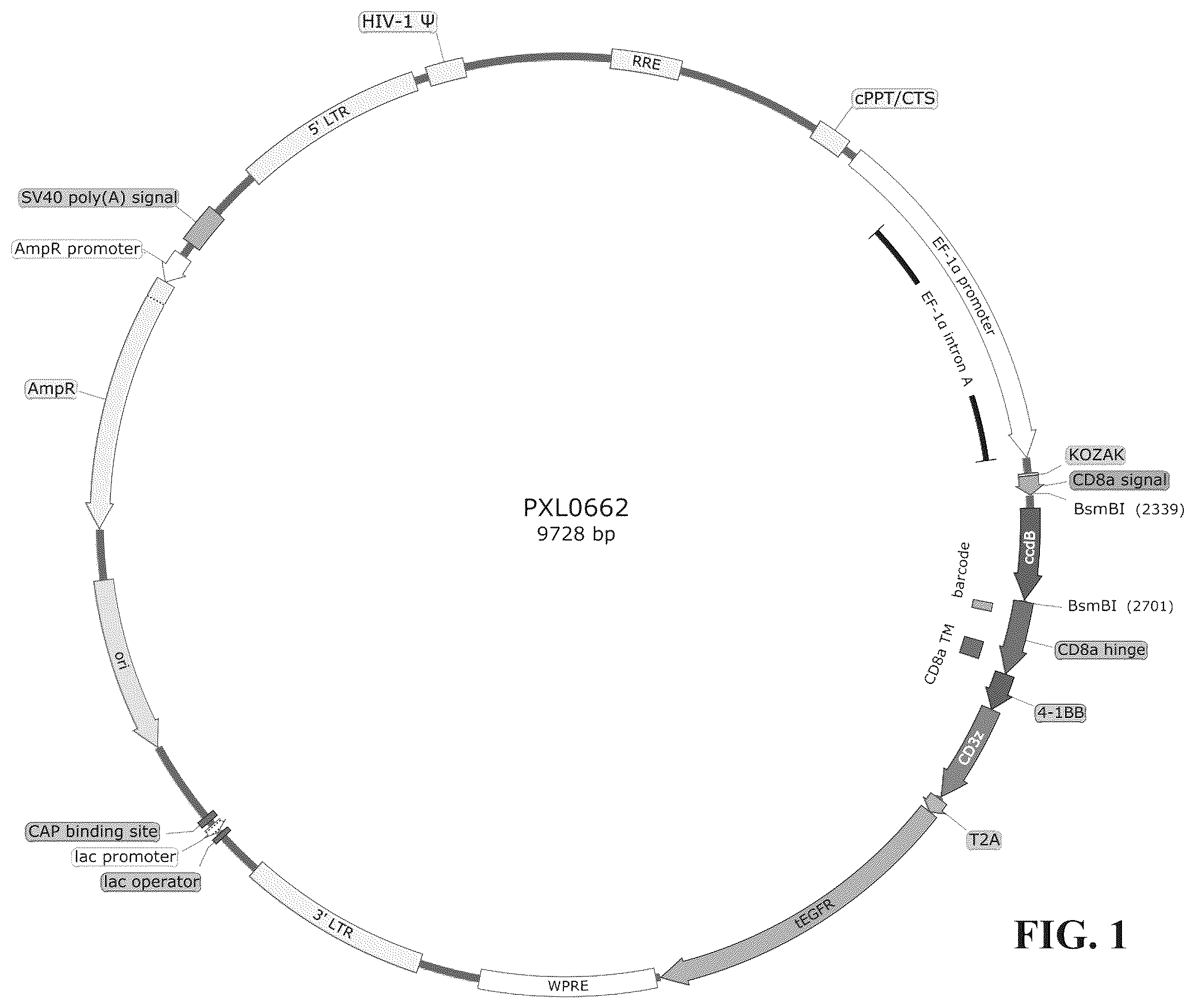
is a schematic diagram of the structure of the plasmid vector used in the present invention.
shows a schematic diagram of the working principle of the reporter gene method.
shows the flow cytometric results of transient expression of CAR molecules with different clone IDs on the reporter cell JLuc307. Indirect characterization was performed by staining with an EGFR antibody (APC anti-human EGFR antibody (cloneAY13)), and cells electro-transfected with a plasmid encoding Renilla luciferase (Promega, pGL4.75) served as a negative control (mock).
shows the expression of CD22 antigen on target cells used in the present invention. 4 A: CD22 antigen on target cells was stained using APC anti-human CD22 antibody (clone S-HCL-1). 4 B: CD22 antigen on target cells was stained using FITC anti-human CD22 antibody (clone HIB22).
shows some of the detection results of the reporter gene method. Among others, the y-axis reading is the flLuc/RLuc ratio (RLU) of the samples normalized to the ffLuc/RLuc ratio of the positive reference (co-incubated samples of Clone 0-2 and Raji cells).
shows the results of flow cytometric analysis of CAR expression in CD22 CAR-T cell samples from donor SXW (day 6). Cell samples were stained with APC mouse anti-human CD8, PE anti-human EGFR and FITC-CD22 protein.
shows the data analysis process of CD107a degranulation assay of CD22 CAR-T cell samples from donor SXW. Taking the data of clone 80 and REH cells co-incubated samples as an example, viable cells were first selected from the SSC vs FSC scatterplot (a), then monodisperse cells were selected from the viable cells (b), then CD8-positive cells were selected from the monodisperse cells (c), and finally the CD107a positive rate in the population of EGFR-positive cells (i.e., CAR-positive cells) was analyzed in the CD8-positive cell population (d).
shows the chemiluminescence results detected after 17 h co-incubation of CD22 CAR-T cell samples from donor SXW with different target cells. Chemiluminescence values were positively correlated with the number of target cells.
shows the inhibitory effect of the CAR-T cells prepared by the present invention on tumors in tumor-bearing NPG mice by in vivo luminescence imaging.
shows the change curve of fluorescence intensity in tumor-bearing NPG mice.
DETAILED DESCRIPTION OF THE INVENTION
Unless otherwise defined, all technical and scientific terms used herein have the meaning as commonly understood by one of ordinary skill in the art.
“Antibody” refers to an immunoglobulin secreted by plasma cells (effector B cells) and used by the body's immune system to neutralize foreign substances (polypeptides, viruses, bacteria, etc.). The foreign substance is correspondingly called an antigen. The basic structure of a classical antibody molecule is a 4-mer consisting of 2 identical heavy chains and 2 identical light chains. According to the conservative differences in amino acid sequences, the heavy and light chains are divided into a variable region (V) at the amino terminus and a constant region (C) at the carboxy terminus. The variable regions of one heavy chain and one light chain interact to form the antigen-binding site (Fv). In the variable region, the composition and arrangement of amino acid residues in certain regions are more variable than other regions (framework regions, FRs) in the variable region, these regions are called hypervariable regions (HVRs) and are actually the key sites for binding of antibodies to antigens. Since these hypervariable regions have their sequences complementary to antigenic determinants, they are also called complementarity-determining regions (CDRs). Both heavy and light chains have three complementarity-determining regions, designated HCDR1, HCDR2, HCDR3 and LCDR1, LCDR2, LCDR3, respectively. In some cases, antibodies may also be used to refer to antibody fragments that have antigen-binding ability, such as scFv, Fab, and F(ab′)2.
“Single chain fragment variable (scFv)” is composed of antibody heavy and light chain variable regions linked by a short peptide into a peptide chain. Through correct folding, the variable regions from the heavy chain and the light chain interact through non-covalent bonds to form the Fv segment, so the scFv can well retain its affinity for the antigen.
“Chimeric antigen receptor (CAR)”, also known as chimeric T cell receptor, and chimeric immunoreceptor, is an engineered membrane protein receptor molecule that confers a desired specificity to immune effector cells, such as the ability to bind to specific tumor antigens. Chimeric antigen receptors generally consist of an extracellular antigen-binding domain, a transmembrane domain, and an intracellular signaling domain. In some cases, the antigen-binding domain is an scFv sequence responsible for recognizing and binding to a specific antigen. Intracellular signaling domains usually comprise immunoreceptor tyrosine activation motifs (ITAMs), such as the signaling domains derived from CD3z molecules, which are responsible for activating immune effector cell to produce killing effects. In addition, the chimeric antigen receptor may also comprise a signal peptide responsible for intracellular localization of the nascent protein at the amino terminus, and a hinge region between the antigen-binding domain and the transmembrane domain. In addition to signaling domains, intracellular signaling domains can also comprise costimulatory domains derived from, for example, 4-1BB or CD28 molecules. When describing CAR structures herein, the abbreviation “bbz” may be used to refer to the intracellular signaling domain that comprises 4-1BB and CD3z, for example, the CAR molecule comprising antibody clone 80 (as the antigen-binding domain) and 4-1BB and CD3z (as the intracellular signaling domain) is abbreviated as “clone80-bbz”.
“CAR-T cells” refer to T cells expressing CARs, which are usually obtained by transducing T cells with an expression vector encoding CARs. Commonly used expression vectors are viral vectors, such as lentiviral expression vectors. Chimeric antigen receptor-modified T cells (CAR-Ts) are not restricted by major histocompatibility complexes, and have specific targeted killing activity and the ability for persistent amplification. In addition to T cells, other lymphocytes such as NK cells can also be transduced with an expression vector encoding a CAR to obtain targeted killer cells expressing the CAR.
“CD22” is a Siglec family lectin, including 7 IgG-like domains in the extramembrane portion, with a molecular weight of about 135 kD. Human CD22 and variants thereof are available in UniProt under accession number P20273. As a transmembrane glycoprotein, it is initially expressed on the surface of B cells at the pre-B cell stage, exists on mature B cells, and disappears on plasma cells. The National Cancer Institute of the United States reported the phase I clinical results of a CD22-targeted chimeric antigen receptor T cell (CAR-T), confirming that CD22 CAR-T is safe and effective, and can induce remission in some patients [2]. Therefore, CD22 protein is an ideal B cell tumor target.
“m971 molecule” is an anti-CD22 antibody panned from a human Fab phage library using a CD22-Fc fusion protein, which binds to the juxtamembrane epitope of the CD22 molecule [3]. The CAR constructed with the m971-derived scFv showed good anti-leukemia activity in preclinical models [4]. In some Examples herein, a CAR constructed with m971 scFv (amino acid sequence SEQ ID NO: 28) is used as the reference to evaluate some biological activities of the CARs provided herein.
“K D ” is the equilibrium dissociation constant, which can be used to measure the binding affinity between an antibody and its antigen. The smaller the K D value, the stronger the affinity.
The term “sequence identity” when referring to amino acid or nucleotide sequences refers to the degree of identity between two amino acid or nucleotide sequences (e.g., a query sequence and a reference sequence), usually expressed as a percentage. Typically, prior to calculating the percentage identity between two amino acid or nucleotide sequences, the sequences are aligned and gaps (if any) are introduced. If at a certain alignment position, the amino acid residues or bases in the two sequences are the same, the two sequences are considered to be identical or matched at that position; and if the amino acid residues or bases in the two sequences are different, they are considered to be non-identical or mismatched at that position. In some algorithms, the number of matched positions is divided by the total number of positions in the alignment window to obtain sequence identity. In other algorithms, the number of gaps and/or the gap length are also taken into account. For the purposes of the present invention, the published alignment software BLAST (available at ncbi.nlm.nih.gov) can be employed to obtain optimal sequence alignments by using default settings and calculate the sequence identity between two amino acid or nucleotide sequences.
In some embodiments, the light chain variable region of the anti-CD22 antibody molecule provided by the invention comprises an amino acid sequence having at least 90% sequence identity (e.g., at least 95%, at least 98%, at least 99% or even 100% sequence identity) with the sequence set forth in SEQ ID NO: 19, and the heavy chain variable region comprises an amino acid sequence having at least 90% sequence identity (e.g., at least 95%, at least 98%, at least 99% or even 100% sequence identity) with the sequence set forth in SEQ ID NO: 20.
In some embodiments, the light chain variable region of the anti-CD22 antibody molecule provided by the invention comprises an amino acid sequence having at least 90% sequence identity (e.g., at least 95%, at least 98%, at least 99% or even 100% sequence identity) with the sequence set forth in SEQ ID NO: 21, and the heavy chain variable region comprises an amino acid sequence having at least 90% sequence identity (e.g., at least 95%, at least 98%, at least 99% or even 100% sequence identity) with the sequence set forth in SEQ ID NO: 22.
In some embodiments, the light chain variable region of the anti-CD22 antibody molecule provided by the invention comprises an amino acid sequence having at least 90% sequence identity (e.g., at least 95%, at least 98%, at least 99% or even 100% sequence identity) with the sequence set forth in SEQ ID NO: 23, and the heavy chain variable region comprises an amino acid sequence having at least 90% sequence identity (e.g., at least 95%, at least 98%, at least 99% or even 100% sequence identity) with the sequence set forth in SEQ ID NO: 24.
In some embodiments, the antigen-binding domain in the CAR provided by the invention comprises an amino acid sequence having at least 90% sequence identity (e.g., at least 95%, at least 98%, at least 99% or even 100% sequence identity) with the sequence set forth in SEQ ID NO: 25, SEQ ID NO: 26 or SEQ ID NO: 27.
In some embodiments, the CAR provided by the invention comprises an amino acid sequence having at least 90% sequence identity (e.g., at least 95%, at least 98%, at least 99% or even 100% sequence identity) with the sequence set forth in SEQ ID NO: 35, SEQ ID NO: 36 or SEQ ID NO: 37.
Those skilled in the art can understand that, on the basis of the specific sequences provided herein, the corresponding variants of the anti-CD22 antibody molecules or CD22-targeted chimeric antigen receptors provided by the invention can be obtained by substituting, deleting, adding a few amino acids, and verifying or screening the resultant product for its binding ability with the corresponding antigen CD22 or its biological activity, and these variants should also be included within the scope of the present invention.
Those skilled in the art can also understand that, on the basis of the specific heavy chain variable region sequences provided herein, an antibody light chain library (such as a human phage light chain library) can be screened by using CD22 as the antigen, so as to obtain light chain variable regions matched with the heavy chain variable region while maintaining CD22 binding ability. Anti-CD22 antibody molecules obtainable in this way and CD22-targeted CARs constructed using the anti-CD22 antibody molecules are also included within the scope of the present invention.
When referring to pharmaceutical compositions, “pharmaceutically acceptable carrier” is used to refer to substances such as solid or liquid diluents, fillers, antioxidants, and stabilizers, which are safe for administration, and which are suitable for administration to humans and/or animals without undue adverse side effects, while being suitable for maintaining the viability of the drug or active agent therein.
A “therapeutically effective amount” refers to an amount of an active compound sufficient to elicit the biological or medical response desired by a clinician in a subject. The “therapeutically effective amount” of the bispecific antibody of the present invention can be determined by those skilled in the art according to the administration route, the subject's body weight, age, condition and other factors. For example, a typical daily dose may range from 0.01 mg to 100 mg of active ingredient per kg of body weight.
The CAR-T cells prepared by using the anti-CD22 antibody molecules screened out in the present invention have better killing activity against CD22-expressed target cells in vitro and in vivo, and are expected to be used for the treatment of some lymphomas and leukemias.
The present invention will be further described below through specific examples.
Example 1 Preparation and Analysis of Anti-CD22 Antibody Molecules
Screening of fully human antibodies against CD22 by yeast surface display technology. An established scFv yeast display library was subjected to multiple rounds of fluorescence-activated cell sorting with biotinylated CD22-llama-Fc or CD22-his protein, and a total of 129 fully human antibody clones against CD22 were obtained. They were sequenced and used for subsequent in vitro and in vivo screening.
The prepared antibodies were prepared in the form of IgG and Fab, respectively, and their binding ability to human CD22 was tested (ForteBio). Part of the results are as shown in Tables 1 and 2.
TABLE 1
Detection results of binding of antibodies in the form of IgG to
human CD22
IgG K D (M) kon koff Response
Clone ID Avid (1/Ms) (1/s) (nm)
17 1.6E−09 1.3E+05 2.0E−04 0.62
28 9.7E−10 2.1E+05 2.0E−04 0.81
80 8.2E−10 2.4E+05 2.0E−04 0.93
TABLE 2
Detection results of binding of antibodies in the form of
Fab to human CD22
Fab K D (M) kon koff Response
Clone ID Monovalent (1/Ms) (1/s) (nm)
17 8.1E−09 2.1E+05 1.7E−03 0.19
28 9.4E−10 2.8E+05 2.7E−04 0.25
80 1.3E−08 3.6E+05 4.6E−03 0.24
All 129 antibodies were grouped into 7 Bins by epitope binning analysis. Among them, Bin 3 is the m971 competition group. The antigen-binding sites of Bins 4 and 5 are the extracellular juxtamembrane regions of CD22. We selected all the antibodies of Bins 3, 4, and 5, as well as the antibodies of 4 other Bins, a total of 62 antibody sequences, for the subsequent preliminary screening by the reporter gene method. Among them, clones 80 and 28 have similar antigenic epitopes binding to m971, all of which belong to Bin 3. The binding epitope of clone 17 is different from that of m971 and it belongs to Bin 4.
Example 2 CD22 CAR Plasmid Vector Construction
First, a nucleotide sequence (SEQ ID NO: 42) was artificially synthesized, which contains KOZAK (bases 1-9), CD8α signal peptide (bases 10-72, and the corresponding amino acid sequence is SEQ ID NO: 30), ccdB screening gene (bases 73-428), CD8α hinge region and transmembrane region (bases 429-677, and the corresponding amino acid sequence is SEQ ID NO: 31), 4-1BB costimulatory factor (bases 678-803, and the corresponding amino acid sequence is SEQ ID NO: 32), CD3z intracellular signaling domain (bases 804-1139, the corresponding amino acid sequence is SEQ ID NO: 33), T2A cleavable peptide (bases 1140-1202, and the corresponding amino acid sequence is SEQ ID NO: 29), and tEGFR (bases 1203-2276, and the corresponding amino acid sequence is SEQ ID NO: 34). By PCR splicing method, the aforementioned synthetic sequence was inserted into the multi-cloning site of the lentiviral vector PLVX-EF1alpha-IRES-Puro plasmid (Clontech, Cat. No. 631988) to obtain the PXL0662 plasmid shown in .
Then, the nucleotide sequences encoding scFv (e.g., SEQ ID NO: 38, SEQ ID NO: 39, SEQ ID NO: 40, SEQ ID NO: 41 nucleotide sequences) were synthesized separately, and through the two type II endonuclease site BsmBIs (sites 2339 and 2701) in the PXL0662 plasmid, the nucleotide sequences of these scFvs were inserted into PXL0662 separately to obtain the plasmid vectors encoding the CD22 CAR.
Example 3 Primary Screening by CD22 CAR Reporter Gene Method
Working Principle
The activation of CAR-T cells is achieved by CD3z and costimulatory factors in the intracellular region of CAR molecules, wherein CD3z can activate the NFAT signaling pathway in the cells, which is a necessary condition for CAR-T cell activation. Therefore, CAR molecules with the function of activating the NFAT signaling pathway can be screened out by the NFAT reporter gene method [5].
In the process of primary screening, Jurkat cells integrated with the NFAT-RE-ffLuc reporter gene are used as reporter cells (as shown in , the cells are named JLuc307). CAR molecules are transiently expressed on the surface of reporter cells by plasmid electroporation. When a reporter cell expressing a CAR molecule and a target cell are co-incubated, the target cell surface antigen can specifically activate the CAR molecule, thereby activating the expression of the reporter gene (ffLuc, firefly luciferase). Then, by detecting the activity of luciferase, the ability of the CAR molecule to activate the NFAT signaling pathway can be evaluated. The plasmid used in this reporter gene method also includes a sequence encoding a truncated EGFR (tEGFR), which can be used to label cells that successfully express CAR when tEGFR is expressed on the cell surface. In addition, since different CAR molecules have different electroporation efficiencies, the internal reference plasmid (CMV-hRLuc, Renilla luciferase) mixed with CAR molecules can be used to calibrate the electroporation efficiency.
Operation Steps
•
• 1) Mix the CAR plasmid to be tested and the internal reference plasmid according to a fixed ratio, and transfect the reporter cells by electroporation method; • 2) 48 h after transfection, take some cells and stain them with PE-anti human EGFR antibody for flow cytometry to evaluate the transient expression of CAR plasmid; • 3) 72 h after transfection, mix the reporter cells and target cells in a ratio of 1:1, and then place them separately in a U-bottom 96-well plate to incubate for 24 h; wherein 3×10 4 reporter cells are added to each well, and 3 duplicate wells are set for each of target cell; and • 4) After completion of incubation, perform centrifugation at 1000 g for 5 min at 4° C., remove the culture supernatant, add 100 μL of lysis buffer to each well to lyse the cells, and take 20 μL of the cell lysate for dual-luciferase activity detection. Screening Criteria
CD22-positive target cells can effectively activate the NFAT-RE-ffLuc reporter gene to generate fluorescent signals. In the absence of stimulation by target cells or CD22-negative target cells, the fluorescent signal resulting from background (tonic effects) or non-specific activation is low.
Results
The primary screening was conducted in 6 batches, PXL0589 (m971-bbz-T2A-tEGFR, clone 0-2) plasmid was used as a positive control and pGL4.75 plasmid (No. PXL0337) as a negative control (mock) in each batch.
Because the koff values of the 62 antibodies to be tested were different, and some antibodies dissociated quickly after binding to the CD22 antigen, the transient expression of CAR molecules on the reporter cell JLuc307 was indirectly characterized by EGFR antibodies. The results of flow cytometry showed that, except for clone 4, the remaining 61 CAR molecules to be tested could be transiently expressed on JLuc307 cells. A representative flow cytometry chart is shown in .
In the process of preliminary screening by the reporter gene method, cells such as Raji, REH, JVM2, K562 and CD22 K/O Raji (clone 3D11 or 3E09, which are CD22 knockout/knockdown Raji cells prepared by us) were selected as target cells. Before the primary screening, we used APC mouse anti-human CD22 antibody (clone S-HCL-1) or FITC mouse anti-human CD22 antibody (clone HIB22) to detect the expression of CD22 antigen on the surface of target cells by flow cytometry individually. The results are shown in , in which Raji cells express CD22 highly; 3E09, REH, and JVM2 cells express CD22 moderately; and K562 and 3D11 cells are CD22 negative cells.
Two sets of fluorescence readings of firefly luciferase (ffLuc) and Renilla luciferase (RLuc) can be obtained by the dual luciferase reporter gene detection kit individually; among them, the Renilla luciferase reading is used as an internal reference to eliminate differences in cell quantity or transfection efficiency. Therefore, the level of transcriptional regulation of NFAT-RE-flLuc produced by each CAR sample as activated by the target cell can be characterized by the ffLuc/RLuc ratio (RLU). Taking the first batch of detections as an example, the results are shown in . Among them, clone 0-2 is a control CAR sample (m971), which could activate NFAT when it was stimulated by positive target cells Raji, 3E09 and JVM2, and the signal intensity was positively correlated with the antigen expression density on target cells; when it was stimulated by negative target cells 3D11, K562 stimulation or in the absence of target cell stimulation, it would not activate NFAT to generate fluorescent signals. Comparing with clone 0-2, clones 28, 36, and 80 were clones specifically recognizing CD22 target cells and activating the NFAT signaling pathway. The rest clones were eliminated.
A total of 10 clones were screened out of 62 clones by the reporter gene method for further function evaluation. The specific steps include preparation of lentiviral vectors, preparation of CAR-T cells, in vitro function evaluation of CAR-T cells and the like.
Example 4. Preparation of Lentiviral Vectors
For the 10 clones obtained in Example 3, the corresponding lentiviral vector preparation process is as follows.
HEK293T cells were thawed and cultured in DMEM medium containing 10% FBS. After 2-3 passages of cell proliferation culture, the cells were seeded into ten layers of cell factories at a density of 6×10 4 cells/cm 2 . Plasmid transfection was performed 3 days after cell seeding. The plasmid transfection liquid was formulated with Opti-MEM, and the final concentration of plasmid was 10 μg/mL. The plasmid transfection liquid contained CAR vector plasmid (T), psPAX2 plasmid (P) and pMD2.G plasmid (E) in a ratio of T:P:E=5:3:2. In addition, PEI with a final concentration of 30 μg/mL was added to the plasmid transfection liquid. The mixture was mixed well, and incubated at room temperature for 30 min before use. Each cell factory was transfected with 100 mL of the plasmid transfection liquid.
After 72 h, the supernatant was collected into a centrifuge tube, centrifuged at 3000 g at 4° C. for 10 min, and the supernatant obtained after centrifugation was filtered with a 0.45 μm filter. The filtered supernatant was centrifuged at 27,000 g for 4 h at 4° C. After centrifugation, the supernatant was discarded, and the virus was re-suspended in PBS pre-cooled at 4° C. The re-suspended virus was aliquoted and stored at −80° C. for later use.
Example 5. Preparation of CAR-T Cells
This example uses healthy donor cells to prepare CAR-Ts, and evaluates the functions of the 10 clones obtained in Example 3. An example of the preparation process of CAR-Ts is as follows.
On day 1, about 80 mL of peripheral blood from healthy donors was collected, and separated by using Ficoll to obtain PBMCs, and T cells were obtained by further sorting by CD3 MicroBeads. Sorted T cells were activated using CD3/CD28 Dynabeads. About 24 h after activation (day 2), the lentiviruses (MOI=3) prepared in Example 4 were added for transduction separately, and the T cell density during transduction was about 1.5×10 6 cells/mL. On day 3, medium change was conducted once for the transduced T cells. After that, the cell density was maintained between (0.6−2.0)×10 6 cells/mL for culture.
When the cells were cultured to the 6th or 7th day, the expression of CAR molecules, tEGFR molecules and CD8 on the cell surface was detected by flow cytometry. Here is an example of CAR-T cells prepared from the peripheral blood of donor SXW to illustrate. On day 6 of cell culture, approximately 5×10 5 cells were removed from each sample individually, and the medium was removed by centrifugation at 500 g. Then, after washing the cells twice with PBS+1% HSA solution, the cells were re-suspended in 50 μL of PBS+1% HSA solution, and 2 μL of CD22-FITC protein (Acro Biosystem, Cat. No. SI2-HF2H6), 2 μL of APC anti-human CD8 antibody (BD, Cat. No. 555369) and 2 μL of PE anti-human EGFR antibody (BioLegend, Cat. No. 352904) were added to each sample individually. The mixtures were mixed well and incubated at 4° C. in the dark for 20 min. After completion of incubation, the cells were washed twice with PBS+1% HSA solution again, re-suspended in 200 μL of PBS+1% HSA solution, and then loaded onto the instrument for testing. Part of the test results are shown in and Table 3. Except for the control T cells, all samples could express both EGFR and CAR molecules, the CAR molecules could normally bind to CD22 protein, the CAR positive rate (CAR %) was between 34.6% and 53.3%, and the CAR molecules could be expressed normally on the CD8-positive cell population.
TABLE 3
Flow cytometry data of CAR-T samples prepared from
donor SXW cells.
CAR % in CD8+
Clone ID CAR % CD8+ % population
28 34.60% 37.00% 34.32%
80 43.00% 35.00% 50.29%
36 42.70% 37.50% 42.67%
27 38.10% 39.80% 36.93%
17 53.30% 33.10% 59.52%
26 47.90% 34.70% 45.24%
0-2 (m971) 40.80% 36.30% 42.98%
T cells N/A 40.91% N/A
Example 6. In Vitro Function Evaluation of CAR-T Cells
The CAR-T cells prepared in Example 5, after cultured for 8 to 12 days, were subjected to in vitro function evaluation using two methods: CD107a degranulation experiment (CD107a degranulation assay) and in vitro cell killing experiment (in vitro cytotoxicity assay). Their working principles and screening criteria are as follows.
6.1 CD107a Degranulation Assay
Working Principle
CD107a is a marker for intracellular microvesicles, and CD107a on the cell membrane increases after granzyme-loaded microvesicles fuse with the cell membrane, and when its recovery is blocked by monesin (purchased from BioLegend), it can quantitatively reflect the strength of microvesicle release [6]. Therefore, when CAR-T cells are stimulated by target cell surface antigens to undergo degranulation effect, the positive rate of CD107a on the surface of CAR-T cells can be detected by flow cytometry to determine the activation of CAR-T cells.
Operation Steps
•
• 1) Centrifuge the CD22 positive and negative target cells separately at room temperature and 300 g for 5 min; discard the supernatant, and re-suspend the cells in T cell culture medium to 2×10 5 cells/mL; • 2) According to the CAR positive rate and E:T value (usually 0.3:1) of the CAR-T cells to be tested, re-suspend the CAR-T cells to an appropriate density, and add monensin and PE/Cy7 mouse anti-human CD107a antibody; • 3) In a U-bottom 96-well plate, add 100 μL/well CAR-T cells to be tested and 100 μL/well target cells individually, mix well, and then place them in an incubator (37° C., 5% CO 2 ) for incubation 3 h; • 4) After completion of incubation, centrifuge at 4° C. and 600 g for 5 min, discard the supernatant, and wash the cells twice with 200 μL/well DPBS+1% HSA; • 5) re-suspend the cells with 20 μL/well DPBS+1% HSA, add APC mouse anti-human CD8 antibody and Alexa Fluor 488 anti-human EGFR antibody (or FITC-CD22 protein), mix the cells well and incubate them on ice in the dark for 20 min; and • 6) After completion of incubation, wash the cells 3 times with 200 μL/well DPBS+1% HSA, and then re-suspend the cells with 200 L/well DPBS+1% HSA for flow cytometry. Screening Criteria
CD22 positive target cells can effectively activate CAR-T cells (in the CD8 + /CAR + cell population, the proportion of CD107a positive cells is high). In the absence of target cell stimulation or CD22-negative target cell stimulation, the CD107a-positive proportion is low in the CD8 + /CAR + cell population.
Results
CAR-T samples from donor SXW were subjected to CD107a degranulation assay on day 8. The CD22 positive target cells used in the CD107a degranulation assay were Raji. NALM6, REH, and JVM2, and the negative target cells were Jurkat, U266, HEK293, Karpas-299, K562, and 3D11, wherein 3D11 was the prepared CD22 knockout/knockdown Raji cell. An example of the CD107a degranulation assay data analysis is shown in . First, in the SSC vs FSC scatterplot ( a ), the viable cell population was selected. Then in the FSC-H vs FSC-A scatterplot of the live cell population ( b ), monodisperse cells were selected. CD8-positive cells were then selected in the SSC vs APC-CD8 scatterplot of the monodisperse cell population ( c ). Finally, in the PECy7-CD107a vs AF488-EGFR scatterplot of CD8-positive cells ( d ), the positive rate of CD107a in the EGFR-positive cell population was analyzed. The positive rate of CD107a was calculated by the ratio of Q2/(Q2+Q3) in d , and the calculation results are shown in Table 2. In addition, since there was no EGFR in T cells, its CD107a positive rate was calculated by Q1/(Q1+Q2) in d.
All cloned CAR-T cells was subjected to degranulation effect when stimulated by positive target cells. Among them, the degranulation effect of clone 80, clone 28, clone 36 and clone 17 was similar to that of the control CAR (m971-bbz). However, under stimulation by CD22-negative target cells, clone 36 had an obvious degranulation effect, so clone 36 may have the problem of non-specific activation and was eliminated.
In addition, the down-regulated expression of CD22 antigen on the surface of tumor cells is one of the main reasons for recurrence after CD22 CAR-T cell therapy 181. Therefore, we hope to obtain CAR molecules that can recognize and kill tumor cells with low CD22 expression. Since 3D11 is a CD22 knockout/knockdown Raji cell, clone 80, clone 28, clone 17 and m971 all had a small degranulation effect under stimulation by 3D11. The strength of the effect indicates the ability of a clone to recognize low-density targets. Therefore, clone 80 may have better low-density target recognition ability.
TABLE 4
The positive rate of CD107a in the CD8+/CAR+ cell population of CAR-T samples
from donor SXW under stimulation by different target cells.
Target cell
Clone NAL HEK2 Karpas-
ID raji REH M6 JVM2 3D11 K562 Jurkat 93 U266 299 Buffer
28 54.82% 70.39% 73.46% 43.50% 16.90% 2.12% 1.30% 2.22% 3.78% 2.16% 2.32%
80 39.69% 62.32% 59.71% 32.82% 18.88% 2.16% 1.47% 2.45% 3.64% 2.93% 3.14%
36 48.80% 67.86% 67.39% 49.09% 29.70% 20.27% 20.84% 15.44% 27.32% 29.03% 29.60%
27 61.89% 58.81% 70.25% 26.57% 1.55% 1.28% 0.94% 1.64% 2.87% 1.42% 2.74%
17 55.53% 64.80% 64.75% 44.30% 14.94% 1.88% 2.69% 1.54% 4.58% 2.64% 2.66%
26 9.61% 28.70% 13.12% 10.74% 0.52% 1.03% 0.78% 1.06% 1.83% 1.13% 1.68%
m971 53.87% 73.53% 72.00% 44.91% 15.93% 2.36% 1.51% 1.22% 2.84% 1.91% 1.83%
T cells 1.94% 1.98% 2.16% 1.63% 1.90% 1.49% 2.02% 2.28% 3.53% 2.56% 3.93%
6.2 In Vitro Cell Killing Experiment Working Principle
In the evaluation of the antigen-specific killing ability of CAR-T cells, NALM6-ffLuc was used as CD22 positive target cells, and K562-ffLuc or Jurkat-flLuc cells were used as CD22 negative target cells. These target cells are cell lines stably expressing firefly luciferase, which are obtained by lentiviral transduction.
In the in vitro cell killing assay, CAR-T cells and target cells were co-incubated with different effector-target ratios (E:T) individually. When target cells are killed by CAR-T cells, luciferase is released and quickly inactivated (firefly luciferase has a half-life of about 0.5 h [7]). If the target cells are not killed or inhibited by CAR-T cells, more luciferases will be produced as the target cells proliferate and continue to express luciferase. Therefore, the killing of target cells by CAR-T can be detected by the activity of luciferase.
Operation steps
•
• 1) Centrifuge NALM6-ffLuc and K562-ffLuc cells at room temperature and 300 g for 5 min separately, discard the supernatant, and then re-suspended the cells in T cell complete medium to 2×10 5 cells/mL; add 100 μL/well target cells to 96 well plates with transparent bottom separately; • 2) According to the CAR positive rate and E:T value (usually 2:1, 1:1, and 0.5:1) of the CAR-T cells to be tested, add 100 μL/well CAR-T cells to the 96-well plate separately, and after well mixing with the target cells, put them in an incubator (3° C., 5% CO 2 ) to incubate for 24 h; • 3) After completion of incubation, centrifuge the cells at room temperature and 800 g for 5 min, collect 100 μL/well supernatant as a reserved sample for cytokine detection (stored at −80° C.); and • 4) Use a luciferase detection kit to detect the luciferase activity of the remaining cells after sample reservation in each well. Screening Criteria
CAR-T cells can effectively kill CD22-positive target cells, and have no non-specific killing effect on CD22-negative target cells.
Results
CAR-T samples from donor SXW were subjected to in vitro cell killing experiment on day 8. Two CD22-positive target cells, NALM6-ffLuc and REH-flLuc, and one CD22-negative target cell, K562-flLuc, were used in the experiment.
The results are shown in Table 5 and a . When the effector-target ratio (E:T) was 1:1 or 2:1, all CAR-T samples could kill CD22-positive target cells NALM6-ffLuc. Among them, m971, clone 80 and clone 17 showed strong killing ability. In the control T cell sample, there was no obvious non-specific killing.
As shown in Table 6 and b , all CAR-T samples could kill CD22-positive target cells REH-ffLuc when the E:T value was 2:1. Among them, m971, clone 80 and clone 17 showed strong killing ability. When the E:T value was 1:1, m971, clone 80, clone 36 and clone 17 still had certain killing ability. However, at an E:T value of 0.5:1, all samples were unable to effectively kill target cells REH-ffLuc. None of the control T cell samples showed non-specific killing, and the target cells REH-ffLuc proliferated rapidly.
As shown in Table 7 and c , all CAR-T/T samples did not kill the CD22-negative target cell K562-ffLuc, and K562-ffLuc showed rapid proliferation due to the MLR effect.
TABLE 5
Detection of in vitro cell killing efficiency after co-incubation of
CAR-T samples of donor SXW and target cells NALM6-ffLuc
for 17 h. wherein negative values represent target cell proliferation.
Effector-target ratio (E:T)
Clone ID 2:1 1:1 0.5:1
28 76.1 ± 2.3% 13.8 ± 4.8% −26.8 ± 1.3%
80 90.5 ± 0.8% 58.5 ± 5.1% 15.3 ± 9.9%
36 88.0 ± 0.4% 48.4 ± 6.8% −6.3 ± 5.6%
27 90.1 ± 0.5% 45.5 ± 4.9% −6.5 ± 7.0%
17 86.8 ± 1.8% 51.1 ± 3.6% 8.7 ± 1.2%
26 70.7 ± 4.8% 24.5 ± 3.4% −12.9 ± 6.8%
m971 87.1 ± 1.2% 60.7 ± 1.7% 21.0 ± 3.5%
T cells −3.1 ± 3.7% −48.4 ± 2.0% −50.8 ± 4.4%
TABLE 6
Detection of in vitro cell killing efficiency after co-incubation of
CAR-T samples of donor SXW and target cells REH-ffLuc for 17 h.
wherein negative values represent target cell proliferation.
Effector-target ratio (E:T)
Clone ID 2:1 1:1 0.5:1
28 63.8 ± 4.1% −23.9 ± 18.1% −50.1 ± 5.4%
80 89.0 ± 1.4% 40.2 ± 7.4% −18.2 ± 2.3%
36 78.9 ± 1.8% 21.5 ± 2.1% −35.8 ± 2.2%
27 64.3 ± 6.2% −12.0 ± 5.3% −55.7 ± 3.1%
17 83.1 ± 0.3% 36.8 ± 2.3% −11.2 ± 2.8%
26 68.4 ± 4.7% −5.3 ± 3.4% −30.4 ± 5.4%
m971 80.9 ± 2.9% 44.3 ± 9.1% −12.0 ± 8.3%
T cells −124.2 ± 10.9% −83.8 ± 3.8% −46.1 ± 3.4%
TABLE 7
Detection of in vitro cell killing efficiency after co-incubation of
CAR-T samples of donor SXW and target cells K562-ffLuc for 17 h.
wherein negative values represent target cell proliferation.
Effector-target ratio (E:T)
Clone ID 2:1 1:1 0.5:1
28 −233.5 ± 25.4% −99.0 ± 20.3% −60.2 ± 10.7%
80 −334.6 ± 83.5% −189.9 ± 14.7% −122.2 ± 4.1%
36 −620.0 ± 68.4% −405.7 ± 20.0% −264.2 ± 24.8%
27 −236.1 ± 80.2% −106.6 ± 15.4% −60.7 ± 7.2%
17 −177.0 ± 24.2% −72.8 ± 10.1% −33.9 ± 7.8%
26 −286.2 ± 19.7% −147.5 ± 6.2% −69.0 ± 9.2%
m971 −179.5 ± 15.7% −65.1 ± 25.3% −55.3 ± 13.9%
T cells −328.7 ± 70.9% −172.6 ± 181.5% −179.2 ± 100.0%
The results of Example 3 (preliminary screening by reporter gene method) and Example 6 (in vitro function evaluation of CAR-T cells) together showed that the CAR-T cells generated by using clone 80, clone 28 and clone 17 showed good in vitro cell functions.
Example 7. In Vivo Tumor Inhibition Experiments in Tumor-Bearing Animal Models
Working Principle
Using the immunodeficient mouse NPG bearing the human acute lymphocytic leukemia cells, Nalm6 cells, which specifically express CD22, as the experimental system, the method in Example 5 to prepare CAR-T cell samples was evaluated, and the efficacy of clone 80, clone 28 and clone 17 in animals was evaluated.
Compared with NOD/SCID mice, NPG mice have the gamma chain of the IL-2 receptor knocked out. IL-2 receptor is the co-receptor subunit of IL-2, IL-4, IL-7, IL-9, IL-15 and IL-21. The knockout of this gene can further reduce the immune function of mice, especially, almost completely eliminate the vitality of NK cells. Therefore, NPG mice are more suitable recipients for cell or tissue transplantation.
Nalm6 is a cell line stably expressing firefly luciferase. Nalm6 cells will proliferate after being injected into mice through tail vein. D-fluorescein potassium is injected intraperitoneally, and chemiluminescence signals are captured by Bruker small animal imager under isoflurane anesthesia. If the target cells are not killed or inhibited by CAR-T cells, more luminescent signals will be detected as the target cells proliferate to continue express luciferase; sites with specific aggregation of target cells can also be observed by the imaging position of the target cells. Therefore, the killing of target cells by CAR-T in animals can be detected by the intensity of the luminescent signal.
Operation steps
•
• 1) Prepare CAR-T cell samples comprising clone 80, clone 28 and clone 17 separately according to the method in Example 5; • 2) Select immunodeficient NPG mice, female, 4-5 weeks old, body weight 20±3 g; get NALM6-LUC cells in logarithmic growth phase and inoculate them in NPG mice by tail vein injection, the inoculum size being 1×10 6 /mouse. After 2 days of tumor cell inoculation, different doses of the test samples were given by tail vein injection, and the grouping scheme is shown in Table 8; • 3) On the 3rd, 7th, 12th, 18th and 28th days after administration, inject D-fluorescein potassium intraperitoneally, and capture chemiluminescence signals by Bruker small animal imager under isoflurane anesthesia, and detect tumor growth inhibition by tumor imaging; and • 4) On the 3rd, 7th, 14th, 17th, 21st, and 28th days after administration, after anesthesia with isoflurane and after the corneal reflex disappears, collect 0.1 mL of blood from the orbital vein and store the blood with EDTA-2K anticoagulation; and detect the copy number of CARs.
TABLE 8
Grouping scheme of animal model experiment
Administrated Dose
dose (x10 6 CAR
(x10 8 CAR + positive Number
CAR + pcs/ rate of
Group pcs/kg) mouse) (%) mice
Vehicle control / / / 3
group
Mock T control 3.7 7.4 / 3
group
Clone 80 low 0.25 0.5 42.8 3
dose group
Clone 80 high 1.0 2 42.8 6
dose group
Clone 28 group 1.0 2 30.2 6
Clone 17 group 1.0 2 50.6 6
Results of Animal Experiments 1) Mouse Imaging Results
As shown in , the tumor cells in the vehicle control group and the MockT control group grew normally, and the model was successful: after D20, the mice in the vehicle control group died due to tumor overload, and the mice in the MockT group showed GvHD before death; the mice in all the administration groups had no abnormality in the clinical indicators; the inhibition of tumor growth phenomenon was observed in all the clone 80 low-dose and high-dose groups, clone 28 group and clone17 group; CR began to appear on the 12th day in each group, and CR was maintained until D28, the end of the observation period; obvious differences in efficacy were observed for different doses of clone 80, and the efficacy was positively correlated with the dose; the clone 28 group had 3/6 CR on D28; and some mice in the clone17 group had tumor cell proliferation on D28.
2) Fluorescence Signal of Tumor Cells
The measured fluorescence signal intensities are shown in Table 9 below.
TABLE 9
Mouse tumor cell imaging fluorescence signal
Luminescent intensity Log 10 (p/s/cm/sr)
Group Day 0 Day 3 Day 7 Day 12 Day 18 Day 28
Vehicle 6.32 ± 0.27 7.51 ± 0.18 9.62 ± 0.04 11.2 ± 0.03 11.61 ± 0.08 /
control
group
Mock T 6.32 ± 0.21 7.22 ± 0.19 9.27 ± 0.23 10.8 ± 0.54 11.45 ± 0.07 11.59
control
group
Clone 80 6.27 ± 0.25 6.98 ± 0.39 8.16 ± 0.22 8.78 ± 0.41 9.43 ± 0.64 9.88 ± 0.11
low dose
group
Clone 80 6.11 ± 0.49 6.88 ± 0.32 7.8 ± 0.27 7.7 ± 0.71 7.95 ± 1.28 9.52 ± 1.31
high dose
group
Clone 28 6.2 ± 0.48 6.71 ± 0.47 7.43 ± 0.59 7.52 ± 1.11 8.37 ± 0.76 8.36 ± 0.77
group
Clone 17 6.15 ± 0.43 6.71 ± 0.45 7.78 ± 0.51 8.41 ± 1.5 9.71 ± 0.96 10.56 ± 0.4
group
The data in the above table is plotted, and the results are shown in . In the vehicle control group and the Mock-T group, the tumor signal gradually increased with time, and there was no significant difference between the two groups, the tumor signal decrease was observed in the clone 80 low-dose and high-dose groups, the clone 28 group and the clone 17 group; there were obvious differences in tumor signal for the clone 80 groups with different doses, at the same time point, and the signal in the high-dose group was significantly lower than that in the low-dose group; the tumor signal in the clone 28 group was always at a low level, and the tumor signal in the clone 17 group gradually increased after D18.
In conclusion, obvious efficacy was observed in the Nalm6 tumor-bearing mouse model in all the clone 80 group, clone 28 group and clone 17 group. Of them, the clone 80 group and clone 28 group had better tumor inhibition than the clone 17 group.
Some of the amino acid or nucleic acid sequences mentioned herein are as follows:
(clone 17 LCDR1 amino acid sequence)
SEQ ID NO: 1
RASQSISSWLA
(clone 17 LCDR2 amino acid sequence)
SEQ ID NO: 2
KASSLES
(clone 17 LCDR3 amino acid sequence)
SEQ ID NO: 3
QQYERFPWT
(clone 17 HCDR1 amino acid sequence)
SEQ ID NO: 4
FTFSSYAMS
(clone 17 HCDR2 amino acid sequence)
SEQ ID NO: 5
AISGSGGSTYYADSVKG
(clone 17 HCDR3 amino acid sequence)
SEQ ID NO: 6
AKVGISSLHGMDV
(clone 28 LCDR1 amino acid sequence)
SEQ ID NO: 7
RASQSISSWLA
(clone 28 LCDR2 amino acid sequence)
SEQ ID NO: 8
DASSLES
(clone 28 LCDR3 amino acid sequence)
SEQ ID NO: 9
QQANTYSPT
(clone 28 HCDR1 amino acid sequence)
SEQ ID NO: 10
GSISSYYWS
(clone 28 HCDR2 amino acid sequence)
SEQ ID NO: 11
RIYTSGSTNYNPSLKS
(clone 28 HCDR3 amino acid sequence)
SEQ ID NO: 12
ARDLYRDGMDV
(clone 80 LCDR1 amino acid sequence)
SEQ ID NO: 13
RASQSVSSSYLA
(clone 80 LCDR2 amino acid sequence)
SEQ ID NO: 14
GASSRAT
(clone 80 LCDR3 amino acid sequence)
SEQ ID NO: 15
QQAGLFPYT
(clone 80 HCDR1 amino acid sequence)
SEQ ID NO: 16
GSISSSNWWS
(clone 80 HCDR2 amino acid sequence)
SEQ ID NO: 17
EIYHSGSTNYNPSLKS
(clone 80 HCDR3 amino acid sequence)
SEQ ID NO: 18
ARLPGYESAFDI
(clone 17 VL amino acid sequence)
SEQ ID NO: 19
DIQMTQSPSTLSASVGDRVTITCRASQSISSWLAWYQQKPGKAPKLLIYKASSLESGV
PSRFSGSGSGTEFTLTISSLQPDDFATYYCQQYERFPWTFGGGTKVEIK
(clone 17 VH amino acid sequence)
SEQ ID NO: 20
EVQLLESGGGLVQPGGSLRLSCAASGFTFSSY AMSWVRQAPGKGLEWVSAISGSGGS
TYYADSVKGRFTISRDNSKNTLYLQMNSLRAEDTAVYYCAKVGISSLHGMDVWGQGTT
VTVSS
(clone 28 VL amino acid sequence)
SEQ ID NO: 21
DIQMTQSPSTLSASVGDRVTITCRASQSISSWLAWYQQKPGKAPKLLISDASSLESGV
PSRFSGSGSGTEFTLTISSLQPDDFATYYCQQANTYSPTFGGGTKVEIK
(clone 28 VH amino acid sequence)
SEQ ID NO: 22
QVQLQESGPGLVKPSETLSLTCTVSGGSISSYYWSWIRQPAGKGLEWIGRIYTSGSTN
YNPSLKSRVTMSVDTSKNQFSLKLSSVTAADTAVYYCARDLYRDGMDVWGQGTTVTV
SS
(clone 80 VL amino acid sequence)
SEQ ID NO: 23
EIVLTQSPGTLSLSPGERATLSCRASQSVSSSYLAWYQQKPGQAPRLLIYGASSRATGI
PDRFSGSGSGTDFTLTISRLEPEDFAVYYCQQAGLFPYTFGGGTKVEIK
(clone 80 VH amino acid sequence)
SEQ ID NO: 24
QVQLQESGPGLVKPSGTLSLTCAVSGGSISSSNWWSWVRQPPGKGLEWIGEIYHSGS
TNYNPSLKSRVTISVDKSKNQFSLKLSSVTAADTAVYYCARLPGYESAFDIWGQGTMVT
VSS
(clone 17 scFv amino acid sequence)
SEQ ID NO: 25
EVQLLESGGGLVQPGGSLRLSCAASGFTFSSYAMSWVRQAPGKGLEWVSAISGSGGS
TYYADSVKGRFTISRDNSKNTLYLQMNSLRAEDTAVYYCAKVGISSLHGMDVWGQGTT
VTVSSGGGGSGGGGSGGGGSDIQMTQSPSTLSASVGDRVTITCRASQSISSWLAWYQQK
PGKAPKLLIYKASSLESGVPSRFSGSGSGTEFTLTISSLQPDDFATYYCQQYERFPWTFGG
GTKVEIK
(clone 28 scFv amino acid sequence)
SEQ ID NO: 26
QVQLQESGPGLVKPSETLSLTCTVSGGSISSYYWSWIRQPAGKGLEWIGRIYTSGSTN
YNPSLKSRVTMSVDTSKNQFSLKLSSVTAADTAVYYCARDLYRDGMDVWGQGTTVTV
SSGGGGSGGGGSGGGGSDIQMTQSPSTLSASVGDRVTITCRASQSISSWLAWYQQKPGK
APKLLISDASSLESGVPSRFSGSGSGTEFTLTISSLQPDDFATYYCQQANTYSPTFGGGTK
VEIK
(clone 80 scFv amino acid sequence)
SEQ ID NO: 27
QVQLQESGPGLVKPSGTLSLTCAVSGGSISSSNWWSWVRQPPGKGLEWIGEIYHSGS
TNYNPSLKSRVTISVDKSKNQFSLKLSSVTAADTAVYYCARLPGYESAFDIWGQGTMVT
VSSGGGGSGGGGSGGGGSEIVLTQSPGTLSLSPGERATLSCRASQSVSSSYLAWYQQKPG
QAPRLLIYGASSRATGIPDRFSGSGSGTDFTLTISRLEPEDFAVYYCQQAGLFPYTFGGGT
KVEIK
(M971 scFv amino acid sequence)
SEQ ID NO: 28
QVQLQQSGPGLVKPSQTLSLTCAISGDSVSSNSAAWNWIRQSPSRGLEWLGRTYYRS
KWYNDYAVSVKSRITINPDTSKNQFSLQLNSVTPEDTAVYYCAREVTGDLEDAFDIWGQ
GTMVTVSSGGGGSGGGGSGGGGSDIQMTQSPSSLSASVGDRVTITCRASQTIWSYLNWY
QQRPGKAPNLLIYAASSLQSGVPSRFSGRGSGTDFTLTISSLQAEDFATYYCQQSYSIPQT
FGQGTKLEIK
(T2A amino acid sequence)
SEQ ID NO: 29
EGRGSLLTCGDVEENPGP
(CD8α signal peptide amino acid sequence)
SEQ ID NO: 30
MALPVTALLLPLALLLHAARP
(CD8α hinge region and transmembrane region amino acid sequences)
SEQ ID NO: 31
FVPVFLPAKPTTTPAPRPPTPAPTIASQPLSLRPEACRPAAGGAVHTRGLDFACDIYIW
APLAGTCGVLLLSLVITLYCNHRN
(4-1BB costimulatory signaling domain amino acid sequence)
SEQ ID NO: 32
KRGRKKLLYIFKQPFMRPVQTTQEEDGCCRFPEEEEGGCEL
(CD3z intracellular signaling domain amino acid sequence)
SEQ ID NO: 33
RVKFSRSADAPAYQQGQNQLYNELNLGRREEYDVLDKRRGRDPEMGGKPRRKNPQ
EGLYNELQKDKMAEAYSEIGMKGERRRGKGHDGLYQGLSTATKDTYDALHMQALPPR
(tEGFR amino acid sequence)
SEQ ID NO: 34
MLLLVTSLLLCELPHPAFLLIPRKVCNGIGIGEFKDSLSINATNIKHFKNCTSISGDLHIL
PVAFRGDSFTHTPPLDPQELDILKTVKEITGFLLIQAWPENRTDLHAFENLEIIRGRTKQH
GQFSLAVVSLNITSLGLRSLKEISDGDVIISGNKNLCYANTINWKKLFGTSGQKTKIISNRG
ENSCKATGQVCHALCSPEGCWGPEPRDCVSCRNVSRGRECVDKCNLLEGEPREFVENSE
CIQCHPECLPQAMNITCTGRGPDNCIQCAHYIDGPHCVKTCPAGVMGENNTLVWKYAD
AGHVCHLCHPNCTYGCTGPGLEGCPTNGPKIPSIATGMVGALLLLLVVALGIGLFM
(clone 17 CAR amino acid sequence)
SEQ ID NO: 35
MALPVTALLLPLALLLHAARPEVQLLESGGGLVQPGGSLRLSCAASGFTFSSYAMSW
VRQAPGKGLEWVSAISGSGGSTYYADSVKGRFTISRDNSKNTLYLQMNSLRAEDTAVY
YCAKVGISSLHGMDVWGQGTTVTVSSGGGGSGGGGSGGGGSDIQMTQSPSTLSASVGD
RVTITCRASQSISSWLAWYQQKPGKAPKLLIYKASSLESGVPSRFSGSGSGTEFTLTISSLQ
PDDFATYYCQQYERFPWTFGGGTKVEIKFVPVFLPAKPTTTPAPRPPTPAPTIASQPLSLR
PEACRPAAGGAVHTRGLDFACDIYIWAPLAGTCGVLLLSLVITLYCNHRNKRGRKKLLY
IFKQPFMRPVQTTQEEDGCSCRFPEEEEGGCELRVKFSRSADAPAYQQGQNQLYNELNL
GRREEYDVLDKRRGRDPEMGGKPRRKNPQEGLYNELQKDKMABAYSEIGMKGERRRG
KGHDGLYQGLSTATKDTYDALHMQALPPRGSGEGRGSLLTCGDVEENPG
(clone 28 CAR amino acid sequence)
SEQ ID NO: 36
MALPVTALLLPLALLLHAARPQVQLQESGPGLVKPSETLSLTCTVSGGSISSYYWSWI
RQPAGKGLEWIGRIYTSGSTNYNPSLKSRVTMSVDTSKNQFSLKLSSVTAADTAVYYCA
RDLYRDGMDVWGQGTTVTVSSGGGGSGGGGSGGGGSDIQMTQSPSTLSASVGDRVTIT
CRASQSISSWLAWYQQKPGKAPKLLISDASSLESGVPSRFSGSGSGTEFTLTISSLQPDDF
ATYYCQQANTYSPTFGGGTKVEIKFVPVFLPAKPTTTPAPRPPTP APTIASQPLSLRPEAC
RPAAGGAVHTRGLDFACDIYIWAPLAGTCGVLLLSLVITLYCNHRNKRGRKKLLYIFKQ
PFMRPVQTTQEEDGCSCRFPEEEEGGCELRVKFSRSADAPAYQQGQNQLYNELNLGRRE
EYDVLDKRRGRDPEMGGKPRRKNPQEGLYNELQKDKMAEAYSEIGMKGERRRGKGHD
GLYQGLSTATKDTYDALHMQALPPRGSGEGRGSLLTCGDVEENPG
(clone 80 CAR amino acid sequence)
SEQ ID NO: 37
MALPVTALLLPLALLLHAARPQVQLQESGPGLVKPSGTLSLTCAVSGGSISSSNWWS
WVRQPPGKGLEWIGEIYHSGSTNYNPSLKSRVTISVDKSKNQFSLKLSSVTAADTAVYY
CARLPGYESAFDIWGQGTMVTVSSGGGGSGGGGGGGGSEIVLTQSPGTLSLSPGERAT
LSCRASQSVSSSYLAWYQQKPGQAPRLLIYGASSRATGIPDRFSGSGSGTDFTLTISRLEP
EDFAVYYCQQAGLFPYTFGGGTKVEIKFVPVFLPAKPTTTPAPRPPTPAPTIASQPLSLRP
EACRPAAGGAVHTRGLDFACDIYIWAPLAGTCGVLLLSLVITLYCNHRNKRGRKKLLYI
FKQPFMRPVQTTQEEDGCSCRFPEEEEGGCELRVKFSRSADAPAYQQGQNQLYNELNLG
RREEYDVLDKRRGRDPEMGGKPRRKNPQEGLYNELQKDKMAEAYSEIGMKGERRRGK
GHDGLYQGLSTATKDTYDALHMQALPPRGSGEGRGSLLTCGDVEENPG
(clone 17 scFv nucleotide sequence)
SEQ ID NO: 38
GAGGTGCAGCTGTTGGAGTCTGGGGGAGGCTTGGTACAGCCTGGGGGGTCCCTG
AGACTCTCCTGTGCAGCCTCTGGATTCACCTTTAGCAGCTATGCCATGAGCTGGGTCC
GCCAGGCTCCAGGGAAGGGGCTGGAGTGGGTCTCAGCTATTAGTGGTAGTGGTGGT
AGCACATACTACGCAGACTCCGTGAAGGGCCGGTTCACCATCTCCAGAGACAATTCC
AAGAACACGCTGTATCTGCAAATGAACAGCCTGAGAGCCGAGGACACGGCGGTGTA
CTACTGCGCCAAGGTAGGAATATCCAGCTTACACGGAATGGACGTATGGGGCCAGG
GAACAACTGTCACCGTCAGCTCAGGTGGCGGGGGCAGCGGCGGAGGCGGATCCGGA
GGCGGAGGGAGTGACATCCAGATGACCCAGTCTCCTTCCACCCTGTCTGCATCTGTA
GGAGACAGAGTCACCATCACTTGCCGGGCCAGTCAGAGTATTAGTAGCTGGTTGGCC
TGGTATCAGCAGAAACCAGGGAAAGCCCCTAAGCTCCTGATCTATAAAGCCTCCAGT
TTGGAAAGTGGGGTCCCATCAAGGTTCAGCGGCAGTGGATCTGGGACAGAATTCACT
CTCACCATCAGCAGCCTGCAGCCTGATGATTTTGCAACTTATTACTGCCAGCAGTAC
GAACGCTTCCCTTGGACTTTTGGCGGAGGGACCAAGGTTGAGATCAAA
(clone 28 scFv nucleotide sequence)
SEQ ID NO: 39
CAGGTGCAGCTGCAGGAGTCGGGCCCAGGACTGGTGAAGCCTTCGGAGACCCTG
TCCCTCACCTGCACTGTCTCTGGTGGCTCCATCAGTAGTTACTACTGGAGCTGGATCC
GGCAGCCCGCCGGGAAGGGACTGGAGTGGATTGGGCGTATCTATACCAGTGGGAGC
ACCAACTACAACCCCTCCCTCAAGAGTCGAGTCACCATGTCAGTAGACACGTCCAAG
AACCAGTTCTCCCTGAAGCTGAGCTCTGTGACCGCCGCGGACACGGCGGTGTACTAC
TGCGCCAGAGACTTGTACAGAGATGGAATGGACGTATGGGGCCAGGGAACAACTGT
CACCGTCAGCTCAGGTGGCGGGGGCAGCGGCGGAGGCGGATCCGGAGGCGGAGGG
AGTGACATCCAGATGACCCAGTCTCCTTCCACCCTGTCTGCATCTGTAGGAGACAGA
GTCACCATCACTTGCCGGGCCAGTCAGAGTATTAGTAGCTGGTTGGCCTGGTATCAG
CAGAAACCAGGGAAAGCCCCTAAGCTCCTGATCTCCGATGCCTCCAGTTTGGAAAGT
GGGGTCCCATCAAGGTTCAGCGGCAGTGGATCTGGGACAGAATTCACTCTCACCATC
AGCAGCCTGCAGCCTGATGATTTTGCAACTTATTACTGCCAGCAGGCCAATACCTAC
TCTCCTACTTTTGGCGGAGGGACCAAGGTTGAGATCAAA
(clone 80 scFv nucleotide sequence)
SEQ ID NO: 40
CAGGTGCAGCTGCAGGAGTCGGGCCCAGGACTGGTGAAGCCTTCGGGGACCCTG
TCCCTCACCTGCGCTGTCTCTGGTGGCTCCATCAGCAGTAGTAACTGGTGGAGTTGG
GTCCGCCAGCCCCCAGGGAAGGGGCTGGAGTGGATTGGGGAAATCTATCATAGTGG
GAGCACCAACTACAACCCGTCCCTCAAGAGTCGAGTCACCATATCAGTAGACAAGT
CCAAGAACCAGTTCTCCCTGAAGCTGAGCTCTGTGACCGCCGCGGACACGGCGGTGT
ACTACTGCGCCAGACTTCCTGGATACGAGTCAGCTTTCGACATATGGGGTCAGGGTA
CAATGGTCACCGTCAGCTCAGGTGGCGGGGGCAGCGGCGGAGGCGGATCCGGAGGC
GGAGGGAGTGAAATTGTGTTGACGCAGTCTCCAGGCACCCTGTCTTTGTCTCCAGGG
GAAAGAGCCACCCTCTCCTGCAGGGCCAGTCAGAGTGTTAGCAGCAGCTACTTAGCC
TGGTACCAGCAGAAACCTGGCCAGGCTCCCAGGCTCCTCATCTATGGTGCATCCAGC
AGGGCCACTGGCATCCCAGACAGGTTCAGTGGCAGTGGGTCTGGGACAGACTTCACT
CTCACCATCAGCAGACTGGAGCCTGAAGATTTTGCAGTGTATTACTGTCAGCAGGCC
GGACTCTTCCCTTACACTTTTGGCGGAGGGACCAAGGTTGAGATCAAA
(M971 scFv nucleotide sequence)
SEQ ID NO: 41
CAGGTGCAGCTCCAGCAGAGCGGCCCCGGCCTGGTAAAGCCCAGCCAAACCCTC
TCCCTGACCTGCGCTATCAGCGGCGATTCCGTGAGCAGCAACAGCGCCGCCTGGAAT
TGGATCCGTCAGAGCCCCAGCAGGGGCCTGGAGTGGCTGGGGCGGACCTATTACCG
GAGTAAGTGGTACAACGACTACGCCGTAAGCGTGAAGAGCCGCATCACCATTAATC
CTGACACCAGCAAGAACCAGTTCAGTCTGCAGCTGAACAGCGTGACTCCCGAGGAC
ACCGCCGTGTACTACTGCGCCCGCGAGGTGACTGGAGACCTGGAAGACGCCTTCGA
CATCTGGGGCCAGGGCACAATGGTGACCGTCAGCAGCGGTGGCGGGGGCAGCGGCG
GAGGCGGATCCGGAGGCGGAGGGAGTGACATACAGATGACCCAGAGCCCTAGCAGC
CTCTCTGCCAGCGTGGGAGACCGGGTGACCATCACCTGCCGCGCCAGTCAGACCATC
TGGTCTTATCTGAACTGGTACCAGCAACGGCCCGGCAAGGCCCCTAACCTGTTGATC
TACGCCGCCAGCAGTCTCCAGAGCGGCGTTCCATCTCGCTTCAGCGGCCGCGGCAGC
GGCACAGACTTCACCCTGACCATCAGCAGCCTGCAGGCCGAGGACTTCGCCACCTAC
TACTGCCAGCAGAGCTACAGCATCCCCCAGACTTTOGGACAGGGCACCAAGTTGGA
GATCAAA
(nucleotide sequence of CAR portion in PXL0662)
SEQ ID NO: 42
GCCGCCACCATGGCCCTGCCTGTGACAGCTCTGCTCCTCCCTCTGGCCCTGCTGCT
CCATGCCGCCAGACCCGAGACGTGAgAATTAATACGACTCACTATAGAGGGACTGGT
GAAATGCAGTTCAAGGTTTACACCTATAAAAGAGAGAGCCGCTATCGCCTGTTTGTG
GATGTACAGAGTGATATTATTGACACGCCCGGGCGACGGATGGTGATCCCCCTGGCC
AGTGCACGTCTGCTGTCAGATAAAGTCTCCCGTGAACTTTACCCGGTGGTGCATATC
GGGGATGAAAGCTGGCGCATGATGACCACCCAGATGGTCAGTGTGCCGGTCTCCGTC
ATCGGAGAAGAAGTGGCTGATCTCAGCCACCGCGAAAATGACATCAAAAACGCCAT
TAATCTGATGTTCTGGGGAATATAACGTCTCTTCGTGCCCGTGTTCCTGCCCGCCAAA
CCTACCACCACCCCTGCCCCTAGACCTCCCACCCCAGCCCCAACAATCGCCAGCCAG
CCTCTGTCTCTGCGGCCCGAAGCCTGTAGACCTGCTGCCGGCGGAGCCGTGCACACC
AGAGGCCTGGACTTCGCCTGCGACATCTACATCTGGGCCCCTCTGGCCGGCACCTGT
GGCGTGCTGCTGCTGAGCCTGGTGATCACCCTGTACTGCAACCACCGGAACAAACGG
GGCAGAAAGAAACTCCTGTATATATTCAAACAACCATTTATGAGACCAGTACAAACT
ACTCAAGAGGAAGATGGCTGTAGCTGCCGATTTCCAGAAGAAGAAGAAGGAGGATG
TGAACTGAGAGTGAAGTTCAGCAGATCCGCCGACGCCCCTGCCTACCAGCAGGGAC
AGAACCAGCTGTACAACGAGCTGAACCTGGGCAGACGGGAAGAGTACGACGTGCTG
GACAAGCGGAGAGGCCGGGACCCCGAGATGGGCGGAAAGCCCAGACGGAAGAACC
CCCAGGAAGGCCTGTATAACGAACTGCAGAAAGACAAGATGGCCGAGGCCTACAGC
GAGATCGGCATGAAGGGCGAGCGGAGGCGCGGCAAGGGCCACGATGGCCTGTACCA
GGGCCTGAGCACCGCCACCAAGGACACCTACGACGCCCTGCACATGCAGGCCCTGC
CCCCCAGAGGATCCGGAGAGGGAAGGGGCAGCTTATTAACATGTGGCGATGTGGAA
GAGAACCCCGGTCCCATGCTGCTGCTCGTGACCTCTTTACTGTTATGTGAGCTGCCCC
ACCCCGCTTTTTTACTGATCCCTCGTAAGGTGTGTAACGGAATCGGCATTGGCGAGTT
CAAGGACTCTTTAAGCATCAACGCCACAAACATCAAGCACTTCAAGAATTGTACCTC
CATCAGCGGCGATTTACACATTCTCCCCGTGGCTTTTCGTGGCGATTCCTTCACCCAC
ACCCCCCCTCTGGACCCCCAAGAGCTGGACATTTTAAAAACCGTGAAGGAGATCACC
GGCTTTCTGCTGATCCAAGCTTGGCCCGAGAATCGTACCGACCTCCACGCCTTCGAG
AATTTAGAGATTATTCGTGGAAGGACCAAGCAGCACGGCCAGTTCTCTTTAGCCGTC
GTGTCTTTAAACATTACCAGCCTCGGTTTAAGGTCTTTAAAGGAGATTAGCGACGGC
GACGTGATCATCTCCGGCAACAAGAACCTCTGCTACGCCAACACCATCAACTGGAA
GAAGCTGTTCGGAACCAGCGGCCAAAAGACCAAGATCATCAGCAATCGTGGAGAGA
ACTCTTGTAAGGCCACTGGTCAAGTTTGCCACGCCCTCTGTAGCCCCGAAGGATGTT
GGGGCCCCGAGCCTAGGGACTGTGTTAGCTGCAGAAACGTGAGCAGAGGCAGAGAG
TGTGTGGACAAATGCAATTTACTGGAAGGAGAGCCTAGGGAGTTCGTGGAGAACAG
CGAATGTATCCAGTGCCACCCCGAGTGTTTACCTCAAGCCATGAACATCACTTGTAC
CGGAAGGGGCCCCGATAACTGCATCCAATGCGCCCACTACATCGACGGACCCCACT
GCGTGAAAACTTGTCCCGCCGGAGTGATGGGAGAGAATAACACTTTAGTGTGGAAG
TACGCCGACGCTGGCCACGTCTGCCATCTGTGCCACCCCAACTGTACCTACGGCTGC
ACTGGTCCCGGTTTAGAGGGATGTCCTACCAACGGCCCCAAGATCCCCTCCATCGCC
ACCGGAATGGTGGGCGCTCTGTTATTACTGCTGGTGGTGGCTCTGGGCATCGGTTTA
TTCATGTGA
(clone 17 CAR nucleotide sequence)
SEQ ID NO: 43
ATGGCCCTGCCTGTGACAGCTCTGCTCCTCCCTCTGGCCCTGCTGCTCCATGCCGC
CAGACCCGAGGTGCAGCTGTTGGAGTCTGGGGGAGGCTTGGTACAGCCTGGGGGGT
CCCTGAGACTCTCCTGTGCAGCCTCTGGATTCACCTTTAGCAGCTATGCCATGAGCTG
GGTCCGCCAGGCTCCAGGGAAGGGGCTGGAGTGGGTCTCAGCTATTAGTGGTAGTG
GTGGTAGCACATACTACGCAGACTCCGTGAAGGGCCGGTTCACCATCTCCAGAGACA
ATTCCAAGAACACGCTGTATCTGCAAATGAACAGCCTGAGAGCCGAGGACACGGCG
GTGTACTACTGCGCCAAGGTAGGAATATCCAGCTTACACGGAATGGACGTATGGGG
CCAGGGAACAACTGTCACCGTCAGCTCAGGTGGCGGGGGCAGCGGCGGAGGCGGAT
CCGGAGGCGGAGGGAGTGACATCCAGATGACCCAGTCTCCTTCCACCCTGTCTGCAT
CTGTAGGAGACAGAGTCACCATCACTTGCCGGGCCAGTCAGAGTATTAGTAGCTGGT
TGGCCTGGTATCAGCAGAAACCAGGGAAAGCCCCTAAGCTCCTGATCTATAAAGCCT
CCAGTTTGGAAAGTGGGGTCCCATCAAGGTTCAGCGGCAGTGGATCTGGGACAGAA
TTCACTCTCACCATCAGCAGCCTGCAGCCTGATGATTTTGCAACTTATTACTGCCAGC
AGTACGAACGCTTCCCTTGGACTTTTGGCGGAGGGACCAAGGTTGAGATCAAATTCG
TGCCCGTGTTCCTGCCCGCCAAACCTACTACTACCCCTGCACCTAGGCCTCCCACCCC
AGCCCCAACAATCGCCAGCCAGCCTCTGTCTCTGCGGCCCGAAGCCTGTAGACCTGC
TGCCGGCGGAGCCGTGCACACCAGAGGCCTGGACTTCGCCTGCGACATCTACATCTG
GGCCCCTCTGGCCGGCACCTGTGGCGTGCTGCTGCTGAGCCTGGTGATCACCCTGTA
CTGCAACCACCGGAACAAACGGGGCAGAAAGAAACTCCTGTATATATTCAAACAAC
CATTTATGAGACCAGTACAAACTACTCAAGAGGAAGATGGCTGTAGCTGCCGATTTC
CAGAAGAAGAAGAAGGAGGATGTGAACTGAGAGTGAAGTTCAGCAGATCCGCCGA
CGCCCCTGCCTACCAGCAGGGACAGAACCAGCTGTACAACGAGCTGAACCTGGGCA
GACGGGAAGAGTACGACGTGCTGGACAAGCGGAGAGGCCGGGACCCCGAGATGGG
CGGAAAGCCCAGACGGAAGAACCCCCAGGAAGGCCTGTATAACGAACTGCAGAAA
GACAAGATGGCCGAGGCCTACAGCGAGATCGGCATGAAGGGCGAGCGGAGGCGCG
GCAAGGGCCACGATGGCCTGTACCAGGGCCTGAGCACCGCCACCAAGGACACCTAC
GACGCCCTGCACATGCAGGCCCTGCCCCCCAGAGGATCCGGAGAGGGAAGGGGCAG
CTTATTAACATGTGGCGATGTGGAAGAGAACCCCGGTCCCATGCTGCTGCTCGTGAC
CTCTTTACTGTTATGTGAGCTGCCCCACCCCGCTTTTTTACTGATCCCTCGTAAGGTG
TGTAACGGAATCGGCATTGGCGAGTTCAAGGACTCTTTAAGCATCAACGCCACAAAC
ATCAAGCACTTCAAGAATTGTACCTCCATCAGCGGCGATTTACACATTCTCCCCGTG
GCTTTTCGTGGCGATTCCTTCACCCACACCCCCCCTCTGGACCCCCAAGAGCTGGAC
ATTTTAAAAACCGTGAAGGAGATCACCGGCTTTCTGCTGATCCAAGCTTGGCCCGAG
AATCGTACCGACCTCCACGCCTTCGAGAATTTAGAGATTATTCGTGGAAGGACCAAG
CAGCACGGCCAGTTCTCTTTAGCCGTCGTGTCTTTAAACATTACCAGCCTCGGTTTAA
GGTCTTTAAAGGAGATTAGCGACGGCGACGTGATCATCTCCGGCAACAAGAACCTCT
GCTACGCCAACACCATCAACTGGAAGAAGCTGTTCGGAACCAGCGGCCAAAAGACC
AAGATCATCAGCAATCGTGGAGAGAACTCTTGTAAGGCCACTGGTCAAGTTTGCCAC
GCCCTCTGTAGCCCCGAAGGATGTTGGGGCCCCGAGCCTAGGGACTGTGTTAGCTGC
AGAAACGTGAGCAGAGGCAGAGAGTGTGTGGACAAATGCAATTTACTGGAAGGAGA
GCCTAGGGAGTTCGTGGAGAACAGCGAATGTATCCAGTGCCACCCCGAGTGTTTACC
TCAAGCCATGAACATCACTTGTACCGGAAGGGGCCCCGATAACTGCATCCAATGCGC
CCACTACATCGACGGACCCCACTGCGTGAAAACTTGTCCCGCCGGAGTGATGGGAG
AGAATAACACTTTAGTGTGGAAGTACGCCGACGCTGGCCACGTCTGCCATCTGTGCC
ACCCCAACTGTACCTACGGCTGCACTGGTCCCGGTTTAGAGGGATGTCCTACCAACG
GCCCCAAGATCCCCTCCATCGCCACCGGAATGGTGGGCGCTCTGTTATTACTGCTGG
TGGTGGCTCTGGGCATCGGTTTATTCATGTGA
(clone 28 CAR nucleotide sequence)
SEQ ID NO: 44
ATGGCCCTGCCTGTGACAGCTCTGCTCCTCCCTCTGGCCCTGCTGCTCCATGCCGC
CAGACCCCAGGTGCAGCTGCAGGAGTCGGGCCCAGGACTGGTGAAGCCTTCGGAGA
CCCTGTCCCTCACCTGCACTGTCTCTGGTGGCTCCATCAGTAGTTACTACTGGAGCTG
GATCCGGCAGCCCGCCGGGAAGGGACTGGAGTGGATTGGGCGTATCTATACCAGTG
GGAGCACCAACTACAACCCCTCCCTCAAGAGTCGAGTCACCATGTCAGTAGACACGT
CCAAGAACCAGTTCTCCCTGAAGCTGAGCTCTGTGACCGCCGCGGACACGGCGGTGT
ACTACTGCGCCAGAGACTTGTACAGAGATGGAATGGACGTATGGGGCCAGGGAACA
ACTGTCACCGTCAGCTCAGGTGGCGGGGGCAGCGGCGGAGGCGGATCCGGAGGCGG
AGGGAGTGACATCCAGATGACCCAGTCTCCTTCCACCCTGTCTGCATCTGTAGGAGA
CAGAGTCACCATCACTTGCCGGGCCAGTCAGAGTATTAGTAGCTGGTTGGCCTGGTA
TCAGCAGAAACCAGGGAAAGCCCCTAAGCTCCTGATCTCCGATGCCTCCAGTTTGGA
AAGTGGGGTCCCATCAAGGTTCAGCGGCAGTGGATCTGGGACAGAATTCACTCTCAC
CATCAGCAGCCTGCAGCCTGATGATTTTGCAACTTATTACTGCCAGCAGGCCAATAC
CTACTCTCCTACTTTTGGCGGAGGGACCAAGGTTGAGATCAAATTCGTGCCCGTGTT
CCTGCCCGCCAAACCCACTACTACACCAGCACCCAGACCTCCCACCCCAGCCCCAAC
AATCGCCAGCCAGCCTCTGTCTCTGCGGCCCGAAGCCTGTAGACCTGCTGCCGGCGG
AGCCGTGCACACCAGAGGCCTGGACTTCGCCTGCGACATCTACATCTGGGCCCCTCT
GGCCGGCACCTGTGGCGTGCTGCTGCTGAGCCTGGTGATCACCCTGTACTGCAACCA
CCGGAACAAACGGGGCAGAAAGAAACTCCTGTATATATTCAAACAACCATTTATGA
GACCAGTACAAACTACTCAAGAGGAAGATGGCTGTAGCTGCCGATTTCCAGAAGAA
GAAGAAGGAGGATGTGAACTGAGAGTGAAGTTCAGCAGATCCGCCGACGCCCCTGC
CTACCAGCAGGGACAGAACCAGCTGTACAACGAGCTGAACCTGGGCAGACGGGAAG
AGTACGACGTGCTGGACAAGCGGAGAGGCCGGGACCCCGAGATGGGCGGAAAGCC
CAGACGGAAGAACCCCCAGGAAGGCCTGTATAACGAACTGCAGAAAGACAAGATG
GCCGAGGCCTACAGCGAGATCGGCATGAAGGGCGAGCGGAGGCGCGGCAAGGGCC
ACGATGGCCTGTACCAGGGCCTGAGCACCGCCACCAAGGACACCIACGACGCCCTG
CACATGCAGGCCCTGCCCCCCAGAGGATCCGGAGAGGGAAGGGGCAGCTTATTAAC
ATGTGGCGATGTGGAAGAGAACCCCGGTCCCATGCTGCTGCTCGTGACCTCTTTACT
GTTATGTGAGCTGCCCCACCCCGCTTTTTTACTGATCCCTCGTAAGGTGTGTAACGGA
ATCGGCATTGGCGAGTTCAAGGACTCTTTAAGCATCAACGCCACAAACATCAAGCAC
TTCAAGAATTGTACCTCCATCAGCGGCGATTTACACATTCTCCCCGTGGCTTTTCGTG
GCGATTCCTTCACCCACACCCCCCCTCTGGACCCCCAAGAGCTGGACATTTTAAAAA
CCGTGAAGGAGATCACCGGCTTTCTGCTGATCCAAGCTTGGCCCGAGAATCGTACCG
ACCTCCACGCCTTCGAGAATTTAGAGATTATTCGTGGAAGGACCAAGCAGCACGGCC
AGTTCTCTTTAGCCGTCGTGTCTTTAAACATTACCAGCCTCGGTTTAAGGTCTTTAAA
GGAGATTAGCGACGGCGACGTGATCATCTCCGGCAACAAGAACCTCTGCTACGCCA
ACACCATCAACTGGAAGAAGCTGTTCGGAACCAGCGGCCAAAAGACCAAGATCATC
AGCAATCGTGGAGAGAACTCTTGTAAGGCCACTGGTCAAGTTTGCCACGCCCTCTGT
AGCCCCGAAGGATGTTGGGGCCCCGAGCCTAGGGACTGTGTTAGCTGCAGAAACGT
GAGCAGAGGCAGAGAGTGTGTGGACAAATGCAATTTACTGGAAGGAGAGCCTAGGG
AGTTCGTGGAGAACAGCGAATGTATCCAGTGCCACCCCGAGTGTTTACCTCAAGCCA
TGAACATCACTTGTACCGGAAGGGGCCCCGATAACTGCATCCAATGCGCCCACTACA
TCGACGGACCCCACTGCGTGAAAACTTGTCCCGCCGGAGTGATGGGAGAGAATAAC
ACTTTAGTGTGGAAGTACGCCGACGCTGGCCACGTCTGCCATCTGTGCCACCCCAAC
TGTACCTACGGCTGCACTGGTCCCGGTTTAGAGGGATGTCCTACCAACGGCCCCAAG
ATCCCCTCCATCGCCACCGGAATGGTGGGCGCTCTGTTATTACTGCTGGTGGTGGCTC
TGGGCATCGGTTTATTCATGTGA
(clone 80 CAR nucleotide sequence)
SEQ ID NO: 45
ATGGCCCTGCCTGTGACAGCTCTGCTCCTCCCTCTGGCCCTGCTGCTCCATGCCGC
CAGACCCCAGGTGCAGCTGCAGGAGTCGGGCCCAGGACTGGTGAAGCCTTCGGGGA
CCCTGTCCCTCACCTGCGCTGTCTCTGGTGGCTCCATCAGCAGTAGTAACTGGTGGA
GTTGGGTCCGCCAGCCCCCAGGGAAGGGGCTGGAGTGGATTGGGGAAATCTATCAT
AGTGGGAGCACCAACTACAACCCGTCCCTCAAGAGTCGAGTCACCATATCAGTAGA
CAAGTCCAAGAACCAGTTCTCCCTGAAGCTGAGCTCTGTGACCGCCGCGGACACGGC
GGTGTACTACTGCGCCAGACTTCCTGGATACGAGTCAGCTTTCGACATATGGGGTCA
GGGTACAATGGTCACCGTCAGCTCAGGTGGCGGGGGCAGCGGCGGAGGCGGATCCG
GAGGCGGAGGGAGTGAAATTGTGTTGACGCAGTCTCCAGGCACCCTGTCTTTGTCTC
CAGGGGAAAGAGCCACCCTCTCCTGCAGGGCCAGTCAGAGTGTTAGCAGCAGCTAC
TTAGCCTGGTACCAGCAGAAACCTGGCCAGGCTCCCAGGCTCCTCATCTATGGTGCA
TCCAGCAGGGCCACTGGCATCCCAGACAGGTTCAGTGGCAGTGGGTCTGGGACAGA
CTTCACTCTCACCATCAGCAGACTGGAGCCTGAAGATTTTGCAGTGTATTACTGTCA
GCAGGCCGGACTCTTCCCTTACACTTTTGGCGGAGGGACCAAGGTTGAGATCAAATT
CGTGCCCGTGTTCCTGCCCGCCAAACCAACTACTACTCCTGCCCCAAGGCCACCCAC
CCCAGCCCCAACAATCGCCAGCCAGCCTCTGTCTCTGCGGCCCGAAGCCTGTAGACC
TGCTGCCGGCGGAGCCGTGCACACCAGAGGCCTGGACTTCGCCTGCGACATCTACAT
CTGGGCCCCTCTGGCCGGCACCTGTGGCGTGCTGCTGCTGAGCCTGGTGATCACCCT
GTACTGCAACCACCGGAACAAACGGGGCAGAAAGAAACTCCTGTATATATTCAAAC
AACCATTTATGAGACCAGTACAAACTACTCAAGAGGAAGATGGCTGTAGCTGCCGA
TTTCCAGAAGAAGAAGAAGGAGGATGTGAACTGAGAGTGAAGTTCAGCAGATCCGC
CGACGCCCCTGCCTACCAGCAGGGACAGAACCAGCTGTACAACGAGCTGAACCTGG
GCAGACGGGAAGAGTACGACGTGCTGGACAAGCGGAGAGGCCGGGACCCCGAGAT
GGGCGGAAAGCCCAGACGGAAGAACCCCCAGGAAGGCCTGTATAACGAACTGCAG
AAAGACAAGATGGCCGAGGCCTACAGCGAGATCGGCATGAAGGGCGAGCGGAGGC
GCGGCAAGGGCCACGATGGCCTGTACCAGGGCCTGAGCACCGCCACCAAGGACACC
TACGACGCCCTGCACATGCAGGCCCTGCCCCCCAGAGGATCCGGAGAGGGAAGGGG
CAGCTTATTAACATGTGGCGATGTGGAAGAGAACCCCGGTCCCATGCTGCTGCTCGT
GACCTCTTTACTGTTATGTGAGCTGCCCCACCCCGCTTTTTTACTGATCCCTCGTAAG
GTGTGTAACGGAATCGGCATTGGCGAGTTCAAGGACTCTTTAAGCATCAACGCCACA
AACATCAAGCACTTCAAGAATTGTACCTCCATCAGCGGCGATTTACACATTCTCCCC
GTGGCTTTTCGTGGCGATTCCTTCACCCACACCCCCCCTCTGGACCCCCAAGAGCTG
GACATTTTAAAAACCGTGAAGGAGATCACCGGCTTTCTGCTGATCCAAGCTTGGCCC
GAGAATCGTACCGACCTCCACGCCTTCGAGAATTTAGAGATTATTCGTGGAAGGACC
AAGCAGCACGGCCAGTTCTCTTTAGCCGTCGTGTCTTTAAACATTACCAGCCTCGGTT
TAAGGTCTTTAAAGGAGATTAGCGACGGCGACGTGATCATCTCCGGCAACAAGAAC
CTCTGCTACGCCAACACCATCAACTGGAAGAAGCTGTTCGGAACCAGCGGCCAAAA
GACCAAGATCATCAGCAATCGTGGAGAGAACTCTTGTAAGGCCACTGGTCAAGTTTG
CCACGCCCTCTGTAGCCCCGAAGGATGTTGGGGCCCCGAGCCTAGGGACTGTGTTAG
CTGCAGAAACGTGAGCAGAGGCAGAGAGTGTGTGGACAAATGCAATTTACTGGAAG
GAGAGCCTAGGGAGTTCGTGGAGAACAGCGAATGTATCCAGTGCCACCCCGAGTGT
TTACCTCAAGCCATGAACATCACTTGTACCGGAAGGGGCCCCGATAACTGCATCCAA
TGCGCCCACTACATCGACGGACCCCACTGCGTGAAAACTTGTCCCGCCGGAGTGATG
GGAGAGAATAACACTTTAGTGTGGAAGTACGCCGACGCTGGCCACGTCTGCCATCTG
TGCCACCCCAACTGTACCTACGGCTGCACTGGTCCCGGTTTAGAGGGATGTCCTACC
AACGGCCCCAAGATCCCCTCCATCGCCACCGGAATGGTGGGCGCTCTGTTATTACTG
CTGGTGGTGGCTCTGGGCATCGGTTTATTCATGTGA
REFERENCES
• 1. Shah, N. N., et al., Characterization of CD22 expression in acute lymphoblastic leukemia Pediatr Blood Cancer, 2015. 62(6): p. 964-9. • 2. Fry, T. J., et al., CD22-targeted CAR T cells induce remission in B-ALL that is naive or resistant to CD19-targeted CAR immunotherapy. Nat Med, 2018. 24(1): p. 20-28. • 3. Xiao, X., et al., Identification and characterization of fully human anti-CD22 monoclonal antibodies. MAbs, 2009. 1(3): p. 297-303. • 4. Haso, W, et al., Anti-CD22-chimeric antigen receptors targeting B-cell precursor acute lymphoblastic leukemia. Blood, 2013. 121(7): p. 1165-74. • 5. Rydzek, J., et al., Chimeric Antigen Receptor Library Screening Using a Novel NF-kappaB/NFAT Reporter Cell Platform. Mol Ther, 2019. 27(2): p. 287-299. • 6. Alter, G., J. M. Malenfant, and M. Altfeld, CD107a as a functional marker for the identification of natural killer cell activity. J Immunol Methods, 2004. 294(1-2): p. 15-22. • 7. Matta, H., et al., Development and characterization of a novel luciferase based cytotoxicity assay. Sci Rep, 2018. 8(1): p. 199. • 8. MAJZNER R. G. AND C. L. MACKALL, TUMOR ANTIGEN ESCAPE FROM CAR T-CELL THERAPY. CANCER DISCOV, 2018. 8(10): P. 1219-1226.
Figures (9)
Citations
This patent cites (7)
- US8481683
- US9856323
- US2011/0020344
- US2023/0241212
- US109836495
- US2019/191704
- US2020/014482