Fusosome Compositions and Uses Thereof
Abstract
The present disclosure provides, at least in part, methods and compositions for in vivo fusosome delivery. In some embodiments, the fusosome comprises a combination of elements that promote specificity for target cells, e.g., one or more of a fusogen, a positive target cell-specific regulatory element, and a non-target cell-specific regulatory element. In some embodiments, the fusosome comprises one or more modifications that decrease an immune response against the fusosome.
Claims (31)
1. A fusosome comprising: a) a lipid bilayer comprising a paramyxovirus fusogen, wherein the paramyxovirus fusogen comprises a targeting moiety that binds a cell surface marker on a liver cell for re-targeted delivery to the liver cell; and b) a nucleic acid that comprises: (i) a payload gene encoding an exogenous agent; and (ii) a positive liver cell-specific regulatory element operatively linked to the payload gene, wherein the positive liver cell-specific regulatory element increases expression of the payload gene in a liver cell relative to an otherwise similar fusosome lacking the positive liver cell-specific regulatory element; and (iii) a non-target cell-specific regulatory element (NTCSRE) operatively linked to the payload gene, wherein the NTCSRE decreases expression of the payload gene in a non-liver cell relative to an otherwise similar fusosome lacking the NTCSRE, wherein the NTCSRE comprises a non-liver cell specific miRNA recognition sequence.
Show 30 dependent claims
2. The fusosome of claim 1 , wherein the fusosome further comprises one or both of: (i) a first exogenous or overexpressed immunosuppressive protein on the lipid bilayer; or (ii) a first immunostimulatory protein that is absent or present at reduced levels compared to a fusosome generated from an otherwise similar, unmodified source cell.
3. The fusosome of claim 2 , wherein: the immunosuppressive protein is a complement regulatory protein or CD47; or the immunostimulatory protein is an MHC I or MHC II protein.
4. The fusosome of claim 1 , wherein the payload gene is a gene that treats a genetic deficiency.
5. The fusosome of claim 1 , wherein the payload gene is selected from among OTC, CPS1, NAGS, BCKDHA, BCKDHB, DBT, DLD, MUT, MMAA, MMAB, MMACHC, MMADHC, MCEE, PCCA, PCCB, UGT1A1, ASS1, PAH, PAL, ATP8B1, ABCB11, ABCB4, TJP2, IVD, GCDH, ETFA, ETFB, ETFDH, ASL, D2HGDH, HMGCL, MCCC1, MCCC2, ABCD4, HCFC1, LNBRD1, ARG1, SLC25A15, SLC25A13, ALAD, CPOX, HMBS, PPOX, BTD, HLCS, PC, SLC7A7, CPT2, ACADM, ACADS, ACADVL, AGL, G6PC, GBE1, PHKA1, PHKA2, PHKB, PHKG2, SLC37A4, PMM2, CBS, FAH, TAT, GALT, GALK1, GALE, G6PD, SLC3A1, SLC7A9, MTHFR, MTR, MTRR, ATP7B, HPRT1, HJV, HAMP, JAG1, TTR, AGXT, LIPA, SERPING1, HSD17B4, UROD, HFE, LPL, GRHPR, HOGA1, LDLR, ACAD8, ACADSB, ACAT1, ACSF3, ASPA, AUH, DNAJC19, ETHE1, FBP1, FTCD, GSS, HIBCH, IDH2, L2HGDH, MLYCD, OPA3, OPLAH, OXCT1, POLG, PPM1K, SERAC1, SLC25A1, SUCLA2, SUCLG1, TAZ, AGK, CLPB, TMEM70, ALDH18A1, OAT, CA5A, GLUD1, GLUL, UMPS, SLC22A5, CPT1A, HADHA, HADH, SLC52A1, SLC52A2, SLC52A3, HADHB, GYS2, PYGL, SLC2A2, ALG1, ALG2, ALG3, ALG6, ALG8, ALG9, ALG11, ALG12, ALG13, ATP6V0A2, B3GLCT, CHST14, COG1, COG2, COG4, COG5, COG6, COG7, COG8, DOLK, DHDDS, DPAGT1, DPM1, DPM2, DPM3, G6PC3, GFPT1, GMPPA, GMPPB, MAGT1, MAN1B1, MGAT2, MOGS, MPDU1, MPI, NGLY1, PGM1, PGM3, RFT1, SEC23B, SLC35A1, SLC35A2, SLC35C1, SSR4, SRD5A3, TMEM165, TRIP11, TUSC3, ALG14, B4GALT1, DDOST, NUS1, RPN2, SEC23A, SLC35A3, ST3GAL3, STT3A, STT3B, AGA, ARSA, ARSB, ASAH1, ATP13A2, CLN3, CLN5, CLN6, CLN8, CTNS, CTSA, CTSD, CTSF, CTSK, DNAJC5, FUCA1, GAA, GALC, GALNS, GLA, GLB1, GM2A, GNPTAB, GNPTG, GNS, GRN, GUSB, HEXA, HEXB, HGSNAT, HYAL1, IDS, IDUA, KCTD7, LAMP2, MAN2B1, MANBA, MCOLN1, MFSD8, NAGA, NAGLU, NEU1, NPC1, NPC2, SGSH, PPT1, PSAP, SLC17A5, SMPD1, SUMF1, TPP1, AHCY, GNMT, MAT1A, GCH1, PCBD1, PTS, QDPR, SPR, DNAJC12, ALDH4A1, PRODH, HPD, GBA, HGD, AMN, CD320, CUBN, GIF, TCN1, TCN2, PREPL, PHGDH, PSAT1, PSPH, AMT, GCSH, GLDC, LIAS, NFU1, SLC6A9, SLC2A1, ATP7A, AP1S1, CP, SLC33A1, PEX7, PHYH, AGPS, GNPAT, ABCD1, ACOX1, PEX1, PEX2, PEX3, PEX5, PEX6, PEX10, PEX12, PEX13, PEX14, PEX16, PEX19, PEX26, AMACR, ADA, ADSL, AMPD1, GPHN, MOCOS, MOCS1, PNP, XDH, SUOX, OGDH, SLC25A19, DHTKD1, SLC13A5, FH, DLAT, MPC1, PDHA1, PDHB, PDHX, PDP1, ABCC2, SLCO1B1, SLCO1B3, HFE2, ADAMTS13, PYGM, COL1A2, TNFRSF11B, TSC1, TSC2, DHCR7, PGK1, VLDLR, KYNU, F5, C3, COL4A1, CFH, SLC12A2, GK, SFTPC, CRTAP, P3H1, COL7A1, PKLR, TALDO1, TF, EPCAM, VHL, GC, SERPINA1, ABCC6, F8, F9, ApoB, PCSK9, LDLRAP1, ABCG5, ABCG8, LCAT, SPINK5 and GNE.
6. The fusosome of claim 1 , wherein the payload gene encodes an exogenous agent comprising the sequence set forth in any one of SEQ ID NOS: 161-518, a functional fragment thereof, or a functional variant thereof comprising an amino acid sequence having at least 70%, 75%, 80%, 85%, 90%, 95%, 96%, 97%, 98%, or 99%, identity to an amino acid sequence set forth in any one of SEQ ID NOS: 161-518.
7. The fusosome of claim 1 , wherein the payload gene encoding an exogenous agent is chosen from: OTC, CPS1, NAGS, BCKDHA, BCKDHB, DBT, DLD, MUT, MMAA, MMAB, MMACHC, MMADHC, MCEE, PCCA, PCCB, UGT1A1, ASS1, PAH, ATP8B1, ABCB11, ABCB4, TJP2, IVD, GCDH, ETFA, ETFB, ETFDH, ASL, D2HGDH, HMGCL, MCCC1, MCCC2, ABCD4, HCFC1, LMBRD1, ARG1, SLC25A15, SLC25A13, ALAD, CPOX, HMBS, PPOX, BTD, HLCS, PC, SLC7A7, CPT2, ACADM, ACADS, ACADVL, AGL, G6PC, GBE1, PHKA1, PHKA2, PHKB, PHKG2, SLC37A4, PMM2, CBS, FAH, TAT, GALT, GALK1, GALE, G6PD, SLC3A1, SLC7A9, MTHFR, MTR, MTRR, ATP7B, HPRT1, HJV, HAMP, JAG1, TTR, AGXT, LIPA, SERPING1, HSD17B4, UROD, HFE, LPL, GRHPR, HOGA1, or LDLR.
8. The fusosome of claim 1 , wherein the paramyxovirus fusogen is a viral envelope protein.
9. The fusosome of claim 1 , wherein the paramyxovirus fusogen comprises a paramyxovirus F and G proteins or F and H proteins or F and HN proteins, or a derivative thereof, or any combination thereof.
10. The fusosome of claim 1 , wherein the paramyxovirus fusogen comprises a sequence chosen from Nipah virus F and G proteins, measles virus F and H proteins, tupaia paramyxovirus F and H proteins, Hendra virus F and G proteins, Henipavirus F and G proteins, Morbilivirus F and H proteins, respirovirus F and HN protein, a Sendai virus F and HN protein, rubulavirus F and HN proteins, canine distemper virus F and H proteins, or avulavirus F and HN proteins, or a derivative thereof, or any combination thereof.
11. The fusosome of claim 1 , wherein the paramyxovirus fusogen comprises a sequence chosen from a Nipah virus F and G protein or a derivative thereof.
12. The fusosome of claim 1 , wherein the positive liver cell-specific regulatory element comprises a liver-specific promoter, a liver-specific enhancer, a liver-specific splice site, a liver-specific site extending half-life of an RNA or protein, a liver-specific mRNA nuclear export promoting site, a liver-specific translational enhancing site, or a liver-specific post-translational modification site.
13. The fusosome of claim 12 , wherein the positive liver cell-specific regulatory element comprises a hepatocyte-specific promoter.
14. The fusosome of claim 12 , wherein the positive liver cell-specific regulatory element comprises a promoter selected from an enhanced transthyretin (ET), Alb, Apoa2, Cyp3a4, LP1B, MIR122, hemopexin, SERPINA1, or HLP promoter.
15. The fusosome of claim 14 , wherein the promoter has the sequence set forth in any of SEQ ID NO: 133-136, or 519-525 or a sequence having at least 70%, 75%, 80%, 85%, 90%, 95%, 96%, 97%, 98%, or 99% identity thereto.
16. The fusosome of claim 12 , wherein the positive liver cell-specific regulatory element comprises a ApoE.HCR-hAAT promoter.
17. The fusosome of claim 16 wherein the promoter comprises the sequence set forth in SEQ ID NO:133, or a sequence having at least 70%, 75%, 80%, 85%, 90%, 95%, 96%, 97%, 98%, or 99% identity to the sequence set forth in SEQ ID NO:133.
18. The fusosome of claim 1 , wherein the non-liver cell-specific miRNA recognition sequence is able to be bound by one or more of miR-142, mir-181a-2, mir-181b-1, mir-181c, mir-181a-1, mir-181b-2, mir-181d, miR-223, or miR-126.
19. The fusosome of claim 1 , wherein the nucleic acid comprises one or more insulator elements.
20. The fusosome of claim 19 , wherein the nucleic acid comprises two insulator elements, wherein the two insulator elements comprise a first insulator element upstream of the payload gene and a second insulator element downstream of the payload gene.
21. The fusosome of claim 1 , wherein the fusosome is a retroviral vector particle.
22. The fusosome of claim 1 , wherein the fusosome is a lentiviral vector.
23. The fusosome of claim 1 , wherein the liver cell is chosen from a hepatocyte, liver sinusoidal endothelial cell, cholangiocyte, stellate cell, liver-resident antigen-presenting cell, liver-resident immune lymphocyte, or portal fibroblast.
24. A pharmaceutical composition comprising the fusosome of claim 1 , and a pharmaceutically acceptable carrier, diluent, or excipient.
25. A method of delivering an exogenous agent to a subject comprising administering to the subject the fusosome of claim 1 , thereby delivering the exogenous agent to the subject.
26. A method of modulating a function, in a subject, liver or liver cell, comprising contacting the liver or the liver cell of the subject with the fusosome of claim 1 .
27. The method of claim 26 , wherein the liver or the liver cell is present in the subject and/or the contacting is carried out by administering the fusosome to the subject.
28. A method of treating a genetic deficiency in a subject comprising administering to the subject the fusosome of claim 1 .
29. A method of making the fusosome of claim 1 , comprising: a) providing a cell that comprises the nucleic acid and the paramyxovirus fusogen; b) culturing the cell under conditions that allow for production of the fusosome, and c) separating, enriching, or purifying the fusosome from the cell, thereby making the fusosome.
30. The fusosome of claim 1 , wherein the targeting moiety is covalently conjugated to the paramyxovirus fusogen.
31. The fusosome of claim 1 , wherein the targeting moiety is an antibody or antigen-binding fragment, a single domain antibody, a DARPin, or an antigen-binding fibronectin type III (Fn3) scaffold.
Full Description
Show full text →
CROSS-REFERENCE TO RELATED APPLICATIONS
This application is a U.S. National Phase Application under 35 U.S.C. § 371 of International Application No. PCT/US2019/040978, filed Jul. 9, 2019, which claims priority from U.S. provisional applications No. 62/695,537, filed Jul. 9, 2018, entitled “FUSOSOME COMPOSITIONS AND USES THEREOF,” No. 62/767,241, filed Nov. 14, 2018, entitled “FUSOSOME COMPOSITIONS AND USES THEREOF”, No. 62/848,284, filed May 15, 2019, entitled “FUSOSOME COMPOSITIONS AND USES THEREOF”, No. 62/695,650, filed Jul. 9, 2018, entitled “FUSOSOME COMPOSITIONS AND USES THEREOF,” No. 62/767,261, filed Nov. 14, 2018, entitled “FUSOSOME COMPOSITIONS AND USES THEREOF”, and No. 62/848,305, filed May 15, 2019, entitled “FUSOSOME COMPOSITIONS AND USES THEREOF”, the contents of which are incorporated by reference in their entireties.
INCORPORATION BY REFERENCE OF SEQUENCE LISTING
The present application is being filed along with a Sequence Listing in electronic format. The Sequence Listing is provided as a file entitled V2050-7024WO_SeqList.TXT, created on Jul. 9, 2019, which is 2,549,164 bytes in size. The information in the electronic format of the Sequence Listing is incorporated by reference in its entirety.
BACKGROUND
Complex biologics are promising therapeutic candidates for a variety of diseases. However, it is difficult to deliver large biologic agents into a cell because the plasma membrane acts as a barrier between the cell and the extracellular space. There is a need in the art for new methods of delivering complex biologics into cells in a subject.
SUMMARY
The present disclosure provides, at least in part, fusosome methods and compositions for in vivo delivery. In some embodiments, the fusosome comprises a combination of elements that promote specificity for target cells, e.g., one or more of a fusogen, a positive target cell-specific regulatory element, and a non-target cell-specific regulatory element. In some embodiments, the fusosome comprises one or more modifications that decrease an immune response against the fusosome.
Enumerated Embodiments
Provided herein are fusosomes, including retroviral vectors or particles, such as lentiviral vectors or particles, that result in increased expression of a desired exogenous agent (e.g. therapeutic transgene) in liver target cells compared to non-target cells following introduction to cells in a subject. For example, in some cases the increase in expression is following in vivo administration of a provided fusosome (e.g. retroviral vectors or particle) to a subject, e.g. human subject. In particular, one of the major challenges for successful gene therapy is the ability to maintain stable, long-term expression of a therapeutic transgene (e.g. exogenous agent) from genetically modified cells in vivo. Transgene expression in non-target cells such as the antigen-presenting cells (APCs) can, in some aspects, result in activation of the adaptive immune response leading to generation of neutralizing antibodies against the transgene product by B-cells and/or elimination of transgene producing cells by T-cells. Thus, limiting transgene expression to target cells may substantially impact the durability of transgene expression by avoiding immune clearance. Furthermore, cell-type specific transgene expression may be very relevant to disease biology such as limiting expression of pro-apoptotic genes to target liver cells.
In particular, provided herein are fusosomes (e.g. retroviral vector or particles) that include expression of nucleic acid sequences under the control of or that are regulated by a a positive liver cell-specific regulatory element (e.g. liver-cell promoter) and/or a non-liver cell-specific regulatory element. In some embodiments, the non-liver cell-specific regulatory element is by miRNA-mediated gene silencing, such as by nucleic acid sequences complementary to miRNA sequences in a non-liver cell. In some embodiments, the provided fusosomes (e.g. retroviral vectors or particles) can specifically drive transgene (exogenous agent) expression in a liver cell while restricting or limiting expression in non-target (non-liver) cells.
Among the provided embodiments are:
1. A fusosome comprising:
•
• a) a lipid bilayer comprising a fusogen; and • b) a nucleic acid that comprises:
• (i) a payload gene encoding an exogenous agent, e.g. a payload gene encoding an exogenous agent of Table 5, optionally wherein the exogenous agent is set forth in any of SEQ ID NOS: 161-518 or is a functional fragment or functional variant thereof comprising at least 80%, 85%, 90%, 95%, 96%, 97%, 98%, or 99% sequence identity to any one of SEQ ID NOS: 161-518; and • (ii) a positive liver cell-specific regulatory element (e.g., a liver-cell specific promoter) operatively linked to the payload gene, wherein the positive liver cell-specific regulatory element increases expression of the payload gene in a liver cell relative to an otherwise similar fusosome lacking the positive liver cell-specific regulatory element.
2. The fusosome of embodiment 1, wherein the nucleic acid further comprises a non-liver cell-specific regulatory element (e.g., a non-liver cell-specific miRNA recognition sequence), operatively linked to the payload gene, wherein the non-liver cell-specific regulatory element decreases expression of the payload gene in a non-liver cell relative to an otherwise similar fusosome lacking the non-liver cell-specific regulatory element.
3. A fusosome comprising:
•
• a) a lipid bilayer comprising a fusogen; and • b) a nucleic acid that comprises:
• (i) a payload gene encoding an exogenous agent, e.g., an exogenous agent of Table 5, optionally wherein the exogenous agent is set forth in any of SEQ ID NOS: 161-518 or is a functional fragment or functional variant thereof comprising at least 80%, 85%, 90%, 95%, 96%, 97%, 98%, or 99% sequence identity to any one of SEQ ID NOS: 161-518; and • (ii) a promoter operatively linked to the payload gene, wherein the promoter is chosen from an Apoa2, Cyp3a4, LP1B, MIR122, hemopexin, SERPINA1, or HLP promoter, e.g., according to a sequence of Table 3, or a sequence having at least 70%, 75%, 80%, 85%, 90%, 95%, 96%, 97%, 98%, or 99% identity thereto, optionally wherein the promoter comprises the sequence set forth in any of SEQ ID NOS: 133-136, or 519-525 or a sequence having at least 70%, 75%, 80%, 85%, 90%, 95%, 96%, 97%, 98%, or 99% identity to any one of SEQ ID NOS: 133-136, or 519-525.
4. A fusosome comprising:
•
• a) a lipid bilayer comprising a fusogen; and • b) a nucleic acid that comprises:
• (i) a payload gene encoding an exogenous agent, e.g. a payload gene encoding an exogenous agent of Table 5, optionally wherein the exogenous agent is set forth in any of SEQ ID NOS: 161-518 or is a functional fragment or functional variant thereof comprising at least 80%, 85%, 90%, 95%, 96%, 97%, 98%, or 99% sequence identity to any one of SEQ ID NOS: 161-518; and • (ii) a non-target cell-specific regulatory element (NTCSRE) (e.g., a non-target cell-specific miRNA recognition sequence), operatively linked to the payload gene, wherein the NTCSRE decreases expression of the payload gene in a non-target cell or tissue relative to an otherwise similar fusosome lacking the NTCSRE.
5. A fusosome comprising:
•
• a) a lipid bilayer comprising a fusogen; and • b) a nucleic acid that comprises:
• (i) a payload gene encoding an exogenous agent, e.g. a payload gene encoding an exogenous agent of Table 5, optionally wherein the exogenous agent is set forth in any of SEQ ID NOS: 161-518 or is a functional fragment or functional variant thereof comprising at least 80%, 85%, 90%, 95%, 96%, 97%, 98%, or 99% sequence identity to any one of SEQ ID NOS: 161-518; and • (ii) a negative target cell-specific regulatory element (negative TCSRE) (e.g., a tissue-specific miRNA recognition sequence), operatively linked to the payload gene, wherein the negative TCSRE decreases expression of the exogenous agent in a non-target cell or tissue relative to an otherwise similar nucleic acid lacking the negative TCSRE.
6. The fusosome of either embodiment 4 or 5, wherein the nucleic acid further comprises a positive liver cell-specific regulatory element (e.g., a liver-cell specific promoter) operatively linked to the payload gene, wherein the positive liver cell-specific regulatory element increases expression of the payload gene in a liver cell relative to an otherwise similar fusosome lacking the positive liver cell-specific regulatory element.
7. A fusosome comprising:
•
• a) a lipid bilayer comprising a fusogen; • b) a nucleic acid that comprises a payload gene encoding an exogenous agent, e.g. a payload gene encoding an exogenous agent of Table 5, optionally wherein the exogenous agent is set forth in any of SEQ ID NOS: 161-518 or is a functional fragment or functional variant thereof comprising at least 80%, 85%, 90%, 95%, 96%, 97%, 98%, or 99% sequence identity to any one of SEQ ID NOS: 161-518; and • c) one or both of:
• (i) a first exogenous or overexpressed immunosuppressive protein on the lipid bilayer; or • (ii) a first immunostimulatory protein that is absent or present at reduced levels (e.g., reduced by at least 10%, 20%, 30%, 40%, 50%, 60%, 70%, 80%, or 90%) compared to a fusosome generated from an otherwise similar, unmodified source cell.
8. The fusosome of any of the preceding embodiments, wherein one or more of:
•
• i) the fusosome fuses at a higher rate with a target cell than with a non-target cell, e.g., by at least at least 1%, 2%, 3%, 4%, 5%, 10%, 20%, 30%, 40%, 50%, 60%, 70%, 80%, 90%, 2-fold, 3-fold, 4-fold, 5-fold, 10-fold, 20-fold, 50-fold, or 100-fold; • ii) the fusosome fuses at a higher rate with a target cell than with another fusosome, e.g., by at least 10%, 20%, 30%, 40%, 50%, 60%, 70%, 80%, or 90%, 2-fold, 3-fold, 4-fold, 5-fold, 10-fold, 20-fold, 50-fold, or 100-fold; • iii) the fusosome fuses with target cells at a rate such that an agent in the fusosome is delivered to at least 10%, 20%, 30%, 40%, 50%, 60%, 70%, 80%, or 90%, of target cells after 24, 48, or 72 hours; • iv) the fusosome delivers the nucleic acid, e.g., retroviral nucleic acid, to a target cell at a higher rate than to a non-target cell, e.g., by at least at least 1%, 2%, 3%, 4%, 5%, 10%, 20%, 30%, 40%, 50%, 60%, 70%, 80%, 90%, 2-fold, 3-fold, 4-fold, 5-fold, 10-fold, 20-fold, 50-fold, or 100-fold; • v) the fusosome delivers the nucleic acid, e.g., retroviral nucleic acid, to a target cell at a higher rate than to another fusosome, e.g., by at least 10%, 20%, 30%, 40%, 50%, 60%, 70%, 80%, or 90%, 2-fold, 3-fold, 4-fold, 5-fold, 10-fold, 20-fold, 50-fold, or 100-fold; or • vi) the fusosome delivers the nucleic acid, e.g., retroviral nucleic acid, to a target cell at a rate such that an agent in the fusosome is delivered to at least 10%, 20%, 30%, 40%, 50%, 60%, 70%, 80%, or 90%, of target cells after 24, 48, or 72 hours.
9. The fusosome of any of the preceding embodiments, wherein one or more of (e.g., 2 or all 3 of) the following apply: the fusosome is a retroviral vector, the lipid bilayer is comprised by an envelope, e.g., a viral envelope, and the nucleic acid is a retroviral nucleic acid.
10. The fusosome of any of the preceding embodiments, wherein the nucleic acid comprises one or more of (e.g., all of) the following nucleic acid sequences: 5′ LTR (e.g., comprising U5 and lacking a functional U3 domain), Psi packaging element (Psi), Central polypurine tract (cPPT) Promoter operatively linked to the payload gene, payload gene (optionally comprising an intron before the open reading frame), Poly A tail sequence, WPRE, and 3′ LTR (e.g., comprising U5 and lacking a functional U3).
11. The fusosome of any of the preceding embodiments, which comprises one or more of (e.g., all of) a polymerase (e.g., a reverse transcriptase, e.g., pol or a portion thereof), an integrase (e.g., pol or a portion thereof, e.g., a functional or non-functional variant), a matrix protein (e.g., gag or a portion thereof), a capsid protein (e.g., gag or a portion thereof), a nucleocaspid protein (e.g., gag or a portion thereof), and a protease (e.g., pro).
12. The fusosome of embodiment 7, which comprises (i) and (ii).
13. The fusosome of any of embodiments 7-12, which further comprises a second exogenous or overexpressed immunosuppressive protein on the lipid bilayer.
14. The fusosome of any of embodiments 7-13, which further comprises a second immunostimulatory protein that is absent or present at reduced levels (e.g., reduced by at least 10%, 20%, 30%, 40%, 50%, 60%, 70%, 80%, or 90%) compared to a fusosome generated from an otherwise similar, unmodified source cell.
15. The fusosome of any of embodiments 7-14, wherein the nucleic acid, e.g., retroviral vector, further comprises a positive liver cell-specific regulatory element (e.g., a liver-cell specific promoter) operatively linked to the payload gene, wherein the positive liver cell-specific regulatory element increases expression of the payload gene in a liver cell relative to an otherwise similar fusosome lacking the positive liver cell-specific regulatory element.
16. The fusosome of any of embodiments 7-15, wherein the nucleic acid, e.g., retroviral nucleic acid, further comprises a non-target cell-specific regulatory element (NTCSRE) (e.g., a non-target cell-specific miRNA recognition sequence), operatively linked to the payload gene, wherein the NTCSRE decreases expression of the payload gene in a non-target cell or tissue relative to an otherwise similar fusosome lacking the NTCSRE.
17. The fusosome of any of embodiments 7-15, wherein the nucleic acid, e.g., retroviral nucleic acid, further comprises a negative target cell-specific regulatory element (negative TCSRE) (e.g., a tissue-specific miRNA recognition sequence), operatively linked to the payload gene, wherein the negative TCSRE decreases expression of the exogenous agent in a non-target cell or tissue relative to an otherwise similar nucleic acid, e.g., retroviral nucleic acid, lacking the negative TCSRE.
18. The fusosome of any of embodiments 7-17, wherein, when administered to a subject (e.g., a human subject or a mouse), one or more of:
•
• i) the fusosome does not produce a detectable antibody response (e.g., after a single administration or a plurality of administrations), or antibodies against the fusosome are present at a level of less than 10%, 5%, 4%, 3%, 2%, or 1% above a background level, e.g., by a FACS antibody detection assay, e.g., an assay of Example 13 or Example 14); • ii) the fusosome does not produce a detectable cellular immune response (e.g., T cell response, NK cell response, or macrophage response), or a cellular immune response against the fusosome is present at a level of less than 10%, 5%, 4%, 3%, 2%, or 1% above a background level, e.g., by a PBMC lysis assay (e.g., an assay of Example 5), by an NK cell lysis assay (e.g., an assay of Example 6), by a CD8 killer T cell lysis assay (e.g., an assay of Example 7), or by a macrophage phagocytosis assay (e.g., an assay of Example 8); • iii) the fusosome does not produce a detectable innate immune response, e.g., complement activation (e.g., after a single administration or a plurality of administrations), or the innate immune response against the fusosome is present at a level of less than 10%, 5%, 4%, 3%, 2%, or 1% above a background level, e.g., by a complement activity assay (e.g., an assay of Example 9); • iv) less than 10%, 5%, 4%, 3%, 2%, or 1% of fusosomes are inactivated by serum, e.g., by a serum inactivation assay, e.g., an assay of Example 11 or Example 12; • v) a target cell that has received the exogenous agent from the fusosome does not produce a detectable antibody response (e.g., after a single administration or a plurality of administrations), or antibodies against the target cell are present at a level of less than 10%, 5%, 4%, 3%, 2%, or 1% above a background level, e.g., by a FACS antibody detection assay, e.g., an assay of Example 15; or • vi) a target cell that has received the exogenous agent from the fusosome does not produce a detectable cellular immune response (e.g., T cell response, NK cell response, or macrophage response), or a cellular response against the target cell is present at a level of less than 10%, 5%, 4%, 3%, 2%, or 1% above a background level, e.g., by a macrophage phagocytosis assay (e.g., an assay of Example 16), by a PBMC lysis assay (e.g., an assay of Example 17), by an NK cell lysis assay (e.g., an assay of Example 18), or by a CD8 killer T cell lysis assay (e.g., an assay of Example 19).
19. The fusosome of embodiment 18, wherein the background level is the corresponding level in the same subject prior to administration of the fusosome.
20. The fusosome of any of embodiments 7-19, wherein the immunosuppressive protein (e.g., first immunosuppressive protein or second immunosuppressive protein) is a complement regulatory protein or CD47.
21. The fusosome of any of embodiments 7-20, wherein the immunostimulatory protein (e.g., first immunostimulatory protein or second immunostimulatory protein) is an MHC I (e.g., HLA-A, HLA-B, HLA-C, HLA-E, or HLA-G) or MHC II (e.g., HLA-DP, HLA-DM, HLA-DOA, HLA-DOB, HLA-DQ, or HLA-DR) protein.
22. The fusosome of any of the preceding embodiments, wherein the exogenous agent is chosen from: OTC, CPS1, NAGS, BCKDHA, BCKDHB, DBT, DLD, MUT, MMAA, MMAB, MMACHC, MMADHC, MCEE, PCCA, PCCB, UGT1A1, ASS1, PAH, ATP8B1, ABCB11, ABCB4, TJP2, IVD, GCDH, ETFA, ETFB, ETFDH, ASL, D2HGDH, HMGCL, MCCC1, MCCC2, ABCD4, HCFC1, LMBRD1, ARG1, SLC25A15, SLC25A13, ALAD, CPOX, HMBS, PPOX, BTD, HLCS, PC, SLC7A7, CPT2, ACADM, ACADS, ACADVL, AGL, G6PC, GBE1, PHKA1, PHKA2, PHKB, PHKG2, SLC37A4, PMM2, CBS, FAH, TAT, GALT, GALK1, GALE, G6PD, SLC3A1, SLC7A9, MTHFR, MTR, MTRR, ATP7B, HPRT1, HJV, HAMP, JAG1, TTR, AGXT, LIPA, SERPING1, HSD17B4, UROD, HFE, LPL, GRHPR, HOGA1, or LDLR.
23. The fusosome of any of the preceding embodiments, wherein the fusogen comprises VSV-G.
24. The fusosome of any embodiments 1, 2, 6, 15, 22, or 23, wherein the positive liver-specific regulatory element comprises a liver-specific promoter, a liver-specific enhancer, a liver-specific splice site, a liver-specific site extending half-life of an RNA or protein, a liver-specific mRNA nuclear export promoting site, a liver-specific translational enhancing site, or a liver-specific post-translational modification site.
25. The fusosome of any embodiments 1, 2, 6, 15, or 22-24, wherein the positive liver-specific regulatory element comprises a hepatocyte-specific promoter.
26. The fusosome of embodiment 25, wherein the hepatocyte-specific promoter comprises a motif of Table 3, optionally wherein the promoter is set forth in any of SEQ ID NOS: 133-136, or 519-525 or a sequence that has at least 80%, 85%, 90%, 95%, 96%, 97%, 98%, or 99% sequence identity to any one of SEQ ID NOS: 133-136, or 519-525
27. The fusosome of embodiment 25 or 26, wherein the positive liver-specific regulatory element comprises a promoter selected from an enhanced transthyretin (ET), hAAT, Alb, Apoa2, Cyp3a4, LP1B, MIR122, hemopexin, SERPINA1, or HLP promoter.
28. The fusosome of any of embodiments 4-6, or 16-21, wherein the negative TCSRE or NTCSRE comprises a non-target cell-specific miRNA recognition sequence, non-target cell-specific protease recognition site, non-target cell-specific ubiquitin ligase site, non-target cell-specific transcriptional repression site, or non-target cell-specific epigenetic repression site.
29. The fusosome of any of embodiments 4-6, 16-21, or 28, wherein the negative TCSRE or NTCSRE comprises a tissue-specific miRNA recognition sequence, tissue-specific protease recognition site, tissue-specific ubiquitin ligase site, tissue-specific transcriptional repression site, or tissue-specific epigenetic repression site.
30. The fusosome of any of embodiments 4-6, 16-21, 28, or 29, wherein the negative TCSRE or NTCSRE comprises a non-liver cell-specific miRNA recognition sequence, non-liver cell-specific protease recognition site, non-liver cell-specific ubiquitin ligase site, non-liver cell-specific transcriptional repression site, or non-liver cell-specific epigenetic repression site.
31. The fusosome of any of embodiments 4-6, 16-21, or 28-30, wherein the negative TCSRE or NTCSRE comprises a non-liver cell-specific miRNA recognition sequence bound by a miRNA of Table 4, e.g., by one or more of (e.g., two or more of) miR-142, mir-181a-2, mir-181b-1, mir-181c, mir-181a-1, mir-181b-2, mir-181d, miR-223, or miR-126.
32. The fusosome of any of embodiments 28-31, wherein the negative TCSRE or NTCSRE is situated or encoded within a transcribed region (e.g., the transcribed region encoding the exogenous agent), e.g., such that an RNA produced by the transcribed region comprises the miRNA recognition sequence within a UTR or coding region.
33. The fusosome of any of the preceding embodiments, wherein the nucleic acid, e.g., retroviral nucleic acid, comprises one or more insulator elements.
34. The fusosome of embodiment 33, wherein the nucleic acid, e.g., retroviral nucleic acid, comprises two insulator elements, e.g., a first insulator element upstream of the payload gene and a second insulator element downstream of the payload gene, e.g., wherein the first insulator element and second insulator element comprise the same or different sequences.
35. The fusosome of any of the preceding embodiments, which is not genotoxic or does not increase the rate of tumor formation in target cells.
36. The fusosome of any of the preceding embodiments, wherein the nucleic acid, e.g., retroviral nucleic acid, is capable of integrating into the genome of a target cell.
37. The fusosome of embodiment 36, wherein the nucleic acid, e.g., retroviral nucleic acid, is an integration-competent lentivirus or an integration-deficient lentivirus.
38. The fusosome of any of the preceding embodiments, wherein the target cell is chosen from a hepatocyte, liver sinusoidal endothelial cell, cholangiocyte, stellate cell, liver-resident antigen-presenting cell (e.g., Kupffer Cell), liver-resident immune lymphocyte (e.g., T cell, B cell, or NK cell), or portal fibroblast.
39. The fusosome of any of embodiments 4-6 and 9-38,wherein one or more of:
•
• i) less than 10%, 5%, 4%, 3%, 2%, or 1% of the exogenous agent detectably present in the subject is in non-target cells; • ii) at least 90%, 95%, 96%, 97%, 98%, or 99% of the cells of the subject that detectably comprise the exogenous agent, are target cells (e.g., cells of a single cell type, e.g., T cells); • iii) less than 1,000,000, 500,000, 200,000, 100,000, 50,000, 20,000, or 10,000 cells of the cells of the subject that detectably comprise the exogenous agent are non-target cells; • iv) average levels of the exogenous agent in all target cells in the subject are at least 100-fold, 200-fold, 500-fold, or 1,000-fold higher than average levels of the exogenous agent in all non-target cells in the subject; or • v) the exogenous agent is not detectable in any non-target cell in the subject.
40. The fusosome of any of the preceding embodiments, wherein the nucleic acid, e.g., retroviral nucleic acid, encodes a positive TCSRE and/or a NTCSRE or negative TCSRE.
41. The fusosome of any of the preceding embodiments, wherein the nucleic acid, e.g., retroviral nucleic acid, comprises the complement of a positive TCSRE and/or a NTCSRE or negative TCSRE.
42. The fusosome of either embodiment 40 or 41, wherein the positive TCSRE comprises a liver-specific promoter that is at least 10%, 25%, 50%, 75%, 100%, 150%, 200%, 250%, 300%, 400%, 500%, 750%, 1000% or more active in a liver cell (e.g., hepatocyte) than a non-liver cell.
43. The fusosome of any of embodiments 40-42, wherein the negative TCSRE or NTCSRE comprises a miRNA recognition sequence that decreases gene expression by at least 10%, 25%, 50%, 75%, or 100% in hematopoietic cells compared to hepatocytes.
44. The fusosome of any of the preceding embodiments, which does not deliver nucleic acid, e.g., retroviral nucleic acid, to a non-target cell, e.g., an antigen presenting cell, an MHC class II+ cell, a professional antigen presenting cell, an atypical antigen presenting cell, a macrophage, a dendritic cell, a myeloid dendritic cell, a plasmacyteoid dendritic cell, a CD11c+ cell, a CD11b+ cell, a splenocyte, a B cell, a hepatocyte, a endothelial cell, or a non-cancerous cell.
45. The fusosome of any of the preceding embodiments, wherein less than 10%, 5%, 2.5%, 1%, 0.5%, 0.1%, 0.01%, 0.001%, 0.0001%, 0.00001%, or 0.000001% of a non-target cell type (e.g., one or more of an antigen presenting cell, an MHC class II+ cell, a professional antigen presenting cell, an atypical antigen presenting cell, a macrophage, a dendritic cell, a myeloid dendritic cell, a plasmacyteoid dendritic cell, a CD11c+ cell, a CD11b+ cell, a splenocyte, a B cell, a hepatocyte, a endothelial cell, or a non-cancerous cell) comprise the nucleic acid, e.g., retroviral nucleic acid, e.g., using quantitative PCR, e.g., using an assay of Example 1.
46. The fusosome of any of the preceding embodiments, wherein the target cells comprise 0.00001-10, 0.0001-10, 0.001-10, 0.01-10, 0.1-10, 0.5-5, 1-4, 1-3, or 1-2 copies of the nucleic acid, e.g., retroviral nucleic acid, or a portion thereof, per host cell genome, e.g., wherein copy number of the nucleic acid, e.g., retroviral nucleic acid, is assessed after administration in vivo.
47. The fusosome of any of the preceding embodiments, wherein:
•
• less than 10%, 5%, 2.5%, 1%, 0.5%, 0.1%, 0.01% of the non-target cells (e.g., an antigen presenting cell, an MHC class II+ cell, a professional antigen presenting cell, an atypical antigen presenting cell, a macrophage, a dendritic cell, a myeloid dendritic cell, a plasmacyteoid dendritic cell, a CD11c+ cell, a CD11b+ cell, a splenocyte, a B cell, a hepatocyte, a endothelial cell, or a non-cancerous cell) comprise the exogenous agent; or • the exogenous agent (e.g., protein) is not detectably present in a non-target cell, e.g an antigen presenting cell, an MHC class II+ cell, a professional antigen presenting cell, an atypical antigen presenting cell, a macrophage, a dendritic cell, a myeloid dendritic cell, a plasmacyteoid dendritic cell, a CD11c+ cell, a CD11b+ cell, a splenocyte, a B cell, a hepatocyte, a endothelial cell, or a non-cancerous cell.
48. The fusosome of any of the preceding embodiments, wherein the fusosome delivers the nucleic acid, e.g., retroviral nucleic acid, to a target cell, e.g., a T cell, a CD3+ T cell, a CD4+ T cell, a CD8+ T cell, a hepatocyte, a haematepoietic stem cell, a CD34+ haematepoietic stem cell, a CD105+ haematepoietic stem cell, a CD117+ haematepoietic stem cell, a CD105+ endothelial cell, a B cell, a CD20+B cell, a CD19+B cell, a cancer cell, a CD133+ cancer cell, an EpCAM+ cancer cell, a CD19+ cancel cell, a Her2/Neu+ cancer cell, a GluA2+ neuron, a GluA4+ neuron, a NKG2D+ natural killer cell, a SLC1A3+ astrocyte, a SLC7A10+ adipocyte, or a CD30+ lung epithelial cell.
49. The fusosome of any of the preceding embodiments, wherein at least 0.00001%, 0.0001%, 0.001%, 0.001%, 0.01%, 0.1%, 1%, 2%, 5%, 10%, 20%, 30%, 40%, 50%, 60%, 70%, 80%, or 90% of target cells (e.g., one or more of a T cell, a CD3+ T cell, a CD4+ T cell, a CD8+ T cell, a hepatocyte, a haematepoietic stem cell, a CD34+ haematepoietic stem cell, a CD105+ haematepoietic stem cell, a CD117+ haematepoietic stem cell, a CD105+ endothelial cell, a B cell, a CD20+B cell, a CD19+B cell, a cancer cell, a CD133+ cancer cell, an EpCAM+ cancer cell, a CD19+ cancel cell, a Her2/Neu+ cancer cell, a GluA2+ neuron, a GluA4+ neuron, a NKG2D+ natural killer cell, a SLC1A3+ astrocyte, a SLC7A10+ adipocyte, or a CD30+ lung epithelial cell) comprise the nucleic acid, e.g., retroviral nucleic acid, e.g., using quantitative PCR, e.g., using an assay of Example 3.
50. The fusosome of any of the preceding embodiments, wherein at least 0.00001%, 0.0001%, 0.001%, 0.001%, 0.01%, 0.1%, 1%, 2%, 5%, 10%, 20%, 30%, 40%, 50%, 60%, 70%, 80%, or 90% of target cells (e.g., a T cell, a CD3+ T cell, a CD4+ T cell, a CD8+ T cell, a hepatocyte, a haematepoietic stem cell, a CD34+ haematepoietic stem cell, a CD105+ haematepoietic stem cell, a CD117+ haematepoietic stem cell, a CD105+ endothelial cell, a B cell, a CD20+B cell, a CD19+B cell, a cancer cell, a CD133+ cancer cell, an EpCAM+ cancer cell, a CD19+ cancel cell, a Her2/Neu+ cancer cell, a GluA2+ neuron, a GluA4+ neuron, a NKG2D+ natural killer cell, a SLC1A3+ astrocyte, a SLC7A10+ adipocyte, or a CD30+ lung epithelial cell) comprise the exogenous agent.
51. The fusosome of any of the preceding embodiments, wherein, upon administration, the ratio of target cells comprising the nucleic acid, e.g., retroviral nucleic acid, to non-target cells comprising the nucleic acid, e.g., retroviral nucleic acid, is at least 1.5, 2, 3, 4, 5, 10, 25, 50, 100, 500, 1000, 5000, 10,000, e.g., according to a quantitative PCR assay, e.g., using assays of Example 1 and Example 3.
52. The fusosome of any of the preceding embodiments, wherein the ratio of the average copy number of nucleic acid, e.g., retroviral nucleic acid, or a portion thereof in target cells to the average copy number of nucleic acid, e.g., retroviral nucleic acid, or a portion thereof in non-target cells is at least 1.5, 2, 3, 4, 5, 10, 25, 50, 100, 500, 1000, 5000, 10,000, e.g., according to a quantitative PCR assay, e.g., using assays of Example 1 and Example 3.
53. The fusosome of any of the preceding embodiments, wherein the ratio of the median copy number of of nucleic acid, e.g., retroviral nucleic acid, or a portion thereof in target cells to the median copy number of nucleic acid, e.g., retroviral nucleic acid, or a portion thereof in non-target cells is at least 1.5, 2, 3, 4, 5, 10, 25, 50, 100, 500, 1000, 5000, 10,000, e.g., according to a quantitative PCR assay, e.g., using assays of Example 1 and Example 3.
54. The fusosome of any of the preceding embodiments, wherein the ratio of target cells comprising the exogenous RNA agent to non-target cells comprising the exogenous RNA agent is at least 1.5, 2, 3, 4, 5, 10, 25, 50, 100, 500, 1000, 5000, 10,000, e.g., according to a reverse transcription quantitative PCR assay.
55. The fusosome of any of the preceding embodiments, wherein the ratio of the average exogenous RNA agent level of target cells to the average exogenous RNA agent level of non-target cells is at least 1.5, 2, 3, 4, 5, 10, 25, 50, 100, 500, 1000, 5000, 10,000, e.g., according to a reverse transcription quantitative PCR assay.
56. The fusosome of any of the preceding embodiments, wherein the ratio of the median exogenous RNA agent level of target cells to the median exogenous RNA agent level of non-target cells is at least 1.5, 2, 3, 4, 5, 10, 25, 50, 100, 500, 1000, 5000, 10,000, e.g., according to a reverse transcription quantitative PCR assay.
57. The fusosome of any of the preceding embodiments, wherein the ratio of target cells comprising the exogenous protein agent to non-target cells comprising the exogenous protein agent is at least 1.5, 2, 3, 4, 5, 10, 25, 50, 100, 500, 1000, 5000, 10,000, e.g., according to a FACS assay, e.g., using assays of Example 2 and Example 4.
58. The fusosome of any of the preceding embodiments, wherein the ratio of the average exogenous protein agent level of target cells to the average exogenous protein agent level of non-target cells is at least 1.5, 2, 3, 4, 5, 10, 25, 50, 100, 500, 1000, 5000, 10,000, e.g., according to a FACS assay, e.g., using assays of Example 2 and Example 4.
59. The fusosome of any of the preceding embodiments, wherein the ratio of the median exogenous protein agent level of target cells to the median exogenous protein agent level of non-target cells is at least 1.5, 2, 3, 4, 5, 10, 25, 50, 100, 500, 1000, 5000, 10,000, e.g., according to a FACS assay, e.g., using assays of Example 2 and Example 4.
60. The fusosome of any of the preceding embodiments, which comprises one or both of:
•
• i) an exogenous or overexpressed immunosuppressive protein on the lipid bilayer, e.g., envelope; and • ii) an immunostimulatory protein that is absent or present at reduced levels (e.g., reduced by at least 10%, 20%, 30%, 40%, 50%, 60%, 70%, 80%, or 90%) compared to a fusosome generated from an otherwise similar, unmodified source cell.
61. The fusosome of any of the preceding embodiments, which comprises one or more of:
•
• i) a first exogenous or overexpressed immunosuppressive protein on the lipid bilayer, e.g., envelope, and a second exogenous or overexpressed immunosuppressive protein on the lipid bilayer, e.g., envelope; • ii) a first exogenous or overexpressed immunosuppressive protein on the lipid bilayer, e.g., envelope, and a second immunostimulatory protein that is absent or present at reduced levels (e.g., reduced by at least 10%, 20%, 30%, 40%, 50%, 60%, 70%, 80%, or 90%) compared to a fusosome generated from an otherwise similar, unmodified source cell; or • iii) a first immunostimulatory protein that is absent or present at reduced levels (e.g., reduced by at least 10%, 20%, 30%, 40%, 50%, 60%, 70%, 80%, or 90%) compared to a fusosome generated from an otherwise similar, unmodified source cell and a second immunostimulatory protein that is absent or present at reduced levels (e.g., reduced by at least 10%, 20%, 30%, 40%, 50%, 60%, 70%, 80%, or 90%) compared to a fusosome generated from an otherwise similar, unmodified source cell.
62. The fusosome of any of the preceding embodiments, wherein the fusosome is in circulation at least 0.5, 1, 2, 3, 4, 6, 12, 18, 24, 36, or 48 hours after administration to the subject.
63. The fusosome of any of the preceding embodiments, wherein at least 0.001%, 0.01%, 0.1%, 1%, 10%, 20%, 30%, 40%, 50%, 60%, 70%, 80%, 90%, or 100% of fusosomes are in circulation 30 minutes after administration.
64. The fusosome of any of the preceding embodiments, wherein at least 0.001%, 0.01%, 0.1%, 1%, 10%, 20%, 30%, 40%, 50%, 60%, 70%, 80%, 90%, or 100% of fusosomes are in circulation 1 hour after administration.
65. The fusosome of any of the preceding embodiments, wherein at least 0.001%, 0.01%, 0.1%, 1%, 10%, 20%, 30%, 40%, 50%, 60%, 70%, 80%, 90%, or 100% of fusosomes are in circulation 2 hours after administration.
66. The fusosome of any of the preceding embodiments, wherein at least 0.001%, 0.01%, 0.1%, 1%, 10%, 20%, 30%, 40%, 50%, 60%, 70%, 80%, 90%, or 100% of fusosomes are in circulation 4 hours after administration.
67. The fusosome of any of the preceding embodiments, wherein at least 0.001%, 0.01%, 0.1%, 1%, 10%, 20%, 30%, 40%, 50%, 60%, 70%, 80%, 90%, or 100% of fusosomes are in circulation 8 hours after administration.
68. The fusosome of any of the preceding embodiments, wherein at least 0.001%, 0.01%, 0.1%, 1%, 10%, 20%, 30%, 40%, 50%, 60%, 70%, 80%, 90%, or 100% of fusosomes are in circulation 12 hours after administration.
69. The fusosome of any of the preceding embodiments, wherein at least 0.001%, 0.01%, 0.1%, 1%, 10%, 20%, 30%, 40%, 50%, 60%, 70%, 80%, 90%, or 100% of fusosomes are in circulation 18 hours after administration.
70. The fusosome of any of the preceding embodiments, wherein at least 0.001%, 0.01%, 0.1%, 1%, 10%, 20%, 30%, 40%, 50%, 60%, 70%, 80%, 90%, or 100% of fusosomes are in circulation 24 hours after administration.
71. The fusosome of any of the preceding embodiments, wherein at least 0.001%, 0.01%, 0.1%, 1%, 10%, 20%, 30%, 40%, 50%, 60%, 70%, 80%, 90%, or 100% of fusosomes are in circulation 36 hours after administration.
72. The fusosome of any of the preceding embodiments, wherein at least 0.001%, 0.01%, 0.1%, 1%, 10%, 20%, 30%, 40%, 50%, 60%, 70%, 80%, 90%, or 100% of fusosomes are in circulation 48 hours after administration.
73. The fusosome of any of the preceding embodiments, which has a reduction in immunogenicity as measured by a reduction in humoral response following one or more administration of the fusosome to an appropriate animal model, e.g., an animal model described herein, compared to reference fusosome, e.g., an unmodified fusosome otherwise similar to the fusosome.
74. The fusosome of embodiment 73, wherein the reduction in humoral response is measured in a serum sample by an anti-cell antibody titre, e.g., anti-retroviral antibody titre, e.g., by ELISA.
75. The fusosome of any of the preceding embodiments, wherein a serum sample from animals administered the fusosome has a reduction of 1%, 5%, 10%, 20%, 30%, 40%, 50%, 60%, 70%, 80%, 90%, or more of an anti-fusosome antibody titer compared to the serum sample from a subject administered an unmodified cell.
76. The fusosome of any of the preceding embodiments, wherein a serum sample from a subject administered the fusosome has an increased anti-cell antibody titre, e.g., increased by 1%, 2%, 5%, 10%, 20%, 30%, or 40% from baseline, e.g., wherein baseline refers to serum sample from the same subject before administration of the fusosome.
77. The fusosome of any of the preceding embodiments, wherein:
•
• the subject to be administered the fusosome or a pharmaceutical composition comprising the fusosome has, or is known to have, or is tested for, a pre-existing antibody (e.g., IgG or IgM) reactive with the fusosome; • the subject to be administered the fusosome does not have detectable levels of a pre-existing antibody reactive with the fusosome; • a subject that has received the fusosome or a pharmaceutical composition comprising the fusosome has, or is known to have, or is tested for, an antibody (e.g., IgG or IgM) reactive with the fusosome; • the subject that received the fusosome or a pharmaceutical composition comprising the fusosome (e.g., at least once, twice, three times, four times, five times, or more) does not have detectable levels of antibody reactive with the fusosome; or • levels of antibody do not rise more than 1%, 2%, 5%, 10%, 20%, or 50% between two timepoints, the first timepoint being before the first administration of the fusosome, and the second timepoint being after one or more administrations of the fusosome.
78. The fusosome of any of the preceding embodiments, wherein the fusosome is produced by the methods of Example 5, 6, or 7, e.g., from cells transfected with HLA-G or HLA-E cDNA.
79. The fusosome of any of the preceding embodiments, wherein fusosomes generated from NMC-HLA-G cells have a decreased percentage of lysis, e.g., PBMC mediated lysis, NK cell mediated lysis, and/or CD8+ T cell mediated lysis, at specific timepoints as compared to fusosomes generated from NMCs or NMC-empty vector.
80. The fusosome of any of the preceding embodiments, wherein the modified fusosome evades phagocytosis by macrophages.
81. The fusosome of any of the preceding embodiments, wherein the fusosome is produced by the methods of Example 8, e.g., from cells transfected with CD47 cDNA.
82. The fusosome of any of the preceding embodiments, wherein the phagocytic index is reduced when macrophages are incubated with fusosomes derived from NMC-CD47, versus those derived from NMC, or NMC-empty vector.
83. The fusosome of any of the preceding embodiments, which has a reduction in macrophage phagocytosis, e.g., a reduction of 1%, 5%, 10%, 20%, 30%, 40%, 50%, 60%, 70%, 80%, 90%, or more in macrophage phagocytosis compared to a reference fusosome, e.g., an unmodified fusosome otherwise similar to the fusosome, wherein the reduction in macrophage phagocytosis is determined by assaying the phagocytosis index in vitro, e.g., as described in Example 8.
84. The fusosome of any of the preceding embodiments, wherein the fusosome composition has a phagocytosis index of 0, 1, 10, 100, or more, e.g., as measured by an assay of Example 8, when incubated with macrophages in an in vitro assay of macrophage phagocytosis.
85. The fusosome of any of the preceding embodiments, which is modified and has reduced complement activity compared to an unmodified fusosome.
86. The fusosome of any of the preceding embodiments, which is produced by the methods of Example 9, e.g., from cells transfected with a cDNA coding for a complement regulatory protein, e.g., DAF.
87. The fusosome of any of the preceding embodiments, wherein the dose of fusosome at which 200 pg/ml of C3a is present is greater for the modified fusosome (e.g., HEK293-DAF) incubated with corresponding mouse sera (e.g., HEK-293 DAF mouse sera) than for the reference fusosome (e.g., HEK293 retroviral vector) incubated with corresponding mouse sera (e.g., HEK293 mouse sera).
88. The fusosome of any of the preceding embodiments, wherein the dose of fusosome at which 200 pg/ml of C3a is present is greater for for the modified fusosome (e.g., HEK293-DAF) incubated with naive mouse sera than for the reference fusosome (e.g., HEK293 retroviral vector) incubated with naive mouse sera.
89. The fusosome of any of the preceding embodiments, wherein the fusosome is resistant to complement mediated inactivation in patient serum 30 minutes after administration according to an assay of Example 9.
90. The fusosome of any of the preceding embodiments, wherein at least 0.001%, 0.01%, 0.1%, 1%, 10%, 20%, 30%, 40%, 50%, 60%, 70%, 80%, 90%, or 100% of fusosomes are resistant to complement mediated inactivation.
91. The fusosome of any of embodiments 86-90, wherein the complement regulatory protein comprises one or more of proteins that bind decay-accelerating factor (DAF, CD55), e.g. factor H (FH)-like protein-1 (FHL-1), e.g. C4b-binding protein (C4BP), e.g. complement receptor 1 (CD35), e.g. Membrane cofactor protein (MCP, CD46), eg. Protectin (CD59), e.g. proteins that inhibit the classical and alternative complement pathway CD/C5 convertase enzymes, e.g. proteins that regulate MAC assembly.
92. The fusosome of any of the preceding embodiments, which is produced by the methods of Example 10, e.g., from cells transfected with a DNA coding for an shRNA targeting MHC class I, e.g., wherein retroviral vectors derived from NMC-shMHC class I has lower expression of MHC class I compared to NMCs and NMC-vector control.
93. The fusosome of any of the preceding embodiments, wherein a measure of immunogenicity for fusosomes is serum inactivation, e.g., serum inactivation measured as described herein, e.g., as described in Example 11.
94. The fusosome of any of the preceding embodiments, wherein the percent of cells which receive the exogenous agent is not different between fusosome samples that have been incubated with serum and heat-inactivated serum from fusosome naïve mice.
95. The fusosome of any of the preceding embodiments, wherein the percent of cells which receive the exogenous agent is not different between fusosome samples that have been incubated with serum from fusosome naïve mice and no-serum control incubations.
96. fusosome of any of the preceding embodiments, wherein the percent of cells which receive the exogenous agent is less in fusosome samples that have been incubated with positive control serum than in fusosome samples that have been incubated with serum from fusosome naïve mice.
97. The fusosome of any of the preceding embodiments, wherein a modified fusosome, e.g., modified by a method described herein, has a reduced (e.g., reduced compared to administration of an unmodified fusosome) serum inactivation following multiple (e.g., more than one, e.g., 2 or more), administrations of the modified fusosome.
98. The fusosome of any of the preceding embodiments, wherein a fusosome described herein is not inactivated by serum following multiple administrations.
99. The fusosome of any of the preceding embodiments, wherein a measure of immunogenicity for the fusosome is serum inactivation, e.g., after multiple administrations, e.g., serum inactivation after multiple administrations measured as described herein, e.g., as described in Example 12.
100. The fusosome of any of the preceding embodiments, wherein the percent of cells which receive the exogenous agent is not different between fusosome samples that have been incubated with serum and heat-inactivated serum from mice treated with modified (e.g., HEK293-HLA-G) fusosomes.
101. The fusosome of any of the preceding embodiments, wherein the percent of cells which receive the exogenous agent is not different between fusosome samples that have been incubated from mice treated 1, 2, 3, 5 or 10 times with modified (e.g., HEK293-HLA-G) fusosomes.
102. The fusosome of any of the preceding embodiments, wherein the percent of cells which receive the exogenous agent is not different between fusosome samples that have been incubated with serum from mice treated with vehicle and from mice treated with modified (e.g., HEK293-HLA-G) fusosomes.
103. The fusosome of any of the preceding embodiments, wherein the percent of cells which receive the exogenous agent is less for fusosomes derived from a reference cell (e.g., HEK293) than for modified (e.g., HEK293-HLA-G) fusosomes.
104. The fusosome of any of the preceding embodiments, wherein a measure of immunogenicity for a fusosome is antibody response.
105. The fusosome of any of the preceding embodiments, wherein a subject that receives a fusosome described herein has pre-existing antibodies which bind to and recognize fusosome, e.g., measured as described herein, e.g., as described in Example 13.
106. The fusosome of any of the preceding embodiments, wherein serum from fusosome-naïve mice shows more signal (e.g., fluorescence) than the negative control, e.g., serum from a mouse depleted of IgM and IgG, e.g., indicating that in immunogenicity has occurred.
107. The fusosome of any of the preceding embodiments, wherein serum from fusosome-naïve mice shows similar signal (e.g., fluorescence) compared to the negative control, e.g., indicating that immunogenicity did not detectably occur.
108. The fusosome of any of the preceding embodiments, which is a modified fusosome, e.g., modified by a method described herein, and which has a reduced (e.g., reduced compared to administration of an unmodified fusosome) humoral response following multiple (e.g., more than one, e.g., 2 or more), administrations of the modified fusosome, e.g., measured as described herein, e.g., as described in Example 14.
109. The fusosome of any of the preceding embodiments, wherein the fusosome is produced by the methods of Example 5, 6, 7, or 14, e.g., from cells transfected with HLA-G or HLA-E cDNA.
110. The fusosome of any of the preceding embodiments, wherein humoral response is assessed by determining a value for the level of anti-fusosome antibodies (e.g., IgM, IgG1, and/or IgG2 antibodies).
111. The fusosome of any of the preceding embodiments, wherein modified (e.g., NMC-HLA-G) fusosomes have decreased anti-viral IgM or IgG1/2 antibody titers (e.g., as measured by fluorescence intensity on FACS) after injections, as compared to a control, e.g., NMC fusosomes or NMC-empty fusosomes.
112. The fusosome of any of the preceding embodiments, wherein recipient cells are not targeted by an antibody response, or an antibody response will be below a reference level, e.g., measured as described herein, e.g., as described in Example 15.
113. The fusosome of any of the preceding embodiments, signal (e.g., mean fluorescence intensity) is similar for recipient cells from mice treated with fusosomes and mice treated with PBS.
114. The fusosome of any of the preceding embodiments, wherein a measure of the immunogenicity of recipient cells is the macrophage response.
115. The fusosome of any of the preceding embodiments, wherein recipient cells are not targeted by macrophages, or are targeted below a reference level.
116. The fusosome of any of the preceding embodiments, wherein the phagocytic index, e.g., measured as described herein, e.g., as described in Example 16, is similar for recipient cells derived from mice treated with fusosomes and mice treated with PBS.
117. The fusosome of any of the preceding embodiments, wherein a measure of the immunogenicity of recipient cells is the PBMC response.
118. The fusosome of any of the preceding embodiments, wherein recipient cells do not elicit a PBMC response.
119. The fusosome of any of the preceding embodiments, wherein the percent of CD3+/CMG+ cells is similar for recipient cells derived from mice treated with fusosome and mice treated with PBS, e.g., as measured as described herein, e.g., as described in Example 17.
120. The fusosome of any of the preceding embodiments, wherein a measure of the immunogenicity of recipient cells is the natural killer cell response.
121. The fusosome of any of the preceding embodiments, wherein recipient cells do not elicit a natural killer cell response or elicit a lower natural killer cell response, e.g., lower than a reference value.
122. The fusosome of any of the preceding embodiments, wherein the percent of CD3+/CMG+ cells is similar for recipient cells derived from mice treated with fusosome and mice treated with PBS, e.g., as measured as described herein, e.g., as described in Example 18.
123. The fusosome of any of the preceding embodiments, wherein a measure of the immunogenicity of recipient cells is the CD8+ T cell response.
124. The fusosome of any of the preceding embodiments, wherein recipient cells do not elicit a CD8+ T cell response or elicit a lower CD8+ T cell response, e.g., lower than a reference value.
125. The fusosome of any of the preceding embodiments, wherein the percent of CD3+/CMG+ cells is similar for recipient cells derived from mice treated with fusosome and mice treated with PBS, e.g., as measured as described herein, e.g., as described in Example 19.
126. The fusosome of any of the preceding embodiments, wherein the fusogen is a re-targeted fusogen.
127. The fusosome of any of the preceding embodiments, which comprises a nucleic acid, e.g., retroviral nucleic acid, that encodes one or both of: (i) a positive target cell-specific regulatory element operatively linked to a nucleic acid encoding an exogenous agent, or (ii) a non-target cell-specific regulatory element or negative TCSRE operatively linked to the nucleic acid encoding the exogenous agent.
128. A pharmaceutical composition comprising the fusosome of any of the preceding embodiments, and a pharmaceutically acceptable carrier, diluent, or excipient.
129. A method of delivering an exogenous agent to a subject (e.g., a human subject) comprising administering to the subject a fusosome of any of embodiments 1-127 or a pharmaceutical composition of claim 128, thereby delivering the exogenous agent to the subject.
130. A method of modulating a function, in a subject (e.g., a human subject), target tissue (e.g., liver) or target cell (e.g., liver cell, e.g., hepatocyte), comprising contacting, e.g., administering to, the subject, the target tissue or the target cell a fusosome of any of embodiments 1-127, or the pharmaceutical composition of embodiment 128.
131. The method of embodiment 130, wherein the target tissue or the target cell is present in a subject.
132. A method of treating a genetic deficiency in a subject (e.g., a human subject) comprising administering to the subject a fusosome of any of embodiments 1-127, or the pharmaceutical composition of embodiment 128.
133. The method of embodiment 132, wherein the genetic deficiency is a genetic deficiency of Table 5.
134. The method of embodiment 132 or 133, wherein the genetic deficiency is a genetic deficiency able to be treated by the payload gene encoding the exogenous agent.
135. A fusosome of any of embodiments 1-127 or pharmaceutical composition of embodiment 128 for use in treating a subject (e.g. a human subject) with a genetic deficiency.
136. Use of a fusosome of any of embodiments 1-127 or pharmaceutical composition of embodiment 128 for manufacture of a medicament for use in treating a subject (e.g. a human subject) with a genetic deficiency.
137. The fusosome or pharmaceutical composition for use of embodiment 135 or the use of embodiments 136, wherein the fusosome comprises a payload gene encoding an exogenous agent for treating the genetic deficiency.
138. A method of making a fusosome of any of embodiments 1-127, comprising:
•
• a) providing a cell that comprises the nucleic acid, e.g., retroviral nucleic acid, and the fusogen; • b) culturing the cell under conditions that allow for production of the fusosome, and • c) separating, enriching, or purifying the fusosome from the cell, thereby making the fusosome.
Other features, objects, and advantages of the invention will be apparent from the description and drawings, and from the claims.
Unless otherwise defined, all technical and scientific terms used herein have the same meaning as commonly understood by one of ordinary skill in the art to which this invention belongs. All publications, patent applications, patents, and other references mentioned herein are incorporated by reference in their entirety. For example, all GenBank, Unigene, and Entrez sequences referred to herein, e.g., in any Table herein, are incorporated by reference. Unless otherwise specified, the sequence accession numbers specified herein, including in any Table herein, refer to the database entries current as of May 15, 2018. When one gene or protein references a plurality of sequence accession numbers, all of the sequence variants are encompassed. In addition, the materials, methods, and examples are illustrative only and not intended to be limiting.
BRIEF DESCRIPTION OF THE DRAWINGS
The following detailed description of the invention will be better understood when read in conjunction with the appended drawings. For the purpose of illustrating the invention, there are shown in the drawings described herein certain embodiments, which are presently exemplified. It should be understood, however, that the invention is not limited to the precise arrangement and instrumentalities of the embodiments shown in the drawings.
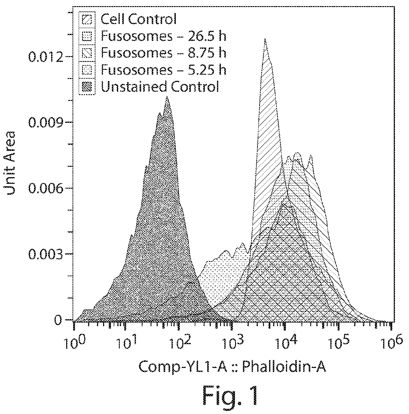
quantifies staining of fusosomes with a dye for F-actin.
is a graph showing the capacity for fusosomes and parent cells to polymerase actin over a period of 3, 5, and 24 hours.
is a table showing size distribution statistics of fusosomes and parental cells as measured by NTA and microscopy.
is a table showing the average size and volume of fusosomes and parental cells.
is a series of diagrams showing the soluble:insoluble ratio observed for fusosomes or a cell preparation.
is a series of diagrams showing MvH(CD8)+F fusosome fusion to target or non-target cells and absolute amount of targeted fusion.
is a diagram showing 2-NBDG mean fluorescence intensity in VSV-G fusosomes.
is a diagram showing esterase activity in the cytosol of VSV-G fusosomes.
A- 9 B are a series of diagrams showing Cre recombinase delivery by fusosomes as detected by bioluminescent imaging in mice. (A) Ventral image and luminescent signal overlay of exposed liver and spleen of IV fusosome treated mice (1× and 3× concentration). Lower portion is luminescent signal alone. (B) Total flux signal of fusosome targeted spleen and liver; y-scale is on log 10 scale. Mice treated with a concentration of 3× fusosome treatment had a significantly greater signal in the spleen (p=0.0004) than background 72 hours post-treatment.
A- 10 B are a series of diagrams showing Cre recombinase to murine liver and spleen by fusosomes as detected by bioluminescent imaging. (A) From left to right; dorsal image and luminescent signal overlay of excised liver, heart, lungs, kidney, small intestines, pancreas, and spleen collected and imaged within 5 minutes of euthanasia. Lower portion is luminescent signal alone. (B) Total flux signal of fusosome targeted spleen and liver and other tissues; y-scale is on log 10 scale. Mice treated with a concentration of 3× fusosome treatment had a significantly greater signal in the spleen (p<0.0001) as compared to the tissue with the lowest signal (heart).
is a table showing delivery of Cre cargo by NivG+F fusosomes via a non-endocytic pathway.
is a graph showing GAPDH: Total protein ratios measured by bicinchoninic acid assay in fusosomes and parental cells.
is a graph showing lipid: protein ratios measured by bicinchoninic acid assay in fusosomes and parental cells.
is a graph showing protein:DNA ratios measured by bicinchoninic acid assay in fusosomes and parental cells.
is a graph showing lipids: DNA ratios measured by bicinchoninic acid assay in fusosomes and parental cells.
is a graph showing protein levels of the exosome marker CD63 in exosomes and fusosomes.
is a graph showing the intensity of calnexin signal detected in fusosomes and parental cells.
is a graph showing lipid:DNA ratios determined for fusosomes and parental cells.
A- 19 B are a series of graphs showing the proportion of lipid species as a percentage of total lipids in parental cells, exosomes, and fusosomes.
is a series of graphs showing the protein content of parental cells, exosomes, and fusosomes with respect to proteins associated with specific compartments, as indicated.
is a series of graphs showing the level of ARRDC1 (left panel) or TSG101 (right panel) as a percentage of total protein content in parental cells, exosomes, and fusosomes.
A- 22 C show results for cell lines, including target human hepatoma cell lines (HepG2) and non-target (non-hepatic) cell lines, transduced with lentivirus (LV) encoding nucleic acid constructs containing positive TCSREs or NTCSREs. A shows GFP expression in human hepatoma cell line (HepG2), human embryonic kidney cell line (293LX), human T-cell line of hematopoietic origin (Molt4.8) and endothelial cell line derived from mouse brain (bEND.3) transduced with LV generated with miRT sequences (hPGK-eGFP+miRT) or without miRT sequences (hPGK-eGFP), under the control of the PGK promoter. B shows GFP expression in HepG2 and 293LX cells transduced with LV generated under the control of the PGK promoter (hPGK-eGFP) or LVs containing mirT sequences and GFP under the control of the hepatocyte specific promoter ApoE (hApoE-eGFP+miRT). C shows quantification of Phenylalanine (Phe) in supernatant of HepG2 and 293LX cells transduced with LVs containing the transgene phenylalanine ammonia lyase (PAL) under the control of the SFFV promoter (SFFV-PAL), or LVs containing mirT sequences and under the control of the hApoE promoter (hApoE-PAL+miRT).
DETAILED DESCRIPTION
The present disclosure provides, at least in part, fusosome methods and compositions for in vivo delivery. In some embodiments, the fusosome comprises a combination of elements that promote specificity for target cells, e.g., one or more of a re-targeted fusogen, a positive target cell-specific regulatory element, and a non-target cell-specific regulatory element. In some embodiments, the fusosome comprises one or more modifications that decrease an immune response against the fusosome.
Definitions
Terms used in the claims and specification are defined as set forth below unless otherwise specified.
As used herein, “detectably present”, when used in the context of an exogenous agent being detectably present, means that the exogenous agent itself is detectably present. For instance, if the exogenous agent is a protein, the exogenous protein agent can be detectably present regardless of whether a nucleic acid that encodes it is detectably present or not.
As used herein, “fusosome” refers to a bilayer of amphipathic lipids enclosing a lumen or cavity and a fusogen that interacts with the amphipathic lipid bilayer. In embodiments, the fusosome comprises a nucleic acid. In some embodiments, the fusosome is a membrane enclosed preparation. In some embodiments, the fusosome is derived from a source cell.
As used herein, “fusosome composition” refers to a composition comprising one or more fusosomes.
As used herein, “fusogen” refers to an agent or molecule that creates an interaction between two membrane enclosed lumens. In embodiments, the fusogen facilitates fusion of the membranes. In other embodiments, the fusogen creates a connection, e.g., a pore, between two lumens (e.g., a lumen of a retroviral vector and a cytoplasm of a target cell). In some embodiments, the fusogen comprises a complex of two or more proteins, e.g., wherein neither protein has fusogenic activity alone. In some embodiments, the fusogen comprises a targeting domain.
As used herein, an “insulator element” refers to a nucleotide sequence that blocks enhancers or prevents heterochromatin spreading. An insulator element can be wild-type or mutant.
The term “effective amount” as used herein means an amount of a pharmaceutical composition which is sufficient enough to significantly and positively modify the symptoms and/or conditions to be treated (e.g., provide a positive clinical response). The effective amount of an active ingredient for use in a pharmaceutical composition will vary with the particular condition being treated, the severity of the condition, the duration of treatment, the nature of concurrent therapy, the particular active ingredient(s) being employed, the particular pharmaceutically-acceptable excipient(s) and/or carrier(s) utilized, and like factors with the knowledge and expertise of the attending physician.
An “exogenous agent” as used herein with reference to a virus, VLP or fusosome, refers to an agent that is neither comprised by nor encoded in the corresponding wild-type virus or fusogen made from a corresponding wild-type source cell. In some embodiments, the exogenous agent does not naturally exist, such as a protein or nucleic acid that has a sequence that is altered (e.g., by insertion, deletion, or substitution) relative to a naturally occurring protein. In some embodiments, the exogenous agent does not naturally exist in the source cell. In some embodiments, the exogenous agent exists naturally in the source cell but is exogenous to the virus. In some embodiments, the exogenous agent does not naturally exist in the recipient cell. In some embodiments, the exogenous agent exists naturally in the recipient cell, but is not present at a desired level or at a desired time. In some embodiments, the exogenous agent comprises RNA or protein.
The term “pharmaceutically acceptable” as used herein, refers to excipients, compositions and/or dosage forms which are, within the scope of sound medical judgment, suitable for use in contact with the tissues of human beings and animals without excessive toxicity, irritation, allergic response, or other problem or complication, commensurate with a reasonable benefit/risk ratio.
As used herein, a “promoter” refers to a cis-regulatory DNA sequence that, when operably linked to a gene coding sequence, drives transcription of the gene. The promoter may comprise a transcription factor binding sites. In some embodiments, a promoter works in concert with one or more enhancers which are distal to the gene.
As used herein, a “positive target cell-specific regulatory element” (or positive TCSRE) refers to a nucleic acid sequence that increases the level of an exogenous agent in a target cell compared to in a non-target cell, wherein the nucleic acid encoding the exogenous agent is operably linked to the positive TCSRE. In some embodiments, the positive TCSRE is a functional nucleic acid sequence, e.g., the positive TCSRE can comprise a promoter or enhancer. In some embodiments, the positive TCSRE encodes a functional RNA sequence, e.g., the positive TCSRE can encode a splice site that promotes correct splicing of the RNA in the target cell. In some embodiments, the positive TCSRE encodes a functional protein sequence, or the positive TCSRE can encode a protein sequence that promotes correct post-translational modification of the protein. In some embodiments, the positive TCSRE decreases the level or activity of a downregulator or inhibitor of the exogenous agent. In some embodiments, the target cell is a liver cell and the positive target-cell-specific regulatory element is a positive liver cell-specific regulatory element.
As used herein, a “negative target cell-specific regulatory element” (or negative TCSRE) refers to a nucleic acid sequence that decreases the level of an exogenous agent in a non-target cell compared to in a target cell, wherein the nucleic acid encoding the exogenous agent is operably linked to the negative TCSRE. In some embodiments, the negative TCSRE is a functional nucleic acid sequence, e.g., a miRNA recognition site that causes degradation or inhibition of the retroviral nucleic acid in a non-target cell. In some embodiments, the nucleic acid sequence encodes a functional RNA sequence, e.g., the nucleic acid encodes an miRNA sequence present in an mRNA encoding an exogenous protein agent, such that the mRNA is degraded or inhibited in a non-target cell. In some embodiments, the negative TCSRE increases the level or activity of a downregulator or inhibitor of the exogenous agent. In some embodiment, the non-target cell is a non-liver cell.
As used herein, a “non-target cell-specific regulatory element” (or NTCSRE) refers to a nucleic acid sequence that decreases the level of an exogenous agent in a non-target cell compared to in a target cell, wherein the nucleic acid encoding the exogenous agent is operably linked to the NTCSRE. In some embodiments, the NTCSRE is a functional nucleic acid sequence, e.g., a miRNA recognition site that causes degradation or inhibition of the retroviral nucleic acid in a non-target cell. In some embodiments, the nucleic acid sequence encodes a functional RNA sequence, e.g., the nucleic acid encodes an miRNA sequence present in an mRNA encoding an exogenous protein agent, such that the mRNA is degraded or inhibited in a non-target cell. In some embodiments, the NTCSRE increases the level or activity of a downregulator or inhibitor of the exogenous agent. In some embodiments, the non-target cell is a non-liver cell and the non-target cell-specific regulatory element is a non-liver cell-specific regulatory element. The terms “negative TCSRE” and “NTCSRE” are used interchangeably herein.
As used herein, a “non-liver cell specific regulatory element” refers to a non-target cell-specific regulatory element (NTCSRE), wherein the target cell is a liver cell. Thus, a non-liver cell specific regulatory element refers to a nucleic acid sequence that decreases the level of an exogenous agent in a non-liver cell (e.g., in an immune cell) or tissue compared to in a liver cell, wherein the nucleic acid encoding the exogenous agent is operably linked to the non-liver cell-specific regulatory element.
As used herein, a “re-targeted fusogen” refers to a fusogen that comprises a targeting moiety having a sequence that is not part of the naturally-occurring form of the fusogen. In embodiments, the fusogen comprises a different targeting moiety relative to the targeting moiety in the naturally-occurring form of the fusogen. In embodiments, the naturally-occurring form of the fusogen lacks a targeting domain, and the re-targeted fusogen comprises a targeting moiety that is absent from the naturally-occurring form of the fusogen. In embodiments, the fusogen is modified to comprise a targeting moiety. In embodiments, the fusogen comprises one or more sequence alterations outside of the targeting moiety relative to the naturally-occurring form of the fusogen, e.g., in a transmembrane domain, fusogenically active domain, or cytoplasmic domain.
As used herein, a “retroviral nucleic acid” refers to a nucleic acid containing at least the minimal sequence requirements for packaging into a retrovirus or retroviral vector, alone or in combination with a helper cell, helper virus, or helper plasmid. In some embodiments, the retroviral nucleic acid further comprises or encodes an exogenous agent, a positive target cell-specific regulatory element, a non-target cell-specific regulatory element, or a negative TCSRE. In some embodiments, the retroviral nucleic acid comprises one or more of (e.g., all of) a 5′ LTR (e.g., to promote integration), U3 (e.g., to activate viral genomic RNA transcription), R (e.g., a Tat-binding region), U5, a 3′ LTR (e.g., to promote integration), a packaging site (e.g., psi (Ψ)), RRE (e.g., to bind to Rev and promote nuclear export). The retroviral nucleic acid can comprise RNA (e.g., when part of a virion) or DNA (e.g., when being introduced into a source cell or after reverse transcription in a recipient cell). In some embodiments, the retroviral nucleic acid is packaged using a helper cell, helper virus, or helper plasmid which comprises one or more of (e.g., all of) gag, pol, and env.
As used herein, a “target cell” refers to a cell of a type to which it is desired that a fusosome (e.g., lentiviral vector) deliver an exogenous agent. In embodiments, a target cell is a cell of a specific tissue type or class, e.g., an immune effector cell, e.g., a T cell. In some embodiments, a target cell is a diseased cell, e.g., a cancer cell. In some embodiments, the fusogen, e.g., re-targeted fusogen (alone or in combination with the positive TCSRE, NTCSRE, negative TCSRE, or any combination thereof) leads to preferential delivery of the exogenous agent to a target cell compared to a non-target cell.
As used herein a “non-target cell” refers to a cell of a type to which it is not desired that a lentiviral vector delivers an exogenous agent. In some embodiments, a non-target cell is a cell of a specific tissue type or class. In some embodiments, a non-target cell is a non-diseased cell, e.g., a non-cancerous cell. In some embodiments, the fusogen, e.g., re-targeted fusogen (alone or in combination with the positive TCSRE, NTCSRE, negative TCSRE or any combination thereof) leads to lower delivery of the exogenous agent to a non-target cell compared to a target cell.
As used herein, the terms “treat,” “treating,” or “treatment” refer to ameliorating a disease or disorder, e.g., slowing or arresting or reducing the development of the disease or disorder, e.g., a root cause of the disorder or at least one of the clinical symptoms thereof.
As used herein, “cytobiologic” refers to a portion of a cell that comprises a lumen and a cell membrane, or a cell having partial or complete nuclear inactivation. In some embodiments, the cytobiologic comprises one or more of a cytoskeleton component, an organelle, and a ribosome. In embodiments, the cytobiologic is an enucleated cell, a microvesicle, or a cell ghost.
Fusosomes, e.g., Cell-Derived Fusosomes
Fusosomes can take various forms. For example, in some embodiments, a fusosome described herein is derived from a source cell. A fusosome may be or comprise, e.g., an extracellular vesicle, a microvesicle, a nanovesicle, an exosome, an apoptotic body (from apoptotic cells), a microparticle (which may be derived from, e.g., platelets), an ectosome (derivable from, e.g., neutrophiles and monocytes in serum), a prostatosome (obtainable from prostate cancer cells), a cardiosome (derivable from cardiac cells), or any combination thereof. In some embodiments, a fusosome is released naturally from a source cell, and in some embodiments, the source cell is treated to enhance formation of fusosomes. In some embodiments, the fusosome is between about 10-10,000 nm in diameter, e.g., about 30-100 nm in diameter. In some embodiments, the fusosome comprises one or more synthetic lipids.
In some embodiments, the fusosome is or comprises a virus, e.g., a retrovirus, e.g., a lentivirus. In accordance with one embodiment of the invention, a fusosome comprising a lipid bilayer comprises a retroviral vector comprising an envelope. For instance, in some embodiments, the fusosome's bilayer of amphipathic lipids is or comprises the viral envelope. The viral envelope may comprise a fusogen, e.g., a fusogen that is endogenous to the virus or a pseudotyped fusogen. In some embodiments, the fusosome's lumen or cavity comprises a viral nucleic acid, e.g., a retroviral nucleic acid, e.g., a lentiviral nucleic acid. The viral nucleic acid may be a viral genome. In some embodiments, the fusosome further comprises one or more viral non-structural proteins, e.g., in its cavity or lumen.
Fusosomes may have various properties that facilitate delivery of a payload, such as a desired transgene or encoding an exogenous agent, to a target cell. For instance, in some embodiments, the fusosome and the source cell together comprise nucleic acid(s) sufficient to make a particle that can fuse with a target cell. In embodiments, these nucleic acid(s) encode proteins having one or more of (e.g., all of) the following activities: gag polyprotein activity, polymerase activity, integrase activity, protease activity, and fusogen activity.
Fusosomes may also comprise various structures that facilitate delivery of a payload to a target cell. For instance, in some embodiments, the fusosome (e.g., virus, e.g., retrovirus, e.g., lentivirus) comprises one or more of (e.g., all of) the following proteins: gag polyprotein, polymerase (e.g., pol), integrase (e.g., a functional or non-functional variant), protease, and a fusogen. In some embodiments, the fusosome further comprises rev. In some embodiments, one or more of the aforesaid proteins are encoded in the retroviral genome, and in some embodiments, one or more of the aforesaid proteins are provided in trans, e.g., by a helper cell, helper virus, or helper plasmid. In some embodiments, the fusosome nucleic acid (e.g., retroviral nucleic acid) comprises one or more of (e.g., all of) the following nucleic acid sequences: 5′ LTR (e.g., comprising U5 and lacking a functional U3 domain), Psi packaging element (Psi), Central polypurine tract (cPPT) Promoter operatively linked to the payload gene, payload gene (optionally comprising an intron before the open reading frame), Poly A tail sequence, WPRE, and 3′ LTR (e.g., comprising U5 and lacking a functional U3). In some embodiments the fusosome nucleic acid (e.g., retroviral nucleic acid) further comprises one or more insulator element. In some embodiments the fusosome nucleic acid (e.g., retroviral nucleic acid) further comprises one or more miRNA recognition sites. In some embodiments, one or more of the miRNA recognition sites are situated downstream of the poly A tail sequence, e.g., between the poly A tail sequence and the WPRE.
In some embodiments, a fusosome provided herein is administered to a subject, e.g., a mammal, e.g., a human. In such embodiments, the subject may be at risk of, may have a symptom of, or may be diagnosed with or identified as having, a particular disease or condition (e.g., a disease or condition described herein). In one embodiment, the subject has a genetic deficiency, such as any listed in Table 5. In some embodiments, the fusosome contains nucleic acid sequences encoding an exogenous agent for treating the disease or condition, such as for treating the genetic deficiency.
Lentiviral Components and Helper Cells
In some embodiments, the retroviral nucleic acid comprises one or more of (e.g., all of): a 5′ promoter (e.g., to control expression of the entire packaged RNA), a 5′ LTR (e.g., that includes R (polyadenylation tail signal) and/or U5 which includes a primer activation signal), a primer binding site, a psi packaging signal, a RRE element for nuclear export, a promoter directly upstream of the transgene to control transgene expression, a transgene (or other exogenous agent element), a polypurine tract, and a 3′ LTR (e.g., that includes a mutated U3, a R, and U5). In some embodiments, the retroviral nucleic acid further comprises one or more of a cPPT, a WPRE, and/or an insulator element.
A retrovirus typically replicates by reverse transcription of its genomic RNA into a linear double-stranded DNA copy and subsequently covalently integrates its genomic DNA into a host genome. Illustrative retroviruses suitable for use in particular embodiments, include, but are not limited to: Moloney murine leukemia virus (M-MuLV), Moloney murine sarcoma virus (MoMSV), Harvey murine sarcoma virus (HaMuSV), murine mammary tumor virus (MuMTV), gibbon ape leukemia virus (GaLV), feline leukemia virus (FLV), spumavirus, Friend murine leukemia virus, Murine Stem Cell Virus (MSCV) and Rous Sarcoma Virus (RSV)) and lentivirus.
In some embodiments the retrovirus is a Gammretrovirus. In some embodiments the retrovirus is an Epsilonretrovirus. In some embodiments the retrovirus is an Alpharetrovirus. In some embodiments the retrovirus is a Betaretrovirus. In some embodiments the retrovirus is a Deltaretrovirus. In some embodiments the retrovirus is a Lentivirus. In some embodiments the retrovirus is a Spumaretrovirus. In some embodiments the retrovirus is an endogenous retrovirus.
Illustrative lentiviruses include, but are not limited to: HIV (human immunodeficiency virus; including HIV type 1, and HIV type 2); visna-maedi virus (VMV) virus; the caprine arthritis-encephalitis virus (CAEV); equine infectious anemia virus (EIAV); feline immunodeficiency virus (FIV); bovine immune deficiency virus (BIV); and simian immunodeficiency virus (SIV). In some embodiments, HIV based vector backbones (i.e., HIV cis-acting sequence elements) are used.
In some embodiments, a vector herein is a nucleic acid molecule capable transferring or transporting another nucleic acid molecule. The transferred nucleic acid is generally linked to, e.g., inserted into, the vector nucleic acid molecule. A vector may include sequences that direct autonomous replication in a cell, or may include sequences sufficient to allow integration into host cell DNA. Useful vectors include, for example, plasmids (e.g., DNA plasmids or RNA plasmids), transposons, cosmids, bacterial artificial chromosomes, and viral vectors. Useful viral vectors include, e.g., replication defective retroviruses and lentiviruses.
A viral vector can comprise, e.g., a nucleic acid molecule (e.g., a transfer plasmid) that includes virus-derived nucleic acid elements that typically facilitate transfer of the nucleic acid molecule or integration into the genome of a cell or to a viral particle that mediates nucleic acid transfer. Viral particles will typically include various viral components and sometimes also host cell components in addition to nucleic acid(s). A viral vector can comprise, e.g., a virus or viral particle capable of transferring a nucleic acid into a cell, or to the transferred nucleic acid (e.g., as naked DNA). Viral vectors and transfer plasmids can comprise structural and/or functional genetic elements that are primarily derived from a virus. A retroviral vector can comprise a viral vector or plasmid containing structural and functional genetic elements, or portions thereof, that are primarily derived from a retrovirus. A lentiviral vector can comprise a viral vector or plasmid containing structural and functional genetic elements, or portions thereof, including LTRs that are primarily derived from a lentivirus.
In embodiments, a lentiviral vector (e.g., lentiviral expression vector) may comprise a lentiviral transfer plasmid (e.g., as naked DNA) or an infectious lentiviral particle. With respect to elements such as cloning sites, promoters, regulatory elements, heterologous nucleic acids, etc., it is to be understood that the sequences of these elements can be present in RNA form in lentiviral particles and can be present in DNA form in DNA plasmids.
In some vectors described herein, at least part of one or more protein coding regions that contribute to or are essential for replication may be absent compared to the corresponding wild-type virus. This makes the viral vector replication-defective. In some embodiments, the vector is capable of transducing a target non-dividing host cell and/or integrating its genome into a host genome.
The structure of a wild-type retrovirus genome often comprises a 5′ long terminal repeat (LTR) and a 3′ LTR, between or within which are located a packaging signal to enable the genome to be packaged, a primer binding site, integration sites to enable integration into a host cell genome and gag, pol and env genes encoding the packaging components which promote the assembly of viral particles. More complex retroviruses have additional features, such as rev and RRE sequences in HIV, which enable the efficient export of RNA transcripts of the integrated provirus from the nucleus to the cytoplasm of an infected target cell. In the provirus, the viral genes are flanked at both ends by regions called long terminal repeats (LTRs). The LTRs are involved in proviral integration and transcription. LTRs also serve as enhancer-promoter sequences and can control the expression of the viral genes. Encapsidation of the retroviral RNAs occurs by virtue of a psi sequence located at the 5′ end of the viral genome.
The LTRs themselves are typically similar (e.g., identical) sequences that can be divided into three elements, which are called U3, R and U5. U3 is derived from the sequence unique to the 3′ end of the RNA. R is derived from a sequence repeated at both ends of the RNA and U5 is derived from the sequence unique to the 5′ end of the RNA. The sizes of the three elements can vary considerably among different retroviruses.
For the viral genome, the site of transcription initiation is typically at the boundary between U3 and R in one LTR and the site of poly (A) addition (termination) is at the boundary between R and U5 in the other LTR. U3 contains most of the transcriptional control elements of the provirus, which include the promoter and multiple enhancer sequences responsive to cellular and in some cases, viral transcriptional activator proteins. Some retroviruses comprise any one or more of the following genes that code for proteins that are involved in the regulation of gene expression: tot, rev, tax and rex.
With regard to the structural genes gag, pol and env themselves, gag encodes the internal structural protein of the virus. Gag protein is proteolytically processed into the mature proteins MA (matrix), CA (capsid) and NC (nucleocapsid). The pol gene encodes the reverse transcriptase (RT), which contains DNA polymerase, associated RNase H and integrase (IN), which mediate replication of the genome. The env gene encodes the surface (SU) glycoprotein and the transmembrane (TM) protein of the virion, which form a complex that interacts specifically with cellular receptor proteins. This interaction promotes infection, e.g., by fusion of the viral membrane with the cell membrane.
In a replication-defective retroviral vector genome gag, pol and env may be absent or not functional. The R regions at both ends of the RNA are typically repeated sequences. U5 and U3 represent unique sequences at the 5′ and 3′ ends of the RNA genome respectively.
Retroviruses may also contain additional genes which code for proteins other than gag, pol and env. Examples of additional genes include (in HIV), one or more of vif, vpr, vpx, vpu, tat, rev and nef. EIAV has (amongst others) the additional gene S2. Proteins encoded by additional genes serve various functions, some of which may be duplicative of a function provided by a cellular protein. In EIAV, for example, tat acts as a transcriptional activator of the viral LTR (Derse and Newbold 1993 Virology 194:530-6; Maury et al. 1994 Virology 200:632-42). It binds to a stable, stem-loop RNA secondary structure referred to as TAR. Rev regulates and co-ordinates the expression of viral genes through rev-response elements (RRE) (Martarano et al. 1994 J. Virol. 68:3102-11). The mechanisms of action of these two proteins are thought to be broadly similar to the analogous mechanisms in the primate viruses. In addition, an EIAV protein, Ttm, has been identified that is encoded by the first exon of tat spliced to the env coding sequence at the start of the transmembrane protein.
In addition to protease, reverse transcriptase and integrase, non-primate lentiviruses contain a fourth pol gene product which codes for a dUTPase. This may play a role in the ability of these lentiviruses to infect certain non-dividing or slowly dividing cell types.
In embodiments, a recombinant lentiviral vector (RLV) is a vector with sufficient retroviral genetic information to allow packaging of an RNA genome, in the presence of packaging components, into a viral particle capable of infecting a target cell. Infection of the target cell can comprise reverse transcription and integration into the target cell genome. The RLV typically carries non-viral coding sequences which are to be delivered by the vector to the target cell. In embodiments, an RLV is incapable of independent replication to produce infectious retroviral particles within the target cell. Usually the RLV lacks a functional gag-pol and/or env gene and/or other genes involved in replication. The vector may be configured as a split-intron vector, e.g., as described in PCT patent application WO 99/15683, which is herein incorporated by reference in its entirety.
In some embodiments, the lentiviral vector comprises a minimal viral genome, e.g., the viral vector has been manipulated so as to remove the non-essential elements and to retain the essential elements in order to provide the required functionality to infect, transduce and deliver a nucleotide sequence of interest to a target host cell, e.g., as described in WO 98/17815, which is herein incorporated by reference in its entirety.
A minimal lentiviral genome may comprise, e.g., (5′)R-U5-one or more first nucleotide sequences-U3-R(3′). However, the plasmid vector used to produce the lentiviral genome within a source cell can also include transcriptional regulatory control sequences operably linked to the lentiviral genome to direct transcription of the genome in a source cell. These regulatory sequences may comprise the natural sequences associated with the transcribed retroviral sequence, e.g., the 5′ U3 region, or they may comprise a heterologous promoter such as another viral promoter, for example the CMV promoter. Some lentiviral genomes comprise additional sequences to promote efficient virus production. For example, in the case of HIV, rev and RRE sequences may be included. Alternatively or combination, codon optimization may be used, e.g., the gene encoding the exogenous agent may be codon optimized, e.g., as described in WO 01/79518, which is herein incorporated by reference in its entirety. Alternative sequences which perform a similar or the same function as the rev/RRE system may also be used. For example, a functional analogue of the rev/RRE system is found in the Mason Pfizer monkey virus. This is known as CTE and comprises an RRE-type sequence in the genome which is believed to interact with a factor in the infected cell. The cellular factor can be thought of as a rev analogue. Thus, CTE may be used as an alternative to the rev/RRE system. In addition, the Rex protein of HTLV-I can functionally replace the Rev protein of HIV-I. Rev and Rex have similar effects to IRE-BP.
In some embodiments, a retroviral nucleic acid (e.g., a lentiviral nucleic acid, e.g., a primate or non-primate lentiviral nucleic acid) (1) comprises a deleted gag gene wherein the deletion in gag removes one or more nucleotides downstream of about nucleotide 350 or 354 of the gag coding sequence; (2) has one or more accessory genes absent from the retroviral nucleic acid; (3) lacks the tat gene but includes the leader sequence between the end of the 5′ LTR and the ATG of gag; and (4) combinations of (1), (2) and (3). In an embodiment the lentiviral vector comprises all of features (1) and (2) and (3). This strategy is described in more detail in WO 99/32646, which is herein incorporated by reference in its entirety.
In some embodiments, a primate lentivirus minimal system requires none of the HIV/SIV additional genes vif, vpr, vpx, vpu, tat, rev and nef for either vector production or for transduction of dividing and non-dividing cells. In some embodiments, an EIAV minimal vector system does not require S2 for either vector production or for transduction of dividing and non-dividing cells.
The deletion of additional genes may permit vectors to be produced without the genes associated with disease in lentiviral (e.g. HIV) infections. In particular, tat is associated with disease. Secondly, the deletion of additional genes permits the vector to package more heterologous DNA. Thirdly, genes whose function is unknown, such as S2, may be omitted, thus reducing the risk of causing undesired effects. Examples of minimal lentiviral vectors are disclosed in WO 99/32646 and in WO 98/17815.
In some embodiments, the retroviral nucleic acid is devoid of at least tat and S2 (if it is an EIAV vector system), and possibly also vif, vpr, vpx, vpu and nef. In some embodiments, the retroviral nucleic acid is also devoid of rev, RRE, or both.
In some embodiments the retroviral nucleic acid comprises vpx. The Vpx polypeptide binds to and induces the degradation of the SAMHD1 restriction factor, which degrades free dNTPs in the cytoplasm. Thus, the concentration of free dNTPs in the cytoplasm increases as Vpx degrades SAMHD1 and reverse transcription activity is increased, thus facilitating reverse transcription of the retroviral genome and integration into the target cell genome.
Different cells differ in their usage of particular codons. This codon bias corresponds to a bias in the relative abundance of particular tRNAs in the cell type. By altering the codons in the sequence so that they are tailored to match with the relative abundance of corresponding tRNAs, it is possible to increase expression. By the same token, it is possible to decrease expression by deliberately choosing codons for which the corresponding tRNAs are known to be rare in the particular cell type. Thus, an additional degree of translational control is available. An additional description of codon optimization is found, e.g., in WO 99/41397, which is herein incorporated by reference in its entirety.
Many viruses, including HIV and other lentiviruses, use a large number of rare codons and by changing these to correspond to commonly used mammalian codons, increased expression of the packaging components in mammalian producer cells can be achieved.
Codon optimization has a number of other advantages. By virtue of alterations in their sequences, the nucleotide sequences encoding the packaging components may have RNA instability sequences (INS) reduced or eliminated from them. At the same time, the amino acid sequence coding sequence for the packaging components is retained so that the viral components encoded by the sequences remain the same, or at least sufficiently similar that the function of the packaging components is not compromised. In some embodiments, codon optimization also overcomes the Rev/RRE requirement for export, rendering optimized sequences Rev independent. In some embodiments, codon optimization also reduces homologous recombination between different constructs within the vector system (for example between the regions of overlap in the gag-pol and env open reading frames). In some embodiments, codon optimization leads to an increase in viral titer and/or improved safety.
In some embodiments, only codons relating to INS are codon optimized. In other embodiments, the sequences are codon optimized in their entirety, with the exception of the sequence encompassing the frameshift site of gag-pol.
The gag-pol gene comprises two overlapping reading frames encoding the gag-pol proteins. The expression of both proteins depends on a frameshift during translation. This frameshift occurs as a result of ribosome “slippage” during translation. This slippage is thought to be caused at least in part by ribosome-stalling RNA secondary structures. Such secondary structures exist downstream of the frameshift site in the gag-pol gene. For HIV, the region of overlap extends from nucleotide 1222 downstream of the beginning of gag (wherein nucleotide 1 is the A of the gag ATG) to the end of gag (nt 1503). Consequently, a 281 bp fragment spanning the frameshift site and the overlapping region of the two reading frames is preferably not codon optimized. In some embodiments, retaining this fragment will enable more efficient expression of the gag-pol proteins. For EIAV, the beginning of the overlap is at nt 1262 (where nucleotide 1 is the A of the gag ATG). The end of the overlap is at nt 1461. In order to ensure that the frameshift site and the gag-pol overlap are preserved, the wild type sequence may be retained from nt 1156 to 1465.
Derivations from optimal codon usage may be made, for example, in order to accommodate convenient restriction sites, and conservative amino acid changes may be introduced into the gag-pol proteins.
In some embodiments, codon optimization is based on codons with poor codon usage in mammalian systems. The third and sometimes the second and third base may be changed.
Due to the degenerate nature of the genetic code, it will be appreciated that numerous gag-pol sequences can be achieved by a skilled worker. Also, there are many retroviral variants described which can be used as a starting point for generating a codon optimized gag-pol sequence. Lentiviral genomes can be quite variable. For example there are many quasi-species of HIV-I which are still functional. This is also the case for EIAV. These variants may be used to enhance particular parts of the transduction process. Examples of HIV-I variants may be found in the HIV databases maintained by Los Alamos National Laboratory. Details of EIAV clones may be found at the NCBI database maintained by the National Institutes of Health.
The strategy for codon optimized gag-pol sequences can be used in relation to any retrovirus, e.g., EIAV, FIV, BIV, CAEV, VMR, SIV, HIV-I and HIV-2. In addition this method could be used to increase expression of genes from HTLV-I, HTLV-2, HFV, HSRV and human endogenous retroviruses (HERV), MLV and other retroviruses.
As described above, the packaging components for a retroviral vector can include expression products of gag, pol and env genes. In addition, packaging can utilize a short sequence of 4 stem loops followed by a partial sequence from gag and env as a packaging signal. Thus, inclusion of a deleted gag sequence in the retroviral vector genome (in addition to the full gag sequence on the packaging construct) can be used. In embodiments, the retroviral vector comprises a packaging signal that comprises from 255 to 360 nucleotides of gag in vectors that still retain env sequences, or about 40 nucleotides of gag in a particular combination of splice donor mutation, gag and env deletions. In some embodiments, the retroviral vector includes a gag sequence which comprises one or more deletions, e.g., the gag sequence comprises about 360 nucleotides derivable from the N-terminus.
The retroviral vector, helper cell, helper virus, or helper plasmid may comprise retroviral structural and accessory proteins, for example gag, pol, env, tat, rev, vif, vpr, vpu, vpx, or nef proteins or other retroviral proteins. In some embodiments the retroviral proteins are derived from the same retrovirus. In some embodiments the retroviral proteins are derived from more than one retrovirus, e.g. 2, 3, 4, or more retroviruses.
The gag and pol coding sequences are generally organized as the Gag-Pol Precursor in native lentivirus. The gag sequence codes for a 55-kD Gag precursor protein, also called p55. The p55 is cleaved by the virally encoded protease4 (a product of the pol gene) during the process of maturation into four smaller proteins designated MA (matrix [p17]), CA (capsid [p24]), NC (nucleocapsid [p9]), and p6. The pol precursor protein is cleaved away from Gag by a virally encoded protease, and further digested to separate the protease (p10), RT (p50), RNase H (p15), and integrase (p31) activities.
Native Gag-Pol sequences can be utilized in a helper vector (e.g., helper plasmid or helper virus), or modifications can be made. These modifications include, chimeric Gag-Pol, where the Gag and Pol sequences are obtained from different viruses (e.g., different species, subspecies, strains, clades, etc.), and/or where the sequences have been modified to improve transcription and/or translation, and/or reduce recombination.
In various examples, the retroviral nucleic acid includes a polynucleotide encoding a 150-250 (e.g., 168) nucleotide portion of a gag protein that (i) includes a mutated INS1 inhibitory sequence that reduces restriction of nuclear export of RNA relative to wild-type INS1, (ii) contains two nucleotide insertion that results in frame shift and premature termination, and/or (iii) does not include INS2, INS3, and INS4 inhibitory sequences of gag.
In some embodiments, a vector described herein is a hybrid vector that comprises both retroviral (e.g., lentiviral) sequences and non-lentiviral viral sequences. In some embodiments, a hybrid vector comprises retroviral e.g., lentiviral, sequences for reverse transcription, replication, integration and/or packaging.
According to certain specific embodiments, most or all of the viral vector backbone sequences are derived from a lentivirus, e.g., HIV-1. However, it is to be understood that many different sources of retroviral and/or lentiviral sequences can be used, or combined and numerous substitutions and alterations in certain of the lentiviral sequences may be accommodated without impairing the ability of a transfer vector to perform the functions described herein. A variety of lentiviral vectors are described in Naldini et al., (1996a, 1996b, and 1998); Zufferey et al., (1997); Dull et al., 1998, U.S. Pat. Nos. 6,013,516; and 5,994,136, many of which may be adapted to produce a retroviral nucleic acid.
At each end of the provirus, long terminal repeats (LTRs) are typically found. An LTR typically comprises a domain located at the ends of retroviral nucleic acid which, in their natural sequence context, are direct repeats and contain U3, R and U5 regions. LTRs generally promote the expression of retroviral genes (e.g., promotion, initiation and polyadenylation of gene transcripts) and viral replication. The LTR can comprise numerous regulatory signals including transcriptional control elements, polyadenylation signals and sequences for replication and integration of the viral genome. The viral LTR is typically divided into three regions called U3, R and U5. The U3 region typically contains the enhancer and promoter elements. The U5 region is typically the sequence between the primer binding site and the R region and can contain the polyadenylation sequence. The R (repeat) region can be flanked by the U3 and U5 regions. The LTR is typically composed of U3, R and U5 regions and can appear at both the 5′ and 3′ ends of the viral genome. In some embodiments, adjacent to the 5′ LTR are sequences for reverse transcription of the genome (the tRNA primer binding site) and for efficient packaging of viral RNA into particles (the Psi site).
A packaging signal can comprise a sequence located within the retroviral genome which mediate insertion of the viral RNA into the viral capsid or particle, see e.g., Clever et al., 1995. J. of Virology, Vol. 69, No. 4; pp. 2101-2109. Several retroviral vectors use a minimal packaging signal (a psi [Ψ] sequence) for encapsidation of the viral genome.
In various embodiments, retroviral nucleic acids comprise modified 5′ LTR and/or 3′ LTRs. Either or both of the LTR may comprise one or more modifications including, but not limited to, one or more deletions, insertions, or substitutions. Modifications of the 3′ LTR are often made to improve the safety of lentiviral or retroviral systems by rendering viruses replication-defective, e.g., virus that is not capable of complete, effective replication such that infective virions are not produced (e.g., replication-defective lentiviral progeny).
In some embodiments, a vector is a self-inactivating (SIN) vector, e.g., replication-defective vector, e.g., retroviral or lentiviral vector, in which the right (3′) LTR enhancer-promoter region, known as the U3 region, has been modified (e.g., by deletion or substitution) to prevent viral transcription beyond the first round of viral replication. This is because the right (3′) LTR U3 region can be used as a template for the left (5′) LTR U3 region during viral replication and, thus, absence of the U3 enhancer-promoter inhibits viral replication. In embodiments, the 3′ LTR is modified such that the U5 region is removed, altered, or replaced, for example, with an exogenous poly(A) sequence The 3′ LTR, the 5′ LTR, or both 3′ and 5′ LTRs, may be modified LTRs.
In some embodiments, the U3 region of the 5′ LTR is replaced with a heterologous promoter to drive transcription of the viral genome during production of viral particles. Examples of heterologous promoters which can be used include, for example, viral simian virus 40 (SV40) (e.g., early or late), cytomegalovirus (CMV) (e.g., immediate early), Moloney murine leukemia virus (MoMLV), Rous sarcoma virus (RSV), and herpes simplex virus (HSV) (thymidine kinase) promoters. In some embodiments, promoters are able to drive high levels of transcription in a Tat-independent manner. In certain embodiments, the heterologous promoter has additional advantages in controlling the manner in which the viral genome is transcribed. For example, the heterologous promoter can be inducible, such that transcription of all or part of the viral genome will occur only when the induction factors are present. Induction factors include, but are not limited to, one or more chemical compounds or the physiological conditions such as temperature or pH, in which the host cells are cultured.
In some embodiments, viral vectors comprise a TAR (trans-activation response) element, e.g., located in the R region of lentiviral (e.g., HIV) LTRs. This element interacts with the lentiviral trans-activator (tat) genetic element to enhance viral replication. However, this element is not required, e.g., in embodiments wherein the U3 region of the 5′ LTR is replaced by a heterologous promoter.
The R region, e.g., the region within retroviral LTRs beginning at the start of the capping group (i.e., the start of transcription) and ending immediately prior to the start of the poly A tract can be flanked by the U3 and U5 regions. The R region plays a role during reverse transcription in the transfer of nascent DNA from one end of the genome to the other.
The retroviral nucleic acid can also comprise a FLAP element, e.g., a nucleic acid whose sequence includes the central polypurine tract and central termination sequences (cPPT and CTS) of a retrovirus, e.g., HIV-1 or HIV-2. Suitable FLAP elements are described in U.S. Pat. No. 6,682,907 and in Zennou, et al., 2000, Cell, 101:173, which are herein incorporated by reference in their entireties. During HIV-1 reverse transcription, central initiation of the plus-strand DNA at the central polypurine tract (cPPT) and central termination at the central termination sequence (CTS) can lead to the formation of a three-stranded DNA structure: the HIV-1 central DNA flap. In some embodiments, the retroviral or lentiviral vector backbones comprise one or more FLAP elements upstream or downstream of the gene encoding the exogenous agent. For example, in some embodiments a transfer plasmid includes a FLAP element, e.g., a FLAP element derived or isolated from HIV-1.
In embodiments, a retroviral or lentiviral nucleic acid comprises one or more export elements, e.g., a cis-acting post-transcriptional regulatory element which regulates the transport of an RNA transcript from the nucleus to the cytoplasm of a cell. Examples of RNA export elements include, but are not limited to, the human immunodeficiency virus (HIV) rev response element (RRE) (see e.g., Cullen et al., 1991. J. Virol. 65: 1053; and Cullen et al., 1991. Cell 58: 423), and the hepatitis B virus post-transcriptional regulatory element (HPRE), which are herein incorporated by reference in their entireties. Generally, the RNA export element is placed within the 3′ UTR of a gene, and can be inserted as one or multiple copies.
In some embodiments, expression of heterologous sequences in viral vectors is increased by incorporating one or more of, e.g., all of, posttranscriptional regulatory elements, polyadenylation sites, and transcription termination signals into the vectors. A variety of posttranscriptional regulatory elements can increase expression of a heterologous nucleic acid at the protein, e.g., woodchuck hepatitis virus posttranscriptional regulatory element (WPRE; Zufferey et al., 1999, J. Virol., 73:2886); the posttranscriptional regulatory element present in hepatitis B virus (HPRE) (Huang et al., Mol. Cell. Biol., 5:3864); and the like (Liu et al., 1995, Genes Dev., 9:1766), each of which is herein incorporated by reference in its entirety. In some embodiments, a retroviral nucleic acid described herein comprises a posttranscriptional regulatory element such as a WPRE or HPRE
In some embodiments, a retroviral nucleic acid described herein lacks or does not comprise a posttranscriptional regulatory element such as a WPRE or HPRE.
Elements directing the termination and polyadenylation of the heterologous nucleic acid transcripts may be included, e.g., to increases expression of the exogenous agent. Transcription termination signals may be found downstream of the polyadenylation signal. In some embodiments, vectors comprise a polyadenylation sequence 3′ of a polynucleotide encoding the exogenous agent. A polyA site may comprise a DNA sequence which directs both the termination and polyadenylation of the nascent RNA transcript by RNA polymerase II. Polyadenylation sequences can promote mRNA stability by addition of a polyA tail to the 3′ end of the coding sequence and thus, contribute to increased translational efficiency. Illustrative examples of polyA signals that can be used in a retroviral nucleic acid, include AATAAA, ATTAAA, AGTAAA, a bovine growth hormone polyA sequence (BGHpA), a rabbit β-globin polyA sequence (rpgpA), or another suitable heterologous or endogenous polyA sequence.
In some embodiments, a retroviral or lentiviral vector further comprises one or more insulator elements, e.g., an insulator element described herein.
In various embodiments, the vectors comprise a promoter operably linked to a polynucleotide encoding an exogenous agent. The vectors may have one or more LTRs, wherein either LTR comprises one or more modifications, such as one or more nucleotide substitutions, additions, or deletions. The vectors may further comprise one of more accessory elements to increase transduction efficiency (e.g., a cPPT/FLAP), viral packaging (e.g., a Psi (Ψ) packaging signal, RRE), and/or other elements that increase exogenous gene expression (e.g., poly (A) sequences), and may optionally comprise a WPRE or HPRE.
In some embodiments, a lentiviral nucleic acid comprises one or more of, e.g., all of, e.g., from 5′ to 3′, a promoter (e.g., CMV), an R sequence (e.g., comprising TAR), a U5 sequence (e.g., for integration), a PBS sequence (e.g., for reverse transcription), a DIS sequence (e.g., for genome dimerization), a psi packaging signal, a partial gag sequence, an RRE sequence (e.g., for nuclear export), a cPPT sequence (e.g., for nuclear import), a promoter to drive expression of the exogenous agent, a gene encoding the exogenous agent, a WPRE sequence (e.g., for efficient transgene expression), a PPT sequence (e.g., for reverse transcription), an R sequence (e.g., for polyadenylation and termination), and a U5 signal (e.g., for integration).
Vectors Engineered to Remove Splice Sites
Some lentiviral vectors integrate inside active genes and possess strong splicing and polyadenylation signals that could lead to the formation of aberrant and possibly truncated transcripts.
Mechanisms of proto-oncogene activation may involve the generation of chimeric transcripts originating from the interaction of promoter elements or splice sites contained in the genome of the insertional mutagen with the cellular transcriptional unit targeted by integration (Gabriel et al. 2009. Nat Med 15: 1431-1436; Bokhoven, et al. J Virol 83:283-29). Chimeric fusion transcripts comprising vector sequences and cellular mRNAs can be generated either by read-through transcription starting from vector sequences and proceeding into the flanking cellular genes, or vice versa.
In some embodiments, a lentiviral nucleic acid described herein comprises a lentiviral backbone in which at least two of the splice sites have been eliminated, e.g., to improve the safety profile of the lentiviral vector. Species of such splice sites and methods of identification are described in WO2012156839A2, all of which is included by reference.
Retroviral Production Methods
Large scale viral particle production is often useful to achieve a desired viral titer. Viral particles can be produced by transfecting a transfer vector into a packaging cell line that comprises viral structural and/or accessory genes, e.g., gag, pol, env, tat, rev, vif, vpr, vpu, vpx, or nef genes or other retroviral genes.
In embodiments, the packaging vector is an expression vector or viral vector that lacks a packaging signal and comprises a polynucleotide encoding one, two, three, four or more viral structural and/or accessory genes. Typically, the packaging vectors are included in a packaging cell, and are introduced into the cell via transfection, transduction or infection. A retroviral, e.g., lentiviral, transfer vector can be introduced into a packaging cell line, via transfection, transduction or infection, to generate a source cell or cell line. The packaging vectors can be introduced into human cells or cell lines by standard methods including, e.g., calcium phosphate transfection, lipofection or electroporation. In some embodiments, the packaging vectors are introduced into the cells together with a dominant selectable marker, such as neomycin, hygromycin, puromycin, blastocidin, zeocin, thymidine kinase, DHFR, Gln synthetase or ADA, followed by selection in the presence of the appropriate drug and isolation of clones. A selectable marker gene can be linked physically to genes encoding by the packaging vector, e.g., by IRES or self cleaving viral peptides.
Packaging cell lines include cell lines that do not contain a packaging signal, but do stably or transiently express viral structural proteins and replication enzymes (e.g., gag, pol and env) which can package viral particles. Any suitable cell line can be employed, e.g., mammalian cells, e.g., human cells. Suitable cell lines which can be used include, for example, CHO cells, BHK cells, MDCK cells, C3H 10T1/2 cells, FLY cells, Psi-2 cells, BOSC 23 cells, PA317 cells, WEHI cells, COS cells, BSC 1 cells, BSC 40 cells, BMT 10 cells, VERO cells, W138 cells, MRC5 cells, A549 cells, HT1080 cells, 293 cells, 293T cells, B-50 cells, 3T3 cells, NIH3T3 cells, HepG2 cells, Saos-2 cells, Huh7 cells, HeLa cells, W163 cells, 211 cells, and 211A cells. In embodiments, the packaging cells are 293 cells, 293T cells, or A549 cells.
A source cell line includes a cell line which is capable of producing recombinant retroviral particles, comprising a packaging cell line and a transfer vector construct comprising a packaging signal. Methods of preparing viral stock solutions are illustrated by, e.g., Y. Soneoka et al. (1995) Nucl. Acids Res. 23:628-633, and N. R. Landau et al. (1992) J. Virol. 66:5110-5113, which are incorporated herein by reference. Infectious virus particles may be collected from the packaging cells, e.g., by cell lysis, or collection of the supernatant of the cell culture. Optionally, the collected virus particles may be enriched or purified.
Packaging Plasmids and Cell Lines
In some embodiments, the source cell comprises one or more plasmids coding for viral structural proteins and replication enzymes (e.g., gag, pol and env) which can package viral particles. In some embodiments, the sequences coding for at least two of the gag, pol, and env precursors are on the same plasmid. In some embodiments, the sequences coding for the gag, pol, and env precursors are on different plasmids. In some embodiments, the sequences coding for the gag, pol, and env precursors have the same expression signal, e.g., promoter. In some embodiments, the sequences coding for the gag, pol, and env precursors have a different expression signal, e.g., different promoters. In some embodiments, expression of the gag, pol, and env precursors is inducible. In some embodiments, the plasmids coding for viral structural proteins and replication enzymes are transfected at the same time or at different times. In some embodiments, the plasmids coding for viral structural proteins and replication enzymes are transfected at the same time or at a different time from the packaging vector.
In some embodiments, the source cell line comprises one or more stably integrated viral structural genes. In some embodiments expression of the stably integrated viral structural genes is inducible.
In some embodiments, expression of the viral structural genes is regulated at the transcriptional level. In some embodiments, expression of the viral structural genes is regulated at the translational level. In some embodiments, expression of the viral structural genes is regulated at the post-translational level.
In some embodiments, expression of the viral structural genes is regulated by a tetracycline (Tet)-dependent system, in which a Tet-regulated transcriptional repressor (Tet-R) binds to DNA sequences included in a promoter and represses transcription by steric hindrance (Yao et al, 1998; Jones et al, 2005). Upon addition of doxycycline (dox), Tet-R is released, allowing transcription. Multiple other suitable transcriptional regulatory promoters, transcription factors, and small molecule inducers are suitable to regulate transcription of viral structural genes.
In some embodiments, the third-generation lentivirus components, human immunodeficiency virus type 1 (HIV) Rev, Gag/Pol, and an envelope under the control of Tet-regulated promoters and coupled with antibiotic resistance cassettes are separately integrated into the source cell genome. In some embodiments the source cell only has one copy of each of Rev, Gag/Pol, and an envelope protein integrated into the genome.
In some embodiments a nucleic acid encoding the exogenous agent (e.g., a retroviral nucleic acid encoding the exogenous agent) is also integrated into the source cell genome. In some embodiments a nucleic acid encoding the exogenous agent is maintained episomally. In some embodiments a nucleic acid encoding the exogenous agent is transfected into the source cell that has stably integrated Rev, Gag/Pol, and an envelope protein in the genome. See, e.g., Milani et al. EMBO Molecular Medicine, 2017, which is herein incorporated by reference in its entirety.
In some embodiments, a retroviral nucleic acid described herein is unable to undergo reverse transcription. Such a nucleic acid, in embodiments, is able to transiently express an exogenous agent. The retrovirus or VLP, may comprise a disabled reverse transcriptase protein, or may not comprise a reverse transcriptase protein. In embodiments, the retroviral nucleic acid comprises a disabled primer binding site (PBS) and/or att site. In embodiments, one or more viral accessory genes, including rev, tat, vif, nef, vpr, vpu, vpx and S2 or functional equivalents thereof, are disabled or absent from the retroviral nucleic acid. In embodiments, one or more accessory genes selected from S2, rev and tat are disabled or absent from the retroviral nucleic acid.
Strategies for Packaging a Retroviral Nucleic Acid
Typically, modern retroviral vector systems consist of viral genomes bearing cis-acting vector sequences for transcription, reverse-transcription, integration, translation and packaging of viral RNA into the viral particles, and (2) producer cells lines which express the trans-acting retroviral gene sequences (e.g., gag, pol and env) needed for production of virus particles. By separating the cis- and trans-acting vector sequences completely, the virus is unable to maintain replication for more than one cycle of infection. Generation of live virus can be avoided by a number of strategies, e.g., by minimizing the overlap between the cis- and trans-acting sequences to avoid recombination.
A viral vector particle which comprises a sequence that is devoid of or lacking viral RNA may be the result of removing or eliminating the viral RNA from the sequence. In one embodiment this may be achieved by using an endogenous packaging signal binding site on gag. Alternatively, the endogenous packaging signal binding site is on pol. In this embodiment, the RNA which is to be delivered will contain a cognate packaging signal. In another embodiment, a heterologous binding domain (which is heterologous to gag) located on the RNA to be delivered, and a cognate binding site located on gag or pol, can be used to ensure packaging of the RNA to be delivered. The heterologous sequence could be non-viral or it could be viral, in which case it may be derived from a different virus. The vector particles could be used to deliver therapeutic RNA, in which case functional integrase and/or reverse transcriptase is not required. These vector particles could also be used to deliver a therapeutic gene of interest, in which case pol is typically included.
In an embodiment, gag-pol are altered, and the packaging signal is replaced with a corresponding packaging signal. In this embodiment, the particle can package the RNA with the new packaging signal. The advantage of this approach is that it is possible to package an RNA sequence which is devoid of viral sequence for example, RNAi.
An alternative approach is to rely on over-expression of the RNA to be packaged. In one embodiment the RNA to be packaged is over-expressed in the absence of any RNA containing a packaging signal. This may result in a significant level of therapeutic RNA being packaged, and that this amount is sufficient to transduce a cell and have a biological effect.
In some embodiments, a polynucleotide comprises a nucleotide sequence encoding a viral gag protein or retroviral gag and pol proteins, wherein the gag protein or pol protein comprises a heterologous RNA binding domain capable of recognising a corresponding sequence in an RNA sequence to facilitate packaging of the RNA sequence into a viral vector particle.
In some embodiments, the heterologous RNA binding domain comprises an RNA binding domain derived from a bacteriophage coat protein, a Rev protein, a protein of the U1 small nuclear ribonucleoprotein particle, a Nova protein, a TF111A protein, a TIS11 protein, a trp RNA-binding attenuation protein (TRAP) or a pseudouridine synthase.
In some embodiments, a method herein comprises detecting or confirming the absence of replication competent retrovirus. The methods may include assessing RNA levels of one or more target genes, such as viral genes, e.g. structural or packaging genes, from which gene products are expressed in certain cells infected with a replication-competent retrovirus, such as a gammaretrovirus or lentivirus, but not present in a viral vector used to transduce cells with a heterologous nucleic acid and not, or not expected to be, present and/or expressed in cells not containing replication-competent retrovirus. Replication competent retrovirus may be determined to be present if RNA levels of the one or more target genes is higher than a reference value, which can be measured directly or indirectly, e.g. from a positive control sample containing the target gene. For further disclosure, see WO2018023094A1.
Repression of a Gene Encoding an Exogenous Agent in a Source Cell
(Over-)expressed protein in the source cell may have an indirect or direct effect on vector virion assembly and/or infectivity. Incorporation of the exogenous agent into vector virions may also impact downstream processing of vector particles.
In some embodiments, a tissue-specific promoter is used to limit expression of the exogenous agent in source cells. In some embodiments, a heterologous translation control system is used in eukaryotic cell cultures to repress the translation of the exogenous agent in source cells. More specifically, the retroviral nucleic acid may comprise a binding site operably linked to the gene encoding the exogenous agent, wherein the binding site is capable of interacting with an RNA-binding protein such that translation of the exogenous agent is repressed or prevented in the source cell.
In some embodiments, the RNA-binding protein is tryptophan RNA-binding attenuation protein (TRAP), for example bacterial tryptophan RNA-binding attenuation protein. The use of an RNA-binding protein (e.g. the bacterial trp operon regulator protein, tryptophan RNA-binding attenuation protein, TRAP), and RNA targets to which it binds, will repress or prevent transgene translation within a source cell. This system is referred to as the Transgene Repression In vector Production cell system or TRIP system.
In embodiments, the placement of a binding site for an RNA binding protein (e.g., a TRAP-binding sequence, tbs) upstream of the NOI translation initiation codon allows specific repression of translation of mRNA derived from the internal expression cassette, while having no detrimental effect on production or stability of vector RNA. The number of nucleotides between the tbs and translation initiation codon of the gene encoding the exogenous agent may be varied from 0 to 12 nucleotides. The tbs may be placed downstream of an internal ribosome entry site (IRES) to repress translation of the gene encoding the exogenous agent in a multicistronic mRNA.
Kill Switch Systems and Amplification
In some embodiments, a polynucleotide or cell harboring the gene encoding the exogenous agent utilizes a suicide gene, e.g., an inducible suicide gene, to reduce the risk of direct toxicity and/or uncontrolled proliferation. In specific aspects, the suicide gene is not immunogenic to the host cell harboring the exogenous agent. Examples of suicide genes include caspase-9, caspase-8, or cytosine deaminase. Caspase-9 can be activated using a specific chemical inducer of dimerization (CID).
In certain embodiments, vectors comprise gene segments that cause target cells, e.g., immune effector cells, e.g., T cells, to be susceptible to negative selection in vivo. For instance, the transduced cell can be eliminated as a result of a change in the in vivo condition of the individual. The negative selectable phenotype may result from the insertion of a gene that confers sensitivity to an administered agent, for example, a compound. Negative selectable genes are known in the art, and include, inter alia the following: the Herpes simplex virus type I thymidine kinase (HSV-I TK) gene (Wigler et al., Cell 11:223, 1977) which confers ganciclovir sensitivity; the cellular hypoxanthine phosphribosyltransferase (HPRT) gene, the cellular adenine phosphoribosyltransferase (APRT) gene, and bacterial cytosine deaminase, (Mullen et al., Proc. Natl. Acad. Sci. USA. 89:33 (1992)).
In some embodiments, transduced cells, e.g., immune effector cells, such as T cells, comprise a polynucleotide further comprising a positive marker that enables the selection of cells of the negative selectable phenotype in vitro. The positive selectable marker may be a gene which, upon being introduced into the target cell, expresses a dominant phenotype permitting positive selection of cells carrying the gene. Genes of this type include, inter alia, hygromycin-B phosphotransferase gene (hph) which confers resistance to hygromycin B, the amino glycoside phosphotransferase gene (neo or aph) from Tn5 which codes for resistance to the antibiotic G418, the dihydrofolate reductase (DHFR) gene, the adenosine deaminase gene (ADA), and the multi-drug resistance (MDR) gene.
In some embodiments, the positive selectable marker and the negative selectable element are linked such that loss of the negative selectable element necessarily also is accompanied by loss of the positive selectable marker. For instance, the positive and negative selectable markers can be fused so that loss of one obligatorily leads to loss of the other. An example of a fused polynucleotide that yields as an expression product a polypeptide that confers both the desired positive and negative selection features described above is a hygromycin phosphotransferase thymidine kinase fusion gene (HyTK). Expression of this gene yields a polypeptide that confers hygromycin B resistance for positive selection in vitro, and ganciclovir sensitivity for negative selection in vivo. See Lupton S. D., et al, Mol. and Cell. Biology 1 1:3374-3378, 1991. In addition, in embodiments, the polynucleotides encoding the chimeric receptors are in retroviral vectors containing the fused gene, particularly those that confer hygromycin B resistance for positive selection in vitro, and ganciclovir sensitivity for negative selection in vivo, for example the HyTK retroviral vector described in Lupton, S. D. et al. (1991), supra. See also the publications of PCT U591/08442 and PCT/US94/05601, describing the use of bifunctional selectable fusion genes derived from fusing dominant positive selectable markers with negative selectable markers.
Suitable positive selectable markers can be derived from genes selected from the group consisting of hph, nco, and gpt, and suitable negative selectable markers can be derived from genes selected from the group consisting of cytosine deaminase, HSV-I TK, VZV TK, HPRT, APRT and gpt. Other suitable markers are bifunctional selectable fusion genes wherein the positive selectable marker is derived from hph or neo, and the negative selectable marker is derived from cytosine deaminase or a TK gene or selectable marker.
Strategies for Regulating Lentiviral Integration
Retroviral and lentiviral nucleic acids are disclosed which are lacking or disabled in key proteins/sequences so as to prevent integration of the retroviral or lentiviral genome into the target cell genome. For instance, viral nucleic acids lacking each of the amino acids making up the highly conserved DDE motif (Engelman and Craigie (1992) J. Virol. 66:6361-6369; Johnson et al. (1986) Proc. Natl. Acad. Sci. USA 83:7648-7652; Khan et al. (1991) Nucleic Acids Res. 19:851-860) of retroviral integrase enables the production of integration defective retroviral nucleic acids.
For instance, in some embodiments, a retroviral nucleic acid herein comprises a lentiviral integrase comprising a mutation that causes said integrase to be unable to catalyze the integration of the viral genome into a cell genome. In some embodiments, said mutations are type I mutations which affect directly the integration, or type II mutations which trigger pleiotropic defects affecting virion morphogenesis and/or reverse transcription. Illustrative non-limitative examples of type I mutations are those mutations affecting any of the three residues that participate in the catalytic core domain of the integrase: DX 39-58 DX 35 E (D64, D116 and E152 residues of the integrase of the HIV-1). In a particular embodiment, the mutation that causes said integrase to be unable to catalyze the integration of the viral genome into a cell genome is the substitution of one or more amino acid residues of the DDE motif of the catalytic core domain of the integrase, preferably the substitution of the first aspartic residue of said DEE motif by an asparagine residue. In some embodiment the retroviral vector does not comprise an integrase protein.
In some embodiments the retrovirus integrates into active transcription units. In some embodiments the retrovirus does not integrate near transcriptional start sites, the 5′ end of genes, or DNAse1 cleavage sites. In some embodiments the retrovirus integration does not active proto-oncogenes or inactive tumor suppressor genes. In some embodiments the retrovirus is not genotoxic. In some embodiments the lentivirus integrates into introns.
In some embodiments, the retroviral nucleic acid integrates into the genome of a target cell with a particular copy number. The average copy number may be determined from single cells, a population of cells, or individual cell colonies. Exemplary methods for determining copy number include polymerase chain reaction (PCR) and flow cytometry.
In some embodiments DNA encoding the exogenous agent is integrated into the genome. In some embodiments DNA encoding the exogenous agent is maintained episomally. In some embodiments the ratio of integrated to episomal DNA encoding the exogenous agent is at least 0.01, 0.1, 0.5, 1.0, 2, 5, 10, 100.
In some embodiments DNA encoding the exogenous agent is linear. In some embodiments DNA encoding the exogenous agent is circular. In some embodiments the ratio of linear to circular copies of DNA encoding the exogenous agent is at least 0.01, 0.1, 0.5, 1.0, 2, 5, 10, 100.
In embodiments the DNA encoding the exogenous agent is circular with 1 LTR. In some embodiments the DNA encoding the exogenous agent is circular with 2 LTRs. In some embodiments the ratio of circular, 1 LTR-comprising DNA encoding the exogenous agent to circular, 2 LTR-comprising DNA encoding the exogenous agent is at least 0.1, 0.5, 1.0, 2, 5, 10, 20, 50, 100.
Maintenance of an Episomal Virus
In retroviruses deficient in integration, circular cDNA off-products of the retrotranscription (e.g., 1-LTR and 2-LTR) can accumulate in the cell nucleus without integrating into the host genome (see Yinez-Munoz R J et al., Nat. Med. 2006, 12: 348-353). Like other exogenous DNA those intermediates can then integrate in the cellular DNA at equal frequencies (e.g., 10 3 to 10 5 /cell).
In some embodiments, episomal retroviral nucleic acid does not replicate. Episomal virus DNA can be modified to be maintained in replicating cells through the inclusion of eukaryotic origin of replication and a scaffold/matrix attachment region (S/MAR) for association with the nuclear matrix.
Thus, in some embodiments, a retroviral nucleic acid described herein comprises a eukaryotic origin of replication or a variant thereof. Examples of eukaryotic origins of replication of interest are the origin of replication of the β-globin gene as have been described by Aladjem et al (Science, 1995, 270: 815-819), a consensus sequence from autonomously replicating sequences associated with alpha-satellite sequences isolated previously from monkey CV-1 cells and human skin fibroblasts as has been described by Price et al Journal of Biological Chemistry, 2003, 278 (22): 19649-59, the origin of replication of the human c-myc promoter region has have been described by McWinney and Leffak (McWinney C. and Leffak M., Nucleic Acid Research 1990, 18(5): 1233-42). In embodiments, the variant substantially maintains the ability to initiate the replication in eukaryotes. The ability of a particular sequence of initiating replication can be determined by any suitable method, for example, the autonomous replication assay based on bromodeoxyuridine incorporation and density shift (Araujo F. D. et al., supra; Frappier L. et al., supra).
In some embodiments, the retroviral nucleic acid comprises a scaffold/matrix attachment region (S/MAR) or variant thereof, e.g., a non-consensus-like AT-rich DNA element several hundred base pairs in length, which organizes the nuclear DNA of the eukaryotic genome into chromatin domains, by periodic attachment to the protein scaffold or matrix of the cell nucleus. They are typically found in non-coding regions such as flanking regions, chromatin border regions, and introns. Examples of S/MAR regions are 1.8 kbp S/MAR of the human IFN-γ gene (hIFN-γ large ) as described by Bode et al (Bode J. et al., Science, 1992, 255: 195-7), the 0.7 Kbp minimal region of the S/MAR of the human IFN-γ gene (hIFN-γ short ) as has have been described by Ramezani (Ramezani A. et al., Blood 2003, 101: 4717-24), the 0.2 Kbp minimal region of the S/MAR of the human dehydrofolate reductase gene (hDHFR) as has been described by Mesner L. D. et al., Proc Natl Acad Sci USA, 2003, 100: 3281-86). In embodiments, the functionally equivalent variant of the S/MAR is a sequence selected based on the set six rules that together or alone have been suggested to contribute to S/MAR function (Kramer et al (1996) Genomics 33, 305; Singh et al (1997) Nucl. Acids Res 25, 1419). These rules have been merged into the MAR-Wiz computer program freely available at genomecluster.secs.oakland.edu/MAR-Wiz. In embodiments, the variant substantially maintains the same functions of the S/MAR from which it derives, in particular, the ability to specifically bind to the nuclear the matrix. The skilled person can determine if a particular variant is able to specifically bind to the nuclear matrix, for example by the in vitro or in vivo MAR assays described by Mesner et al. (Mesner L. D. et al, supra). In some embodiments, a specific sequence is a variant of a S/MAR if the particular variant shows propensity for DNA strand separation. This property can be determined using a specific program based on methods from equilibrium statistical mechanics. The stress-induced duplex destabilization (SIDD) analysis technique “[ . . . ] calculates the extent to which the imposed level of superhelical stress decreases the free energy needed to open the duplex at each position along a DNA sequence. The results are displayed as an SIDD profile, in which sites of strong destabilization appear as deep minima [ . . . ]” as defined in Bode et al (2005) J. Mol. Biol. 358,597. The SIDD algorithm and the mathematical basis (Bi and Benham (2004) Bioinformatics 20, 1477) and the analysis of the SIDD profile can be performed using the freely available internet resource at WebSIDD (www.genomecenter.ucdavis.edu/benham). Accordingly, in some embodiment, the polynucleotide is considered a variant of the S/MAR sequence if it shows a similar SIDD profile as the S/MAR.
Fusogens and Pseudotyping
The fusosomes (e.g., retroviral vectors) described herein can comprise a fusogen, e.g., an endogenous fusogen or a pseudotyped fusogen.
In some embodiments, the fusogen comprises a protein (e.g., glycoprotein), lipid, or small molecule. A fusogen can be, for instance, a mammalian fusogen or a viral fusogen. In some embodiments, the fusogen is a protein fusogen, e.g., a mammalian protein or a homologue of a mammalian protein (e.g., having 50%, 60%, 70%, 80%, 85%, 90%, 95%, 96%, 97%, 98%, 99%, or greater identity), a non-mammalian protein such as a viral protein or a homologue of a viral protein (e.g., having 50%, 60%, 70%, 80%, 85%, 90%, 95%, 96%, 97%, 98%, 99%, or greater identity), a native protein or a derivative of a native protein, a synthetic protein, a fragment thereof, a variant thereof, a protein fusion comprising one or more of the fusogens or fragments, and any combination thereof. In some embodiments, a viral fusogen is a Class I viral membrane fusion protein, a Class II viral membrane protein, a Class III viral membrane fusion protein, a viral membrane glycoprotein, or other viral fusion proteins, or a homologue thereof, a fragment thereof, a variant thereof, or a protein fusion comprising one or more proteins or fragments thereof.
Fusogens, which include viral envelope proteins (env), generally determine the range of host cells which can be infected and transformed by fusosomes. In the case of lentiviruses, such as HIV-1, HIV-2, SIV, FIV and EIV, the native env proteins include gp41 and gp120. In some embodiments, the viral env proteins expressed by source cells described herein are encoded on a separate vector from the viral gag and pol genes, as has been previously described.
Illustrative examples of retroviral-derived env genes which can be employed include, but are not limited to: MLV envelopes, 10A1 envelope, BAEV, FeLV-B, RD114, SSAV, Ebola, Sendai, FPV (Fowl plague virus), and influenza virus envelopes. Similarly, genes encoding envelopes from RNA viruses (e.g., RNA virus families of Picornaviridae, Calciviridae, Astroviridae, Togaviridae, Flaviviridae, Coronaviridae, Paramyxoviridae, Rhabdoviridae, Filoviridae, Orthomyxoviridae, Bunyaviridae, Arenaviridae, Reoviridae, Birnaviridae, Retroviridae) as well as from the DNA viruses (families of Hepadnaviridae, Circoviridae, Parvoviridae, Papovaviridae, Adenoviridae, Herpesviridae, Poxyiridae, and Iridoviridae) may be utilized. Representative examples include, FeLV, VEE, HFVW, WDSV, SFV, Rabies, ALV, BIV, BLV, EBV, CAEV, SNV, ChTLV, STLV, MPMV, SMRV, RAV, FuSV, MH2, AEV, AMV, CT10, and EIAV.
In some embodiments, envelope proteins for display on a fusosome include, but are not limited to any of the following sources: Influenza A such as H1N1, H1N2, H3N2 and H5N1 (bird flu), Influenza B, Influenza C virus, Hepatitis A virus, Hepatitis B virus, Hepatitis C virus, Hepatitis D virus, Hepatitis E virus, Rotavirus, any virus of the Norwalk virus group, enteric adenoviruses, parvovirus, Dengue fever virus, Monkey pox, Mononegavirales, Lyssavirus such as rabies virus, Lagos bat virus, Mokola virus, Duvenhage virus, European bat virus 1 & 2 and Australian bat virus, Ephemerovirus, Vesiculovirus, Vesicular Stomatitis Virus (VSV), Herpesviruses such as Herpes simplex virus types 1 and 2, varicella zoster, cytomegalovirus, Epstein-Bar virus (EBV), human herpesviruses (HHV), human herpesvirus type 6 and 8, Human immunodeficiency virus (HIV), papilloma virus, murine gammaherpesvirus, Arenaviruses such as Argentine hemorrhagic fever virus, Bolivian hemorrhagic fever virus, Sabia-associated hemorrhagic fever virus, Venezuelan hemorrhagic fever virus, Lassa fever virus, Machupo virus, Lymphocytic choriomeningitis virus (LCMV), Bunyaviridiae such as Crimean-Congo hemorrhagic fever virus, Hantavirus, hemorrhagic fever with renal syndrome causing virus, Rift Valley fever virus, Filoviridae (filovirus) including Ebola hemorrhagic fever and Marburg hemorrhagic fever, Flaviviridae including Kaysanur Forest disease virus, Omsk hemorrhagic fever virus, Tick-borne encephalitis causing virus and Paramyxoviridae such as Hendra virus and Nipah virus, variola major and variola minor (smallpox), alphaviruses such as Venezuelan equine encephalitis virus, eastern equine encephalitis virus, western equine encephalitis virus, SARS-associated coronavirus (SARS-CoV), West Nile virus, any encephaliltis causing virus.
In some embodiments, a source cell described herein produces a fusosome, e.g., recombinant retrovirus, e.g., lentivirus, pseudotyped with the VSV-G glycoprotein.
A fusosome or pseudotyped virus generally has a modification to one or more of its envelope proteins, e.g., an envelope protein is substituted with an envelope protein from another virus. For example, HIV can be pseudotyped with a fusion protein from rhabdovirus, e.g., vesicular stomatitis virus G-protein (VSV-G) envelope proteins, which allows HIV to infect a wider range of cells because HIV envelope proteins (encoded by the env gene) normally target the virus to CD4+ presenting cells. In some embodiments, lentiviral envelope proteins are pseudotyped with VSV-G. In one embodiment, source cells produce recombinant retrovirus, e.g., lentivirus, pseudotyped with the VSV-G envelope glycoprotein.
Furthermore, a fusogen or viral envelope protein can be modified or engineered to contain polypeptide sequences that allow the transduction vector to target and infect host cells outside its normal range or more specifically limit transduction to a cell or tissue type. For example, the fusogen or envelope protein can be joined in-frame with targeting sequences, such as receptor ligands, antibodies (using an antigen-binding portion of an antibody or a recombinant antibody-type molecule, such as a single chain antibody), and polypeptide moieties or modifications thereof (e.g., where a glycosylation site is present in the targeting sequence) that, when displayed on the transduction vector coat, facilitate directed delivery of the virion particle to a target cell of interest. Furthermore, envelope proteins can further comprise sequences that modulate cell function. Modulating cell function with a transducing vector may increase or decrease transduction efficiency for certain cell types in a mixed population of cells. For example, stem cells could be transduced more specifically with envelope sequences containing ligands or binding partners that bind specifically to stem cells, rather than other cell types that are found in the blood or bone marrow. Non-limiting examples are stem cell factor (SCF) and Flt-3 ligand. Other examples, include, e.g., antibodies (e.g., single-chain antibodies that are specific for a cell-type), and essentially any antigen (including receptors) that binds tissues as lung, liver, pancreas, heart, endothelial, smooth, breast, prostate, epithelial, vascular cancer, etc.
Exemplary Fusogens
In some embodiments, the fusosome includes one or more fusogens, e.g., to facilitate the fusion of the fusosome to a membrane, e.g., a cell membrane.
In some embodiments, the fusosome comprises one or more fusogens on its envelope to target a specific cell or tissue type. Fusogens include without limitation protein based, lipid based, and chemical based fusogens. In some embodiments, the fusosome includes a first fusogen which is a protein fusogen and a second fusogen which is a lipid fusogen or chemical fusogen. The fusogen may bind a fusogen binding partner on a target cell's surface. In some embodiments, the fusosome comprising the fusogen will integrate the membrane into a lipid bilayer of a target cell.
In some embodiments, one or more of the fusogens described herein may be included in the fusosome.
Protein Fusogens
In some embodiments, the fusogen is a protein fusogen, e.g., a mammalian protein or a homologue of a mammalian protein (e.g., having 50%, 60%, 70%, 80%, 85%, 90%, 95%, 96%, 97%, 98%, 99%, or greater identity), a non-mammalian protein such as a viral protein or a homologue of a viral protein (e.g., having 50%, 60%, 70%, 80%, 85%, 90%, 95%, 96%, 97%, 98%, 99%, or greater identity), a native protein or a derivative of a native protein, a synthetic protein, a fragment thereof, a variant thereof, a protein fusion comprising one or more of the fusogens or fragments, and any combination thereof.
In some embodiments, the fusogen results in mixing between lipids in the fusosome and lipids in the target cell. In some embodiments, the fusogen results in formation of one or more pores between the interior of the fusosome and the cytosol of the target cell.
Mammalian Proteins
In some embodiments, the fusogen may include a mammalian protein, see Table 1A. Examples of mammalian fusogens may include, but are not limited to, a SNARE family protein such as vSNAREs and tSNAREs, a syncytin protein such as Syncytin-1 (DOI: 10.1128/JVI.76.13.6442-6452.2002), and Syncytin-2, myomaker (biorxiv.org/content/early/2017/04/02/123158, doi.org/10.1101/123158, doi: 10.1096/fj.201600945R, doi:10.1038/nature12343), myomixer (www.nature.com/nature/journal/v499/n7458/full/nature12343.html, doi:10.1038/nature12343), myomerger (science.sciencemag.org/content/early/2017/04/05/science.aam9361, DOI: 10.1126/science.aam9361), FGFRL1 (fibroblast growth factor receptor-like 1), Minion (doi.org/10.1101/122697), an isoform of glyceraldehyde-3-phosphate dehydrogenase (GAPDH) (e.g., as disclosed in U.S. Pat. No. 6,099,857A), a gap junction protein such as connexin 43, connexin 40, connexin 45, connexin 32 or connexin 37 (e.g., as disclosed in US 2007/0224176, Hap2, any protein capable of inducing syncytium formation between heterologous cells (see Table 2), any protein with fusogen properties (see Table 3), a homologue thereof, a fragment thereof, a variant thereof, and a protein fusion comprising one or more proteins or fragments thereof. In some embodiments, the fusogen is encoded by a human endogenous retroviral element (hERV) found in the human genome. Additional exemplary fusogens are disclosed in U.S. Pat. No. 6,099,857A and US 2007/0224176, the entire contents of which are hereby incorporated by reference.
TABLE 1A
Non-limiting examples of human and non-human fusogens.
Human and Non-Human Fusogen Classes
Fusogen Class Uniprot Protein Family ID # of sequences
EFF-AFF PF14884 191
SNARE PF05739 5977
DC-STAMP PF07782 633
ENV PF00429 312
TABLE 1B
Genes that encode proteins with fusogen properties.
Human genes with the gene ontology annotation of:
Syncytium formation by plasma membrane fusion
proteins
ID Symbol
A0A024R0I0 DYRK1B
A0A024R1N1 MYH9
A0A024R2D8 CAV3
A0A096LNV2 FER1L5
A0A096LPA8 FER1L5
A0A096LPB1 FER1L5
A0AVI2 FER1L5
A6NI61 TMEM8C (myomaker)
B3KSL7 —
B7ZLI3 FER1L5
HOYD14 MYOF
O43184 ADAM12
O60242 ADGRB3
O60500 NPHS1
O95180 CACNA1H
O95259 KCNH1
P04628 WNT1
P15172 MYOD1
P17655 CAPN2
P29475 NOS1
P35579 MYH9
P56539 CAV3
Q2NNQ7 FER1L5
Q4KMG0 CDON
Q53GL0 PLEKHO1
Q5TCZ1 SH3PXD2A
Q6YHK3 CD109
Q86V25 VASH2
Q99697 PITX2
Q9C0D5 TANCI
Q9H295 DCSTAMP
Q9NZM1 MYOF
Q9Y463 DYRK1B
TABLE 1C
Human Fusogen Candidates
Fusogen Class Gene ID
SNARE O15400
Q16623
K7EQB1
Q86Y82
E9PN33
Q96NA8
H3BT82
Q9UNK0
P32856
Q13190
O14662
P61266
O43752
O60499
Q13277
B7ZBM8
A0AVG3
Q12846
DC-STAMP Q9H295
Q5T1A1
Q5T197
E9PJX3
Q9BR26
ENV Q9UQF0
Q9N2K0
P60507
P60608
B6SEH9
P60508
B6SEH8
P61550
P60509
Q9N2J8
Muscle Fusion H0Y5B2
(Myomaker)
H7C1S0
Q9HCN3
A6NDV4
K4DI83
Muscle Fusion NP_001302423.1
(Myomixer)
ACT64390.1
XP_018884517.1
XP_017826615.1
XP_020012665.1
XP_017402927.1
XP_019498363.1
ELW65617.1
ERE90100.1
XP_017813001.1
XP_017733785.1
XP_017531750.1
XP_020142594.1
XP_019649987.1
XP_019805280.1
NP_001170939.1
NP_001170941.1
XP_019590171.1
XP_019062106.1
EPQ04443.1
EPY76709.1
XP_017652630.1
XP_017459263.1
OBS58441.1
XP_017459262.1
XP_017894180.1
XP_020746447.1
ELK00259.1
XP_019312826.1
XP_017200354.1
HA BAH40091.1
P03452
Q9Q0U6
P03460
GAP JUNCTION P36382
P17302
P36383
P08034
P35212
Other FGFRL1
GAPDH
In some embodiments, the fusosome comprises a curvature-generating protein, e.g., Epsin1, dynamin, or a protein comprising a BAR domain. See, e.g., Kozlov et al, CurrOp StrucBio 2015, Zimmerberg et al. Nat Rev 2006, Richard et al, Biochem J 2011.
Non-Mammalian Proteins
Viral Proteins
In some embodiments, the fusogen may include a non-mammalian protein, e.g., a viral protein. In some embodiments, a viral fusogen is a Class I viral membrane fusion protein, a Class II viral membrane protein, a Class III viral membrane fusion protein, a viral membrane glycoprotein, or other viral fusion proteins, or a homologue thereof, a fragment thereof, a variant thereof, or a protein fusion comprising one or more proteins or fragments thereof.
In some embodiments, Class I viral membrane fusion proteins include, but are not limited to, Baculovirus F protein, e.g., F proteins of the nucleopolyhedrovirus (NPV) genera, e.g., Spodoptera exigua MNPV (SeMNPV) F protein and Lymantria dispar MNPV (LdMNPV), and paramyxovirus F proteins.
In some embodiments, Class II viral membrane proteins include, but are not limited to, tick bone encephalitis E (TBEV E), Semliki Forest Virus E1/E2.
In some embodiments, Class III viral membrane fusion proteins include, but are not limited to, rhabdovirus G (e.g., fusogenic protein G of the Vesicular Stomatatis Virus (VSV-G)), herpesvirus glycoprotein B (e.g., Herpes Simplex virus 1 (HSV-1) gB)), Epstein Barr Virus glycoprotein B (EBV gB), thogotovirus G, baculovirus gp64 (e.g., Autographa California multiple NPV (AcMNPV) gp64), and Borna disease virus (BDV) glycoprotein (BDV G).
Examples of other viral fusogens, e.g., membrane glycoproteins and viral fusion proteins, include, but are not limited to: viral syncytia proteins such as influenza hemagglutinin (HA) or mutants, or fusion proteins thereof; human immunodeficiency virus type 1 envelope protein (HIV-1 ENV), gp120 from HIV binding LFA-1 to form lymphocyte syncytium, HIV gp41, HIV gp160, or HIV Trans-Activator of Transcription (TAT); viral glycoprotein VSV-G, viral glycoprotein from vesicular stomatitis virus of the Rhabdoviridae family; glycoproteins gB and gH-gL of the varicella-zoster virus (VZV); murine leukaemia virus (MLV)-10A1; Gibbon Ape Leukemia Virus glycoprotein (GaLV); type G glycoproteins in Rabies, Mokola, vesicular stomatitis virus and Togaviruses; murine hepatitis virus JHM surface projection protein; porcine respiratory coronavirus spike- and membrane glycoproteins; avian infectious bronchitis spike glycoprotein and its precursor; bovine enteric coronavirus spike protein; the F and H, HN or G genes of Measles virus; canine distemper virus, Newcastle disease virus, human parainfluenza virus 3, simian virus 41, Sendai virus and human respiratory syncytial virus; gH of human herpesvirus 1 and simian varicella virus, with the chaperone protein gL; human, bovine and cercopithicine herpesvirus gB; envelope glycoproteins of Friend murine leukaemia virus and Mason Pfizer monkey virus; mumps virus hemagglutinin neuraminidase, and glyoproteins F1 and F2; membrane glycoproteins from Venezuelan equine encephalomyelitis; paramyxovirus F protein; SIV gp160 protein; Ebola virus G protein; or Sendai virus fusion protein, or a homologue thereof, a fragment thereof, a variant thereof, and a protein fusion comprising one or more proteins or fragments thereof.
Non-mammalian fusogens include viral fusogens, homologues thereof, fragments thereof, and fusion proteins comprising one or more proteins or fragments thereof. Viral fusogens include class I fusogens, class II fusogens, class III fusogens, and class IV fusogens. In embodiments, class I fusogens such as human immunodeficiency virus (HIV) gp41, have a characteristic postfusion conformation with a signature trimer of α-helical hairpins with a central coiled-coil structure. Class I viral fusion proteins include proteins having a central postfusion six-helix bundle. Class I viral fusion proteins include influenza HA, parainfluenza F, HIV Env, Ebola GP, hemagglutinins from orthomyxoviruses, F proteins from paramyxoviruses (e.g. Measles, (Katoh et al. BMC Biotechnology 2010, 10:37)), ENV proteins from retroviruses, and fusogens of filoviruses and coronaviruses. In embodiments, class II viral fusogens such as dengue E glycoprotein, have a structural signature of β-sheets forming an elongated ectodomain that refolds to result in a trimer of hairpins. In embodiments, the class II viral fusogen lacks the central coiled coil. Class II viral fusogen can be found in alphaviruses (e.g., E1 protein) and flaviviruses (e.g., E glycoproteins). Class II viral fusogens include fusogens from Semliki Forest virus, Sinbis, rubella virus, and dengue virus. In embodiments, class III viral fusogens such as the vesicular stomatitis virus G glycoprotein, combine structural signatures found in classes I and II. In embodiments, a class III viral fusogen comprises a helices (e.g., forming a six-helix bundle to fold back the protein as with class I viral fusogens), and R sheets with an amphiphilic fusion peptide at its end, reminiscent of class II viral fusogens. Class III viral fusogens can be found in rhabdoviruses and herpesviruses. In embodiments, class IV viral fusogens are fusion-associated small transmembrane (FAST) proteins (doi:10.1038/sj.emboj.7600767, Nesbitt, Rae L., “Targeted Intracellular Therapeutic Delivery Using Liposomes Formulated with Multifunctional FAST proteins” (2012). Electronic Thesis and Dissertation Repository. Paper 388), which are encoded by nonenveloped reoviruses. In embodiments, the class IV viral fusogens are sufficiently small that they do not form hairpins (doi: 10.1146/annurev-cellbio-101512-122422, doi:10.1016/j.devce1.2007.12.008).
Protein fusogens or viral envelope protein may be re-targeted by mutating amino acid residues in a fusion protein or a targeting protein (e.g. the hemagglutinin protein). In some embodiments the fusogen is randomly mutated. In some embodiments the fusogen is rationally mutated. In some embodiments the fusogen is subjected to directed evolution. In some embodiments the fusogen is truncated and only a subset of the peptide is used in the retroviral vector or VLP. For example, amino acid residues in the measles hemagglutinin protein may be mutated to alter the binding properties of the protein, redirecting fusion (doi:10.1038/nbt942, Molecular Therapy vol. 16 no. 8, 1427-1436 August 2008, doi:10.1038/nbt1060, DOI: 10.1128/JVI.76.7.3558-3563.2002, DOI: 10.1128/JVI.75.17.8016-8020.2001, doi: 10.1073pnas.0604993103).
In some embodiments, the protein fusogen or viral envelope protein is re-targeted by i) mutating amino acid resides in the natural fusogen protein sequence or viral envelope protein sequence and/or ii) engineering the fusogen protein or viral envelope protein to contain polypeptide sequences that allow the fusogen or viral envelope protein to target and fuse or infect host cells outside its normal range.
In some embodiments, the fusosomes comprise one or more fusogens on their exterior surface (e.g., integrated into the cell membrane) to target a specific cell or tissue type. Fusogens include without limitation protein based, lipid based, and chemical based fusogens. The fusogen may bind a partner on a target cells' surface. In some embodiments, the fusosome comprising the fusogen will integrate the membrane into a lipid bilayer of a target cell.
In some embodiments the fusogen is a paramyxovirus fusogen. In some embodiments the fusogen is a Nipah virus protein F, a measles virus F protein, a tupaia paramyxovirus F protein, a paramyxovirus F protein, a Hendra virus F protein, a Henipavirus F protein, a Morbilivirus F protein, a respirovirus F protein, a Sendai virus F protein, a rubulavirus F protein, or an avulavirus F protein.
In some embodiments, the fusogen is a poxviridae fusogen.
Additional exemplary fusogens are disclosed in U.S. Pat. No. 9,695,446, US 2004/0028687, U.S. Pat. Nos. 6,416,997, 7,329,807, US 2017/0112773, US 2009/0202622, WO 2006/027202, and US 2004/0009604, the entire contents of all of which are hereby incorporated by reference.
In some embodiments, a fusogen described herein comprises an amino acid sequence of Table 1D, or an amino acid sequence having at least 80%, 85%, 90%, 95%, 96%, 97%, 98%, or 99% sequence identity thereto, or an amino acid sequence having at least 80%, 85%, 90%, 95%, 96%, 97%, 98%, or 99% sequence identity to a portion of the sequence, e.g., a portion of 100, 200, 300, 400, 500, or 600 amino acids in length. For instance, in some embodiments, a fusogen described herein comprises an amino acid sequence having at least 80% identity to any amino acid sequence of Table 1D. In some embodiments, a nucleic acid sequence described herein encodes an amino acid sequence of Table 1D, or an amino acid sequence having at least 80%, 85%, 90%, 95%, 96%, 97%, 98%, or 99% sequence identity thereto, or an amino acid sequence having at least 80%, 85%, 90%, 95%, 96%, 97%, 98%, or 99% sequence identity to a portion of the sequence, e.g., a portion of 40, 50, 60, 80, 100, 200, 300, 400, 500, or 600 amino acids in length.
In some embodiments, a fusogen described herein comprises an amino acid sequence set forth in any one of SEQ ID NOS: 1-56, or an amino acid sequence having at least 80%, 85%, 90%, 95%, 96%, 97%, 98%, or 99% sequence identity thereto, or an amino acid sequence having at least 80%, 85%, 90%, 95%, 96%, 97%, 98%, or 99% sequence identity to a portion of the sequence, e.g., a portion of 100, 200, 300, 400, 500, or 600 amino acids in length. For instance, in some embodiments, a fusogen described herein comprises an amino acid sequence having at least 80% identity to an amino acid sequence set forth in any one of SEQ ID NOS: 1-56. In some embodiments, a nucleic acid sequence described herein encodes an amino acid sequence set forth in any one of SEQ ID NOS: 1-56, or an amino acid sequence having at least 80%, 85%, 90%, 95%, 96%, 97%, 98%, or 99% sequence identity thereto, or an amino acid sequence having at least 80%, 85%, 90%, 95%, 96%, 97%, 98%, or 99% sequence identity to a portion of the sequence, e.g., a portion of 40, 50, 60, 80, 100, 200, 300, 400, 500, or 600 amino acids in length. Table 1D. Paramyxovirus F sequence clusters. Column 1, Genbank ID includes the Genbank ID of the whole genome sequence of the virus that is the centroid sequence of the cluster. Column 2, Nucleotides of CDS provides the nucleotides corresponding to the CDS of the gene in the whole genome. Column 3, Full Gene Name, provides the full name of the gene including Genbank ID, virus species, strain, and protein name. Column 4, Sequence, provides the amino acid sequence of the gene. Column 5, #Sequences/Cluster, provides the number of sequences that cluster with this centroid sequence.
Nucleo- #
Gen tides Sequen- SEQ
bank of ces/ ID
ID CDS Full Gene Name Sequence Cluster NO
KP3 5630- gb:KP317927:563 MIPQARTELNLGQITMELLIHRSSAIFLTLAINALYLTSSQ 993 1
1792 7399 0- NITEEFYQSTCSAVSRGYLSALRTGWYTSVITIELSNIKET
7 7399|Organism: KCNGTDTKVKLIKQELDKYKNAVTELQLLMQNTPAAN
Human respiratory NRARREAPQYMNYTINTTGSLNVSISKKRKRRFLGFLLG
syncytial VGSAIASGIAVSKVLHLEGEVNKIKNALLSTNKAVVSLS
virus|Strain NGVSVLTSKVLDLKNYINNQLLPIVNQQSCRISNIETVIE
Name:Kilifi_ FQQKNSRLLEITREFSVNAGVTTPLSTYMLTNSELLSLIN
9465_7_RSVB_2011| DMPITNDQKKLMSSNVQIVRQQSYSIMSIIKEEVLAYVV
Protein Name:fusion QLPIYGVIDTPCWKLHTSPLCTTNIKEGSNICLTRTDRGW
glycoprotein|Gene YCDNAGSVSFFPQADTCKVQSNRVFCDTMNSLTLPSEV
Symbol:F SLCNTDIFNSKYDCKIMTSKTDISSSVITSLGAIVSCYGKT
KCTASNKNRGIIKTFSNGCDYVSNKGVDTVSVGNTLYY
VNKLEGKNLYVKGEPIINYYDPLVFPSDEFDASISQVNE
KINQSLAFIRRSDELLHNVNTGKSTTNIMITAIIIVIIVVLL
SLIAIGLLLYCKAKNTPVTLSKDQLSGINNIAFSK
AB5 4556- gb:AB524405:455 MDPKPSTSYLHAFPLIFVAISLVFMAGRASALDGRPLAA 418 2
2440 6217 6- AGIVVTGDKAVNIYTSSQTGTIIIKLLPNMPKDKEQCAKS
5 6217/Organism: PLDAYNRTLTTLLAPLGDSIRRIQESVTTSGGERQERLVG
Newcastle disease AIIGGVALGVATAAQITAASALIQANQNAANILKLKESIA
virus|Strain ATNEAVHEVTSGLSQLAVAVGKMQQFVNDQFNKTAQE
Name:Goose/Alaska/ IDCIKITQQVGVELNLYLTELTTVFGPQITSPALTQLTIQA
415/91|Protein LYNLAGGNMDYMLTKLGVGNNQLSSLISSGLISGNPILY
Name:fusion DSQTQLLGIQVTLPSVGNLNNMRATYLETLSVSTNKGF
protein|Gene ASALVPKVVTQVGSVIEELDTSYCIETDLDLYCTRIVTFP
Symbol:F MSPGIFSCLGGNTSACMYSKTEGALTTPYMTLKGSVIAN
CKMTTCRCADPPGIISQNYGEAVSLIDKKVCNILTLDGIT
LRLSGEFDATYQKNISIQDSQVVITGNLDISTELGNVNNS
ISNALDKLEESNSKLDKVNVRLTSTSALITYIVLTTIALIC
GIVSLVLACYIMYKQKAQQKTLLWLGNNTLDQMRATT
KM
AF2 4875- gb:AF266286:487 MSIMGLKVNVSAIFMAVLLTLQTPTGQIHWGNLSKIGV 128 3
6628 7247 5- VGIGSASYKVMTRSSHQSLVIKLMPNITLLNNCTRVEIAE
6 7247|Organism: YRRLLRTVLEPIRDALNAMTQNIRPVQSVASSRRHKRFA
Measles virus strain GVVLAGAALGVATAAQITAGIALHQSMLNSQAIDNLRA
AIK-C|Strain SLETTNQAIEAIRQAGQEMILAVQGVQDYINNELIPSMN
Name:Measles QLSCDLIGQKLGLKLLRYYTEILSLFGPSLRDPISAEISIQ
virus strain ALSYALGGDINKVLEKLGYSGGDLLGILESRGIKARITH
Edmonston (AIK- VDTESYFIVLSIAYPTLSEIKGVIVHRLEGVSYNIGSQEW
C vaccine)|Protein YTTVPKYVATQGYLISNFDESSCTFMPEGTVCSQNALYP
Name:fusion MSPLLQECLRGYTKSCARTLVSGSFGNRFILSQGNLIAN
protein|Gene CASILCKCYTTGTIINQDPDKILTYIAADNCPVVEVNGVT
Symbol:F IQVGSRRYPDAVYLHRIDLGPPILLERLDVGTNLGNAIA
KLEDAKELLESSDQILRSMKGLSSTCIVYILIAVCLGGLIG
IPALICCCRGRCNKKGEQVGMSRPGLKPDLTGTSKSYVR
SL
AB5 3068- gb:AB503857:306 MSWKVVIIFSLLITPQHGLKESYLEESCSTITEGYLSVLRT 125 4
0385 4687 8- GWYTNVFTLEVGDVENLTCSDGPSLIKTELDLTKSALRE
7 4687|Organism: LKTVSADQLAREEQIEKPRQSRFVLGAIALGVATAAAVT
Human AGVAIAKTIRLESEVTAIKNALKTTNEAVSTLGNGVRVL
metapneumovirus| ATAVRELKDFVSKNLTRAINKNKCDIDDLKMAVSFSQF
Strain NRRFLNVVRQFSDNAGITPAISLDLMTDAELARAVSNM
Name:Jpn03- PTSAGQIKLMLENRAMVRRKGFGILIGVYGSSVIYMVQL
1|Protein PIFGVIDTPCWIVKAAPSCSEKKGNYACLLREDQGWYC
Name:fusion QNAGSTVYYPNEKDCETRGDHVFCDTAAGINVAEQSKE
glycoprotein CNINISTTNYPCKVSTGRHPISMVALSPLGALVACYKGV
precursor|Gene SCSIGSNRVGIIKQLNKGCSYITNQDADTVTIDNTVYQLS
Symbol:F KVEGEQHVIKGRPVSSSFDPIKFPEDQFNVALDQVFENIE
NSQALVDQSNRILSSAEKGNTGFIIVIILIAVLGSSMILVSI
FIIIKKTKKPTGAPPELSGVTNNGFIPHS
EU2 5078- gb:EU277658:507 MIIIVITMILSLTPSSLCQIDITKLQSVGVLVNSPKGIKISQ 93 5
7765 6700 8- NFETRYLILSLIPKIEDSHSCGNQQIDQYKKLLDRLIIPLY
8 6700|Organism: DGLKLQKDVIVVNHESHNNTNLRTKRFFGEIIGTIAIGIA
Bovine parainfluenza TSAQITAAVALVEAKQARSDIDKLKEAIKDTNKAVQSIQ
virus 3|Strain SSVGNLIVAVKSVQDYVNNEIVPSITRLGCEAAGLQLGI
Name:Q5592| ALTQHYSELTNIFGDNIGTLGEKGVKLQGIASLYRTNITE
Protein Name:fusion VFTTSTVDQYDIYDLLFTESIKMRVIDVDLSDYSITLQVR
protein|Gene LPLLTKVSNTQIYKVDSISYNIQGKEWYIPLPHHIMTKGA
Symbol:F FLGGADIKECIESFSNYICPSDPGFILNHEMENCLSGNITQ
CPKTIVTSDIVPRYAFVDGGVIANCIPTTCTCNGIDNRIN
QSPDQGIKIITYKECQIVGINGMLFKTNQEGTLAKYTFDN
IKLNNSVALNPIDISLELNKAKSDLEESKRWIEKSNQKLD
SIGSWHQSSVTIIIIIVMIVVLLIINAIIIMIMIRYLRDRNRH
LNNKDSEPYVLTNRQ
AB0 4546- gb:AB040874:454 MKVFLVTCLGFAVFSSSVCVNINILQQIGYIKQQVRQLS 89 6
4087 6162 6- YYSQSSSSYIVVKLLPNIQPTDNSCEFKSVTQYNKTLSNL
4 6162|Organism: LLPIAENINNIASPSSGSRRHKRFAGIAIGIAALGVATAAQ
Mumps virus|Strain VTAAVSLVQAQTNARAIAAMKNSIQATNRAVFEVKEGT
Name:Miyahara| QRLAIAVQAIQDHINTIMNTQLNNMSCQILDNQLATSLG
Protein Name:fusion LYLTELTTVFQPQLINPALSPISIQALRSLLGSMTPAVVQ
protein|Gene ATLSTSISAAEILSAGLMEGQIVSVLLDEMQMIVKINIPTI
Symbol:F VTQSNALVIDFYSISSFINNQESIIQLPDRILEIGNEQWSYP
AKNCKLTRHHIFCQYNEAERLSLESKLCLAGNISACVFS
PIAGSYMRRFVALDGTIVANCRSLTCLCKSPSYPIYQPDH
HAVTTIDLTACQTLSLDGLDFSIVSLSNITYAENLTISLSQ
TINTQPIDISTELSKVNASLQNAVKYIKESNHQLQSVNVN
SKIGAIIVAALVLSILSIIISLLFCCWAYVATKEIRRINFKT
NHINTISSSVDDLIRY
AB4 4908- gb:AB475097:490 MNPHEQTIPMHEKIPKRSKTQTHTQQDLPQQHSTKSAES 46 7
7509 6923 8- KTSRARHSITSAQRSTHYDPRTADWPDYYIMKRTRSCK
7 6923|Organism: QASYRSDNIPAHGDHDGIIHHTPESVSQGAKSRLKMGQS
Canine distemper NAVKSGSQCTWLVLWCIGVASLFLCSKAQIHWNNLSTI
virus|Strain GIIGTDSVHYKIMTRPSHQYLVIKLMPNVSLIDNCTKAEL
Name:M25CR| DEYEKLLSSILEPINQALTLMTKNVKPLQSVGSGRRQRR
Protein Name:fusion FAGVVLAGAALGVATAAQITAGIALHQSNLNAQAIQSL
protein|Gene RTSLEQSNKAIEEIREATQETVIAVQGVQDYVNNELVPA
Symbol:F MQHMSCELVGQRLGLKLLRYYTELLSIFGPSLRDPISAEI
SIQALSYALGGEIHKILEKLGYSGNDMIAILESRGIKTKIT
HVDLPGKFIILSVSYPTLSEVKGVIVHRLEAVSYNIGSQE
WYTTVPRYVATNGYLISNFDESSCVFVSESAICSQNSLY
PMSPLLQQCIRGDTSSCARTLVSGTMGNKFILSKGNIVA
NCASILCKCYSTSTIINQSPDKLLTFIASDTCPLVEIDGVTI
QVGSRQYPDMVYESKVALGPAISLERLDVGTNLGNALK
KLDDAKVLIDSSNQILETVRRSSFNFGSLLSVPILSCTALA
LLLLICCCKRRYQQTHKQNTKVDPTFKPDLTGTSRSYVR
SL
AJ8 5526- gb:AJ849636:5526 MTRVAILTFLFLFPNAVACQIHWGNLSKIGIVGTGSASY 34 8
4963 7166 1 KVMTRPSHQTLVIKLMPNITAIDNCTKSEIAEYKRLLITV
6 7166|Organism: LKPVEDALSVITKNVRPIQTLTPGRRTRRFAGAVLAGVA
Peste-des-petits- LGVATAAQITAGVALHQSLMNSQAIESLKTSLEKSNQAI
ruminants EEIRLANKETILAVQGVQDYINNELVPSVHRMSCELVGH
virus|Strain KLGLKLLRYYTEILSIFGPSLRDPIAAEISIQALSYALGGDI
Name:Turkey NRILDKLGYSGGDFLAILESKGIKARVTYVDTRDYFIILSI
2000|Protein AYPTLSEIKGVIVHKIEAITYNIGAQEWYTTIPKYVATQG
Name:fusion YLISNFDETSCVFTPDGTVCSQNALYPMSPLLQECFQGS
protein|Gene TKSCARTLVSGTISNRFILSKGNLIANCASVLCKCYTTET
Symbol:F VISQDPDKLLTVVASDKCPVVEVDGVTIQVGSREYPDSV
YLHKIDLGPAISLEKLDVGTNLGNAVTRLENAKELLDAS
DQILKTVKGVPFGGNMYIALAACIGVSLGLVTLICCCKG
RCKNKEVPISKINPGLKPDLTGTSKSYVRSL
AF0 6618- gb:AF017149| MATQEVRLKCLLCGIIVLVLSLEGLGILHYEKLSKIGLVK 29 9
1714 8258 Organism:Hendra GITRKYKIKSNPLTKDIVIKMIPNVSNVSKCTGTVMENY
9 virus|Strain KSRLTGILSPIKGAIELYNNNTHDLVGDVKLAGVVMAGI
Name:UNKNOW AIGIATAAQITAGVALYEAMKNADNINKLKSSIESTNEA
N- VVKLQETAEKTVYVLTALQDYINTNLVPTIDQISCKQTE
AF017149|Protein LALDLALSKYLSDLLFVFGPNLQDPVSNSMTIQAISQAF
Name:fusion|Gene GGNYETLLRTLGYATEDFDDLLESDSIAGQIVYVDLSSY
Symbol:F YIIVRVYFPILTEIQQAYVQELLPVSFNNDNSEWISIVPNF
VLIRNTLISNIEVKYCLITKKSVICNQDYATPMTASVREC
LTGSTDKCPRELVVSSHVPRFALSGGVLFANCISVTCQC
QTTGRAISQSGEQTLLMIDNTTCTTVVLGNIIISLGKYLG
SINYNSESIAVGPPVYTDKVDISSQISSMNQSLQQSKDYI
KEAQKILDTVNPSLISMLSMIILYVLSIAALCIGLITFISFVI
VEKKRGNYSRLDDRQVRPVSNGDLYYIGT
AB0 4866- gb:AB005795:486 MATYIQRVQCISALLSVVLTTLVSCQIPRDRLSNIGVIVD 23 10
0579 6563 6- EGKSLKIAGSHESRYIVLSLVPGIDLENGCGTAQVIQYKS
5 6563|Organism: LLNRLLIPLRDALDLQEALITVINDTMTGADVPQSRFFG
Sendai virus|Strain AVIGTIALGVATSAQITAGIALAEAREAKRDIALIKESMT
Name:Ohita| KTHKSIELLQNAVGEQILALKTLQDFVNDEIKPAISELGC
Protein Name:fusion ETAALRLGIKLTQHYSELLTAFGSNFGTIGEKSLTLQALS
protein|Gene SLYSANITEIMTTIRTGQSNIYDVIYTEQIKGTVIDVDLER
Symbol:F YMVTLSVKIPILSEVPGVLIHKASSISYNIDGEEWYVTVP
SHILSRASFLGGANIADCVESRLTYICPRDPAQLIPDSQQ
KCILGDTTRCPVTKVVDNIIPKFAFVNGGVVANCIASTC
TCGTGRRPISQDRSKGVVFLTHDNCGLIGVNGIELYANR
KGHDATWGVQNLTVGPAIAIRPVDISLNLAAATDFLQD
SRAELEKARKILSEVGRWYNSGATLITIIVVMIVVLVVII
VIVIVLYRLRRSMLMSNPAGRISRDTYTLEPKIRHMYTN
GGFDAMTEKR
AF4 5088- gb:AF457102| MQKSEILFLVYSSLLLSSSLCQIPVEKLSNVGVIINEGKLL 21 11
5710 6755 Organism:Human KIAGSYESRYIVLSLVPSIDLQDGCGTTQIIQYKNLLNRL
2 parainfluenza virus LIPLKDALDLQESLITITNDTTVTNDNPQTRFFGAVIGTIA
1 strain LGVATAAQITAGIALAEAREARKDIALIKDSIVKTHNSVE
Washington/1964| LIQRGIGEQIIALKTLQDFVNDEIRPAIGELRCETTALKLG
Strain IKLTQHYSELATAFSSNLGTIGEKSLTLQALSSLYSANITE
Name:Washington ILSTTKKDKSDIYDIIYTEQVKGTVIDVDLEKYMVTLLV
1964|Protein KIPILSEIPGVLIYRASSISYNIEGEEWHVAIPNYIINKASSL
Name:F GGADVTNCIESKLAYICPRDPTQLIPDNQQKCILGDVSK
glycoprotein|Gene CPVTKVINNLVPKFAFINGGVVANCIASTCTCGTNRIPVN
Symbol:F QDRSRGVTFLTYTNCGLIGINGIELYANKRGRDTTWGN
QIIKVGPAVSIRPVDISLNLASATNFLEESKTELMKARAII
SAVGGWHNTESTQIIMIIIVCILIIIICGILYYLYRVRRLLV
MINSTHNSPVNAYTLESRMRNPYMGNNSN
AB9 4951- gb:AB910309:495 MGKIRVIIISSLLLSNITTAQVGWDNLTSIGVISTKQYDYK 12 12
1030 6582 1- ITTLNTNQLMVIKMVPNISSIINCTKPELMKYRELVLGVI
9 6582|Organism: RPINESLELMNSYINMRAGSERFIGAVIAGVALGVATAA
Feline QITSGIALHNSIMNKRQIQELRKALSTTNKAIDEIRIAGER
morbillivirus| TLIAVQGVQDYINNIIIPMQDKLQCDILSSQLAIALLRYY
Strain TNILTVFGPSIRDPVTSIISIQALSQAFNGNLQALLDGLGY
Name:SS1|Protein TGRDLRDLLESRSITGQIIHADMTDLFLVLRINYPSITEM
Name:fusion QGVTIYELNSITYHIGPEEWYTIMPNFIAVQGFLTSNFDE
protein|Gene RKCSITKSSILCQQNSIYPMSTEMQRCIKGEIRFCPRSKAV
Symbol:F GTLVNRFILTKGNLMANCLGVICRCYSSGQIITQDPSKLI
TIISQEECKEVGVDGIRIMVGPRKLPDVIFNARLEVGVPIS
LSKLDVGTDLAIASAKLNNSKALLEQSDKILDSMSKLDS
INSRITGLILAIMAIFIITVTIIWIIYKRCRNKDNKFSTSIEPL
YIPPSYNSPHSVVKSI
KT0 4310- gb:KT071755:431 MIAALFISLFATCGALDNSVLAPVGIASAQEWQLAAYTN 12 13
7175 6070 0- TLSGTIAVRFVPVLPGNLSTCAQATLAEYNKTVTNILGP
5 6070|Organism: LKENLETLLSEPTKTAARFVGAIIGTVALGVATSAQITAA
Avian paramyxovirus VALNQAQENARNIWRLKESIRKTNEAVLELKDGLASTAI
2|Strain ALDKVQKFINEDIIPQIKEIDCQVVANKLGVYLSLYLTEL
Name:APMV- TTIFGAQITNPALTPLSYQALYNLCGGDMGKLTELIGVK
2/Procarduelis AKDINSLYEANLITGQVIGYDSESQIILIQVSYPSVSEVTG
nipalensis/China/ VRATELVTVSVTTPKGEGRAIAPKYVAQSRVVTEELDTS
Suiling/53/2013| TCRFSKTTLYCRSIITRPLPPLIANCLNGLYQDCQYTTEIG
Protein Name:fusion ALSSRFITVNGGIIANCRATICKCVNPPKIIVQSDASSLTVI
protein|Gene DSAICKDVVLDNVQLRLEGKLSAQYFTNITIDLSQITTSG
Symbol:F SLDISSEIGSINNTVNKVEELIAESNAWLQAVNPHLVNNT
SIIVLCVLAAIFVVWLVALTGCLAYYIKKSSATRMVGIG
SSPAGNPYVAQSATKM
AY0 4598- gb:AY029299| MGARLGPLAMAPGRYVIIFNLILLHRVVSLDNSRLLQQG 11 14
2929 6265 Organism:Avian IMSATEREIKVYTNSITGSIAVRLIPNLPQEVLKCSAGQIK
9 paramyxovirus SYNDTLNRIFTPIKANLERLLATPSMLEDNQNPAPEPRLI
6|Strain GAIIGTAALGLATAAQVTAALALNQAQDNAKAILNLKE
Name:APMV- SITKTNEAVLELKDATGQIAIALDKTQRFINDNILPAINNL
6/duck/Taiwan/Y1/ TCEVAGAKVGVELSLYLTELSTVFGSQITNPALSTLSIQA
98|Protein LMSLCGNDFNYLLNLMGAKHSDLGALYEANLINGRIIQ
Name:fusion YDQASQIMVIQVSVPSISSISGLRLTELFTLSIETPVGEGK
protein|Gene AVVPQFVVESGQLLEEIDTQACTLTDTTAYCTIVRTKPL
Symbol:F PELVAQCLRGDESRCQYTTGIGMLESRFGVFDGLVIANC
KATICRCLAPEMIITQNKGLPLTVISQETCKRILIDGVTLQ
IEAQVSGSYSRNITVGNSQIAPSGPLDISSELGKVNQSLSN
VEDLIDQSNQLLNRVNPNIVNNTAIIVTIVLLVLLVLWCL
ALTISILYVSKHAVRMIKTVPNPYVMQAKSPGSATQF
AY1 5028- gb:AY141760| MTRITILQUILTLTLPVMCQVSFDNLEQVGVMFDKPKFLK 8 15
4176 6665 Organism:Fer-de- ITGPASTATMIIKLIPTLGTMESCGTSAVNEYKKTLDTIL
0 Lance VPLRDTINKLSTDITVVEGTSNISNKREKRFVGIAIAVGA
paramyxovirus| VALATSAQITAGIALSNTIKNAEAIESIKSSIQASNQAIQK
Strain Name:ATCC VIDAQGRTVTVINGIQDHINSVINPALNQLGCDVAKNTL
VR-895|Protein AISLTQYFSKLSLLFGPNLRNPVEQPLSVQAIAGLMDGDI
Name:fusion NAVVSQLGYTQSDLLDLLSTESIVGTVTAIDMVNYMIQI
protein F|Gene EMSFPQYITIPDTKVLEGHKITFNDKGSEWQTQVPSTIAV
Symbol:F RDILIAGVDPDGCSITSTSYICKNDPTYAMSEVLTNCFRG
NTQECPRARITSTFATRFAIARSTVIANCVAAVCLCGDPG
IPVVQKAEVTLTAMTLDQCSLITVDGLQIKPSKSIANVTA
NFGNITLGPVVSVGDLDLSAELTKVQSDLKEAQDKLDE
SNAILQGINNKILTAPTSIALIVVSVVVILLIIGMISWLVW
LTKAVRRSNTRSERVTPSAYNNLGFIK
EU8 4330- gb:EU877976:433 MRLSRTILTLILGTLTGYLMGAHSTNVNEGPKSEGIRGD 8 16
7797 6410 0- LIPGAGIFVTQVRQLQIYQQSGYHDLVIRLLPLLPAELND
6 6410|Organism: CQREVVTEYNNTVSQLLQPIKTNLDTLLADGGTRDADI
Avian paramyxovirus QPRFIGAIIATGALAVATVAEVTAAQALSQSKTNAQNIL
4|Strain KLRDSIQATNQAVFEISQGLEATATVLSKLQTELNENIIP
Name:APMV- SLNNLSCAAMGNRLGVSLSLYLTLMTTLFGDQITNPVLT
4/KR/YJ/06|Protein PISYSTLSAMAGGHIGPVMSKILAGSVTSQLGAEQLIASG
Name:fusion LIQSQVVGYDSQYQLLVIRVNLVRIQEVQNTRVVSLRTL
protein|Gene AVNRDGGLYRAQVPPEVVERSGIAERFYADDCVLTTTD
Symbol:F YICSSIRSSRLNPELVKCLSGALDSCTFERESALLSTPFFV
YNKAVVANCKAATCRCNKPPSIIAQYSASALVTITTDTC
ADLEIEGYRFNIQTESNSWVAPNETVSTSQIVSVDPIDISS
DIAKINSSIEAAREQLELSNQILSRINPRIVNDESLIAIIVTI
VVLSLLVIGLIVVLGVMYKNLKKVQRAQAAMMMQQM
SSSQPVTTKLGTPF
AB1 4793- gb:AB176531:479 MHHLHPMIVCIFVMYTGIVGSDAIAGDQLLNIGVIQSKIR 7 17
7653 6448 3- SLMYYTDGGASFIVVKLLPNLPPSNGTCNITSLDAYNVT
1 6448|Organism: LFKLLTPLIENLSKISTVTDTKTRQKRFAGVVVGLAALG
Human parainfluenza VATAAQITAAVAIVKANANAAAINNLASSIQSTNKAVSD
virus 2|Strain VIDASRTIATAVQAIQDRINGAIVNGITSASCRAHDALIG
Name:Nishio| SILNLYLTELTTIFHNQITNPALTPLSIQALRILLGSTLPIVI
Protein Name:fusion ESKLNTNFNTAELLSSGLLTGQIISISPMYMQMLIQINVPT
protein|Gene FIMQPGAKVIDLIAISANHKLQEVVVQVPNRILEYANEL
Symbol:F QNYPANDCVVTPNSVFCRYNEGSPIPESQYQCLRGNLNS
CTFTPIIGNFLKRFAFANGVLYANCKSLLCRCADPPHVV
SQDDTQGISIIDIKRCSEMMLDTFSFRITSTFNATYVTDFS
MINANIVHLSPLDLSNQINSINKSLKSAEDWIADSNFFAN
QARTAKTLYSLSAIALILSVITLVVVGLLIAYIIKLVSQIH
QFRSLAATTMFHRENPAFFSKNNHGNIYGIS
BK0 4677- gb:BK005918| MPQQQVAHTCVMLWGIISTVSGINTEALSQYGVVVTNV 7 18
0591 6302 Organism:Porcine RQLTYYTQAGSTYLAVRLLPSLASPDQSCALHSIINYNA
8 rubulavirus|Strain TLQAILSPIAENLNLISTALREQHRKKRFAGVAIGLTALG
Name:UNKNOW VATAAQATAAVALVRANKNAEKVEQLSQALGETNAAI
N- SDLIDATKNLGFAVQAIQNQINTAILPQIHNLSCQVIDAQ
BK005918|Protein LGNILSLYLTELTTVFQPQLTNPALSPLTIQALRAVLGTT
Name:fusion LPALLSEKLKSNIPLGDLMSSGLLKGQLVGLNLQNMLMI
protein|Gene IELYIPTLSTHSTAKVLDLVTISSHVNGREVEIQVPNRVLE
Symbol:F LGSEVLGYGGSECALTMSHILCPFNDARVLSTDMKYCL
QGNITHCIFSPVVGSFLRRFALVNGVVIANCADMSCVCF
DPQEIIYQNFQEPTTVIDIKKCGKVQLDTLTFTISTFANRT
YGPPAYVPPDNIIQSEPLDISGNLIAVNNSLSSALNHLATS
EILRNEQIWTSSLGISTIVALVIIGILIICLVVTWAALWALL
KEVRGLNSAVNSQLSSYVMGDKFIRY
KC2 4530- gb:KC237063:453 MGTRIQFLMVSCLLAGTGSLDPAALMQIGVIPTNVRQL 7 19
3706 6185 0- MYYTEASSAFIVVKLMPTIDSPISGCNITSISSYNATMTK
3 6185|Organism: LLQPIGENLETIRYQLIPTRRRRRFVGVVIGLAALGVATA
Parainfluenza virus AQVTAAVALVKANKNAAAILNLKNAIQKTNAAVADVV
5|Strain Name:08- QATQSLGTAVQAVQDHINSVVSPAITAANCKAQDAIIGS
1990|Protein ILNLYLTELTTIFHNQITNPALSPITIQALRILLGSTLPTVV
Name:fusion RKSFNTQISAAELLSSGLLTGQIVGLDLTYMQMVIKIELP
protein|Gene TLTVQPATQIIDLVTISAFINNREVMAQLPTRIIVTGSLIQ
Symbol:F|Segment:4 AYPASQCTITPNTVYCRYNDAQVLSDDTMACLQGNLTR
CTFSPVVGSFLTRFVLFDGIVYANCRSMLCKCMQPAAVI
LQPSSSPVTVIDMHKCVSLQLDNLRFTITQLANITYNSTI
KLETSQILPIDPLDISQNLAAVNKSLSDALQHLAQSDTYL
SAITSATTTSVLSIIAICLGSLGLILIILISVVVWKLLTIVAA
NRNRMENFVYHNSAFHHSRSDLSEKNQPATLGTR
AY7 5862- gb:AY729016:586 MIPGRIFLVLLVIFNTKPIHPNTLTEKFYESTCSVETAGYK 6 20
2901 7523 2- SALRTGWHMTVMSIKLSQINIESCKSSNSLLAHELAIYSS
6 7523|Organism: AVDELRTLSSNALKSKRKKRFLGLILGLGAAVTAGVAL
Murine pneumonia AKTVQLESEIALIRDAVRNTNEAVVSLINGMSVLAKVV
virus|Strain DDLKNFISKELLPKINRVSCDVHDITAVIRFQQLNKRLLE
Name:15; ATCC VSREFSSNAGLTHTVSSFMLTDRELTSIVGGMAVSAGQK
VR-25|Protein EIMLSSKAIMRRNGLAILSSVNADTLVYVIQLPLFGVMD
Name:fusion TDCWVIRSSIDCHNIADKYACLARADNGWYCHNAGSLS
glycoprotein YFPSPTDCEIHNGYAFCDTLKSLTVPVTSRECNSNMYTT
precursor|Gene NYDCKISTSKTYVSTAVLTTMGCLVSCYGHNSCTVINN
Symbol:F DKGIIRTLPDGCHYISNKGVDRVQVGNTVYYLSKEVGK
SIVVRGEPLVLKYDPLSFPDDKFDVAIRDVEHSINQTRTF
LKASDQLLDLSENRENKNLNKSYILTTLLFVVMLIIIMAV
IGFILYKVLKMIRDNKLKSKSTPGLTVLS
AB5 5174- gb:AB543336:517 MGVKGLSLIMIGLLISPITNLDITHLMNLGTVPTAIRSLV 5 21
4333 6805 4- YYTYTKPSYLTVDLIPNLKNLDQNCNYSSLNYYNKTAL
6 6805|Organism: SLIQPIADNINRLTKPITSSEIQSRFFGAVIGTIALGVATAA
Human parainfluenza QVTAAIGLAKAQENAKLILTLKKAATETNEAVRDLANS
virus 4a|Strain NKIVVKMISAIQNQINTIIQPAIDQINCQIKDLQVANILNL
Name:M- YLTEITTVFHNQLTNPALESISIQALKSLLGPTLPEVLSKL
25|Protein DLNNISAASVMASGLIKGQIIAVDIPTMTLVLMVQIPSISP
Name:fusion LRQAKIIDLTSITIHTNSQEVQAVVPARFLEIGSEILGFDG
protein|Gene SVCQITKDTIFCPYNDAYELPIQQKRCLQGQTRDCVFTP
Symbol:F VAGTFPRRFLTTYGTIVANCRDLVCSCLRPPQIIYQPDEN
PVTIIDKDLCTTLTLDSITIEIQKSINSTFRREVVLESTQVR
SLTPLDLSTDLNQYNQLLKSAEDHIQRSTDYLNSINPSIV
NNNAIIILIILCILLILTVTICIIWLKYLTKEVKNVARNQRL
NRDADLFYKIPSQIPVPR
AF2 4834- gb:AF298895| MRIALTAVIVSIHFDLAFPMNKNSLLSVGLVHKSVKNLY 5 22
9889 6450 Organism:Tioman FYSQGSPSYIVVKLVPTLGNVPGNCTLNSLVRYKSTVSS
5 virus|Strain LLSPLAENLEYLQKTLTVSRGGRRRRFAGVAIGLAALGV
Name:UNKNOW AAAAQATAAVALVEARQNAAQIQSLSEAIQNTNLAVNE
N- LKTAIGASATAIQAIQTQINEVINPAINRLSCEILDAQLAS
AF298895|Protein MLNLYLIHLTTVFQNQLTNPALTPLSIQSLQSLLQGTSSV
Name:fusion LTNITSSSKLALNDALVTGLITGQVVGLNMTSLQIVIAAY
protein|Gene VPSVAKLSNAVVHNFIRITTSVNGTEVIIQSPTIIMEQNEV
Symbol:F MYDLKTGHCTESDLNIYCPYVDAQLLSPGMTNCINGRL
NDCTFSKVVGSFPTRFAAVEGAILANCKYLQCNCLTPPY
IITPLNGEMISMIDLSKCQRLDLGTIVFDINNPVNVTFNG
NYRADVGQMIVTNPLDISAELNQINTSLSNAQGFLSKSD
AWLHVSQWVTNSGTIFIILIIGLIVGIVYMIINTYVVVQIIK
EINRMRTSDRAHLLKGSISSIST
FJ21 4499- gb:FJ215863:4499- MGQISVYLINSVLLLLVYPVNSIDNTLIAPIGVASANEWQ 5 23
5863 6130 6130|Organism: LAAYTTSLSGTIAVRFLPVLPDNMTTCLRETITTYNNTV
Avian paramyxovirus NNILGPLKSNLDALLSSETYPQTRLIGAVIGSIALGVATS
8|Strain AQITAAVALKQAQDNARNILALKEALSKTNEAVKELSS
Name:goose/Delaware/ GLQQTAIALGKIQSFVNEEILPSINQLSCEVTANKLGVYL
1053/76|Protein SLYLTELTTIFGAQLTNPALTSLSYQALYNLCGGNMAM
Name:fusion LTQKIGIKQQDVNSLYEAGLITGQVIGYDSQYQLLVIQV
protein|Gene NYPSISEVTGVRATELVTVSVTTDKGEGKAIVPQFVAES
Symbol:F RVTIEELDVASCKFSSTTLYCRQVNTRALPPLVASCLRG
NYDDCQYTTEIGALSSRYITLDGGVLVNCKSIVCRCLNP
SKIISQNTNAAVTYVDATICKTIQLDDIQLQLEGSLSSVY
ARNISIEISQVTTSGSLDISSEIGNINNTVNRVEDLIHQSEE
WLAKVNPHIVNNTTLIVLCVLSALAVIWLAVLTAIIIYLR
TKLKTISALAVINTIQSNPYVNQTKRESKF
JN6 4689- gb:JN689227:4689 MKLSVVYTTLLVSTFYSDLARSQLALSELTKIGVIPGRSY 5 24
8922 6521 6521|Organism: DLKISTQASYQYMVVKLIPNLTGLNNCTNGTIEAYKKM
7 Tailam virus|Strain LNRLLSPIDAALRKMKDAVNDKPPESVGNVKFWGAVIG
Name:TL8K|Protein GVALGVATSAQITAGVALHNSIQNANAILALKDSIRQSN
Name:fusion KAIQELQTAMSTTVVVLNALQDQINNQLVPAINSLGCQ
protein|Gene VVANTLGLKLNQYFSEISLVFGPNLRDPTSETLSIQALSR
Symbol:F AFNGDFDSMLSKLKYDDSDFLDLLESDSIRGRIIDVSLSD
YLITIQIEYPALLSIKDAVIQTFNLISYNTRGTEWISIFPKQ
LLVRGTYISNIDISQCVIAATSIICKSDTSTPISSATWSCAT
GNITNCARTRVVNAHVPRFALYGGVVFANCAPVVCKC
QDPLYSINQEPKVTNVMVDVDACKEMYLDGLYITLGKT
QISRAMYAEDVSLGGPISVDPIDLGNEINSINSAINRSEEH
LNHANELLDKVNPRIVNVKTFGVMIGLLVLVVLWCVIT
LVWLICLTKQLARTAYAGSMGSRASTVNSLSGFVG
JX8 4831- gb:JX857409:4831 MQVTTLRPAIILSIALLVTGQVPRDKLANLGIIIKDSKAL 5 25
5740 6615 6615|Organism: KIAGSYENRYIVLSLVPTIDNVNGCGSIQIAKYKEMLERL
9 Porcine parainfluenza LIPIKDALDLQESLIVIDNETVNNNYSPQYRFVGAIIGTIA
virus 1|Strain LGVATAAQVTAGVALMEAREAKRDISMLKEAIEKTQNS
Name:S206N| IEKLQNSAGEQILALKMLQDYVNGEIKPAIEELGCETAA
Protein Name:fusion LKLGIALTQHYTELTNAFGSNLGSIGEKSLTLQALSSLYK
protein|Gene TNITNILTATNLGKTDIYDIIYAEQVKGRVIDVDLKRYM
Symbol:F VTISVKIPILSEIPGVLIYEVSSISYNIDGAEWYAAVPDHIL
SKSAYIGGADISDCIESRLTYICPQDPAQIIADNQQQCFFG
HLDKCPITKVIDNLVPKFAFINGGVVANCIASTCTCGEER
IQVSQDRNKGVTFLTHNNCGLIGINGIEFHANKKGSDAT
WNVSPIGVGPAVSLRPVDISLQIVAATNFLNSSRKDLMK
AKEILNQVGNLKDLTTITIINIVIIIILLICVIGLGILYHQLRS
ALGMRDKMSVLNNSSYSLEPRTAQVQVIKPTSFMG
AY6 2932- gb:AY640317:293 MDVRICLLLFLISNPSSCIQETYNEESCSTVTRGYKSVLR 4 26
4031 4571 2- TGWYTNVFNLEIGNVENITCNDGPSLIDTELVLTKNALR
7 4571|Organism: ELKTVSADQVAKESRLSSPRRRRFVLGAIALGVATAAA
Avian VTAGVALAKTIRLEGEVKAIKNALRNTNEAVSTLGNGV
metapneumovirus| RVLATAVNDLKEFISKKLTPAINQNKCNIADIKMAISFGQ
Strain Name:LAH NNRRFLNVVRQFSDSAGITSAVSLDLMTDDELVRAINR
A|Protein MPTSSGQISLMLNNRAMVRRKGFGILIGVYDGTVVYMV
Name:F|Gene QLPIFGVIETPCWRVVAAPLCRKRRGNYACILREDQGW
Symbol:F YCTNAGSTAYYPNKDDCEVRDDYVFCDTAAGINVALE
VDQCNYNISTSKYPCKVSTGRHPVSMVALTPLGGLVSC
YESVSCSIGSNKVGIIKQLGKGCTHIPNNEADTITIDNTV
YQLSKVVGEQRTIKGAPVVNNFNPILFPVDQFNVALDQ
VFESIDRSQDLIDKSNDLLGADAKSKAGIAIAIVVLVILGI
FFLLAVIYYCSRVRKTKPKHDYPATTGHSSMAYVS
KU6 4641- gb:KU646513:464 MARFSWEIFRLSTILLIAQTCQGSIDGRLTLAAGIVPVGD 4 27
4651 6498 1- RPISIYTSSQTGIIVVKLIPNLPDNKKDCAKQSLQSYNETL
3 6498|Organism: SRILTPLATAMSAIRGNSTTQVRENRLVGAIIGSVALGVA
Avian paramyxovirus TAAQITAATALIQANQNAANIARLANSIAKTNEAVTDLT
13 EGLGTLAIGVGKLQDYVNEQFNNTAVAIDCLTLESRLGI
goose/Kazakhstan/ QLSLYLTELMGVFGNQLTSPALTPITIQALYNLAGGNLN
5751/2013|Strain ALLSRLGASETQLGSLINSGLIKGMPIMYDDANKLLAVQ
Name:APMV- VELPSIGKLNGARSTLLETLAVDTTRGPSSPIIPSAVIEIGG
13/white fronted AMEELDLSPCITTDLDMFCTKIISYPLSQSTLSCLNGNLS
goose/Northern DCVFSRSEGVLSTPYMTIKGKIVANCKQVICRCMDPPQI
Kazakhstan/5751/ LSQNYGEALLLIDENTCRSLELSGVILKLAGTYESEYTRN
2013|Protein LTVDPSQVIITGPLDISAELSKVNQSIDSAKENIAESNKFL
Name:fusion SQVNVKLLSSSAMITYIVATVVCLIIAITGCVIGIYTLTKL
protein|Gene KSQQKTLLWLGNNAEMHGSRSKTSF
Symbol:F
AF3 4818- gb:AF326114| MMPRVLGMIVLYLTHSQILCINRNTLYQIGLIHRSVKKV 3 28
2611 6482 Organism:Menangle NFYSQGSPSYIVVKLVPTLAAIPPNCSIKSLQRYKETVTS
4 virus|Strain LVQPISDNLGYLQDKLVTGQSRRRRRFAGVAIGLAALG
Name:UNKNOW VAAAAQATAAVALVETRENAGKIQALSESIQNTNQAVH
N- SLKTALGFSATAIQAIQNQVNEVINPAINKLSCEVLDSQL
AF326114|Protein ASMLNLYLIHLTTVFQTQLTNPALTPLSIQALTSVLQGTS
Name:fusion GVLMNSTNSTLTQPIDLLATGLITGQIISVNMTSLQLIIAT
protein|Gene FMPSIAELPNAVLHSFFRITTSVNLTEVMIQSPEFIMEQN
Symbol:F GVFYDFNTAHCQLGDNNVYCPYIDAARLSSMMTNCING
NLGECVFSRVIGSFPSRFVSLNGAILANCKFMRCNCLSPE
KIITPLDGEMISLIDLRVCQKLTLGTITFEISQPVNVSFQG
GFVANAGQIIVTNPFDISAELGQINNSLNDAQGFLDQSN
NWLKVSGWINNSGSLFIAGIVVIGLIVLCIVIIIYINVQIIRE
VNRLRSFIYRDYVLDHDKAPYSPESSSPHRKSLKTVS
GU2 5441- gb:GU206351:544 MLQLPLTILLSILSAHQSLCLDNSKLIHAGIMSTTEREVN 3 29
0635 7468 1- VYAQSITGSIVVRLIPNIPSNHKSCATSQIKLYNDTLTRLL
1 7468|Organism: TPIKANLEGLISAVSQDQSQNSGKRKKRFVGAVIGAAAL
Avian paramyxovirus GLATAAQVTATVALNQAQENARNILRLKNSIQKTNEAV
5|Strain MELKDAVGQTAVAIDKTQAFINNQILPAISNLSCEVLGN
Name:budgerigar/ KIGVQLSLYLTELTTVFGNQLTNPALTTLSLQALYNLCG
Kunitachi/74|Prote DDFNYLINLLNAKNRNLASLYEANLIQGRITQYDSMNQ
in Name:fusion LLIIQVQIPSISTVSGMRVTELFTLSVDTPIGEGKALVPKY
protein|Gene VLSSGRIMEEVDLSSCAITSTSVFCSSIISRPLPLETINCLN
Symbol:F GNVTQCQFTANTGTLESRYAVIGGLVIANCKAIVCRCLN
PPGVIAQNLGLPITIISSNTCQRINLEQITLSLGNSILSTYSA
NLSQVEMNLAPSNPLDISVELNRVNTSLSKVESLIKESNS
ILDSVNPQILNVKTVIILAVIIGLIVVWCFILTCLIVRGFML
LVKQQKFKGLSVQNNPYVSNNSH
JQ0 6129- gb:JQ001776:6129 MSNKRTTVLIIISYTLFYLNNAAIVGFDFDKLNKIGVVQG 3 30
0177 8166 8166|Organism: RVLNYKIKGDPMTKDLVLKFIPNIVNITECVREPLSRYNE
6 Cedar virus|Strain TVRRLLLPIHNMLGLYLNNTNAKMTGLMIAGVIMGGIAI
Name:CG1a|Protein GIATAAQITAGFALYEAKKNTENIQKLTDSIMKTQDSID
Name:fusion KLTDSVGTSILILNKLQTYINNQLVPNLELLSCRQNKIEF
glycoprotein|Gene DLMLTKYLVDLMTVIGPNINNPVNKDMTIQSLSLLFDG
Symbol:F NYDIMMSELGYTPQDFLDLIESKSITGQIIYVDMENLYV
VIRTYLPTLIEVPDAQIYEFNKITMSSNGGEYLSTIPNFILI
RGNYMSNIDVATCYMTKASVICNQDYSLPMSQNLRSCY
QGETEYCPVEAVIASHSPRFALTNGVIFANCINTICRCQD
NGKTITQNINQFVSMIDNSTCNDVMVDKFTIKVGKYMG
RKDINNINIQIGPQIIIDKVDLSNEINKMNQSLKDSIFYLRE
AKRILDSVNISLISPSVQLFLIIISVLSFIILLIIIVYLYCKSK
HSYKYNKFIDDPDYYNDYKRERINGKASKSNNIYYVGD
LC1 4869- gb:LC168749:486 MGILFAALLAMTNPHLATGQIHWGNLSKIGVVGTGSAS 2 31
6874 7235 9- YKVMTQSSHQSLVIKLMPNVTAIDNCTKTEIMEYKRLL
9 7235|Organism: GTVLKPIREALNAITKNIKPIQSSTTSRRHKRFAGVVLAG
Rinderpest AALGVATAAQITAGIALHQSMMNSQAIESLKASLETTN
morbillivirus|Strain QAIEEIRQAGQEMVLAVQGVQDYINNELVPAMGQLSCE
Name:Lv|Protein IVGQKLGLKLLRYYTEILSLFGPSLRDPVSAELSIQALSY
Name:F ALGGDINKILEKLGYSGSDLLAILESKGIKAKITYVDIESY
protein|Gene FIVLSIAYPSLSEIKGVIVHRLESVSYNIGSQEWYTTVPRY
Symbol:F VATQGYLISNFDDTPCAFTPEGTICSQNALYPMSPLLQEC
FRGSTRSCARTLVSGSIGNRFILSKGNLIANCASILCKCYT
TGSIISQDPDKILTYIAADQCPVVEVGGVTIQVGSREYSD
AVYLHEIDLGPPISLEKLDVGTNLWNAVTKLEKAKDLL
DSSDLILENIKGVSVTNTGYILVGVGLIAVVGILIITCCCK
KRRSDNKVSTMVLNPGLRPDLTGTSKSYVRSL
LC1 6250- gb:LC187310:625 MTRTRLLFLLTCYIPGAVSLDNSILAPAGIISASERQIAIY 2 32
8731 7860 0- TQTLQGTIALRFIPVLPQNLSSCAKDTLESYNSTVSNLLL
0 7860|Organism: PIAENLNALLKDADKPSQRIIGAIIGSVALGVATTAQVTA
Avian paramyxovirus ALAMTQAQQNARNIWKLKESIKNTNQAVLELKDGLQQ
10|Strain SAIALDKVQSFINSEILPQINQLGCEVAANKLGIFLSLYLT
Name:rAPMV-10- EITTVFKNQITNPALSTLSYQALYNLCGGNMAALTKQIG
FI324/YmHA| IKDTEINSLYEAELITGQVIGYDSADQILLIQVSYPSVSRV
Protein Name:fusion QGVRAVELLTVSVATPKGEGKAIAPSFIAQSNIIAEELDT
protein|Gene QPCKFSKTTLYCRQVNTRTLPVRVANCLKGKYNDCQYT
Symbol:F TEIGALASRYVTITNGVVANCRSIICRCLDPEGIVAQNSD
AAITVIDRSTCKLIQLGDITLRLEGKLSSSYSKNITIDISQV
TTSGSLDISSELGSINNTITKVEDLISKSNDWLSKVNPTLI
SNDTIIALCVIAGIVVIWLVIITILSYYILIKLKNVALLSTM
PKKDLNPYVNNTKF
NC_ 5277- gb:NC_005283:52 MAASNGGVMYQSFLTIIILVIMTEGQIHWGNLSKIGIVGT 2 33
0052 6935 77- GSASYKVMTRPNHQYLVIKLMPNVTMIDNCTRTEVTEY
83 6935|Organism: RKLLKTVLEPVKNALTVITKNIKPIQSLTTSRRSKRFAGV
Dolphin VLAGVALGVATAAQITAGVALHQSIMNSQSIDNLRTSLE
morbillivirus| KSNQAIEEIRQASQETVLAVQGVQDFINNELIPSMHQLSC
Strain EMLGQKLGLKLLRYYTEILSIFGPSLRDPVSAEISIQALSY
Name:UNKNOW ALGGDINKILEKLGYSGADLLAILESRGIKAKVTHVDLE
N- GYFIVLSIAYPTLSEVKGVIVHKLEAVSYNLGSQEWYTT
NC_005283|Protein LPKYVATNGYLISNFDESSCAFMSEVTICSQNALYPMSP
Name:fusion LLQQCLRGSTASCARSLVSGTIGNRFILSKGNLIANCASV
protein|Gene LCKCYSTGTIISQDPDKLLTFVAADKCPLVEVDGITIQVG
Symbol:F SREYPDSVYVSRIDLGPAISLEKLDVGTNLGSALTKLDN
AKDLLDSSNQILENVRRSSFGGAMYIGILVCAGALVILC
VLVYCCRRHCRKRVQTPPKATPGLKPDLTGTTKSYVRS
L
NC_ 5374- gb:NC_005339:53 MSNYFPARVIIIVSLITAVSCQISFQNLSTIGVFKFKEYDY 2 34
0053 7602 74- RVSGDYNEQFLAIKMVPNVTGVENCTASLIDEYRHVIY
39 7602|Organism: NLLQPINTTLTASTSNVDPYAGNKKFFGAVIAGVALGVA
Mossman TAAQVTAGVALYEARQNAAAIAEIKESLHYTHKAIESLQ
virusIStrain ISQKQTVVAIQGIQDQINTNIIPQINALTCEIANQRLRLML
Name:UNKNOW LQYYTEMLSSFGPIIQDPLSGHITVQALSQAAGGNITGLM
N- RELGYSSKDLRYILSVNGISANIIDADPEIGSIILRIRYPSMI
NC_005339| KIPDVAVMELSYLAYHAAGGDWLTVGPRFILKRGYSLS
Protein Name:fusion NLDITSCTIGEDFLLCSKDVSSPMSLATQSCLRGDTQMC
protein|Gene SRTAVQDREAPRFLLLQGNLIVNCMSVNCKCEDPEETIT
Symbol:F QDPAYPLMVLGSDTCKIHYIDGIRIKLGKVQLPPITVLNT
LSLGPIVVLNPIDVSNQLSLVETTVKESEDHLKNAIGALR
SQSRVGGVGIVAIVGLIIATVSLVVLVISGCCLVKYFSRT
ATLESSLTTIEHGPTLAPKSGPIIPTYINPVYRHD
NC_ 4635- gb:NC_007454:46 MKPVALIYLTILAFTVKVRSQLALSDLTKIGIIPAKSYEL 2 35
0074 6384 35- KISTQAAQQLMVIKLIPNVNGLTNCTIPVMDSYKKMLD
54 6384|Organism:J- RILKPIDDALNHVKNAIQDKQGDGVPGVRFWGAIIGGV
virus|Strain ALGVATSAQITAGVALHNSIQNANAILQLKESIRNSNKAI
Name:UNKNOW EELQAGLQSTVLVINALQDQINSQLVPAINTLGCSVIANT
N- LGLRLNQYFSEISLVFGPNLRDPTSQTLSIQAIAKAFNGD
NC_007454|Protein FDSMMKKMHYTDSDFLDLLESDSIRGRIISVSLEDYLIIIQ
Name:fusion IDYPGLTTIPNSVVQTFNLITYNYKGTEWESIFPRELLIRG
protein|Gene SYISNIDISQCVGTSKSMICKSDTSTTISPATWACATGNLT
Symbol:F SCARTRVVNSHSTRFALSGGVLFANCAPIACRCQDPQYS
INQEPKTTNVMVTSEDCKELYIDGFYLTLGKKMLDRAM
YAEDVALGGSVSVDPIDIGNELNSINESINKSHEYLDKA
NELLEQVNPNIVNVSSFSFILVISILLIIWFIVTLVWLIYLT
KHMNFIVGKVAMGSRSSTVNSLSGFVG
NC_ 4620- gb:NC_009489:46 MRSSLFLVLTLLVPFAHSIDSITLEQYGTVITSVRSLAYFL 2 36
0094 6500 20- ETNPTYISVRLMPAIQTDSSHCSYHSIENYNLTLTKLLLP
89 6500|Organism: LQENLHQITDSLSSRRRKKRFAGVAVGLAALGVATAAQ
Mapuera virus|Strain VTAAIAVVKAKENSAKIAQLTSAISETNRAVQDLIEGSK
Name:BeAnn QLAVAVQAIQDQINNVIQPQLTNLSCQVADAQVGTILN
370284|Protein MYLTELTTVFHPQITNSALTPITIQALRSLLGSTLPQVVTS
Name:fusion TIKTDVPLQDLLTSGLLKGQIVYLDLQSMIMVVSVSVPTI
protein|Gene ALHSMAKVYTLKAISAHVNNAEVQMQVPSRVMELGSEI
Symbol:F MGYDIDQCEETSRYLFCPYNGGSILSATMKMCLNGNISQ
CVFTPIYGSFLQRFVLVDGVIVANCRDMTCACKSPSKIIT
QPDSLPVTIIDSTSCSNLVLDTLELPIISINNATYRPVQYV
GPNQIIFSQPLDLLSQLGKINSSLSDAIEHLAKSDEILEQIQ
WDSPQGYTLIALTSVLAFVVVAIVGLLISTRYLIFEIRRIN
TTLTQQLSSYVLSNKIIQY
NC_ 4534- gb:NC_017937:45 MAEQEKTPLRYKILLIIIVINHYNITNVFGQIHLANLSSIG 2 37
0179 6330 34- VFVTKTLDYRTTSDPTEQLLVINMLPNISNIQDCAQGVV
37 6330|Organism: NEYKHLISSLLTPINDTLDLITSNINPYSGRNKLFGEIIAG
Nariva virus|Strain AALTVATSAQITAGVALYEARQNAKDIAAIKESLGYAY
Name:UNKNOW KAIDKLTTATREITVVINELQDQINNRLIPRINDLACEVW
N- ATRLQAMLLQYYAEIFSVIGPNLQDPLSGKISIQALARAA
NC_017937|Protein GGNIKLMVDELNYSGQDLSRLVKVGAIKGQIIDADPSLG
Name:fusion VVIIKMRYPNIIKIPNVAISELSYVSYSSDGQDWITTGPNY
protein|Gene IVTRGYSIANIQTSSCSVGDDFVLCDRDMTYPMSQVTQD
Symbol:F CLRGNIALCSRMVVRDREAPRYLILQGNMVANCMSITC
RCEEPESEIYQSPDQPLTLLTRDTCDTHVVDGIRIRLGVR
KLPTISVINNITLGPIITTDPIDVSNQLNAVVSTIDQSAELL
HQAQRVLSERARGARDHILATAAIVICVVLAVLILVLLIG
LVYLYRTQNEILVKTTMLEQVPTFAPKSFPMESQIYSGK
TNKGYDPAE
NC_ 6865- gb:NC_025256:68 MKKKTDNPTISKRGHNHSRGIKSRALLRETDNYSNGLIV 2 38
0252 8853 65- ENLVRNCHHPSKNNLNYTKTQKRDSTIPYRVEERKGHY
56 8853|Organism:Bat PKIKHLIDKSYKHIKRGKRRNGHNGNIITIILLLILILKTQ
Paramyxovirus MSEGAIHYETLSKIGLIKGITREYKVKGTPSSKDIVIKLIP
Eid_hel/GH- NVTGLNKCTNISMENYKEQLDKILIPINNIIELYANSTKS
M74a/GHA/2009| APGNARFAGVIIAGVALGVAAAAQITAGIALHEARQNA
Strain ERINLLKDSISATNNAVAELQEATGGIVNVITGMQDYIN
Name:BatPV/Eid_ TNLVPQIDKLQCSQIKTALDISLSQYYSEILTVFGPNLQN
hel/GH- PVTTSMSIQAISQSFGGNIDLLLNLLGYTANDLLDLLESK
M74a/GHA/2009| SITGQITYINLEHYFMVIRVYYPIMTTISNAYVQELIKISF
Protein NVDGSEWVSLVPSYILIRNSYLSNIDISECLITKNSVICRH
Name:fusion DFAMPMSYTLKECLTGDTEKCPREAVVTSYVPRFAISG
protein|Gene GVIYANCLSTTCQCYQTGKVIAQDGSQTLMMIDNQTCSI
Symbol:F VRIEEILISTGKYLGSQEYNTMHVSVGNPVFTDKLDITSQ
ISNINQSIEQSKFYLDKSKAILDKINLNLIGSVPISILFIIAIL
SLILSIITFVIVMIIVRRYNKYTPLINSDPSSRRSTIQDVYIIP
NPGEHSIRSAARSIDRDRD
NC_ 4471- gb:NC_025347:44 MRVRPLIIILVLLVLLWLNILPVIGLDNSKIAQAGIISAQE 2 39
0253 6386 71- YAVNVYSQSNEAYIALRTVPYIPPHNLSCFQDLINTYNT
47 6386|Organism: TIQNIFSPIQDQITSITSASTLPSSRFAGLVVGAIALGVATS
Avian paramyxovirus AQITAAVALTKAQQNAQEIIRLRDSIQNTINAVNDITVGL
7|Strain SSIGVALSKVQNYLNDVINPALQNLSCQVSALNLGIQLN
Name:APMV- LYLTEITTIFGPQITNPSLTPLSIQALYTLAGDNLMQFLTR
7/dove/Tennessee/ YGYGETSVSSILESGLISAQIVSFDKQTGIAILYVTLPSIAT
4/75|Protein LSGSRVTKLMSVSVQTGVGEGSAIVPSYVIQQGTVIEEFI
Name:fusion PDSCIFTRSDVYCTQLYSKLLPDSILQCLQGSMADCQFT
protein|Gene RSLGSFANRFMTVAGGVIANCQTVLCRCYNPVMIIPQN
Symbol:F NGIAVTLIDGSLCKELELEGIRLTMADPVFASYSRDLIIN
GNQFAPSDALDISSELGQLNNSISSATDNLQKAQESLNK
SIIPAATSSWLIILLFVLVSISLVIGCISIYFIYKHSTTNRSR
NLSSDIISNPYIQKAN
NC_ 4790- gb:NC_025348:47 MAPCVLFLSSLLLISTISPSHGINQPALRRIGAIVSSVKQL 2 40
0253 6570 90- KFYSKTKPNYIIVKLLPTINLSKSNCNLTSINRYKESVIEII
48 6570|Organism:Tuhoko KPLADNIDNLNQKLLPKNRRKRMAGVAIGLAALGVAA
virus 2|Strain AAQATAAVALVEARKNTQMIQSLADSIQDTNAAVQAV
Name:UNKNOW NIGLQNSAVAIQAIQNQINNVINPALDRINCEVLDAQIAS
N- ILNLYLIKSVTIFQNQLTNPALQQLSIQMLSIVMQDTAKI
NC_025348|Protein LGNFTIGDKFDQHDLLGSGLITGQVVGVNLTNLQLIIAA
Name:fusion FIPSIAPLPQAYIIDLISITISVNDTEAVIQIPERIMEHGSSIY
protein|Gene QFGGKQCVYGQFSAYCPFSDAVLMTQDLQLCMKGNIE
Symbol:F HCIFSSVLGSFPNRFASVDGVFYANCKYMSCACSDPLQV
IHQDDSVNLMVIDSSVCRSLTLGHVTFPIIAFSNVSYQMK
TNISIEQMIVTSPLDLSTELKQINNSVNIANTFLDSSNRAL
KTSIFGTSSQIILIVLLIFTCLLILYVIFLTYIIKILIKEVKRLR
DGNSRTGSKLSFINPDV
NC_ 4663- gb:NC_025350:46 MLWLTILIALVGNHESTCMNINFLQSLGQINSQKRFLNF 2 41
0253 6428 63- YTQQPPSYMVIRLVPTLQLSANNCTLGSIVRYRNAIKELI
50 6428|Organism: QPMDENLRWLSSNLIPQRRGKRFAGVAVGLAALGVAV
Tuhoko virus 3|Strain AAQATAAVALVEARANAEKIASMSQSIQETNKAVTSLS
Name:UNKNOW QAVSASGIAIQAIQNEINNVIHPILNQVQCDVLDARVGNI
N- LNLYLIKVTTIFQNQLTNPALQRLSTQALSMLMQSTSSY
NC_025350|Protein LRNLSSSESAINADLSMTNLIEAQIVGINMTNLQLVLAVF
Name:fusion IPSIARLNGALLYDFISITISSNQTEVMLQIPHRVLEIGNSL
protein|Gene YTFEGTQCEMTKLNAYCLYSDAIPVTESLRDCMNGLFS
Symbol:F QCGFVRIIGSFANRFASVNGVIYANCKHLTCSCLQPDEII
TQDTNVPLTIIDTKRCTKISLGHLTFTIREYANVTYSLRTE
IANSQITVVSPLDLSSQLTTINNSLADATNHIMNSDRILD
RLNSGLYSKWVIIFLICASIVSLIGLVFLGFLIRGLILELRS
KHRSNLNKASTYSIDSSIGLT
NC_ 5950- gb:NC_025352:59 MALNKNMFSSLFLGYLLVYATTVQSSIHYDSLSKVGVIK 2 42
0253 8712 50- GLTYNYKIKGSPSTKLMVVKLIPNIDSVKNCTQKQYDEY
52 8712|Organism: KNLVRKALEPVKMAIDTMLNNVKSGNNKYRFAGAIMA
Mojiang virus|Strain GVALGVATAATVTAGIALHRSNENAQAIANMKSAIQNT
Name:Tongguan1| NEAVKQLQLANKQTLAVIDTIRGEINNNIIPVINQLSCDTI
Protein GLSVGIRLTQYYSEIITAFGPALQNPVNTRITIQAISSVEN
Name:fusion GNFDELLKIMGYTSGDLYEILHSELIRGNIIDVDVDAGYI
protein|Gene ALEIEFPNLTLVPNAVVQELMPISYNIDGDEWVTLVPRF
Symbol:F VLTRTTLLSNIDTSRCTITDSSVICDNDYALPMSHELIGCL
QGDTSKCAREKVVSSYVPKFALSDGLVYANCLNTICRC
MDTDTPISQSLGATVSLLDNKRCSVYQVGDVLISVGSYL
GDGEYNADNVELGPPIVIDKIDIGNQLAGINQTLQEAED
YIEKSEEFLKGVNPSIITLGSMVVLYIFMILIAIVSVIALVL
SIKLTVKGNVVRQQFTYTQHVPSMENINYVSH
NC_ 4622- gb:NC_025363:46 MAIPVPSSTALMIFNILVSLAPASALDGRLLLGAGIVPTG 2 43
0253 6262 22- DRQVNVYTSSQTGIIALKLLPNLPKDKENCAEVSIRSYN
63 6262|Organism: ETLTRILTPLAQSMAAIRGNSTVSTRGREPRLVGAIIGGV
Avian paramyxovirus ALGVATAAQITAATALIQANQNAENIARLAKGLAATNE
12|Strain AVTDLTKGVGSLAIGVGKLQDYVNEQFNRTGEAIECLTI
Name:Wigeon/Italy/ ESRVGVQLSLYLTEVIGVFGDQITSPALSDISIQALYNLA
3920_1/2005| GGNLNVLLQKMGIEGTQLGSLINSGLIKGRPIMYDDGNK
Protein Name:fusion ILGIQVTLPSVGRINGARATLLEAIAVATPKGNASPLIPRA
protein|Gene VISVGSLVEELDMTPCVLTPTDIFCTRILSYPLSDSLTTCL
Symbol:F KGNLSSCVFSRTEGALSTPYVSVHGKIVANCKSVVCRC
VEPQQIISQNYGEALSLIDESLCRILELNGVILKMDGQFTS
EYTKNITIDPVQVIISGPIDISSELSQVNQSLDSALENIKES
NSYLSKVNVKLISSSAMITYIVITVICLILTFVALVLGIYS
YTKIRSQQKTLIWMGNNIARSKEGNRF
NC_ 4617- gb:NC_025373:46 MASPMVPLLIITVVPALISSQSANIDKLIQAGIIMGSGKEL 2 44
0253 6582 17- HIYQESGSLDLYLRLLPVIPSNLSHCQSEVITQYNSTVTR
73 6582|Organism: LLSPIAKNLNHLLQPRPSGRLFGAVIGSIALGVATSAQIS
Avian paramyxovirus AAIALVRAQQNANDILALKAAIQSSNEAIKQLTYGQEKQ
3|Strain LLAISKIQKAVNEQVIPALTALDCAVLGNKLAAQLNLYL
Name:turkey/ IEMTTIFGDQINNPVLTPIPLSYLLRLTGSELNDVLLQQTR
Wisconsin/68|Protein SSLSLIHLVSKGLLSGQIIGYDPSVQGIIIRIGLIRTQRIDRS
Name:fusion LVFXPYVLPITISSNIATPIIPDCVVKKGVIIEGMLKSNCIE
protein|Gene LERDIICKTINTYQITKETRACLQGNITMCKYQQSRTQLS
Symbol:F TPFITYNGVVIANCDLVSCRCIRPPMIITQVKGYPLTIINR
NLCTELSVDNLILNIETNHNFSLNPTIIDSQSRLIATSPLEI
DALIQDAQHHAAAALLKVEESNAHLLRVTGLGSSSWHII
LILTLLVCTIAWLIGLSIYVCRIKNDDSTDKEPTTQSSNRG
IGVGSIQYMT
NC_ 5548- gb:NC_025386:55 MNPLNQTLIAKVLGFLLLSSSFTVGQIGFENLTRIGVHQV 2 45
0253 7206 48- KQYGYKLAHYNSHQLLLIRMIPTVNGTHNCTHQVITRY
86 7206|Organism: REMVREIITPIKGALDIMKKAVSPDLVGARIFGAIVAGAA
Salem virus|Strain LGIATSAQITAGVALHRTKLNGQEISKLKEAVSLTNEAV
Name:UNKNOW EQLQYSQGKSILAIQGIQDFINFNVVPLLEEHTCGIAKLH
N- LEMALMEYFQKLILVFGPNLRDPIGSTIGIQALATLFQNN
NC_025386|Protein MFEVSLRLGYAGDDLEDVLQSNSIRANIIEAEPDSGFIVL
Name:fusion AIRYPTLTLVEDQVITELAHITFNDGPQEWVATIPQFVTY
protein|Gene RGLVLANIDVSTCTFTERNVICARDQTYPMIIDLQLCMR
Symbol:F GNIAKCGRTRVTGSTASRFLLKDGNMYANCIATMCRC
MSSSSIINQEPSHLTTLIVKETCSEVMIDTIRITLGERKHPP
IDYQTTITLGQPIALAPLDVGTELANAVSYLNKSKVLLE
HSNEVLSSVSTAHTSLTATIVLGIVVGGLAILIVVMFLFL
EAQVIKVQRAMMLCPITNHGYLPNEDLLTRGHSIPTIG
NC_ 4805- gb:NC_025390:48 MGYFHLLLILTAIAISAHLCYTTTLDGRKLLGAGIVITEE 2 46
0253 6460 05- KQVRVYTAAQSGTIVLRSFRVVSLDRYSCMESTIESYNK
90 6460|Organism: TVYNILAPLGDAIRRIQASGVSVERIREGRIFGAILGGVA
Avian paramyxovirus LGVATAAQITAAIALIQANENAKNILRIKDSITKTNEAVR
9|Strain DVTNGVSQLTIAVGKLQDFVNKEFNKTTEAINCVQAAQ
Name:duck/NewYork/ QLGVELSLYLTEITTVFGPQITSPALSKLTIQALYNLAGV
22/1978|Protein SLDVLLGRLGADNSQLSSLVSSGLITGQPILYDSESQILA
Name:fusion LQVSLPSISDLRGVRATYLDTLAVNTAAGLASAMIPKVV
protein|Gene IQSNNIVEELDTTACIAAEADLYCTRITTFPIASAVSACIL
Symbol:F GDVSQCLYSKTNGVLTTPYVAVKGKIVANCKHVTCRC
VDPTSIISQNYGEAATLIDDQLCKVINLDGVSIQLSGTFES
TYVRNVSISANKVIVSSSIDISNELENVNSSLSSALEKLDE
SDAALSKVNVHLTSTSAMATYIVLTVIALILGFVGLGLG
CFAMIKVKSQAKTLLWLGAHADRSYILQSKPAQSST
NC_ 4826- gb:NC_025403:48 MWIMIILSLFQIIPGVTPINSKVLTQLGVITKHTRQLKFYS 2 47
0254 6649 26- HSTPSYLVVKLVPTINTESTVCNFTSLSRYKDSVRELITP
03 6649|Organism: LAKNIDNLNSILTIPKRRKRMAGVVIGLAALGVAAAAQ
Achimota virus ATAAVALIEAKKNTEQIQALSESIQNTNKAVSSIEKGLSS
1|Strain AAIAVQAIQNQINNVINPALTALDCGVTDAQLGNILNLY
Name:UNKNOW LIKTLTVFQKQITNPALQPLSIQALNIIMQETSSVLRNFTK
N- TDEIEHTDLLTSGLITGQVVGVNLTNLQLIIAAFIPSIAPL
NC_025403|Protein NQAYILDFIRITVNINNSESMIQIPERIMEHGISLYQFGGD
Name:fusion QCTFSDWSAYCPYSDATLMAPGLQNCFRGQAADCVFST
protein|Gene VMGSFPNRFVSVQGVFYVNCKFIRCACTQPQRLITQDDS
Symbol:F LSLTQIDAKTCRMLTLGFVQFSINEYANVTYSFKNNVTA
GQLIMTNPIDLSTEIKQMNDSVDEAARYIEKSNAALNKL
MYGGRSDIVTTVLLVGFILLVVYVIFVTYILKILMKEVA
RLRNSNHPDLIKPYNYPM
NC_ 4772- gb:NC_025404:47 MLNSFYQIICLAVCLTTYTVISIDQHNLLKAGVIVKSIKG 2 48
0254 6647 72- LNFYSRGQANYIIVKLIPNVNVTDTDCDIGSIKRYNETVY
04 6647|Organism: SLIKPLADNIDYLRTQFAPTKRKKRFAGVAIGLTALGVA
Achimota virus TAAQVTAAVALVKAQENARKLDALADSIQATNEAVQD
2|Strain LSTGLQAGAIAIQAIQSEINHVINPALERLSCEIIDTRVASI
Name:UNKNOW LNLYLIRLTTVFHRQLVNPALTPLSIQALNHLLQGETEGL
N- VKNESKMTDSKIDLLMSGLITGQVVGVNIKHMQLMIAV
NC_025404|Protein FVPTTAQLPNAYVINLLTITANINNSEVLVQLPNQILERS
Name:fusion GIIYQFRGKDCVSSPNHMYCPYSDASILSPELQLCLQGRL
protein|Gene EMCLFTQVVGSFPTRFASDKGIVYANCRHLQCACSEPEG
Symbol:F IIYQDDTSAITQIDASKCSTLKLDMLTFKLSTYANKTFDA
SFSVGKDQMLVTNLLDLSAELKTMNASVAHANKLIDKS
NLLIQSNALIGHSNTIFIVVIVILAVMVLYLIIVTYIIKVIM
VEVSRLKRMNIYSIDK
NC_ 4958- gb:NC_025410:49 MVTIIKPLILLVTVILQISGHIDTTALTSIGAVIASSKEIMY 2 49
0254 6751 58- YAQSTPNYIVIKLIPNLPNIPSQCNFSSIAYYNKTLLDLFT
10 6751|Organism: PISDNINMLHQRLSNTGRNRRFAGVAIGLAALGVATAA
Tuhoko virus 1|Strain QVTAAFALVEAKSNTAKIAQIGQAIQNTNAAINSLNAGI
Name:UNKNOW GGAVTAIQAIQTQINGIITDQINAATCTALDAQIGTLLNM
N- YLLQLTTTFQPQIQNPALQPLSIQALHRIMQGTSIVLSNL
NC_025410|Protein TDSSKYGLNDALSAGLITGQIVSVDLRLMQITIAANVPTL
Name:fusion SRLENAIAHDIMRITTNVNNTEVIVQLPETIMEHAGRLY
protein|Gene QFNKDHCLSSTQRFFCPYSDAKLLTSKISSCLSGIRGDCIF
Symbol:F SPVVGNFATRFISVKGVIIANCKFIRCTCLQPEGIISQLDD
HTLTVIDLKLCNKLDLGLIQFDLQVLSNISYEMTLNTSQ
NQLILTDPLDLSSELQTMNQSINNAANFIEKSNSLLNSST
YEFNRSVALLVALILLSLTILYVIVLTCVVKLLVHEVSKN
RRHIQDLESHHK
NC_ 4850- gb:NC_028249:48 MTRVKKLPVPTNPPMHHSLDSPFLNPEHATGKISITDDT 2 50
0282 7055 50- SSQLTNFLYHKYHKTTINHLSRTISGTDPPSAKLNKFGSP
49 7055|Organism:Ph ILSTYQIRSALWWIAMVILVHCVMGQIHWTNLSTIGIIGT
ocine distemper DSSHYKIMTRSSHQYLVLKLMPNVSIIDNCTKAELDEYE
virus|Strain KLLNSVLEPINQALTLMTKNVKSLQSLGSGRRQRRFAG
Name:PDV/Wadd VVIAGAALGVATAAQITAGVALYQSNLNAQAIQSLRAS
en_Sea.NLD/1988| LEQSNKAIDEVRQASQNIIIAVQGVQDYVNNEIVPALQH
Protein MSCELIGQRLGLKLLRYYTELLSVFGPSLRDPVSAEISIQ
Name:fusion ALSYALGGEIHKILEKLGYSGNDMVAILETKGIRAKITH
protein|Gene VDLSGKFIVLSISYPTLSEVKGVVVHRLEAVSYNIGSQE
Symbol:F WYTTVPRYVATNGYLISNFDESSCVFVSESAICSQNSLY
PMSPILQQCLRGETASCARTLVSGTLGNKFILSKGNIIAN
CASILCKCHSTSKIINQSPDKLLTFIASDTCSLVEIDGVTIQ
VGSRQYPDVVYASKVILGPAISLERLDVGTNLGSALKKL
NDAKVLIESSDQILDTVKNSYLSLGTLIALPVSIGLGLILL
LLICCCKKRYQHLFSQSTKVAPVFKPDLTGTSKSYVRSL
NC_ 5217- gb:NC_028362:52 MIKKIICIFSMPILLSFCQVDIIKLQRVGILVSKPKSIKISQN 2 51
0283 6842 17- FETRYLVLNLIPNIENAQSCGDQQIKQYKKLLDRLIIPLY
62 6842|Organism: DGLRLQQDIIVVDNNLKNNTNHRAKRFFGEIIGTIALGV
Caprine ATSAQITAAVALVEAKQARSDIERVKNAVRDTNKAVQS
parainfluenza virus IQGSVGNLIVAVKSVQDYVNNEIVPSIKRLGCEAAGLQL
3|Strain GIALTQHYSELTNIFGDNIGTLKEKGIKLQGIASLYHTNIT
Name:JS2013| EIFTTSTVDQYDIYDLLFTESIKMRVIDVDLNDYSITLQV
Protein Name:fusion RLPLLTKISDAQIYNVDSVSYNIGGTEWYIPLPRNIMTKG
protein|Gene AFLGGANLQDCIESFSDYICPSDPGFILNRDIENCLSGNIT
Symbol:F QCPKTLVISDIVPRYAFVDGGVIANCLSTTCTCNGIDNRI
NQAPDQGIKIITYKDCQTIGINGMLFKTNQEGTLAAYTP
VDITLNNSVNLDPIDLSIELNRARSDLAESKEWIKRSEAK
LDSVGSWYQSSTTEIIQIVMIIVLFIINIIVLIVLIKYSRSQN
QSMNNHMNEPYILTNKVQ
AF0 5919- gb:AF079780| MASLLKTICYIYLITYAKLEPTPKSQLDLDSLASIGVVDA 1 52
7978 7580 Organism:Tupaia GKYNYKLMTTGSEKLMVIKLVPNITYATNCNLTAHTAY
0 paramyxovirus| TKMIERLLTPINQSLYEMRSVITERDGGTIFWGAIIAGAA
Strain LGVATAAAITAGVALHRAEQNARNIAALKDALRNSNEA
Name:UNKNOW IQHLKDAQGHTVLAIQGLQEQINNNIIPKLKESHCLGVN
N- NQLGLLLNQYYSEILTVFGPNLQNPVSASLTIQAIAKAFN
AF079780|Protein GDFNSLMTNLNYDPTDLLDILESNSINGRIIDVNLNEKYI
Name:fusion ALSIEIPNFITLTDAKIQTFNRITYGYGSNEWLTLIPDNILE
protein|Gene YGNLISNVDLTSCVKTKSSYICNQDTSYPISSELTRCLRG
Symbol:F DTSSCPRTPVVNSRAPTFALSGGHIYANCAKAACRCEKP
PMAIVQPATSTLTFLTEKECQEVVIDQINIQLAPNRLNKT
IITDGIDLGPEVIINPIDVSAELGNIELEMDKTQKALDRSN
KILDSMITEVTPDKLLIAMIVVFGILLLWLFGVSYYAFKI
WSKLHFLDSYVYSLRNPSHHRSNGHQNHSFSTDISG
EU4 4664- gb:EU403085:466 MQPGSALHLPHLYIIIALVSDGTLGQTAKIDRLIQAGIVL 1 53
0308 6585 4- GSGKELHISQDSGTLDLFVRLLPVLPSNLSHCQLEAITQY
5 6585|Organism: NKTVTRLLAPIGKNLEQVLQARPRGRLFGPIIGSIALGVA
Avian paramyxovirus TSAQITAAIALVRAQQNANDILALKNALQSSNEAIRQLT
3|Strain YGQDKQLLAISKIQKAVNEQILPALDQLDCAVLGTKLA
Name:APMV3/PKT/ VQLNLYLIEMTTIFGEQINNPVLATIPLSYILRLTGAELNN
Netherland/449/ VLMKQARSSLSLVQLVSKGLLSGQVIGYDPSVQGLIIRV
75|Protein NLMRTQKIDRALVYQPYVLPITLNSNIVTPIAPECVIQKG
Name:fusion TIIEGMSRKDCTELEQDIICRTVTTYTLARDTRLCLQGNI
protein|Gene SSCRYQQSGTQLHTPFITYNGAVIANCDLVSCRCLRPPMI
Symbol:F ITQVKGYPLTIITRSVCQELSVDNLVLNIETHHNFSLNPTI
IDPLTRVIATTPLEIDSLIQEAQDHANAALAKVEESDKYL
RAVTGGNYSNWYIVLVIVLLFGNLGWSLLLTVLLCRSR
KQQRRYQQDDSVGSERGVGVGTIQYMS
KX2 4443- gb:KX258200:444 MEKGTVLFLAALTLYNVKALDNTKLLGAGIASGKEHEL 1 54
5820 6068 3- KIYQSSVNGYIAVKLIPFLPSTKRECYNEQLKNYNATINR
0 6068|Organism: LMGPINDNIKLVLSGVKTRTREGKLIGAIIGTAALGLATA
Avian paramyxovirus AQVTAAIALEQAQDNARAILTLKESIRNTNNAVSELKTG
14|Strain LSEVSIALSKTQDYINTQIMPALSNLSCEIVGLKIGIQLSQ
Name:APMV14/ YLTEVTAVFGNQITNPALQPLSMQALYQLCGGDFSLLL
duck/Japan/11OG0 DKIGADRNELESLYEANLVTGRIVQYDTADQLVIIQVSIP
352/2011|Protein SVSTLSGYRVTELQSISVDMDHGEGKAVIPRYIVTSGRVI
Name:fusion EEMDISPCVLTATAVYCNRLLTTSLPESVLKCLDGDHSS
protein|Gene CTYTSNSGVLETRYIAFDGMLIANCRSIVCKCLDPPYIIP
Symbol:F QNKGKPLTIISKEVCKKVTLDGITLLIDAEFTGEYGLNITI
GPDQFAPSGALDISTELGKLNNSINKAEDYIDKSNELLNR
VNVDIVNDTAVIVLCVMSALVVVWCIGLTVGLIYVSKN
TLRAVAIKGTSIENPYVSSGKHAKNSS
KY5 4592- gb:KY511044:459 MIFTMYHVTVLLLLSLLTLPLGIQLARASIDGRQLAAAGI 1 55
1104 6247 2- VVTGEKAINLYTSSQTGTIVVKLLPNVPQGREACMRDPL
4 6247|Organism: TSYNKTLTSLLSPLGEAIRRIHESTTETAGLVQARLVGAII
Avian paramyxovirus GSVALGVATSAQITAAAALIQANKNAENILKLKQSIAAT
UPO216|Strain NEAVHEVTDGLSQLAVAVGKMQDFINTQFNNTAQEIDC
Name:APMV- IRISQQLGVELNLYLTELTTVFGPQITSPALSPLSIQALYN
15/WB/Kr/UPO216/ LAGGNLDVLLSKIGVGNNQLSALISSGLISGSPILYDSQT
2014|Protein QLLGIQVTLPSVSSLNNMRAIFLETLSVSTDKGFAAALIP
Name:fusion KVVTTVGTVTEELDTSYCIETDIDLFCTRIVTFPMSPGIY
protein|Gene ACLNGNTSECMYSKTQGALTTPYMSVKGSIVANCKMT
Symbol:F TCRCADPASIISQNYGEAVSLIDSSVCRVITLDGVTLRLS
GSFDSTYQKNITIRDSQVIITGSLDISTELGNVNNSINNAL
DKIEESNQILESVNVSLTSTNALIVYIICTALALICGITGLI
LSCYIMYKMRSQQKTLMWLGNNTLDQMRAQTKM
NC_ 6104- gb:NC_025360:61 MDGPKFRFVLLILLTAPARGQVDYDKLLKVGIFEKGTA 1 56
0253 8123 04- NLKISVSSQQRYMVIKMMPNLGPMNQCGIKEVNLYKES
60 8123|Organism: ILRLITPISTTLNYIKSEIQVEREVALQPNGTIVRFFGLIVA
Atlantic salmon AGALTLATSAQITAGIALHNSLENAKAIKGLTDAIKESNL
paramyxovirus| AIQKIQDATAGTVIALNALQDQVNTNIIPAINTLGCTAAG
Strain NTLGIALTRYYSELIMIFGPSLGNPVEAPLTIQALAGAFN
Name:ASPV/Yrkje371/ GDLHGMIREYGYTPSDIEDILRTNSVTGRVIDVDLVGMN
95|Protein IVLEINLPTLYTLRDTKIVNLGKITYNVDGSEWQTLVPE
Name:fusion WLAIRNTLMGGVDLSRCVVSSRDLICKQDPVFSLDTSIIS
protein|Gene CLNGNTESCPRNRVVNSVAPRYAVIRGNILANCISTTCL
Symbol:F CGDPGVPIIQKGDNTLTAMSINDCKLVGVDGYVFRPGP
KAVNVTFNLPHLNLGPEVNVNPVDISGALGKVEQDLAS
SRDHLAKSEKILSGINPNIINTEMVLVAVILSLVCAMVVI
GIVCWLSILTKWVRSCRADCRRPNKGPDLGPIMSSQDNL
SF
In some embodiments, a fusogen described herein comprises an amino acid sequence of Table 2, or an amino acid sequence having at least 80%, 85%, 90%, 95%, 96%, 97%, 98%, or 99% sequence identity thereto, or an amino acid sequence having at least 80%, 85%, 90%, 95%, 96%, 97%, 98%, or 99% sequence identity to a portion of the sequence, e.g., a portion of 100, 200, 300, 400, 500, or 600 amino acids in length. For instance, in some embodiments, a fusogen described herein comprises an amino acid sequence having at least 80% identity to any amino acid sequence of Table 2. In some embodiments, a nucleic acid sequence described herein encodes an amino acid sequence of Table 2, or an amino acid sequence having at least 80%, 85%, 90%, 95%, 96%, 97%, 98%, or 99% sequence identity thereto, or an amino acid sequence having at least 80%, 85%, 90%, 95%, 96%, 97%, 98%, or 99% sequence identity to a portion of the sequence, e.g., a portion of 40, 50, 60, 80, 100, 200, 300, 400, 500, or 600 amino acids in length.
In some embodiments, a fusogen described herein comprises an amino acid sequence set forth in any one of SEQ ID NOS: 57-132, or an amino acid sequence having at least 80%, 85%, 90%, 95%, 96%, 97%, 98%, or 99% sequence identity thereto, or an amino acid sequence having at least 80%, 85%, 90%, 95%, 96%, 97%, 98%, or 99% sequence identity to a portion of the sequence, e.g., a portion of 100, 200, 300, 400, 500, or 600 amino acids in length. For instance, in some embodiments, a fusogen described herein comprises an amino acid sequence having at least 80% identity to an amino acid sequence set forth in any one of SEQ ID NOS: 57-132. In some embodiments, a nucleic acid sequence described herein encodes an amino acid sequence set forth in any one of SEQ ID NOS: 57-132, or an amino acid sequence having at least 80%, 85%, 90%, 95%, 96%, 97%, 98%, or 99% sequence identity thereto, or an amino acid sequence having at least 80%, 85%, 90%, 95%, 96%, 97%, 98%, or 99% sequence identity to a portion of the sequence, e.g., a portion of 40, 50, 60, 80, 100, 200, 300, 400, 500, or 600 amino acids in length.
Table 2. Paramyxovirus protein G, H, and HN sequence clusters. Column 1, Genbank ID includes the Genbank ID of the whole genome sequence of the virus that is the centroid sequence of the cluster. Column 2, nucleotides of CDS provides the nucleotides corresponding to the CDS of the gene in the whole genome. Column 3, Full Gene Name, provides the full name of the gene including Genbank ID, virus species, strain, and protein name. Column 4, Sequence, provides the amino acid sequence of the gene. Column 5, #Sequences/Cluster, provides the number of sequences that cluster with this centroid sequence.
Nucleo- #Se- SEQ
Genbank tides quences/ ID
ID of CDS Full sequence ID Sequence Cluster NO
KU950686 4643- gb: KU950686: 4643- MSKTKDQRTAKTLERTWDTLNHLLFISSCLYKLNLKSIA 706 57
56388 5638|Organism: Human QITLSILAMIISTSLIIAAIIFIASANHKVTLTTAIIQDATNQI
respiratory syncytial KNTTPTYLTQNPQLGISFSNLSGTTLQSTTILASTTPSAES
virus|Strain TPQSTTVKIINTTTTQILPSKPTTKQRQNKPQNKPNNDFH
Name: RSVA/Homo FEVFNFVPCSICSNNPTCWAICKRIPNKKPGKKTTTKPTK
sapiens/USA/TH_10506/ KPTLKTTKKDPKPQTTKPKEALTTKPTGKPTINTTKTNIR
2014|Protein TTLLTSNTKGNPEHTSQEETLHSTTSEGYLSPSQVYTTSG
Name: attachment QEETLHSTTSEGYLSPSQVYTTSEYLSQSLSSSNTTK
glycoprotein|Gene
Symbol: G
AB524405 6424- gb: AB524405: 6424 MERGVSQVALENDEREAKNTWRLVFRVTVLFLTIVTLA 418 58
8274 8274|Organism: ISAAALAFSMNASTPQDLEGIPVAISKVEDKITSALGASQ
Newcastle disease DVMDRIYKQVALESPLALLNTESTIMNALTSLSYQINGA
virus|Strain ANASGCGAPVPDPDYIGGIGKELIVDDTSDVTSFYPSAF
Name: Goose/Alaska/ QEHLNFIPAPTTGSGCTRIPSFDMSATHYCYTHNVILSGC
415/91|Protein RDHSHSHQYLALGVLRTSATGRVFFSTLRSINLDDTQNR
Name: hemagglutinin- KSCSVSATPLGCDMLCSKVTETEEEDYQSTDPTLMVHG
neuraminidase RLGFDGQYHERDLDVHTLFGDWVANYPGVGGGSFINN
protein|Gene RVWFPVYGGLKPGSPTDKROEGQY AIYKRYNDTCPDD
Symbol: HN QEYQVRMAKSAYKPNRFGGKRVQQAILSIGVSTTLADD
PVLTVTSNTITLMGAEGRVMTVGTSHYLYQRGSSYYSP
AILYPLTIANKTATLQDPYKFNAFTRPGSVPCQASARCP
NSCVTGVYTDPYPIVFHKNHTLRGVFGTMLDDEQARLN
PVSAVFDSIARSRVTRVSSSSTKAAYTTSTCFKVVKTGK
VYCLSIAEISNTLFGEFRIVPLLVEILRDEGRSEARSALTT
QGHPGWNDEVVDPIFCAVTNQTDHRQKLEEYAQSWP
JQ582844 4686- gb: JQ582844: 4686- MSKNKNQRTARTLEKTWDTLNHLIVISSCLYKLNLKSIA 278 59
5636 5636|Organism: Human QIALSVLAMIISTSLIIAAIIFIISANHKVTLTTVTVQTIKNH
respiratory syncytial TEKNITTYLTQVSPERVSPSKQPTTTPPIHTNSATISPNTK
virus|Strain SETHHTTAQTKGRTTTPTQNNKPSTKPRPKNPPKKPKDD
Name: NH1067|Protein YHFEVFNFVPCSICGNNQLCKSICKTIPNNKPKKKPTTKP
Name: receptor-binding TNKPPTKTTNKRDPKTPAKTLKKETTINPTTKKPTPKTTE
glycoprotein|Gene RDTSTPQSTVLDTTTSKHTERDTSTPQSTVLDTTTSKHTI
Symbol: G QQQSLHSITPENTPNSTQTPTASEPSTSNSTQKL
AB254456 7271- gb: AB254456: 7271- MSPHRDRINAFYRDNPHPKGSRIVINREHLMIDRPYVLL 128 60
9136 9136|Organism: AVLFVMFLSLIGLLAIAGIRLHRAAIYTAEIHKSLSTNLD
Measles virus|Strain VTNSIEHQVKDVLTPLFKIIGDEVGLRTPQRFTDLVKFIS
Name: SSPE-Kobe- DKIKFLNPDREYDFRDLTWCINPPERIKLDYDQYCADVA
1|Protein AEELMNALVNSTLLEARATNQFLAVSKGNCSGPTTIRG
Name: Hemagglutinin| QFSNMSLSLLDLYLSRGYNVSSIVTMTSQGMYGGTYLV
Gene Symbol: H GKPNLSSKGSELSQLSMHRVFEVGVIRNPGLGAPVFHM
TNYFEQPVSNDFSNCMVALGELRFAALCHREDSVTVPY
QGSGKGVSFQLVKLGVWKSPTDMQSWVPLSTDDPVID
RLYLSSHRGVIADNQAKWAVPTTRTDDKLRMETCFQQ
ACKGKNQALCENPEWAPLKDNRIPSYGVLSVNLSLTVE
LKIKIASGFGPLITHGSGMDLYKTNHDNVYWLTIPPMKN
LALGVINTLEWIPRFKVSPNLFTVPIKEAGEDCHAPTYLP
AEVDGDVKLSSNLVILPGQDLQYVLATYDTSRVEHAVV
YYVYSPSRSFSYFYPFRLPIKGVPIELQVECFTWDQKLW
CRHFCVLADSESGGHITHSGMVGMGVSCTVTREDGTNR
RQGCQ
AB040874 6614- gb: AB040874: 6614- MEPSKLFTMSDNATFAPGPVINAADKKTFRTCFRILVLS 87 61
8362 8362|Organism: Mumps VQAVTLILVIVTLGELVRMINDQGLSNQLSSIADKIRESA
virus|Strain TMIASAVGVMNQVIHGVTVSLPLQIEGNQNQLLSTLATI
Name: Miyahara|Protein CTGKKQVSNCSTNIPLVNDLRFINGINKFIIEDY ATHDFSI
Name: hemagglutinin- GHPLNMPSFIPTATSPNGCTRIPSFSLGKTHWCYTHNVIN
neuraminidase|Gene ANCKDHTSSNQYISMGILVQTASGYPMFKTLKIQYLSDG
Symbol: HN LNRKSCSIATVPDGCAMYCYVSTQLETDDYAGSSPPTQ
KLTLLFYNDTVTERTISPTGLEGNWATLVPGVGSGIYFE
NKLIFPAYGGVLPNSSLGVKSAREFFRPVNPYNPCSGPQ
QDLDQRALRSYFPSYFSNRRVQSAFLVCAWNQILVTNC
ELVVPSNNQTLMGAEGRVLLINNRLLYYQRSTSWWPYE
LLYEISFTFTNSGQSSVNMSWIPIYSFTRPGSGNCSGENV
CPTACVSGVYLDPWPLTPYSHQSGINRNFYFTGALLNSS
TTRVNPTLYVSALNNLKVLAPYGNQGLFASYTTTTCFQ
DTGDASVYCVYIMELASNIVGEFQILPVLTRLTIT
AB736166 6709- gb: AB736166: 6709- MEYWKHTNHGKDAGNELETATATHGNRLTNKITYILW 78 62
8427 8427/Organism: Human TITL VLLSIVFIIVLINSIKSEKAHESLLQDINNEFMEVTEK
respirovirus 3|Strain IQVASDNTNDLIQSGVNTRLLTIQSHVQNYIPISLTQQISD
Name: ZMLS/2011| LRKFISEITIRNDNQEVPPQRITHDVGIKPLNPDDFWRCTS
Protein GLPSLMRTPKIRLMPGPGLLAMPTTVDGCVRTPSLVIND
Name: hemagglutinin- LIYAYTSNLITRGCQDIGKSYQVLQIGIITVNSDLVPDLNP
neuraminidase|Gene RISHTFNINDNRKSCSLALLNTDVYQLCSTPKVDERSDY
Symbol: HN ASSGIEDIVLDIVNYDGSISTTRFKNNNISFDQPYAALYPS
VGPGIYYKGKIIFLGYGGLEHPINENAICNTTGCPGKTQR
DCNQASHSPWFSDRRMVNSIIVVDKGLNSVPKLKVWTI
SMRQNYWGSEGRLLLLGNKIYIYTRSTSWHSKLQLGIID
ITDYSDIRIKWTWHNVLSRPGNNECPWGHSCPDGCITGV
YTDAYPLNPTGSIVSSVILDSQKSRVNPVITYSTATERVN
ELAIRNKTLSAGYTTTSCITHYNKGYCFHIVEINHKSLNT
FQPMLFKTEIPKSCS
KJ627396 6166- gb: KJ627396: 6166- MEVKVENIRAIDMLKARVKNRVARSKCFKNASLILIGIT 71 63
6885 6885|Organism: Human TLSIALNIYLIINYTIQKTTSESEHHTSSPPTESNKETSTIPI
metapneumovirus|Strain DNPDITPNSQHPTQQSTESLTLYPASSMSPSETEPASTPGI
Name: HMPV/Homo TNRLSLADRSTTQPSESRTKTNSTVHKKNKKNISSTISRT
sapiens/PER/FLI1305/ QSPPRTTAKAVSRTTALRMSSTGERPTTTSVQSDSSTTA
2010/A|Protein QNHEETGPANPQASVSTM
Name: attachment
glycoprotein
G|Gene Symbol: G
AB475097 7079- gb: AB475097: 7079- MLSYQDKVGAFYKDNARANSSKLSLVTEEQGGRRPPYL 45 64
8902 8902|Organism: Canine LFVLLILLVGILALLAIAGVRFRQVSTSNVEFGRLLKDDL
distemper virus|Strain EKSEAVHHQVMDVLTPLFKIIGDEIGLRLPQKLNEIKQFI
Name: M25CR|Protein LQKTNFFNPNREFDFRDLHWCINPPSKIKVNFTNYCDAI
Name: hemagglutinin| GVRKSIASAANPILLSALSGGRGDIFPPYRCSGATTSVGR
Gene Symbol: H VFPLSVSLSMSLISKTSEIISMLTAISDGVYGKTYLLVPDY
IEREFDTQKIRVFEIGFIKRWLNDMPLLQTTNYMVLPENS
KAKVCTIAVGELTLASLCVDESTVLLYHDSNGSQDSILV
VTLGIFGATPMNQVEEVIPVAHPSVERIHITNHRGFIKDS
VATWMVPALVSEQQEGQKNCLESACORKSYPMCNQTS
WEPFGGVQLPSYGRLTLPLDASIDLQLNISFTYGPVILNG
DGMDYYENPLLDSGWLTIPPKNGTILGLINKASRGDQFT
VTPHVLTFAPRESSGNCYLPIQTSQIMDKDVLTESNLVV
LPTQNFRYVVATYDISRENHAIVYYVYDPIRTISYTYPFR
LTTKGRPDFLRIECFVWDDDLWCHQFYRFESDITNSTTS
VEDLVRIRFSCNRSKP
AJ849636 7326- gb: AJ849636: 7326-9155| MSAQRERINAFYKDNPHNKNHRVILDRERL VIERPYILL 34 65
9155 Organism: Peste- GVLLVMFLSLIGLLAIAGIRLHRATVGTSEIQSRLNTNIEL
des-petits- TESIDHQTKDVLTPLFKIIGDEVGIRIPQKFSDLVKFISDKI
ruminants KFLNPDREYDFRDLRWCMNPPERVKINFDQFCEYKAAV
virus|Strain KSIEHIFESPLNKSKKLQSLTLGPGTGCLGRTVTRAHFSE
Name: Turkey LTLTLMDLDLEMKHNVSSVFTVVEEGLFGRTYTVWRSD
2000|Protein ARDPSTDLGIGHFLRVFEIGLVRDLGLGPPVFHMTNYLT
Name: haemagglutinin| VNMSDDYRRCLLAVGELKLTALCSSSETVTLGERGVPK
Gene Symbol: H REPLVVVILNLAGPTLGGELYSVLPTSDLMVEKLYLSSH
RGIIKDDEANWVVPSTDVRDLQNKGECLVEACKTRPPS
FCNGTGSGPWSEGRIPAYGVIRVSLDLASDPGVVITSVF
GPLIPHLSGMDLYNNPFSRAVWLAVPPYEQSFLGMINTI
GFPNRAEVMPHILTTEIRGPRGRCHVPIELSRRVDDDIKI
GSNMVILPTIDLRYITATYDVSRSEHAIVYYIYDTGRSSS
YFYPVRLNFKGNPLSLRIECFPWRHKVWCYHDCLIYNTI
TDEEVHTRGLTGIEVTCNPV
AB005795 6693- gb: AB005795: 6693- MDGDRSKRDSYWSTSPGGSTTKLVSDSERSGKVDTWLL 23 66
8420 8420|Organism: Sendai ILAFTQWALSIATVIICIVIAARQGYSMERYSMTVEALNT
virus|Strain SNKEVKESLTSLIRQEVITRAANIQSSVQTGIPVLLNKNS
Name: OhitalProtein RDVIRLIEKSCNRQELTQLCDSTIAVHHAEGIAPLEPHSF
Name: hemagglutinin- WRCPAGEPYLSSDPEVSLLPGPSLLSGSTTISGCVRLPSLS
neuraminidase IGEAIYAYSSNLITQGCADIGKSYQVLQLGYISLNSDMFP
protein|Gene DLNPVVSHTYDINDNRKSCSVVATGTRGYQLCSMPIVD
Symbol: HN ERTDYSSDGIEDLVLDILDLKGRTKSHRYSNSEIDLDHPF
SALYPSVGSGIATEGSLIFLGYGGLTTPLOGDTKCRIQGC
QQVSQDTCNEALKITWLGGKQVVSVLIQVNDYLSERPRI
RVTTIPITQNYLGAEGRLLKLGDQVYIYTRSSGWHSQLQ
IGVLDVSHPLTISWTPHEALSRPGNEDCNWYNTCPKECI
SGVYTDAYPLSPDAANVATVTLYANTSRVNPTIMYSNT
TNIINMLRIKDVQLEAAYTTTSCITHFGKGYCFHIIEINQK
SLNTLQPMLFKTSIPKLCKAES
AF457102 6903- gb: AF457102|Organism: MAEKGKTNSSYWSTTRNDNSTVNTHINTPAGRTHIWLL 21 67
8630 Human parainfluenza IATTMHTVLSFIIMILCIDLIIKQDTCMKTNIMTVSSMNES
virus 1 strain AKIIKETITELIRQEVISRTINIQSSVQSGIPILLNKQSRDLT
Washington/1964|Strain QLIEKSCNRQELAQICENTIAIHHADGISPLDPHDFWRCP
Name: Washington VGEPLLSNNPNISLLPGPSLLSGSTTISGCVRLPSLSIGDAI
1964|Protein YAYSSNLITQGCADIGKSYQVLQLGYISLNSDMYPDLNP
Name: HN VISHTYDINDNRKSCSVIAAGTRGYQLCSLPTVNETTDY
glycoprotein|Gene SSEGIEDLVFDILDLKGKTKSHRYKNEDITFDHPFSAMYP
Symbol: HN SVGSGIKIENTLIFLGYGGLTTPLQGDTKCVINRCTNVNQ
SVCNDALKITWLKKRQVVNVLIRINNYLSDRPKIVVETIP
ITQNYLGAEGRLLKLGKKIYIYTRSSGWHSNLQIGSLDIN
NPMTIKWAPHEVLSRPGNQDCNWYNRCPRECISGVYTD
AYPLSPDAVNVATTTLYANTSRVNPTIMYSNTSEIINML
RLKNVQLEAAYTTTSCITHFGKGYCFHIVEINQASLNTL
QPMLFKTSIPKICKITS
KJ627397 6146- gb: KJ627397: 6146- MEVRVENIRAIDMFKAKMKNRIRSSKCYRNATLILIGLT 21 68
6888 6888|Organism: Human ALSMALNIFLIIDY ATLKNMTKVEHCVNMPPVEPSKKSP
metapneumovirus|Strain MTSAADLNTKLNPQQATQLTTEDSTSLAATSENHLHTE
Name: HMPV/Homo TTPTSDATISQQATDEHTTLLRPINRQTTQTTTEKKPTGA
sapiens/PER/FPP00098/ TTKKDKEKETTTRTTSTAATQTLNTTNQTSNGREATTTS
2010/B|Protein ARSRNGATTQNSDQTIQAADPSSKPYHTQTNTTTAHNT
Name: attachment DTSSLSS
glycoprotein
G|Gene Symbol: G
AF017149 8913- gb: AF017149|Organism: MMADSKLVSLNNNLSGKIKDQGKVIKNYYGTMDIKKIN 14 69
10727 Hendra DGLLDSKILGAFNTVIALLGSIIIIVMNIMIIQNYTRTTDN
virus|Strain QALIKESLQSVQQQIKALTDKIGTEIGPKVSLIDTSSTITIP
Name: UNKNOWN- ANIGLLGSKISQSTSSINENVNDKCKFTLPPLKIHECNISC
AF017149|Protein PNPLPFREYRPISQGVSDLVGLPNQICLOKTTSTILKPRLI
Name: glycoprotein| SYTLPINTREGVCITDPLLAVDNGFFAYSHLEKIGSCTRG
Gene Symbol: G IAKQRIIGVGEVLDRGDKVPSMFMTNVWTPPNPSTIHHC
SSTYHEDFYYTLCAVSHVGDPILNSTSWTESLSLIRLAVR
PKSDSGDYNQKYIAITKVERGKYDKVMPYGPSGIKQGD
TLYFPAVGFLPRTEFQYNDSNCPIIHCKYSKAENCRLSM
GVNSKSHYILRSGLLKYNLSLGGDIILQFIEIADNRLTIGS
PSKIYNSLGQPVFYQASYSWDTMIKLGDVDTVDPLRVQ
WRNNSVISRPGQSQCPRFNVCPEVCWEGTYNDAFLIDR
LNWVSAGVYLNSNQTAENPVFAVFKDNEILYQVPLAED
DTNAQKTITDCFLLENVIWCISLVEIYDTGDSVIRPKLFA
VKIPAQCSES
AF212302 8943- gb: AF212302|Organism: MPAENKKVRFENTTSDKGKIPSKVIKSYYGTMDIKKINE 14 70
10751 Nipah virus|Strain GLLDSKILSAFNTVIALLGSIVIIVMNIMIIQNYTRSTDNQ
Name: UNKNOWN- AVIKDALQGIQQQIKGLADKIGTEIGPKVSLIDTSSTITIPA
AF212302|Protein NIGLLGSKISQSTASINENVNEKCKFTLPPLKIHECNISCP
Name: attachment NPLPFREYRPQTEGVSNLVGLPNNICLQKTSNQILKPKLI
glycoprotein|Gene SYTLPVVGQSGTCITDPLLAMDEGYFAYSHLERIGSCSR
Symbol: G GVSKQRIIGVGEVLDRGDEVPSLFMTNVWTPPNPNTVY
HCSAVYNNEFYYVLCAVSTVGDPILNSTYWSGSLMMTR
LAVKPKSNGGGYNQHQLALRSIEKGRYDKVMPYGPSGI
KQGDTLYFPAVGFLVRTEFKYNDSNCPITKCQYSKPENC
RLSMGIRPNSHYILRSGLLKYNLSDGENPKVVFIEISDQR
LSIGSPSKIYDSLGQPVFYQASFSWDTMIKFGDVLTVNPL
VVNWRNNTVISRPGQSQCPRFNTCPEICWEGVYNDAFLI
DRINWIS AGVFLDSNQTAENPVFTVFKDNEILYRAQLAS
EDTNAQKTITNCFLLKNKIWCISL VEIYDTGDNVIRPKLF
AVKIPEQCT
EU439428 6751- gb: EU439428: 6751-8638| MEYWKHTNSTKDTNNELGTTRDRHSSKATNIIMYIFWT 14 71
8638 Organism: Swine TTSTILSVIFIMILINLIQENNHNKLMLQEIKKEFAVIDTKI
parainfluenza QKTSDDISTSIQSGINTRLLTIQSHVQNYIPLSLTQQMSDL
virus 3|Strain RKFINDLTTKREHQEVPIQRMTHDSGIEPLNPDKFWRCT
Name: 92-7783_ISU-92| SGNPSLTSSPKIRLIPGPGLLATSTTVNGCIRIPSLAINNLI
Protein Name: YAYTSNLITQGCQDIGKSYQVLQIGIITINSDLVPDLNPR
hemagglutinin- VTHTFNIDDNRKSCSLALLNTDVYQLCSTPKVDERSDY
neuraminidase ASTGIEDIVLDIVTSNGLIITTRFTNNNITFDKPYAALYPS
HN|Gene VGPGIYYKDKVIFLGYGGLEHEENGDVICNTTGCPGKTQ
Symbol: HN RDCNQASYSPWFSNRRMVNSIIVVDKSIDTTFSLRVWTI
PMRQNYWGSEGRLLLLGDRIYIYTRSTSWHSKLQLGVI
DISDYNNIRINWTWHNVLSRPGNDECPWGHSCPDGCIT
GVYTDAYPLNPSGSVVSSVILDSQKSRENPIITYSTATNR
VNELAIYNRTLPAAYTTTNCITHYDKGYCFHIVEINHRSL
NTFQPMLFKTEVPKNCS
KF530164 6157- gb: KF530164: 6157- MEVRVENIRAIDMFKAKIKNRIRSSRCYRNATLILIGLTA 14 72
6906 6906|Organism: Human LSMALNIFLIIDHATLRNMIKTENCANMPSAEPSKKTPM
metapneumovirus|Strain TSTAGPSTKPNPQQATQWTTENSTSPAATLEGHPYTGTT
Name: HMPV/AUS/ QTPDTTAPQQTTDKHTALPKSTNEQITQTTTEKKTTRAT
172832788/2004/B| TQKREKRKENTNQTTSTAATQTTNTTNQTRNASETITTS
Protein DGPRIDTTTQSSEQTARATEPGSSPYHARRGAGPR
Name: attachment
glycoprotein
G|Gene Symbol: G
AB910309 6960- gb: AB910309: 6960-8747| MKNINIKYYKDSNRYLGKILDEHKIVNSQLYSLSIKVITII 12 73
8747 Organism: Feline AIIVSLIATIMTIINATSGRTTLNSNTDILLNORDEIHSIHE
morbillivirus|Strain MIFDRVYPLITAMSTELGLHIPTLLDELTKAIDQKIKIMN
Name: SS1|Protein PPVDTVTSDLSWCIKPPNGIIIDPKGYCESMELSKTYKLL
Name: hemagglutinin LDQLDVSRKKSLTINRKNINQCQLVDDSEIIFATVNIQST
protein|Gene PRFLNFGHTVSNQRITFGQGTYSSTYILTIQEDGITDVQY
Symbol: H RVFEIGYISDQFGVFPSLIVSRVLPIRMVLGMESCTLTSD
RQGGYFLCMNTLTRSIYDYVNIRDLKSLYITLPHYGKVN
YTYFNFGKIRSPHEIDKLWLTSDRGQIISGYFAAFVTITIR
NYNNYPYKCLNNPCFDNSENYCRGWYKNITGTDDVPIL
AYLLVEMYDEEGPLITLVAIPPYNYTAPSHNSLYYDDKI
NKLIMTTSHIGYIQINEVHEVIVGDNLKAILLNRLSDEHP
NLTACRLNQGIKEQYKSDGMIISNSALIDIQERMYITVKA
IPPVGNYNFTVELHSRSNTSYILLPKQFNAKYDKLHLECF
NWDKSWWCALIPQFSLSWNESLSVDTAIFNLINCK
AB759118 7116- gb: AB759118: 7116- MASPSELNRSQATLYEGDPNSKRTWRTVYRASTLILDL 11 74
8957 8957|Organism: Avian AILCVSIVAIVRMSTLTPSDVTDSISSSITSLSDTYQSVWS
paramyxovirus DTHQKVNSIFKEVGISIPVTLDKMQVEMGTAVNIITDAV
6|Strain Name: red- RQLQGVNGSAGFSITNSPEYSGGIDALIYPQKSLNGKSLA
necked ISDLLEHPSFIPAPTTSHGCTRIPTFHLGYRHWCYSHNTIE
stint/Japan/8KS081 SGCHDAGESIMYLSMGAVGVGHQGKPVFTTSAAVILDD
3/2008|Protein GKNRKSCSVVANPNGCDVLCSLVKQTEDQDYADPTPTP
Name: hemagglutinin- MIHGRLHENGTYTESMLDQSLFTGHWVAQYPAVGSGS
neuraminidase|Gene VSHGRLFFPLYGGISKSSSLFPKLRAHAYFTHNEELECKN
Symbol: HN LTSKQREDLFNAYMPGKIAGSLWAQGIVICNLTTLADC
KIAVANTSTMMMAAEGRLQLVQDKVVLYQRSSSWWP
VLIYYDILVSELVNARHLDIVNWVPYPQSKFPRPTWTKG
LCEKPSICPAVCVTGVYQDVWVVSVGDFSNETVVIGGY
LEAASERKDPWIAAANQYNWLTRRQLFTAQTEAAYSST
TCFRNTHQDKVFCLTIMEVTDNLLGDWRIAPLLYEVTV
VDRQQSSRKAVAMSEAHRTRFKYYSPENKFTPQH
AY141760 6791- gb: AY141760|Organism: MDPKSYYCNEDLRSDGGEKSPGGDLYKGIILVSTVISLII 8 75
8485 Fer-de-Lance AIISLAFIIDNKINIQSLDPLRGLEDSYLVPIKDKSESISQDI
paramyxovirus|Strain QEGIFPRLNLITAATTTTIPRSIAIQTKDLSDLIMNRCYPS
Name: ATCC VR- VVNNDTSCDVLAGAIHSNLFSQLDPSTYWTCSSGTPTM
895|Protein NQTVKLLPDNSQIPGSTYSTGCVRIPTFSLGSMIYSYSHN
Name: hemagglutinin- VIYEGCNDHSKSSQYWQLGYISTSKTGEPLQQVSRTLTL
neuraminidase NNGLNRKSCSTVAQGRGAYLLCTNVVEDERTDYSTEGI
protein HN|Gene QDLTLDYIDIFGAERSYRYTNNEVDLDRPYAALYPSVGS
Symbol: HN GTVYNDRILFLGYGGLMTPYGDQAMCQAPECTSATQE
GCNSNQLIGYFSGRQIVNCIIEIITVGTEKPIIRVRTIPNSQ
VWLGAEGRIQTLGGVLYLYIRSSGWHALAQTGIILTLDP
IRISWIVNTGYSRPGNGPCSASSRCPAQCITGVYTDIFPLS
QNYGYLATVTLLSGVDRVNPVISYGTSTGRVADSQLTSS
SQVAAYTTTTCFTFNQKGYCYHIIELSPATLGIFQPVLVV
TEIPKICS
EU877976 6248- gb: EU877976: 6248- MQGNMEGSRDNLTVDDELKTTWRLAYRVVSLLLMVS 8 76
8161 8161|Organism: Avian ALIISIVILTRDNSQSIITAINQSSDADSKWQTGIEGKITSI
paramyxovirus MTDTLDTRNAALLHIPLQLNTLEANLLSALGGNTGIGPG
4|Strain Name: APMV- DLEHCRYPVHDTAYLHGVNRLLINQTADYTAEGPLDHV
4/KR/YJ/06|Protein NFIPAPVTTTGCTRIPSFSVSSSIWCYTHNVIETGCNDHSG
Name: hemagglutinin- SNQYISMGVIKRAGNGLPYFSTVVSKYLTDGLNRKSCSV
neuraminidase|Gene AAGSGHCYLLCSLVSEPEPDDYVSPDPTPMRLGVLTWD
Symbol: HN GSYTEQAVPERIFKNIWSANYPGVGSGAIVGNKVLFPFY
GGVRNGSTPEVMNRGRYYYIQDPNDYCPDPLQDQILRA
EQSYYPTRFGRRMVMQGVLACPVSNNSTIASQCQSYYF
NNSLGFIGAESRIYYLNGNIYLYQRSSSWWPHPQIYLLDS
RIASPGTQNIDSGVNLKMLNVTVITRPSSGFCNSQSRCPN
DCLFGVYSDIWPLSLTSDSIFAFTMYLQGKTTRIDPAWA
LFSNHAIGHEARLFNKEVSAAYSTTTCFSDTIQNQVYCL
SILEVRSELLGAFKIVPFLYRVL
AB176531 6821- gb: AB176531: 6821- MEDYSNLSLKSIPKRTCRIIFRTATILGICTLIVLCSSILHEI 7 77
8536 8536|Organism: Human IHLDVSSGLMDSDDSQQGIIQPIIESLKSLIALANQILYNV
parainfluenza AIIIPLKIDSIETVIFSALKDMHTGSMSNTNCTPGNLLLHD
virus 2|Strain AAYINGINKFLVLKSYNGTPKYGPLLNIPSFIPSATSPNGC
Name: Nishio|Protein TRIPSFSLIKTHWCYTHNVMLGDCLDFTTSNQYLAMGII
Name: hemagglutinin- QQSAAAFPIFRTMKTIYLSDGINRKSCSVTAIPGGCVLYC
neuraminidase YVATRSEKEDYATTDLAELRLAFYYYNDTFIERVISLPN
protein|Gene TTGQWATINPAVGSGIYHLGFILFPVYGGLISGTPSYNKQ
Symbol: HN SSRYFIPKHPNITCAGNSSEQAAAARSSYVIRYHSNRLIQ
SAVLICPLSDMHTARCNLVMFNNSQVMMGAEGRLYVI
DNNLYYYQRSSSWWSASLFYRINTDFSKGIPPIIEAQWV
PSYQVPRPGVMPCNATSFCPANCITGVYADVWPLNDPE
PTSQNALNPNYRFAGAFLRNESNRTNPTFYTASASALLN
TTGFNNTNHKAAYTSSTCFKNTGTQKIYCLIIIEMGSSLL
GEFQIIPFLRELIP
AF052755 6584- gb: AF052755|Organism: MVAEDAPVRATCRVLFRTTTLIFLCTLLALSISILYESLIT 7 78
8281 Parainfluenza QKQIMSQAGSTGSNSGLGSITDLLNNILSVANQIIYNSAV
virus 5|Strain ALPLQLDTLESTLLTAIKSLOTSDKLEQNCSWSAALIND
Name: W3A|Protein NRYINGINQFYFSIAEGRNLTLGPLLNMPSFIPTATTPEGC
Name: hemagglutinin- TRIPSFSLTKTHWCYTHNVILNGCQDHVSSNQFVSMGIIE
neuraminidase PTSAGFPFFRTLKTLYLSDGVNRKSCSISTVPGGCMMYC
protein|Gene FVSTQPERDDYFSAAPPEQRIIIMYYNDTIVERIINPPGVL
Symbol: HN DVWATLNPGTGSGVYYLGWVLFPIYGGVIKGTSLWNN
QANKYFIPQMVAALCSQNQATQVQNAKSSYYSSWFGN
RMIQSGILACPLRQDLTNECLVLPFSNDQVLMGAEGRLY
MYGDSVYYYQRSNSWWPMTMLYKVTITFTNGQPSAIS
AQNVPTQQVPRPGTGDCSATNRCPGFCLTGVYADAWL
LTNPSSTSTFGSEATFTGSYLNTATQRINPTMYIANNTQII
SSQQFGSSGQEAAYGHTTCFRDTGSVMVYCIYIIELSSSL
LGQFQIVPFIRQVTLS
BK005918 6560- gb: BK005918|Organism: MSQLGTDQIMHLAQPAIARRTWRLCFRIFALFILIAIVITQ 7 79
8290 Porcine rubulavirus|Strain IFMLTFDHTLLTTTQFLTSIGNLQSTITSWTPDVQAMLSIS
Name: UNKNOWN- NQLIYTTSITLPLKISTTEMSILTAIRDHCHCPDCSSACPT
BK005918|Protein RQMLLNDPRYMSGVNQFIGAPTESINITFGPLFGIPSFIPT
Name: attachment STTTQGCTRIPSFALGPSHWCYTHNFITAGCADGGHSNQ
protein|Gene YLAMGTIQSASDGSPLLITARSYYLSDGVNRKSCSIAVV
Symbol: HN PGGCAMYCYVATRSETDYY AGNSPPQQLLTL VFSNDTII
ERTIHPTGLANGWVMLVPGVGSGTLYNEYLLFPAYGG
MQQILANQSGEINQFFTPYNATVRCAMAQPQFSQRAAA
SYYPRYFSNRWIRSAIVACPYRAIYQTQCTLIPLPNRMV
MMGSEGRIFTLGDRLFYYQRSSSWWPYPLLYQVGLNFL
TTPPSVSSMTQVPLEHLARPGKGGCPGNSHCPATCVTG
VYADVWPLTDPRSGVGGTSLVAAGGLDSTSERMAPVN
YLAIGESLLSKTYLLSKTQPAAYTTTTCFRDTDTGKIYCI
TIAELGKVLLGEFQIVPFLREIKIQSRY
EU338414 6015- gb: EU338414: 6015- MDFPSRENLAAGDISGRKTWRLLFRILTLSIGVVCLAINI 7 80
7913 7913|Organism: Avian ATIAKLDHLDNMASNTWTTTEADRVISSITTPLKVPVNQ
paramyxovirus 2|Strain INDMFRIVALDLPLQMTSLQKEITSQVGFLAESINNVLSK
Name: APMV- NGSAGLVLVNDPEYAGGIAVSLYQGDASAGLNFQPISLI
2/Chicken/California/ EHPSFVPGPTTAKGCIRIPTFHMGPSHWCYSHNIIASGCQ
Yucaipa/56|Protein DASHSSMYISLGVLKASQTGSPIFLTTASHLVDDNINRKS
Name: hemagglutinin- CSIVASKYGCDILCSIVIETENEDYRSDPATSMIIGRLFFN
neuraminidase|Gene GSYTESKINTGSIFSLFSANYPAVGSGIVVGDEAAFPIYG
Symbol: HN GVKQNTWLFNQLKDFGYFTHNDVYKCNRTDIQQTILDA
YRPPKISGRLWVQGILLCPVSLRPDPGCRLKVFNTSNVM
MGAEARLIQVGSTVYLYQRSSSWWVVGLTYKLDVSEIT
SQTGNTLNHVDPIAHTKFPRPSFRRDACARPNICPAVCV
SGVYQDIWPISTATNNSNIVWVGQYLEAFYSRKDPRIGI
ATQYEWKVTNQLFNSNTEGGYSTTTCFRNTKRDKAYC
VVISEYADGVFGSYRIVPQLIEIRTTTGKSE
KC403973 6234- gb: KC403973: 6234- MEVKVENIRTIDMLKARVKNRVARSKCFKNASLILIGIT 6 81
6964 6964|Organism: Human TLSIALNIYLIINYTMQENTSESEHHTSSSPMESSRETPTV
metapneumovirus|Strain PIDNSDTNPSSQYPTQQSTEGSTLYFAASASSPETEPTSTP
Name: HMPV/USA/ DTTSRPPFVDTHTTPPSASRTKTSPAVHTKNNPRISSRTH
TN-82-518/1982/A|Protein SPPWAMTRTVRRTTTLRTSSIRKRSSTASVQPDSSATTH
Name: attachment KHEEASPVSPQTSASTTRPQRKSMEASTSTTYNQTS
glycoprotein G|Gene
Symbol: G|Segment: 8
KF015281 4511- gb: KF015281: 4511- MRPAEQLIQENYKLTSLSMGRNFEVSGSTTNLNFERTQY 6 82
5844 5844|Organism: Canine PDTFRAVVKVNQMCKLIAGVLTSAAVAVCVGVIMYSV
pneumovirus|Strain FTSNHKANSMQNATIRNSTSAPPQPTAGPPTTEQGTTPK
Name: dog/Bari/10012/ FTKPPTKTTTHHEITEPAKMVTPSEDPYQCSSNGYLDRP
ITA/2012|Protein DLPEDFKLVLDVICKPPGPEHHSTNCYEKREINLGSVCP
Name: attachment DLVTMKANMGLNNGGGEEAAPYIEVITLSTYSNKRAM
protein|Gene Symbol: G CVHNGCDQGFCFFLSGLSTDQKRAVLELGGQQAIMELH
YDSYWKHYWSNSNCVVPRTNCNLTDQTVILFPSFNNKN
QSQCTTCADSAGLDNKFYLTCDGLSRNLPLVGLPSLSPQ
AHKAALKQSTGTTTAPTPETRNPTPAPRRSKPLSRKKRA
LCGVDSSREPKPTMPYWCPMLQLFPRRSNS
KF973339 4624- gb: KF973339: 4624- MSKTKDQRAAKTLEKTWDTLNHLLFISSCLYKSNLKSIA 6 83
5310 5310|Organism: QITLSILAMTIPTSLIIVATTFIASANNKVTPTTAIIQDATS
Respiratory syncytial QIKNTTPTHLTQNPQPGISFFNLSGTISQTTAILAPTTPSVE
virus type A|Strain PILQSTTVKTKNTTTTQIQPSKLTTKQRQNKPPNKPNDDF
Name: RSV-A/US/BID- HFEVFNFVPCSICSNNPTCWAICKRIPSKKPGKKTTTKPT
V7358/2002|Protein KKQTIKTTKKDLKPQTTKPKEAPTT
Name: truncated
attachment
glycoprotein|Gene
Symbol: G
FJ215864 6383- gb: FJ215864: 6383- MSNIASSLENIVEQDSRKTTWRAIFRWSVLLITTGCLALS 5 84
8116 8116|Organism: Avian IVSIVQIGNLKIPSVGDLADEVVTPLKTTLSDTLRNPINQI
paramyxovirus 8|Strain NDIFRIVALDIPLQVTSIQKDLASQFSMLIDSLNAIKLGNG
Name: pintail/Wakuya/ TNLIIPTSDKEYAGGIGNPVFTVDAGGSIGFKQFSLIEHPS
20/78|Protein FIAGPTTTRGCTRIPTFHMSESHWCYSHNIIAAGCQDASA
Name: hemagglutinin- SSMYISMGVLHVSSSGTPIFLTTASELIDDGVNRKSCSIV
neuraminidase ATQFGCDILCSIVIEKEGDDYWSDTPTPMRHGRFSFNGS
protein|Gene FVETELPVSSMFSSFSANYPAVGSGEIVKDRILFPIYGGIK
Symbol: HN QTSPEFTELVKYGLFVSTPTTVCQSSWTYDQVKAAYRP
DYISGRFWAQVILSCALDAVDLSSCIVKIMNSSTVMMA
AEGRIIKIGIDYFYYQRSSSWWPLAFVTKLDPQELADTN
SIWLTNSIPIPQSKFPRPSYSENYCTKPAVCPATCVTGVY
SDIWPLTSSSSLPSIIWIGQYLDAPVGRTYPRFGIANQSH
WYLQEDILPTSTASAYSTTTCFKNTARNRVFCVTIAEFA
DGLFGEYRITPQLYELVRNN
JX857409 6619- gb: JX857409: 6619- MEETKVKTSEYWARSPQIHATNHPNVQNREKIKEILTILI 5 85
8542 8542|Organism: Porcine SFISSLSLVLVIAVLIMQSLHNGTILRCKDVGLESINKSTY
parainfluenza SISNAILDVIKQELITRIINTQSSVQVALPILINKKIQDLSLII
virus 1|Strain EKSSKVHQNSPTCSGVAALTHVEGIKPLDPDDYWRCPS
Name: S206N|Protein GEPYLEDELTLSLIPGPSMLAGTSTIDGCVRLPSLAIGKSL
Name: haemagglutinin YAYSSNLITKGCQDIGKSYQVLQLGIITLNSDLHPDLNPII
protein|Gene SHTYDINDNRKSCSVAVSETKGYQLCSMPRVNEKTDYT
Symbol: H SDGIEDIVFDVLDLKGSSRSFKFSNNDINFDHPFSALYPS
VGSGIIWKNELYFLGYGALTTALQGNTKCNLMGCPGAT
QDNCNKFISSSWLYSKQMVNVLIQVKGYLSSKPSIIVRTI
PITENYVGAEGKLVGTRERIYIYTRSTGWHTNLQIGVLNI
NHPITITWTDHRVLSRPGRSPCAWNNKCPRNCTTGVYT
DAYPISPDANYVATVTLLSNSTRNNPTIMYSSSDRVYNM
LRLRNTELEAAYTTTSCIVHFDRGYCFHIIEINQKELNTL
QPMLFKTAIPKACRISNL
KF908238 7510- gb: KF908238: 7510- MQDSRGNTQIFSQANSMVKRTWRLLFRIVTLILLISIFVL 5 86
9249 9249|Organism: Human SLIIVLQSTPGNLQSDVDIIRKELDELMENFETTSKSLLSV
parainfluenza ANQITYDVSVLTPIRQEATETNIIAKIKDHCKDRVVKGES
virus 4b|Strain TCTLGHKPLHDVSFLNGFNKFYFTYRDNVQIRLNPLLDY
Name: QLD-01|Protein PNFIPTATTPHGCIRIPSFSLSQTHWCYTHNTILRGCEDTA
Name: hemagglutinin- SSKQYVSLGTLQTLENGDPYFKVEYSHYLNDRKNRKSC
neuraminidase SVVAVLDGCLLYCVIMTKNETENFKDPQLATQLLTYISY
protein|Gene NGTIKERIINPPGSSRDWVHISPGVGSGILYSNYIIFPLYG
Symbol: HN GLMENSMIYNNQSGKYFFPNSTKLPCSNKTSEKITGAKD
SYTITYFSKRLIQSAFLICDLRQFLSEDCEILIPSNDHMLV
GAEGRLYNIENNIFYYQRGSSWWPYPSLYRIKLNSNKK
YPRIIEIKFTKIEIAPRPGNKDCPGNKACPKECITGVYQDI
WPLSYPNTAFPHKKRAYYTGFYLNNSLARRNPTFYTAD
NLDYHQQERLGKFNLTAGYSTTTCFKQTTTARLYCLYIL
EVGDSVIGDFQIFPFLRSIDQAIT
KT071757 6066- gb: KT071757: 6066 MDALSRENLTEISQGGRRTWRMLFRILTLVLTLVCLAINI 5 87
7962 7962|Organism: Avian ATIAKLDSIDTSKVQTWTTTESDRVIGSLTDTLKIPINQV
paramyxovirus NDMFRIVALDLPLQMTTLQKEIASQVGFLAESINNFLSK
2|Strain Name: APMV-2/ NGSAGSVLVNDPEYAGGIGTSLFHGDSASGLDFEAPSLI
Emberiza EHPSFIPGPTTAKGCIRIPTFHMSASHWCYSHNIIASGCQ
spodocephala/China/ DAGHSSMYISMGVLKATQAGSPSFLTTASQLVDDKLNR
Daxing′anling/974/ KSCSIISTTYGCDILCSLVVENEDADYRSDPPTDMILGRL
2013|Protein FFNGTYSESKLNTSAIFQLFSANYPAVGSGIVLGDEIAFP
Name: hemagglutinin- VYGGVKQNTWLFNQLKDYGYFAHNNVYKCNNSNIHQ
neuraminidase TVLNAYRPPKISGRLWSQVVLICPMRLFINTDCRIKVENT
protein|Gene STVMMGAEARLIQVGSDIYLYQRSSSWWVVGLTYKLD
Symbol: HN FQELSSKTGNILNNVSPIAHAKFPRPSYSRDACARPNICP
AVCVSGVYQDIWPISTAHNLSQVVWVGQYLEAFYARK
DPWIGIATQYDWKKNVRLFNANTEGGYSTTTCFRNTKR
DKAFCVIISEYADGVFGSYRIVPQLIEIRTTSKKGLPS
LC041132 6605- gb: LC041132: 6605- MQPGISEVSFVNDERSERGTWRLLFRILTIVLCLTSIGIGI 4 88
8437 8437|Organism: Avian PALIYSKEAATSGDIDKSLEAVKTGMSTLSSKIDESINTE
paramyxovirus QKIYRQVILEAPVSQLNMESNILSAITSLSYQIDGTSNSSG
goose/Shimane/67/ CGSPMHDQDFVGGINKEIWTTDNVNLGEITLTPFLEHLN
2000|Strain FIPAPTTGNGCTRIPSFDLGLTHWCYTHNVILSGCQDYSS
Name: goose/Shimane/ SFQYIALGVLKISATGHVFLSTMRSINLDDERNRKSCSIS
67/2000|Protein ATSIGCDIICSLVTEREVDDYNSPAATPMIHGRLDFSGKY
Name: hemagglutinin- NEVDLNVGQLFGDWSANYPGVGGGSFLNGRVWFPIYG
neuraminidase|Gene GVKEGTPTFKENDGRYAIYTRYNDTCPDSESEQVSRAKS
Symbol: HN SYRPSYFGGKLVQQAVLSIKIDDTLGLDPVLTISNNSITL
MGAESRVLQIEEKLYFYQRGTSWFPSLIMYPLTVDDKM
VRFEPPTIFDQFTRPGNHPCSADSRCPNACVTGVYTDGY
PIVFHNNHSIAAVYGMQLNDVTNRLNPRSAVWYGVSM
SNVIRVSSSTTKAAYTTSTCFKVKKTQRVYCLSIGEIGNT
LFGEFRIVPLLLEVYSEKGKSLKSSFDGWEDISINNPLRPL
DNHRVDPILISNYTSSWP
AF092942 4705- gb: AF092942|Organism: MSNHTHHLKFKTLKRAWKASKYFIVGLSCLYKFNLKSL 3 89
5478 Bovine respiratory VQTALTTLAMITLTSLVITAIIYISVGNAKAKPTSKPTIQQ
syncytial virus|Strain TQQPQNHTSPFFTEHNYKSTHTSIQSTTLSQLPNTDTTRE
Name: ATue519081 TTYSHSINETQNRKIKSQSTLPATRKPPINPSGSNPPENHQ
Protein DHNNSQTLPYVPCSTCEGNLACLSLCQIGPERAPSRAPTI
Name: attachment TLKKTPKPKTTKKPTKTTIHHRTSPEAKLQPKNNTAAPQ
glycoprotein|Gene QGILSSPEHHTNQSTTQI
Symbol: G
AF326114 6691- gb: AF326114|Organism: MWNSIPQLVSDHEEAKGKFTDIPLQDDTDSQHPSGSKST 3 90
847 Menangle virus|Strain CRTLFRTVSIILSLVILVLGVTSTMFSAKYSGGCATNSQL
Name: UNKNOWN- LGVSNLINQIQKSIDSLISEVNQVSITTAVTLPIKIMDFGK
AF326114|Protein SVTDQVTQMIRQCNTVCKGPGQKPGSQNVRIMPSNNLS
Name: attachment TFQNINMSARGIAYQDVPLTFVRPIKNPQSCSRFPSYSVS
protein|Gene FGVHCFANAVTDQTCELNQNTFYRVVLSVSKGNISDPSS
Symbol: HN LETKAETRTPKGTPVRTCSIISSVYGCYLLCSKATVPESE
EMKTIGFSQMFILYLSMDSKRIIYDNIVSSTSAIWSGLYP
GEGAGIWHMGQLFFPLWGGIPFLTPLGQKILNSTLDIPEV
GSKCKSDLTSNPAKTKDMLFSPYYGENVMVFGFLTCYL
LSNVPTNCHADYLNSTVLGFGSKAQFYDYRGIVYMYIQ
SAGWYPFTQIFRITLQLKQNRLQAKSIKRIEVTSTTRPGN
RECSVLRNCPYICATGLFQVPWIVNSDAITSKEVDNMVF
VQAWAADFTEFRKGILSLCSQVSCPINDLLSKDNSYMR
DTTTYCFPQTVPNILSCTSFVEWGGDSGNPINILEIHYEVI
FVAS
GU206351 7500- gb: GU206351: 7500- MDKSYYTEPEDQRGNSRTWRLLFRLIVLTLLCLIACTSV 3 91
9714 9714|Organism: Avian SQLFYPWLPQVLSTLISLNSSIITSSNGLKKEILNQNIKED
paramyxovirus 5|Strain LIYREVAINIPLTLDRVTVEVGTAVNQITDALRQLQSVN
Name: budgerigar/ GSAAFALSNSPDYSGGIEHLVFQRNTLINRSVSVSDLIEH
Kunitachi/74|Protein PSFIPTPTTQHGCTRIPTFHLGTRHWCYSHNIIGQGCADS
Name: hemagglutinin GASMMYISMGALGVSSLGTPTFTTSATSILSDSLNRKSCS
neuraminidase IVATTEGCDVLCSIVTQTEDQDYADHTPTPMIHGRLWFN
protein|Gene GTYTERSLSQSLFLGTWAAQYPAVGSGIMTPGRVIFPFY
Symbol: HN GGVIPNSPLFLDLERFALFTHNGDLECRNLTQYQKEAIY
SAYKPPKIRGSLWAQGFIVCSVGDMGNCSLKVINTSTV
MMGAEGRLQLVGDSVMYYQRSSSWWPVGILYRLSLVD
IIARDIQVVINSEPLPLSKFPRPTWTPGVCQKPNVCPAVC
VTGVYQDLWAISAGETLSEMTFFGGYLEASTQRKDPWI
GVANQYSWFMRRRLFKTSTEAAYSSSTCFRNTRLDRNF
CLLIFELTDNLLGDWRIVPLLFELTIV
JQ001776 8170- gb: JQ001776: 8170- MLSQLQKNYLDNSNQQGDKMNNPDKKLSVNFNPLELD 3 92
10275 10275|Organism: Cedar KGQKDLNKSYYVKNKNYNVSNLLNESLHDIKFCIYCIFS
virus|Strain LLIIITIINIITISIVITRLKVHEENNGMESPNLQSIQDSLSSL
Name: CG1a|Protein TNMINTEITPRIGILVTATSVTLSSSINYVGTKTNQLVNEL
Name: attachment KDYITKSCGFKVPELKLHECNISCADPKISKSAMYSTNA
glycoprotein|Gene YAELAGPPKIFCKSVSKDPDFRLKQIDYVIPVQQDRSICM
Symbol: G NNPLLDISDGFFTYIHYEGINSCKKSDSFKVLLSHGEIVD
RGDYRPSLYLLSSHYHPYSMQVINCVPVTCNQSSFVFCH
ISNNTKTLDNSDYSSDEYYITYFNGIDRPKTKKIPINNMT
ADNRYIHFTFSGGGGVCLGEEFIIPVTTVINTDVFTHDYC
ESFNCSVQTGKSLKEICSESLRSPTNSSRYNLNGIMIISQN
NMTDFKIQLNGITYNKLSFGSPGRLSKTLGQVLYYQSSM
SWDTYLKAGFVEKWKPFTPNWMNNTVISRPNQGNCPR
YHKCPEICYGGTYNDIAPLDLGKDMYVSVILDSDQLAE
NPEITVFNSTTILYKERVSKDELNTRSTTTSCFLFLDEPW
CISVLETNRFNGKSIRPEIYSYKIPKYC
KP271123 6644- gb: KP271123: 6644- MWSTQASKHPAMVNSATNLVDIPLDHPSSAQFPINRKR 3 93
8431 8431|Organism: Teviot TGRLIYRLFSILCNLILISILISLVVIWSRSSRDCAKSDGLS
virus|Strain SVDNQLSSLSRSINSLITEVNQISVTTAINLPIKLSEFGKSV
Name: Geelong|Protein VDQVTQMIRQCNAACKGPGEKPGIQNVRINIPNNFSTYS
Name: attachment ELNRTANSLNFQSRTALFARPNPYPKTCSRFPSYSVYFGI
protein|Gene HCFSHAVTDSSCELSDSTYYRLVIGVADKNLSDPADVK
Symbol: HN YIGETTTPVRVQTRGCSVVSSIYGCYLLCSKSNQDYQDD
FREQGFHQMFILFLSRELKTTFFDDMVSSTTVTWNGLYP
GEGSGIWHMGHLVFPLWGGIRFGTHASEGILNSTLELPP
VGPSCKRSLADNGLINKDVLFSPYFGDSVMVFAYLSCY
MLSNVPTHCQVETMNSSVLGFGSRAQFYDLKGIVYLYI
QSAGWFSYTQLFRLSLQSKGYKLSVKQIKRIPISSTSRPG
TEPCDIIHNCPYTCATGLFQAPWIVNGDSIRDRDVRNMA
FVQAWSGAINTFORPFMSICSQYSCPLSELLDSESSIMRS
TTTYCFPSLTESILQCVSFIEWGGPVGNPISINEVYSSISFR
PD
AY286409 7644- gb: AY286409|Organism: MVDPPAVSYYTGTGRNDRVKVVTTQSTNPYWAHNPNQ 2 94
9542 Mossman virus|Strain GLRRLIDMVVNVIMVTGVIFALINIILGIVIISQSAGSRQD
Name: UNKNOWN- TSKSLDIIQHVDSSVAITKQIVMENLEPKIRSILDSVSFQIP
AY286409|Protein KLLSSLLGPGKTDPPIALPTKASTPVIPTEYPSLNTTTCLR
Name: attachment IEESVTQNAAALFNISFDLKTVMYELVTRTGGCVTLPSY
glycoprotein|Gene SELYTRVRTFSTAIRNPKTCQRAGQETDLNLIP AFIGTDT
Symbol: G GILINSCVRQPVIATGDGIYALTYLTMRGTCQDHRHAVR
HFEIGLVRRDAWWDPVLTPIHHFTEPGTPVFDGCSLTVQ
NQTALALCTLTTDGPETDIHNGASLGLALVHFNIRGEFS
KHKVDPRNIDTQNQGLHLVTTAGKSAVKKGILYSFGYM
VTRSPEPGDSKCVTEECNQNNQEKCNAYSKTTLDPDKP
RSMIIFQIDVGAEYFTVDKVVVVPRTQYYQLTSGDLFYT
GEENDLLYQLHNKGWYNKPIRGRVTFDGQVTLHEHSRT
YDSLSNQRACNPRLGCPSTCELTSMASYFPLDKDFKAA
VGVIALRNGMTPIITYSTDDWRNHWKYIKNADLEFSESS
LSCYSPNPPLDDYVLCTAVITAKVMSNTNPQLLATSWY
QYDKCHT
AY900001 7809- gb: AY900001|Organism: MNPVAMSNFYGINQADHLREKGDQPEKGPSVLTYVSLI 2 95
9938 J-virus|Strain TGLLSLFTIIALNVTNIIYLTGSGGTMATIKDNQQSMSGS
Name: UNKNOWN- MRDISGMLVEDLKPKTDLINSMVSYTIPSQISAMSAMIK
AY900001 |Protein NEVLRQCTPSFMFNNTICPIAEHPVHTSYFEEVGIEAISM
Name: attachment CTGTNRKLVVNQGINFVEYPSFIPGSTKPGGCVRLPSFSL
glycoprotein|Gene GLEVFAYAHAITQDDCTSSSTPDYYFSVGRIADHGTDVP
Symbol: G VFETLAEWFLDDKMNRRSCSVTAAGKGGWLGCSILVG
SFTDELTSPEVNRISLSYMDTFGKKKDWLYTGSEVRAD
QSWSALFFSVGSGVVIGDTVYFLVWGGLNHPINVDAMC
RAPGCQSPTQSLCNYAIKPQEWGGNQIVNGILHFKHDTN
EKPTLHVRTLSPDNNWMGAEGRLFHFHNSGKTFIYTRSS
TWHTLPQVGILTLGWPLSVQWVDITSISRPGQSPCEYDN
RCPHQCVTGVYTDLFPLGVSYEYSVTAYLDQVQSRMNP
KIALVGAQEKIYEKTITTNTQHADYTTTSCFAYKLRVWC
VSIVEMSPGVITTRQPVPFLYHLNLGCQDTSTGSLTPLDA
HGGTYLNTDPVGNKVDCYFVLHEGQIYFGMSVGPINYT
YSIVGRSREIGANMNVSLNQLCHSVYTEFLKEKEHPGTR
NNIDVEGWLLKRIETLNGTKIFGLDDLEGSGPGHQSGPE
DPSIAPIGHN
EF199772 6150- gb: EF199772: 6150- MEVKVENVGKSQELKVKVKNFIKRSDCKKKLFALILGL 2 96
6944 6944|Organism: Avian VSFELTMNIMLSVMYVESNEALSLCRIQGTPAPRDNKTN
metapneumovirus|Strain TENATKETTLHTTTTTRDPEVRETKTTKPQANEGATNPS
Name: PL-2|Protein RNLTTKGDKHQTTRATTEAELEKQSKOTTEPGTSTQKH
Name: attachment TPARPSSKSPTTTQATAQPTTPTAPKASTAPKNRQATTK
glycoprotein|Gene KTETDTTTASRARNTNNPTETATTTPKATTETGKGKEGP
Symbol: G TQHTTKEQPETTARETTTPQPRRTAGASPRAS
JF424833 5981- gb: JF424833: 5981- MGSKLYMVQGTSAYQTAVGFWLDIGRRYILAIVLSAFG 2 97
7156 7156|Organism: Avian LTCTVTIALTVSVIVEQSVLEECRNYNGGDRDWWSTTQ
metapneumovirus|Strain EQPTTAPSATPAGNYGGLQTARTRKSESCLHVQISYGD
Name: IT/Ty/A/259- MYSRSDTVLGGFDCMGLLVLCKSGPICQRDNQVDPTAL
01/03|Protein CHCRVDLSSVDCCKVNKISTNSSTTSEPQKTNPAWPSQD
Name: attachment NTDSDPNPQGITTSTATLLSTSLGLMLTSKTGTHKSGPPQ
protein|Gene ALPGSNTNGKTTTDRELGSTNQPNSTTNGQHNKHTQRM
Symbol: G TLPPSYDNTRTILQHTTPWEKTFSTYKPTHSPTNESDQSL
PTTQNSINCEHFDPQGKEKICYRVGSYNSNITKQCRIDVP
LCSTYNTVCMKTYYTEPFNCWRRIWRCLCDDGVGLVE
WCCTS
JN689227 7918- gb: JN689227: 7918- MSQLAAHNLAMSNFYGIHQGGQSTSQKEEEQPVQGVIR 2 98
12444 12444|Organism: Tailam YASMIVGLLSLFTIIALNVTNIIYMTESGGTMQSIKNAQG
virus|Strain SIDGSMKDLSGTIMEDIKPKTDLINSMVSYNIPAQLSMIH
Name: TL8K|Protein QIIKNDVLKQCTPSFMFNNTICPLAENPTHSRYFEEVNLD
Name: attachment SISECSGNEMSLELGTEPEFIEYPSFAPGSTKPGSCVRLPS
glycoprotein|Gene FSLSSTVFAYTHTIMGHGCSELDVGDHYLAIGRIADAGH
Symbol: G EIPQFETISSWFINDKINRRSCTVAAGVMETWMGCVIMT
ETFYDDLDSLDTGKITISYLDVFGRKKEWIYTRSEILYDY
TYTSVYFSIGSGVVVGDTVYFLLWGSLSSPIEETAYCYA
PGCSNYNQRMCNEAQRPAKFGHRQMANAILRFKTNSM
GKPSISVRTLSPTVIPFGTEGRLIYSDFTKIIYLYLRSTSWY
VLPLTGLLILGPPVSISWVTQEAVSRPGEYPCGASNRCPK
DCITGVYTDLFPLGARYEYAVTVYLNAETYRVNPTLALI
DRSKIIARKKITTESQKAGYTTTTCFVFKLRIWCMSVVEL
APATMTAFEPVPFLYQLDLTCKRNNGTTAMQFSGQDG
MYKSGRYKSPRNECFFEKVSNKYYFVVSTPEGIQPYEVR
DLTPERVSHVIMYISDVCAPALSAFKKLIPAMRPITTLTIG
NWQFRPVDISGGLRVNIYRNLTRYGDLSMSAPEDPGTD
TFPGTHAPSKGHEEVGHYTLPNEKLSEVTTAAVKTKESL
NLIPDTKDTRGEEENGSGLNEIITGHTTPGHIKTHPAETK
VTKHTVIIPQIEEDGSGATTSTELQDETGYHTEDYNTTNT
NGSLTAPNERNNYTSGDHTVSGEDITHTITVSDRTKTTQ
TLPTDNTFNQTPTKIQEGSPKSESTPKDYTAIESEDSHFT
DPTLIRSTPEGTIVQVIGDQFHSAVTQLGESNAIGNSEPID
QGNNLIPTTDRGTMDNTSSQSHSSTTSTQGSHSAGHGSQ
SNMNLTALADTDSVTDQSTSTQEIDHEHENVSSILNPLS
RHTRVMRDTVQEALTGAWGFIRGMIP
KC562242 6178- gb: KC562242: 6178- MEVRVENIRAIDMFKAKIKNRIRNSRCYRNATLILIGLTA 2 99
6926 6926|Organism: Human LSMALNIFLIIDHATLRNMIKTENCANMPSAEPSKKTPM
metapneumovirus|Strain TSIAGPSTKPNPQQATQWTTENSTSPAATLEGHPYTGTT
Name: HMPV/USA/ QTPDTTAPQQTTDKHTALPKSTNEQITQTTTEKKTTRAT
C1-334/2004/B|Protein TQKRKKEKKTQTKPQVQLQPKQPTPPTKSEMQVRQSQH
Name: attachment PTDPELTPLPKAVNRQPGQQNQAPHHIMHGEVQDPGER
glycoprotein NTQVSHPSS
G|Gene Symbol: G
KC915036 6154- gb: KC915036: 6154- MEVKIENVGKSQELRVKVKNFIKRSDCKKKLFALILGLI 2 100
7911 7911|Organism: Avian SFDITMNIMLSVMYVESNEALSSCRVQGTPAPRDNRTNT
metapneumovirus ENTAKETTLHTMTTTRNTEAGGTKTTKPQADERATSPS
type C|Strain KNPTIGADKHKTTRATTEAEQEKQSKQTTEPGTSTPKHI
Name: GDY|Protein PARPSSKSPATTKTTTQPTTPTVAKGGTAPKNRQTTTKK
Name: attachment TEADTPTTSRAKQTNKPTGTETTPPRATTETDKDKEGPT
glycoprotein|Gene QHTTKEQPETTAGGTTTPQPRRTTSRPAPTTNTKEGAET
Symbol: G TGTRTTKSTQTSASPPRPTRSTPSKTATGTNKRATTTKGP
NTASTDRRQQTRTTPKQDQQTQTKAKTTTNKAHAKAA
TTPEHNTDTTDSMKENSKEDKTTRDPSSKATTKQENTS
KGTTATNLGNNTEAGARTPPTTTPTRHTTEPATSTAGGH
TKARTTRWKSTAARQPTRNNTTADTKTAQSKOTTPAQL
GNNTTPENTTPPDNKSNSQTNVAPTEEIEIGSSLWRRRY
VYGPCRENALEHPMNPCLKDNTTWIYLDNGRNLPAGY
YDSKTDKIICYGIYRGNSYCYGRIECTCKNGTGLLSYCC
NSYNWS
LC168749 7239- gb: LC168749: 7239- MSSPRDRVNAFYKDNLQFKNTRVVLNKEQLLIERPYML 2 101
9196 9196|Organism: LAVLFVMFLSLVGLLAIAGIRLHRAAVNTAEINSGLTTSI
Rinderpest DITKSIEYQVKDVLTPLFKIIGDEVGLRTPQRFTDLTKFIS
morbillivirus|Strain DKIKFLNPDKEYDFRDINWCISPPERIKINYDQYCAHTAA
Name: Lv|Protein EELITMLVNSSLAGTAVLRTSLVNLGRSCTGSTTTKGQF
Name: H SNMSLALSGIYSGRGYNISSMITITEKGMYGSTYLVGKH
protein|Gene NOGARRPSTAWQRDYRVFEVGIIRELGVGTPVFHMTNY
Symbol: H LELPRQPELEICMLALGEFKLAALCLADNSVALHYGGLR
DDHKIRFVKLGVWPSPADSDTLATLSAVDPTLDGLYITT
HRGIIAAGKAVWAVPVTRTDDQRKMGQCRREACREKP
PPFCNSTDWEPLEAGRIPAYGILTIRLGLADKPEIDIISEFG
PLITHDSGMDLYTPLDGNEYWLTIPPLONSALGTVNTLV
LEPSLKISPNILTLPIRSGGGDCYTPTYLSDLADDDVKLSS
NLVILPSRNLQYVSATYDTSRVEHAIVYYIYSTGRLSSYY
YPVKLPIKGDPVSLQIGCFPWGLKLWCHHFCSVIDSGTG
KQVTHTGAVGIEITCNSR
LC187310 8144- gb: LC187310: 8144 MDSSQMNILDAMDRESSKRTWRGVFRVTTIIMVVTCVV 2 102
9871 9871|Organism: Avian LSAITLSKVAHPQGFDTNELGNGIVDRVSDKITEALTVP
paramyxovirus NNQIGEIFKIVALDLHVLVSSSQQAIAGQIGMLAESINSIL
10|Strain SQNGSASTILSSSPEYAGGIGVPLFSNKLINGTVIKPITLIE
Name: rAPMV-10- HPSFIPGPTTIGGCTRIPTFHMASSHWCYSHNIIEKGCKDS
FI324/YmHA|Protein GISSMYISLGVLQVLKKGTPVFLVTASAVLSDDRNRKSC
Name: hemagglutinin- SIISSRFGCEILCSLVTEAESDDYKSDTPTGMVHGRLYFN
neuraminidase|Gene GTYREGLVDTETIFRDFSANYPGVGSGEIVEGHIHFPIYG
Symbol: HN GVKQNTGLYNSLTPYWLDAKNKYDYCKLPYTNQTIQN
SYKPPFIHGRFWAQGILSCELDLFNLGNCNLKIIRSDKVM
MGAESRLMLVGSKLLMYQRASSWWPLGITQEIDIAELH
SSNTTILREVKPILSSKFPRPSYQPNYCTKPSVCPAVCVT
GVYTDMWPISITGNISDY AWISHYLDAPTSRQQPRIGIAN
QYFWIHQTTIFPTNTQSSYSTTTCFRNQVRSRMFCLSIAE
FADGVFGEFRIVPLLYELRV
NC_004074 6590- gb: NC_004074: 6590- MWATSESKAPIPANSTLNLVDVPLDEPQTITKHRKQKRT 2 103
8563 8563|Organism: Tioman GRLVFRLLSLVLSLMTVILVLVILASWSQKINACATKEG
virus|Strain FNSLDLQISGLVKSINSLITEVNQISITTAINLPIKLSDFGK
Name: UNKNOWN- SIVDQVTQMIRQCNAVCKGPGEKPGIQNIRINIPNNFSTY
NC_004074|Protein LELNNTVKSIELQRRPALLARPNPIPKSCSRFPSYSVNFGI
Name: attachment HCFAHAITDQSCELSDKTYYRLAIGISDKNLSDPSDVKYI
protein|Gene GEAFTPMGLQARGCSVISSIYGCYLLCSKSNQGYEADFQ
Symbol: HN TQGFHQMYILFLSRDLKTTLFNDMISSTTVVWNGLYPGE
GAGIWHMGYLIFPLWGGIKIGTPASTSILNSTLDLPLVGP
SCKSTLEENNLINKDVLFSPYFGESVMVFGFLSCYMLSN
VPTHCQVEVLNSSVLGFGSRSQLMDLKGIVYLYIQSAG
WYSYTQLFRLSLQSRGYKLTVKQIRRIPISSTTRPGTAPC
DVVHNCPYTCATGLFQAPWIVNGDSILDRDVRNLVFVQ
AWSGNFNTFQKGLISICNQYTCPLTTLLDNDNSIMRSTT
TYCYPSLSEYNLQCQSFIEWGGPVGNPIGILEVHYIIKFK
NC_005283 7091- gb: NC_005283: 7091- MSSPRDKVDAFYKDIPRPRNNRVLLDNERVIIERPLILVG 2 104
8905 8905|Organism: Dolphin VLAVMFLSLVGLLAIAGVRLOKATTNSIEVNRKLSTNLE
morbillivirus|Strain TTVSIEHHVKDVLTPLFKIIGDEVGLRMPQKLTEIMQFIS
Name: UNKNOWN- NKIKFLNPDREYDFNDLHWCVNPPDQVKIDYAQYCNHI
NC_005283|Protein AAEELIVTKFKELMNHSLDMSKGRIFPPKNCSGSVITRG
Name: haemagglutin QTIKPGLTLVNIYTTRNFEVSFMVTVISGGMYGKTYFLK
in protein|Gene PPEPDDPFEFQAFRIFEVGLVRDVGSREPVLQMTNFMVI
Symbol: H DEDEGLNFCLLSVGELRLAAVCVRGRPVVTKDIGGYKD
EPFKVVTLGIIGGGLSNQKTEIYPTIDSSIEKLYITSHRGII
RNSKARWSVPAIRSDDKDKMEKCTQALCKSRPPPSCNS
SDWEPLTSNRIPAYAYIALEIKEDSGLELDITSNYGPLIIH
GAGMDIYEGPSSNQDWLAIPPLSQSVLGVINKVDFTAGF
DIKPHTLTTAVDYESGKCYVPVELSGAKDQDLKLESNL
VVLPTKDFGYVTATYDTSRSEHAIVYYVYDTARSSSYFF
PFRIKARGEPIYLRIECFPWSRQLWCHHYCMINSTVSNEI
VVVDNLVSINMSCSR
NC_007803 7978- gb: NC_007803: 7978- MSQLAAHNLAMSNFYGTHQGDLSGSQKGEEQQVQGVI 2 105
12504 12504|Organism: Beilong RYVSMIVSLLSLFTIIALNVTNIIYMTESGGTMQSIKTAQ
virus|Strain GSIDGSMREISGVIMEDVKPKTDLINSMVSYNIPAQLSMI
Name: Li|Protein HQIIKNDVPKQCTPSFMFNNTICPLAENPTHSRYFEEVNL
Name: attachment DSISECSGPDMHLGLGVNPEFIEFPSFAPGSTKPGSCVRL
glycoprotein|Gene PSFSLSTTVFAYTHTIMGHGCSELDVGDHYFSVGRIADA
Symbol: G GHEIPQFETISSWFINDKINRRSCTVAAGAMEAWMGCVI
MTETFYDDRNSLDTGKLTISYLDVFGRKKEWIYTRSEIL
YDYTYTSVYFSVGSGVVVGDTVYFLIWGSLSSPIEETAY
CFAPDCSNYNQRMCNEAQRPSKFGHRQMVNGILKFKTT
STGKPLLSVGTLSPSVVPFGSEGRLMYSEITKIIYLYLRST
SWHALPLTGLFVLGPPTSISWIVQRAVSRPGEFPCGASN
RCPKDCVTGVYTDLFPLGSRYEYAATVYLNSETYRVNP
TLALINQTNIIASKKVTTESQRAGYTTTTCFVFKLRVWCI
SVVELAPSTMTAYEPIPFLYQLDLTCKGKNGSLAMRFA
GKEGTYKSGRYKSPRNECFFEKVSNKYYFIVSTPEGIQP
YEIRDLTPDRMPHIIMYISDVCAPALSAFKKLLPAMRPIT
TLTIGNWQFRPVEVSGGLRVNIGRNLTKEGDLTMSAPE
DPGSNTFPGNHIPGNGILDAGYYTVEYPKE
NC_009489 6559- gb: NC_009489: 6559- MASLQSEPGSQKPHYQSDDQLVKRTWRSFFRFSVLVVTI 2 106
8512 8512|Organism: Mapuera TSLALSIITLIGVNRISTAKQISNAFAAIQANILSSIPDIRPIN
virus|Strain SLLNQLVYTSSVTLPLRISSLESNVLAAIQEACTYRDSQS
Name: BeAnn SCSATMSVMNDQRYIEGIQVYSGSFLDLQKHTLSPPIAFP
370284|Protein SFIPTSTTTVGCTRIPSFSLTKTHWCYTHNYIKTGCRDAT
Name: attachment QSNQYIALGTIYTDPDGTPGFSTSRSQYLNDGVNRKSCSI
protein|Gene SAVPMGCALYCFISVKEEVDYYKGTVPPAQTLILFFFNG
Symbol: HN TVHEHRIVPSSMNSEWVMLSPGVGSGVFYNNYIIFPLYG
GMTKDKAEKRGELTRFFTPKNSRSLCKMNDSVFSNAAQ
SAYYPPYFSSRWIRSGLLACNWNQIITTNCEILTFSNQVM
MMGAEGRLILINDDLFYYQRSTSWWPRPLVYKLDIELN
YPDSHIQRVDQVEVTFPTRPGWGGCVGNNFCPMICVSG
VYQDVWPVTNPVNTTDSRTLWVGGTLLSNTTRENPAS
VVTSGGSISQTVSWFNQTVPGAYSTTTCFNDQVQGRIFC
LIIFEVGGGLLGEYQIVPFLKELKYQGAVHA
NC_017937 6334- gb: NC_017937: 6334- MAPINYPASYYTNNAERPVVITTKSTESKGQRPLPLGNA 2 107
8544 8544|Organism: Nariva RFWEYFGHVCGTLTFCMSLIGIIVGIIALANYSSDKDWK
virus|Strain GRIGGDIQVTRMATEKTVKLILEDTTPKLRNILDSVLFQL
Name: UNKNOWN- PKMLASIASKINTQTPPPPTTSGHSTALATQCSSNCENRP
NC_017937|Protein EIGYDYLRQVEQSLQRITNISIQLLEASEIHSMAGAYPNA
Name: attachment LYKIRTQDSWSVTAKECPLQAFQPNLNLIPAMIGTATGA
protein|Gene LIRNCVRQPVIVVDDGVYMLTYLAMRGSCQDHQKSVR
Symbol: H HFEMGVITSDPFGDPVPTPLRHWTKRALPAYDGCALAV
KGHAGFALCTETSVGPLRDRTAKRKPNIVLFKASLVGEL
SERVIPPQSWLSGFSFFSVYTVAGKGYAYHSKFHAFGNV
VRVGQSEYQAKCRGTGCPTANQDDCNTAQRVSQEDNT
YLHQAILSVDIDSVIDPEDVVYVIERDQYYQASAGDLYR
VPETGEILYNLHNGGWSNEVQVGRIQPSDRFYMREIQLT
STRVPAPNGCNRVKGCPGGCVAVISPAFTPMHPEFNVG
VGIFPMNQPHNPSIMHVQQQTELFWKPIVGGNITLHESSI
ACYSTVPPNPSYDLCIGVMTLLLHQGQLPQFQALSWYQ
PTMCNGNAPQNRRALIPVIVEDSKAMSVSSDAPRTP
NC_025256 9117- gb: NC_025256: 9117- MPQKTVEFINMNSPLERGVSTLSDKKTLNQSKITKQGYF 2 108
11015 11015|Organism: Bat GLGSHSERNWKKQKNQNDHYMTVSTMILEILVVLGIMF
Paramyxovirus NLIVLTMVYYQNDNINQRMAELTSNITVLNLNLNQLTN
Eid_hel/GH- KIQREIIPRITLIDTATTITIPSAITYILATLTTRISELLPSI
M74a/GHA/2009|Strain NQKCEFKTPTLVLNDCRINCTPPLNPSDGVKMSSLATNLVA
Name: BatPV/Eid_hel/GH- HGPSPCRNFSSVPTIYYYRIPGLYNRTALDERCILNPRLTI
M74a/GHA/2009|Protein SSTKFAYVHSEYDKNCTRGFKYYELMTFGEILEGPEKEP
Name: glycoprotein| RMFSRSFYSPTNAVNYHSCTPIVTVNEGYFLCLECTSSDP
Gene Symbol: G LYKANLSNSTFHLVILRHNKDEKIVSMPSFNLSTDQEYV
QIIPAEGGGTAESGNLYFPCIGRLLHKRVTHPLCKKSNCS
RTDDESCLKSYYNQGSPQHQVVNCLIRIRNAQRDNPTW
DVITVDLTNTYPGSRSRIFGSFSKPMLYQSSVSWHTLLQ
VAEITDLDKYQLDWLDTPYISRPGGSECPFGNYCPTVC
WEGTYNDVYSLTPNNDLFVTVYLKSEQVAENPYFAIFS
RDQILKEFPLDAWISSARTTTISCFMFNNEIWCIAALEITR
LNDDIIRPIYYSFWLPTDCRTPYPHTGKMTRVPLRSTYNY
NC_025347 6398- gb: NC_025347: 6398- MESIGKGTWRTVYRVLTILLDVVIIILSVIALISLGLKPGE 2 109
8418 8418|Organism: Avian RIINEVNGSIHNQLVPLSGITSDIQAKVSSIYRSNLLSIPLQ
paramyxovirus LDQINQAISSSARQIADTINSFLALNGSGTFIYTNSPEFAN
7|Strain GFNRAMFPTLNQSLNMLTPGNLIEFTNFIPTPTTKSGCIRI
Name: APMV- PSFSMSSSHWCYTHNIIASGCQDHSTSSEYISMGVVEVT
7/dove/Tennessee/4 DQAYPNFRTTLSITLADNLNRKSCSIAATGFGCDILCSVV
/75|Protein TETENDDYQSPEPTQMIYGRLFFNGTYSEMSLNVNQMF
Name: hemagglutinin- ADWVANYPAVGSGVELADFVIFPLYGGVKITSTLGASLS
neuraminidase|Gene QYYYIPKVPTVNCSETDAQQIEKAKASYSPPKVAPNIWA
Symbol: HN QAVVRCNKSVNLANSCEILTFNTSTMMMGAEGRLLMIG
KNVYFYQRSSSYWPVGIIYKLDLQELTTFSSNQLLSTIPIP
FEKFPRPASTAGVCSKPNVCPAVCQTGVYQDLWVLYDL
GKLENTTAVGLYLNSAVGRMNPFIGIANTLSWYNTTRL
FAQGTPASYSTTTCFKNTKIDTAYCLSILELSDSLLGSWR
ITPLLYNITLSIMS
NC_025348 6590- gb: NC_025348: 6590- MPPVPTVSQSIDEGSFTDIPLSPDDIKHPLSKKTCRKLFRI 2 110
8548 8548|Organism: Tuhoko VTLIGVGLISILTIISLAQQTGILRKVDSSDFQSYVQESFK
virus 2|Strain QVLNLMKQFSSNLNSLIEITSVTLPFRIDQFGTDIKTQVA
Name: UNKNOWN QLVRQCNAVCRGPIKGPTTQNIVYPALYETSLNKTLETK
NC_025348|Protein NVRIQEVRQEVDPVPGPGLSNGCTRNPSFSVYHGVWCY
Name: hemagglutinin- THATSIGNCNGSLGTSQLFRIGNVLEGDGGAPYHKSLAT
neuraminidase|Gene HLLTTRNVSRQCSATASYYGCYFICSEPVLTERDDYETP
Symbol: HN GIEPITIFRLDPDGNWVVFPNINRFTEYSLKALYPGIGSGV
LFQGKLIFPMYGGIDKERLSALGLGNIGLIERRMADTCN
HTEKELGRSFPGAFSSPYYHDAVMLNFLLICEMIENLPG
DCDLQILNPTNMSMGSESQLSVLDNELFLYQRSASWWP
YTLIYRLNMRYTGKYLKPKSIIPMVIKSNTRPGYEGCNH
ERVCPKVCVTGVFQAPWILSIGRDHKERVSNVTYMVA
WSMDKSDRTYPAVSVCGSDTCKLTVPLGDSKVHSAYS
VTRCYLSRDHMSAYCLVIFELDARPWAEMRIQSFLYKLI
LT
NC_025350 6451- gb: NC_025350: 6451- MHNRTQSVSSIDTSSDVYLPRRKKAVTKFTFKKIFRVLIL 2 111
8341 8341|Organism: Tuhoko TLLLSIIIIIA VIFPKIDHIRETCDNSQILETITNONSEIKNLI
virus 3|Strain NSAITNLNVLLTSTTVDLPIKLNNFGKSIVDQVTMMVRQ
Name: UNKNOWN CNAVCRGPGDRPTQNIELFKGLYHTSPPSNTSTKLSMITE
NC_025350|Protein ASNPDDIVPRPGKLLGCTRFPSFSVHYGLWCYGHMAST
Name: hemagglutinin- GNCSGSSPSVQIIRIGSIGTNKDGTPKYVIIASASLPETTRL
neuraminidase|Gene YHCSVTMTSIGCYILCTTPSVSETDDYSTMGIEKMSISFL
Symbol: HN SLDGYLTQLGQPTGLDNONLYALYPGPGSGVIFRDFLIF
PMMGGIRLMDAQKMLNRNITYRGFPPSETCTESELKLK
QEVANMLTSPYYGEVLVLNFLYVCSLLDNIPGDCSVQLI
PPDNMTLGAESRLYVLNGSLIMYKRGSSWWPYTELYQI
NYRVNNRAFRVRESVRINTTSTSRPGVQGCNLEKVCPK
VCVSGIYQSPGIISAPVNPTRQEEGLLYFLVWTSSMSSRT
GPLSSLCDHSTCRITYPIGDDTIFIGYTDSSCFMSSIKEGIY
CIAFLELDNQPYSMMAIRSLSYIIN
NC_025352 8716- gb: NC_025352: 8716- MATNRDNTITSAEVSQEDKVKKYYGVETAEKVADSISG 2 112
11257 11257|Organism: Mojiang NKVFILMNTLLILTGAIITITLNITNLTAAKSQQNMLKIIQ
virus|Strain DDVNAKLEMFVNLDQLVKGEIKPKVSLINTAVSVSIPGQ
Name: Tongguan 1|Protein ISNLQTKFLQKYVYLEESITKQCTCNPLSGIFPTSGPTYPP
Name: attachment TDKPDDDTTDDDKVDTTIKPIEYPKPDGCNRTGDHFTM
glycoprotein|Gene EPGANFYTVPNLGPASSNSDECYTNPSFSIGSSIYMFSQEI
Symbol: G RKTDCTAGEILSIQIVLGRIVDKGQQGPQASPLLVWAVP
NPKIINSCAVAAGDEMGWVLCSVTLTAASGEPIPHMFD
GFWLYKLEPDTEVVSYRITGYAYLLDKQYDSVFIGKGG
GIQKGNDLYFQMYGLSRNRQSFKALCEHGSCLGTGGGG
YQVLCDRAVMSFGSEESLITNAYLKVNDLASGKPVIIGQ
TFPPSDSYKGSNGRMYTIGDKYGLYLAPSSWNRYLRFGI
TPDISVRSTTWLKSQDPIMKILSTCTNTDRDMCPEICNTR
GYQDIFPLSEDSEYYTYIGITPNNGGTKNFVAVRDSDGHI
ASIDILQNYYSITSATISCFMYKDEIWCIAITEGKKQKDNP
QRIYAHSYKIRQMCYNMKSATVTVGNAKNITIRRY
NC_025363 6503- gb: NC_025363: 6503- MESATSQVSFENDKTSDRRTWRAVFRVLMIILALSSLCV 2 113
8347 8347|Organism: Avian TVAALIYSAKAAIPGNIDASEQRILSSVEAVQVPVSRLED
paramyxovirus TSQKIYRQVILEAPVTQLNMETNILNAITSLSYQIDASAN
12|Strain SSGCGAPVHDSDFTGGVGRELLQEAEVNLTIIRPSKFLEH
Name: Wigeon/Italy/ LNFIPAPTTGNGCTRIPSFDLGQTHWCYTHNVVLNGCRD
3920_1/2005|Protein RGHSFQYVALGILRTSATGSVFLSTLRSVNLDDDRNRKS
Name: hemagglutinin- CSVSATPIGCEMLCSLVTETEEGDYDSIDPTPMVHGRLG
neuraminidase|Gene FDGKYREVDLSEKEIFADWRANYPAVGGGAFFGNRVW
Symbol: HN FPVYGGLKEGTQSERDAEKGYAIYKRFNNTCPDDNTTQI
ANAKASYRPSRFGGRFIQQGILSFKVEGNLGSDPILSLTD
NSITLMGAEARVMNIENKLYLYQRGTSWFPSALVYPLD
VANTAVKVRAPYIFDKFTRPGGHPCSASSRCPNVCVTG
VYTDAYPLVFSRSHDIVAVYGMQLAAGTARLDPQAAI
WYGNEMSTPTKVSSSTTKAAYTTSTCFKVTKTKRIYCISI
AEIGNTLFGEFRIVPLLIEVQKTPLTRRSELRQQMPQPPID
LVIDNPFCAPSGNLSRKNAIDEYANSWP
NC_025373 6619- gb: NC_025373: 6619- MEPTGSKVDIVPSQGTKRTCRTFYRLLILILNLIIIILTIISIY 2 114
8605 8605|Organism: Avian VSISTDQHKLCNNEADSLLHSIVEPITVPLGTDSDVEDEL
paramyxovirus REIRRDTGINIPIQIDNTENIILTTLASINSNIARLHNATDES
3|Strain PTCLSPVNDPRFIAGINKITKGSMIYRNFSNLIEHVNFIPSP
Name: turkey/Wisco TTLSGCTRIPSFSLSKTHWCYSHNVISTGCQDHAASSQYI
nsin/68|Protein SIGIVDTGLNNEPYLRTMSSRLLNDGLNRKSCSVTAGAG
Name: hemagglutinin| VCWLLCSVVTESESADYRSRAPTAMILGRFNFYGDYTE
Gene Symbol: HN SPVPASLFSGRFTANYPGVGSGTQLNGTLYFPIYGGVVN
DSDIELSNRGKSFRPRNPTNPCPDPEVTQSQRAQASYYP
TRFGRLLIQQAILACRISDTTCTDYYLLYFDNNQVMMG
AEARIYYLNNQMYLYQRSSSWWPHPLFYRFSLPHCEPM
SVCMITDTHLILTYATSRPGTSICTGASRCPNNCVDGVY
TDVWPLTEGTTQDPDSYYTVFLNSPNRRISPTISIYSYNQ
KISSRLAVGSEIGAAYTTSTCFSRTDTGALYCITIIEAVNT
IFGQYRIVPILVQLISD
NC_025386 7541- gb: NC_025386: 7541- MKAMHYYKNDFADPGTNDNSSDLTTNPFISNQIKSNLSP 2 115
9403 9403|Organism: Sale PVLAEGHLSPSPIPKFRKILLTISFVSTIVVLTVILLVLTIRI
m virus|Strain LTIIEASAGDEKDIHTILSSLLNTFMNEYIPVFKNLVSIISL
Name: UNKNOWN QIPQMLIDLKTSSTQMMQSLKTFPRDLETLSTVTQSVAV
NC_025386|Protein LLEKAKSTIPDINKFYKNVGKVTFNDPNIKVLTLEVPAW
Name: attachment LPIVRQCLKQDFRQVISNSTGFALIGALPSQLFNEFEGYP
glycoprotein|Gene SLAIVSEVYAITYLKGVMFENQENFLYQYFEIGTISPDGY
Symbol: G NKPYFLRHTSVMLSTFKLSGKCTAAVDYRGGIFLCTPSP
KIPKILQNPPDLPTLTVVSIPFDGRYTIRNISLMLTDEADII
YDLDTLQGRGVLQAMRFYALVRVISSSSPRHFPFCKNS
WCPTADDKICDQSRRLGADGNYPVMYGLISIPAHSSYQ
GNVSLKLIDPKYYAYTRDASLFYNSMTDTYHYSFGTRG
WVSRPIIGELLLGDDIVLTRYTVRSVSRATAGDCTTVSM
CPQACSGGMNSIFYPLNFDKPQVTGVAIRQYERQQEGII
VVTMNDHYYYSVPIIKNGTLLISSVTDCFWLMGDLWCM
SLMEKNNLPLGVRSLAHLTWNIHWSCS
NC_025390 6647- gb: NC_025390: 6647- MESGISQASLVNDNIELRNTWRTAFRVVSLLLGFTSLVL 2 116
8386 8386|Organism: Avian TACALHFALNAATPADLSSIPVAVDQSHHEILQTLSLMS
paramyxovirus DIGNKIYKQVALDSPVALLNTESTLMSAITSLSYQINNAA
9|Strain NNSGCGAPVHDKDFINGVAKELFVGSQYNASNYRPSRF
Name: duck/New LEHLNFIPAPTTGKGCTRIPSFDLAATHWCYTHNVILNG
York/22/1978|Protein CNDHAQSYQYISLGILKVSATGNVFLSTLRSINLDDDEN
Name: hemagglutinin- RKSCSISATPLGCDLLCAKVTEREEADYNSDAATRLVHG
neuraminidase|Gene RLGFDGVYHEQALPVESLFSDWVANYPSVGGGSYFDNR
Symbol: HN VWFGVYGGIRPGSQTDLLQSEKYAIYRRYNNTCPDNNP
TQIERAKSSYRPQRFGQRLVQQAILSIRVEPSLGNDPKLS
VLDNTVVLMGAEARIMTFGHVALMYQRGSSYFPSALL
YPLSLTNGSAAASKPFIFEQYTRPGSPPCQATARCPNSCV
TGVYTDAYPLFWSEDHKVNGVYGMMLDDITSRLNPVA
AIFDRYGRSRVTRVSSSSTKAAYTTNTCFKVVKTKRVY
CLSIAEIENTLFGEFRITPLLSEIIFDPNLEPSDTSRN
NC_025403 6692- gb: NC_025403: 6692- MATNLSTITNGKFSQNSDEGSLTELPFFEHNRKVATTKR 2 117
8645 8645|Organism: Achimota TCRFVFRSVITLCNLTILIVTVVVLFQQAGFIKRTESNQV
virus 1 |Strain CETLQNDMHGVVTMSKGVITTLNNLIEITSVNLPFQMK
Name: UNKNOWN- QFGQGIVTQVTQMVRQCNAVCKGPTIGPDIQNIVYPASY
NC_025403|Protein ESMIKHPVNNSNILLSEIRQPLNFVPNTGKLNGCTRTPSF
Name: attachment SVYNGFWCYTHAESDWNCNGSSPYMQVFRVGVVTSDY
protein|Gene DYNVIHKTLHTKTSRLANVTYQCSTISTGYECYFLCSTP
Symbol: HN NVDEITDYKTPGIESLQIYKIDNRGTFAKFPITDQLNKEL
LTALYPGPGNGVLYQGRLLFPMHGGMQSSELNKVNLN
NTVLSQFNDNKGCNATEIKLESEFPGTFTSPYYSNQVML
NYILICEMIENLPGNCDLQIVAPKNMSMGSESQLYSINN
KLYLYQRSSSRWPYPLIYEVGTRLTNRQFRLRAINRFLIK
STTRPGSEGCNIYRVCPKVCVTGVYQAPWILHVSKAGS
QSIAKVLYAVAWSKDHMSRKGPLFSICDNDTCFLTKSL
ASEHVHSGYSITRCYLENSERHIICVVIMELDASPWAEM
RIQSVIYNITLPS
NC_025404 6655- gb: NC_025404: 6655- MDNSMSISTISLDAQPRIWSRHESRRTWRNIFRITSLVLL 2 118
8586 8586|Organism: Achimota GVTVIICIWLCCEVARESELELLASPLGALIMAINTIKSSV
virus 2|Strain VKMTTELNQVTFTTSIILPNKVDQFGQNVVSQVAQLVK
Name: UNKNOWN- QCNAVCRGHQDTPELEQFINQKNPTWILQPNYTTKLTN
NC_025404|Protein LHEIDSIIPLVDYPGFSKSCTRFPSFSEGSKFWCFTYAVVK
Name: attachment EPCSDISSSIQVVKYGAIKANHSDGNPYLVLGTKVLDDG
protein|Gene KFRRGCSITSSLYGCYLLCSTANVSEVNDYAHTPAYPLT
Symbol: HN LELISKDGITTDLSPTYTVQLDKWSALYPGIGSGVIFKGY
LMFPVYGGLPFKSPLISASWVGPGNKWPVDFSCSEDQY
STFNFSNPYSALYSPHFSNNIVVSALFVCPLNENLPYSCE
VQVLPQGNLTIGAEGRLYVIDQDLYYYQRSTSWWPYLQ
LYKLNIRITNRVFRVRSLSLLPIKSTTRPGYGNCTYFKLC
PHICVTGVYQSPWLISIRDKRPHEEKNILYFIGWSPDEQIR
QNPLVSLCHETACFINRSLATNKTHAGYSESHCVQSFER
NKLTCTVFYELTAKPWAEMRVQSLLFQVDFL
NC_025410 6799- gb: NC_025410: 6799- MDSRSDSFTDIPLDNRIERTVTSKKTWRSIFRVTAIILLIIC 2 119
8869 8869|Organism: Tuhoko VVVSSISLNQHNDAPLNGAGNQATSGFMDAIKSLEKLM
virus 1|Strain SQTINELNQVVMTTSVQLPNRITKFGQDILDQVTQMVR
Name: UNKNOWN- QCNAVCRGPGVGPSIQNYVIQGHAPTVSFDPISAEYQKF
NC_025410|Protein VFGITEKTLITAYHNPWECLRFPSQHLFDTTWCVSYQILT
Name: hemagglutinin- QNCSDHGPRITVIQLGEIMIANNLSTVFRDPVIKYIRHHI
neuraminidase|Gene WLRSCSVVAYYSQCTIFCTSTNKSEPSDYADTGYEQLFL
Symbol: HN ATLQSDGTFTEHSMHGVNIVHQWNAIYGGVGNGVIIGR
NMLIPLYGGINYYDHNTTIVQTVDLRPYPIPDSCSQTDN
YQTNYLPSMFTNSYYGTNLVVSGYLSCRLMAGTPTSCSI
RVIPIENMTMGSEGQFYLINNQLYYYKRSSNWIRDTQVY
LLSYSDKGNIIEITSAERYIFKSVTSPDEGDCVTNHGCPSN
CIGGLFQAPWILNDFKLCGSNITCPKIVTVWADQPDKRS
NPMLSIAETDKLLLHKSYINYHTAVGYSTVLCFDSPKLN
LKTCVVLQELMSDDKLLIRISYSIVSIMVE
NC_028249 7059- gb: NC_028249: 7059- MFSHQDKVGAFYKNNARANSSKLSLVTDEVEERRSPWF 2 120
9010 9010|Organism: Phocine LSILLILLVGILILLAITGIRFHQVVKSNLEFNKLLIEDMEK
distemper TKAVHHQVKDVLTPLFKIIGDEVGLRLPQKLNEIKQFIV
virus|Strain QKTNFFNPNREFDFRELHWCINPPSKVKVNFTQYCEITE
Name: PDV/ FKEATRSVANSILLLTLYRGRDDIFPPYKCRGATTSMGN
Wadden_Sea.NLD/ VFPLAVSLSMSLISKPSEVINMLTAISEGIYGKTYLLVTD
1988|Protein DTEENFETPEIRVFEIGFINRWLGDMPLFQTTNYRIISNNS
Name: hemagglutinin NTKICTIAVGELALASLCTKESTILLNLGDEESQNSVLVV
protein|Gene ILGLFGATHMDQLEEVIPVAHPSIEKIHITNHRGFIKDSVA
Symbol: H TWMVPALALSEQGEQINCLRSACKRRTYPMCNQTSWEP
FGDKRLPSYGRLTLSLDVSTDLSINVSVAQGPIIFNGDGM
DYYEGTLLNSGWLTIPPKNGTILGLINQASKGDQFIVTPH
ILTFAPRESSTDCHLPIQTYQIQDDDVLLESNLVVLPTQSF
EYVVATYDVSRSDHAIVYYVYDPARTVSYTYPFRLRTK
GRPDILRIECFVWDGHLWCHQFYRFQLDATNSTSVVEN
LIRIRFSCDRLDP
NC_028362 6951- gb: NC_028362: 6951- MEYWGHTNNPDKINRKVGVDQVRDRSKTLKIITFIISM 2 121
8675 8675|Organism: Caprine MTSIMSTVALILILIMFIQNNNNNRIILQELRDETDAIEARI
parainfluenza QKASNDIGVSIQSGINTRLLTIQNHVQNYIPLALTQQVSS
virus 3|Strain LRESINDVITKREETQSKMPIQRMTHDDGIEPLIPDNFWK
Name: JS2013|Protein CPSGIPTISASPKIRLIPGPGLLATSTTINGCIRLPSLVINNLI
Name: hemagglutinin- YAYTSNLITQGCQDIGKSYQVLQIGIITINSDLVPDLNPRI
neuraminidase|Gene THTFDIDDNRKSCSLALRNADVYQLCSTPKVDERSDYSS
Symbol: HN IGIEDIVLDIVTSEGTVSTTRFTNNNITFDKPYAALYPSVG
PGIYYDNKIIFLGYGGLEHEENGDVICNITGCPGKTQHDC
NQASYSPWFSNRRMVNAIILVNKGLNKVPSLQVWTIPM
RONYWGSEGRLLLLGNKIYIYTRSTSWHSKLQLGTLDIS
NYNDIRIRWTHHDVLSRPGSEECPWGNTCPRGCITGVY
NDAYPLNPSGSVVSSVILDSRTSRENPIITYSTDTSRVNEL
AIRNNTLSAAYTTTNCVTHYGKGYCFHIIEINHKSLNTL
QPMLFKTEIPKSCN
AB548428 5999- gb: AB548428: 5999- MGSELYIIEGVSSSEIVLKQVLRRSKKILLGLVLSALGLT 1 122
7261 7261|Organism: Avian LTSTIVISICISVEQVKLRQCVDTYWAENGSLHPGQSTEN
metapneumovirus|Strain TSTRGKTTTKDPRRLQATGAGKFESCGYVQVVDGDMH
Name: VCO3/60616| DRSYAVLGGVDCLGLLALCESGPICQGDTWSEDGNFCR
Protein CTFSSHGVSCCKKPKSKATTAQRNSKPANSKSTPPVHSD
Name: attachment RASKEHNPSQGEQPRRGPTSSKTTIASTPSTEDTAKPTIS
glycoprotein|Gene KPKLTIRPSQRGPSGSTKAASSTPSHKTNTRGTSKTTDQR
Symbol: G PRTGPTPERPRQTHSTATPPPTTPIHKGRAPTPKPTTDLK
VNPREGSTSPTAIQKNPTTQSNLVDCTLSDPDEPQRICYQ
VGTYNPSQSGTCNIEVPKCSTYGHACMATLYDTPFNCW
RRTRRCICDSGGELIEWCCTSQ
AF079780 8118- gb: AF079780|Organism: MDYHSHTTQTGSNETLYQDPLQSQSGSRDTLDGPPSTL 1 123
10115 Tupaia QHYSNPPPYSEEDQGIDGPQRSQPLSTPHQYDRYYGVNI
paramyxovirus|Strain QHTRVYNHLGTIYKGLKLAFQILGWVSVIITMIITVTTLK
Name: UNKNOWN- KMSDGNSQDSAMLKSLDENFDAIQEVANLLDNEVRPKL
AF079780|Protein GVTMTQTTFQLPKELSEIKRYLLRLERNCPVCGTEATPQ
Name: hemagglutinin| GSKGNASGDTAFCPPCLTRQCSEDSTHDQGPGVEGTSR
Gene Symbol: H NHKGKINFPHILQSDDCGRSDNLIVYSINLVPGLSFIQLPS
GTKHCIIDVSYTFSDTLAGYLIVGGVDGCQLHNKAIIYLS
LGYYKTKMIYPPDYIAIATYTYDLVPNLRDCSIAVNQTS
LAAICTSKKTKENQDFSTSGVHPFYIFTLNTDGIFTVTVIE
QSQLKLDYQYAALYPATGPGIFIGDHLVFLMWGGLMTK
AEGDAYCQASGCNDAHRTSCNIAQMPSAYGHRQLVNG
LLMLPIKELGSHLIQPSLETISPKINWAGGHGRLYYNWEI
NTTYIYIEGKTWRSRPNLGIISWSKPLSIRWIDHSVARRP
GARPCDSANDCPEDCLVGGYYDMFPMSSDYKTAITIIPT
HHQWPSSPALKLFNTNREVRVVMILRPPNNVKKTTISCI
RIMQTNWCLGFIIFKEGNNAWGQIYSYIYQVESTCPNTK
AY590688 6138- gb: AY590688: 6138- MEVKVENVGKSQELKVKVKNFIKRSDCKKKLFALILGL 1 124
7935 7935|Organism: Avian VSFELTMNIMLSVMYVESNEALSLCRIQGTPAPRDNKTN
metapneumovirus|Strain TENATKETTLHTTTTTRDPEVRETKTTKPQANEGATNPS
Name: Colorado|Protein RNLTTKGDKHQTTRATTEAELEKQSKQTTEPGTSTQKH
Name: attachment TPTRPSSKSPTTTQAIAQLTTPTTPKASTAPKNRQATTKK
glycoprotein|Gene TETDTTTASRARNTNNPTETATTTPKATTETGKSKEGPT
Symbol: G QHTTKEQPETTAGETTTPQPRRTASRPAPTTKIEEEAETT
KTRTTKSTQTSTGPPRPTGGAPSGAATEGSGRAAAAGGP
SAASAGGRRRTEAAAERDRRTRAGAGPTAGGARARTA
AASERGADTAGSAGGGPGGDGATGGLSGGAPAEREDA
SGGTAAAGPGDGTEADGRAPPAAALAGRTTESAAGAA
GDSGRAGTAGWGSAADGRSTGGNAAAEAGAAQSGRA
APRQPSGGTAPESTAPPNSGGSGRADAAPTEEVGVGSGL
WRGRYVCGPCGESVPEHPMNPCFGDGTAWICSDDGGS
LPAGCYDGGTDGVVCCGVCGGNSCCCGRVECTCGGGA
GLLSCCCGSYSWS
EU403085 6620- gb: EU403085: 6620- MESPPSGKDAPAFREPKRTCRLCYRATTLSLNLTIVVLSI 1 125
8593 8593|Organism: Avian ISIYVSTQTGANNSCVNPTIVTPDYLTGSTTGSVEDLADL
paramyxovirus ESQLREIRRDTGINLPVQIDNTENLILTTLASINSNLRFLQ
3|Strain NATTESQTCLSPVNDPRFVAGINRIPAGSMAYNDFSNLIE
Name: APMV3/PKT/ HVNFIPSPTTLSGCTRIPSFSLSKTHWCYTHNVISNGCLD
Netherland/449/75| HAASSQYISIGIVDTGLNNEPYFRTMSSKSLNDGLNRKS
Protein CSVTAAANACWLLCSVVTEYEAADYRSRTPTAMVLGR
Name: hemagglutinin- FDFNGEYTEIAVPSSLFDGRFASNYPGVGSGTQVNGTLY
neuraminidase FPLYGGVLNGSDIETANKGKSFRPQNPKNRCPDSEAIQS
protein|Gene FRAQDSYYPTRFGKVLIQQAIIACRISNKSCTDFYLLYFD
Symbol: HN NNRVMMGAEARLYYLNNQLYLYQRSSSWWPHPLFYSI
SLPSCQALAVCQITEAHLTLTYATSRPGMSICTGASRCPN
NCVDGVYTDVWPLTKNDAQDPNLFYTVYLNNSTRRISP
TISLYTYDRRIKSKLAVGSDIGAAYTTSTCFGRSDTGAV
YCLTIMETVNTIFGQYRIVPILLRVTSR
FJ977568 6139- gb: FJ977568: 6139- MEVKVENVGKSQELKVKVKNFIKRSDCKKKLFALILGL 1 126
7936 7936IOrganism: Avian VSFELTMNIMLSVMYVESNEALSLCRIQGTPAPRDNKTN
metapneumovirus|Strain TENATKETTLHTTTTTRDPEVRETKTTKPQANEGATNPS
Name: aMPV/MN/turkey/ RNLTTKGDKHQTTRATTEAELEKQSKQTTEPGTSTQKH
2a/97|Protein TPARPSSKSPTTTQATAQPTTPTAPKASTAPKNRQATTK
Name: attachment KTETDTTTASRARNTNNPTETATTTPKATTETGKGKEGP
glycoprotein|Gene TQHTTKEQPETTARETTTPQPRRTASRPAPTTKIEEEAET
Symbol: G TKTRTTKNTQTSTGPPRPTRSTPSKTATENNKRTTTTKRP
NTASTDSRQQTRTTAEQDQQTQTRAKPTTNGAHPQTTT
TPEHNTDTTNSTKGSPKEDKTTRDPSSKTPTEQEDASKG
TAAANPGGSAEADRRAPPATTPTGRTTESAAGTTGDDS
GAETTRRRSAADRRPTGGSTAAEAGTAQSGRATPKQPS
GGTAAGNTAPPNNESSGRADAAPAEEAGVGPSIRRGRH
ACGPRRESAPEHPTNPCPGDGTAWTRSDGGGNLPAGRH
DSGADGAARRGARGGNPRRRGRAERTRGGGAGPPSCR
CGSHNRS
HG934339 5997- gb: HG934339: 5997- MGAKLYAISGASDAQLMKKTCAKLLEKVVPIIILAVLGI 1 127
7166 7166|Organism: Avian TGTTTIALSISISIERAVLSDCTTQLRNGTTSGSLSNPTRST
metapneumovirus TSTAVTTRDIRGLQTTRTRELKSCSNVQIAYGYLHDSSN
type D|Strain PVLDSIGCLGLLALCESGPFCQRNYNPRDRPKCRCTLRG
Name: Turkey/1985/ KDISCCKEPPTAVTTSKTTPWGTEVHPTYPTQVTPQSQP
Fr85.1|Protein ATMAHQTATANQRSSTTEPVGSQGNTTSSNPEQQTEPPP
Name: attachment SPQHPPTTTSQDQSTETADGQEHTPTRKTPTATSNRRSPT
glycoprotein|Gene PKRQETGRATPRNTATTQSGSSPPHSSPPGVDANMEGQC
Symbol: G KELQAPKPNSVCKGLDIYREALPRGCDKVLPLCKTSTIM
CVDAYYSKPPICFGYNQRCFCMETFGPIEFCCKS
JN032116 4659- gb: JN032116: 4659- MSKNKNQRTARTLEKTWDTLNHLIVISSCLYKLNLKSIA 1 128
5252 5252Organism: QIALSVLAMIISTSLIIAAIIFIISANHKVTLTTVTVQTIKNH
Respiratory syncytial TEKNITTYLTQVSPERVSPSKQPTTTPPIHTNSATISPNTK
virus|Strain SEIHHTTAQTKGRTSTPTQNNKPNTKPRPKNPPKKDDYH
Name: B/WI/629- FEVFNFVPCSICGNNQLCKSICKTIPSNKPRKNOP
12/06-07|Protein
Name: attachment
glycoprotein|Gene
Symbol: G
KX258200 6254- gb: KX258200: 6254 MEGSRTVIYQGDPNEKNTWRLVFRTLTLILNLAILSVTIA 1 129
7996 7996|Organism: Avian SIIITSKITLSEVTTLKTEGVEEVITPLMATLSDSVQQEKM
paramyxovirus IYKEVAISIPLVLDKIQTDVGTSVAQITDALRQIQGVNGT
14|Strain QAFALSNAPEYSGGIEVPLFQIDSFVNKSMSISGLLEHAS
Name: APMV14/duck/ FIPSPTTLHGCTRIPSFHLGPRHWCYTHNIIGSRCRDEGFS
Japan/11OG0352/ SMYISIGAITVNRDGNPLFITTASTILADDNNRKSCSIIASS
2011|Protein YGCDLLCSIVTESENDDYANPNPTKMVHGRFLYNGSYV
Name: hemagglutinin- EQALPNSLFQDKWVAQYPGVGSGITTHGKVLFPIYGGIK
neuraminidase KNTQLFYELSKYGFFAHNKELECKNMTEEQIRDIKAAY
protein|Gene LPSKTSGNLFAQGIIYCNISKLGDCNVAVLNTSTTMMGA
Symbol: HN EGRLQMMGEYVYYYQRSSSWWPVGIVYKKSLAELMN
GINMEVLSFEPIPLSKFPRPTWTAGLCQKPSICPDVCVTG
VYTDLFSVTIGSTTDKDTYFGVYLDSATERKDPWVAAA
DQYEWRNRVRLFESTTEAAYTTSTCFKNTVNNRVFCVS
IVELRENLLGDWKIVPLLFQIGVSQGPPPK
KX940961 7978- gb: KX940961: 7978- MSQLAAHNLAMSNFYGTHQGDLSGSQKGEEQQVQGVI 1 130
12504 12504|Organism: Beilong RYVSMIVGLLSLFTIIALNVTNIIYMTESGGTMQSIKTAQ
virus|Strain GSIDGSMREISGVIMEDVKPKTDLINSMVSYNIPAQLSMI
Name: ERN081008_1S| HQIIKNDVLKQCTPSFMFNNTICPLAENPTHSRYFEEVNL
Protein DSISECSGPDMHLGLGVNPEFIEFPSFAPGSTKPGSCVRL
Name: attachment PSFSLSTTVFAYTHTIMGHGCSELDVGDHYFSVGRIADA
glycoprotein|Gene GHEIPQFETISSWFINDKINRRSCTVAAGAMEAWMGCVI
Symbol: G MTETFYDDLNSLDTGKLTISYLDVFGRKKEWIYTRSEIL
YDYTYTSVYFSVGSGVVVGDTVYFLIWGSLSSPIEETAY
CFAPDCSNYNQRMCNEAQRPSKFGHRQMVNGILKFKTT
STGKPLLSVGTLSPSVVPFGSEGRLMYSEITKIIYLYLRST
SWHALPLTGLFVLGPPTSISWIVQRAVSRPGEFPCGASN
RCPKDCVTGVYTDLFPLGSRYEYAATVYLNSETYRVNP
TLALINQTNIIASKKVTTESQRAGYTTTTCFVFKLRVWCI
SVVELAPSTMTAYEPIPFLYQLDLTCKGKNGSLAMRFTG
KEGTYKSGRYKSPRNECFFEKVSNKYYFIVSTPEGIQPYE
IRDLTPDRMPHIIMYISDVCAPALSAFKKLLPAMRPITTL
TIGNWQFRPVEVSGGLRVSIGRNLTKEGDLTMSAPEDPG
SNTFPGGHIPGNGLFDAGYYTVEYPKEWKQTTPKPSEG
GNIIDKNKTPVIPSRDNPTSDSSIPHRESIEPVRPTREVLKS
SDYVTIVSTDSGSGSGDFATGVPWTGVSPKAPQNGINLP
GTELPHPTVLDRINTPAPSDPKVSADSDHTRDTIDPTALS
KPLNHDTTGDTDTRINTGTATYGFTPGREATSSGKLAND
LTNSTSVPSEAHPSASTSEASKPEKNTDNRVTQDPTSGT
AERPTTNAPVDGKHSTQLTDARPNTADPERTSQHSSSTT
RDEVKPSLPSTTEASTHQRTEAATPPELVNNTLNPPSTQ
VRSVRSLMQDAIAQAWNFVRGVTP
KY511044 6454- gb: KY511044: 6454 MERGISEVALANDRTEEKNTWRLIFRITVLVVSVITLGLT 1 131
8310 8310|Organism: Avian AASLVYSMNAAQPADFDGIIPAVQQVGTSLTNSIGGMQ
paramyxovirus DVLDRTYKQVALESPLTLLNMESTIMNAITSLSYKINNG
UPO216|Strain GNSSGCGAPIHDPEYIGGIGKELLIDDNVDVTSFYPSAFK
Name: APMV-15/ EHLNFIP APTTGAGCTRIPSFDLSATHYCYTHNVILSGCQ
WB/Kr/UPO216/2014| DHSHSHQYIALGVLKLSDTGNVFFSTLRSINLDDTANRK
Protein SCSISATPLGCDILCSKVTETELEDYKSEEPTPMVHGRLS
Name: hemagglutinin- FDGTYSEKDLDVNNLFSDWTANYPSVGGGSYIGNRVW
neuraminidase YAVYGGLKPGSNTDQSQRDKYVIYKRYNNTCPDPEDY
protein|Gene QINKAKSSYTPSYFGSKRVQQAILSIAVSPTLGSDPVLTP
Symbol: HN LSNDVVLMGAEGRVMHIGGYTYLYQRGTSYYSPALLY
PLNIQDKSATASSPYKFDAFTRPGSVPCQADARCPQSCV
TGVYTDPYPLIFAKDHSIRGVYGMMLNDVTARLNPIAA
VFSNISRSQITRVSSSSTKAAYTTSTCFKVIKTNRIYCMSI
AEISNTLFGEFRIVPLLVEILSNGGNTARSAGGTPVKESP
KGWSDAIAEPLFCTPTNVTRYNADIRRYAYSWP
NC_025360 8127- gb: NC_025360: 8127- MPPAPSPVHDPSSFYGSSLFNEDTASRKGTSEEIHLLGIR 1 132
10158 10158|Organism: WNTVLIVLGLILAIIGIGIGASSFSASGITGNTTKEIRLIVE
Atlantic salmon EMSYGLVRISDSVRQEISPKVTLLONAVLSSIPALVTTET
paramyxovirus|Strain NTIINAVKNHCNSPPTPPPPTEAPLKKHETGMAPLDPTTY
Name: ASPV/ WTCTSGTPRFYSSPNATFIPGPSPLPHTATPGGCVRIPSM
Yrkje371/95|Protein HIGSEIYAYTSNLIASGCQDIGKSYQNVQIGVLDRTPEGN
Name: hemagglutinin- PEMSPMLSHTFPINDNRKSCSIVTLKRAAYIYCSQPKVTE
neuraminidase FVDYQTPGIEPMSLDHINANGTTKTWIYSPTEVVTDVPY
protein|Gene ASMYPSVGSGVVIDGKLVFLVYGGLLNGIQVPAMCLSP
Symbol: HN ECPGIDQAACNASQYNQYLSGRQVVNGIATVDLMNGQ
KPHISVETISPSKNWFGAEGRLVYMGGRLYIYIRSTGWH
SPIQIGVIYTMNPLAITWVTNTVLSRPGSAGCDWNNRCP
KACLSGVYTDAYPISPDYNHLATMILHSTSTRSNPVMVY
SSPTNMVNYAQLTTTAQIAGYTTTSCFTDNEVGYCATA
LELTPGTLSSVQPILVMTKIPKECV
Other Proteins
In some embodiments, the fusogen may include a pH dependent protein, a homologue thereof, a fragment thereof, and a protein fusion comprising one or more proteins or fragments thereof. Fusogens may mediate membrane fusion at the cell surface or in an endosome or in another cell-membrane bound space.
In some embodiments, the fusogen includes a EFF-1, AFF-1, gap junction protein, e.g., a connexin (such as Cn43, GAP43, CX43) (DOI: 10.1021/jacs.6b05191), other tumor connection proteins, a homologue thereof, a fragment thereof, a variant thereof, and a protein fusion comprising one or more proteins or fragments thereof.
Lipid Fusogens
In some embodiments, the fusosome can comprise one or more fusogenic lipids, such as saturated fatty acids. In some embodiments, the saturated fatty acids have between 10-14 carbons. In some embodiments, the saturated fatty acids have longer-chain carboxylic acids. In some embodiments, the saturated fatty acids are mono-esters.
In some embodiments, the fusosome can comprise one or more unsaturated fatty acids. In some embodiments, the unsaturated fatty acids have between C16 and C18 unsaturated fatty acids. In some embodiments, the unsaturated fatty acids include oleic acid, glycerol mono-oleate, glycerides, diacylglycerol, modified unsaturated fatty acids, and any combination thereof.
Without wishing to be bound by theory, in some embodiments negative curvature lipids promote membrane fusion. In some embodiments, the fusosome comprises one or more negative curvature lipids, e.g., exogenous negative curvature lipids, in the membrane. In embodiments, the negative curvature lipid or a precursor thereof is added to media comprising source cells or fusosomes. In embodiments, the source cell is engineered to express or overexpress one or more lipid synthesis genes. The negative curvature lipid can be, e.g., diacylglycerol (DAG), cholesterol, phosphatidic acid (PA), phosphatidylethanolamine (PE), or fatty acid (FA).
Without wishing to be bound by theory, in some embodiments positive curvature lipids inhibit membrane fusion. In some embodiments, the fusosome comprises reduced levels of one or more positive curvature lipids, e.g., exogenous positive curvature lipids, in the membrane. In embodiments, the levels are reduced by inhibiting synthesis of the lipid, e.g., by knockout or knockdown of a lipid synthesis gene, in the source cell. The positive curvature lipid can be, e.g., lysophosphatidylcholine (LPC), phosphatidylinositol (Ptdlns), lysophosphatidic acid (LPA), lysophosphatidylethanolamine (LPE), or monoacylglycerol (MAG).
Chemical Fusogens
In some embodiments, the fusosome may be treated with fusogenic chemicals. In some embodiments, the fusogenic chemical is polyethylene glycol (PEG) or derivatives thereof.
In some embodiments, the chemical fusogen induces a local dehydration between the two membranes that leads to unfavorable molecular packing of the bilayer. In some embodiments, the chemical fusogen induces dehydration of an area near the lipid bilayer, causing displacement of aqueous molecules between two membranes and allowing interaction between the two membranes together.
In some embodiments, the chemical fusogen is a positive cation. Some nonlimiting examples of positive cations include Ca2+, Mg2+, Mn2+, Zn2+, La3+, Sr3+, and H+.
In some embodiments, the chemical fusogen binds to the target membrane by modifying surface polarity, which alters the hydration-dependent intermembrane repulsion.
In some embodiments, the chemical fusogen is a soluble lipid soluble. Some nonlimiting examples include oleoylglycerol, dioleoylglycerol, trioleoylglycerol, and variants and derivatives thereof.
In some embodiments, the chemical fusogen is a water-soluble chemical. Some nonlimiting examples include polyethylene glycol, dimethyl sulphoxide, and variants and derivatives thereof.
In some embodiments, the chemical fusogen is a small organic molecule. A nonlimiting example includes n-hexyl bromide.
In some embodiments, the chemical fusogen does not alter the constitution, cell viability, or the ion transport properties of the fusogen or target membrane.
In some embodiments, the chemical fusogen is a hormone or a vitamin. Some nonlimiting examples include abscisic acid, retinol (vitamin A1), a tocopherol (vitamin E), and variants and derivatives thereof.
In some embodiments, the fusosome comprises actin and an agent that stabilizes polymerized actin. Without wishing to be bound by theory, stabilized actin in a fusosome can promote fusion with a target cell. In embodiments, the agent that stabilizes polymerized actin is chosen from actin, myosin, biotin-streptavidin, ATP, neuronal Wiskott-Aldrich syndrome protein (N-WASP), or formin. See, e.g., Langmuir. 2011 Aug. 16; 27(16):10061-71 and Wen et al., Nat Commun. 2016 Aug. 31; 7. In embodiments, the fusosome comprises exogenous actin, e.g., wild-type actin or actin comprising a mutation that promotes polymerization. In embodiments, the fusosome comprises ATP or phosphocreatine, e.g., exogenous ATP or phosphocreatine.
Small Molecule Fusogens
In some embodiments, the fusosome may be treated with fusogenic small molecules. Some nonlimiting examples include halothane, nonsteroidal anti-inflammatory drugs (NSAIDs) such as meloxicam, piroxicam, tenoxicam, and chlorpromazine.
In some embodiments, the small molecule fusogen may be present in micelle-like aggregates or free of aggregates.
Modifications to Protein Fusogens
Protein fusogens or viral envelope proteins may be re-targeted by mutating amino acid residues in a fusion protein or a targeting protein (e.g. the hemagglutinin protein). In some embodiments the fusogen is randomly mutated. In some embodiments the fusogen is rationally mutated. In some embodiments the fusogen is subjected to directed evolution. In some embodiments the fusogen is truncated and only a subset of the peptide is used in the retroviral vector or VLP. For example, amino acid residues in the measles hemagglutinin protein may be mutated to alter the binding properties of the protein, redirecting fusion (doi:10.1038/nbt942, Molecular Therapy vol. 16 no. 8, 1427-1436 August 2008, doi:10.1038/nbt1060, DOI: 10.1128/JVI.76.7.3558-3563.2002, DOI: 10.1128/JVI.75.17.8016-8020.2001, doi: 10.1073pnas.0604993103).
Protein fusogens may be re-targeted by covalently conjugating a targeting-moiety to the fusion protein or targeting protein (e.g. the hemagglutinin protein). In some embodiments, the fusogen and targeting moiety are covalently conjugated by expression of a chimeric protein comprising the fusogen linked to the targeting moiety. A target includes any peptide (e.g. a receptor) that is displayed on a target cell. In some examples the target is expressed at higher levels on a target cell than non-target cells. For example, single-chain variable fragment (scFv) can be conjugated to fusogens to redirect fusion activity towards cells that display the scFv binding target (doi:10.1038/nbt1060, DOI 10.1182/blood-2012-11-468579, doi:10.1038/nmeth.1514, doi:10.1006/mthe.2002.0550, HUMAN GENE THERAPY 11:817-826, doi:10.1038/nbt942, doi:10.1371/journal.pone.0026381, DOI 10.1186/s12896-015-0142-z). For example, designed ankyrin repeat proteins (DARPin) can be conjugated to fusogens to redirect fusion activity towards cells that display the DARPin binding target (doi:10.1038/mt.2013.16, doi:10.1038/mt.2010.298, doi: 10.4049/jimmunol.1500956), as well as combinations of different DARPins (doi:10.1038/mto.2016.3). For example, receptor ligands and antigens can be conjugated to fusogens to redirect fusion activity towards cells that display the target receptor (DOI: 10.1089/hgtb.2012.054, DOI: 10.1128/JVI.76.7.3558-3563.2002). A targeting protein can also include, e.g., an antibody or an antigen-binding fragment thereof (e.g., Fab, Fab′, F(ab′)2, Fv fragments, scFv antibody fragments, disulfide-linked Fvs (sdFv), a Fd fragment consisting of the VH and CH1 domains, linear antibodies, single domain antibodies such as sdAb (either VL or VH), nanobodies, or camelid VHH domains), an antigen-binding fibronectin type III (Fn3) scaffold such as a fibronectin polypeptide minibody, a ligand, a cytokine, a chemokine, or a T cell receptor (TCRs). Protein fusogens may be re-targeted by non-covalently conjugating a targeting moiety to the fusion protein or targeting protein (e.g. the hemagglutinin protein). For example, the fusion protein can be engineered to bind the Fc region of an antibody that targets an antigen on a target cell, redirecting the fusion activity towards cells that display the antibody's target (DOI: 10.1128/JVI.75.17.8016-8020.2001, doi:10.1038/nm1192). Altered and non-altered fusogens may be displayed on the same retroviral vector or VLP (doi: 10.1016/j.biomaterials.2014.01.051).
A targeting moiety may comprise, e.g., a humanized antibody molecule, intact IgA, IgG, IgE or IgM antibody; bi- or multi-specific antibody (e.g., Zybodies®, etc); antibody fragments such as Fab fragments, Fab′ fragments, F(ab′)2 fragments, Fd′ fragments, Fd fragments, and isolated CDRs or sets thereof; single chain Fvs; polypeptide-Fc fusions; single domain antibodies (e.g., shark single domain antibodies such as IgNAR or fragments thereof); cameloid antibodies; masked antibodies (e.g., Probodies®); Small Modular ImmunoPharmaceuticals (“SMIPs™”); single chain or Tandem diabodies (TandAb®); VHHs; Anticalins®; Nanobodies®; minibodies; BiTE®s; ankyrin repeat proteins or DARPINs®; Avimers®; DARTs; TCR-like antibodies; Adnectins®; Affilins®; Trans-Bodies®; Affibodies®; TrimerX®; MicroProteins; Fynomers®, Centyrins®; and KALBITOR®s.
In embodiments, the re-targeted fusogen binds a cell surface marker on the target cell, e.g., a protein, glycoprotein, receptor, cell surface ligand, agonist, lipid, sugar, class I transmembrane protein, class II transmembrane protein, or class III transmembrane protein.
Fusosomes may display targeting moieties that are not conjugated to protein fusogens in order to redirect the fusion activity towards a cell that is bound by the targeting moiety, or to affect homing.
The targeting moiety added to the fusosome may be modulated to have different binding strengths. For example, scFvs and antibodies with various binding strengths may be used to alter the fusion activity of the fusosome towards cells that display high or low amounts of the target antigen (doi:10.1128/JVI.01415-07, doi:10.1038/cgt.2014.25, DOI: 10.1002/jgm.1151). For example DARPins with different affinities may be used to alter the fusion activity of the retroviral vector or VLP towards cells that display high or low amounts of the target antigen (doi:10.1038/mt.2010.298). Targeting moieties may also be modulated to target different regions on the target ligand, which will affect the fusion rate with cells displaying the target (doi: 10.1093/protein/gzv005).
In some embodiments protein fusogens can be altered to reduce immunoreactivity, e.g., as described herein. For instance, protein fusogens may be decorated with molecules that reduce immune interactions, such as PEG (DOI: 10.1128/JVI.78.2.912-921.2004). Thus, in some embodiments, the fusogen comprises PEG, e.g., is a PEGylated polypeptide. Amino acid residues in the fusogen that are targeted by the immune system may be altered to be unrecognized by the immune system (doi: 10.1016/j.virol.2014.01.027, doi:10.1371/journal.pone.0046667). In some embodiments the protein sequence of the fusogen is altered to resemble amino acid sequences found in humans (humanized). In some embodiments the protein sequence of the fusogen is changed to a protein sequence that binds MHC complexes less strongly. In some embodiments, the protein fusogens are derived from viruses or organisms that do not infect humans (and which humans have not been vaccinated against), increasing the likelihood that a patient's immune system is naïve to the protein fusogens (e.g., there is a negligible humoral or cell-mediated adaptive immune response towards the fusogen) (doi:10.1006/mthe.2002.0550, doi:10.1371/journal.ppat.1005641, doi:10.1038/gt.2011.209, DOI 10.1182/blood-2014-02-558163). In some embodiments, glycosylation of the fusogen may be changed to alter immune interactions or reduce immunoreactivity. Without wishing to be bound by theory, in some embodiments, a protein fusogen derived from a virus or organism that do not infect humans does not have a natural fusion targets in patients, and thus has high specificity.
Positive Target Cell-Specific Regulatory Element
In some embodiments, a retroviral nucleic acid described herein comprises a positive target cell-specific regulatory element such as a tissue-specific promoter, a tissue-specific enhancer, a tissue-specific splice site, a tissue-specific site extending half-life of an RNA or protein, a tissue-specific mRNA nuclear export promoting site, a tissue-specific translational enhancing site, or a tissue-specific post-translational modification site.
A retroviral nucleic acid described herein can comprise regions, e.g., non-translated regions such as origins of replication, selection cassettes, promoters, enhancers, translation initiation signals (Shine Dalgarno sequence or Kozak sequence), introns, a polyadenylation sequence, 5′ and 3′ untranslated regions—which interact with host cellular proteins to carry out transcription and translation, and which are capable of directing, increasing, regulating, or controlling the transcription or expression of an operatively linked polynucleotide. Such elements may vary in their strength and specificity. Depending on the vector system and host utilized, any number of suitable transcription and translation elements, including ubiquitous promoters and inducible promoters may be used.
In particular embodiments, control elements are capable of directing, increasing, regulating, or controlling the transcription or expression of an operatively linked polynucleotide in a cell-specific manner. In particular embodiments, retroviral nucleic acids comprise one or more expression control sequences that are specific to particular cells, cell types, or cell lineages e.g., target cells; that is, expression of polynucleotides operatively linked to an expression control sequence specific to particular cells, cell types, or cell lineages is expressed in target cells and not (or at a lower level) in non-target cells.
In particular embodiments, a retroviral nucleic acid can include exogenous, endogenous, or heterologous control sequences such as promoters and/or enhancers.
In embodiments, the promoter comprises a recognition site to which an RNA polymerase binds. An RNA polymerase initiates and transcribes polynucleotides operably linked to the promoter. In particular embodiments, promoters operative in mammalian cells comprise an AT-rich region located approximately 25 to 30 bases upstream from the site where transcription is initiated and/or another sequence found 70 to 80 bases upstream from the start of transcription, a CNCAAT region where N may be any nucleotide.
In embodiments, an enhancer comprises a segment of DNA which contains sequences capable of providing enhanced transcription and in some instances can function independent of orientation relative to another control sequence. An enhancer can function cooperatively or additively with promoters and/or other enhancer elements. In some embodiments, a promoter/enhancer segment of DNA contains sequences capable of providing both promoter and enhancer functions.
Illustrative ubiquitous expression control sequences include, but are not limited to, a cytomegalovirus (CMV) immediate early promoter, a viral simian virus 40 (SV40) (e.g., early or late), a Moloney murine leukemia virus (MoMLV) LTR promoter, a Rous sarcoma virus (RSV) LTR, a herpes simplex virus (HSV) (thymidine kinase) promoter, H5, P7.5, and P11 promoters from vaccinia virus, an elongation factor 1-alpha (EF1a) promoter, early growth response 1 (EGR1), ferritin H (FerH), ferritin L (FerL), Glyceraldehyde 3-phosphate dehydrogenase (GAPDH), eukaryotic translation initiation factor 4A1 (EIF4A1), heat shock 70 kDa protein 5 (HSPA5), heat shock protein 90 kDa beta, member 1 (HSP90B1), heat shock protein 70 kDa (HSP70), β-kinesin (β-KIN), the human ROSA 26 locus Orions et al., Nature Biotechnology 25, 1477-1482 (2007)), a Ubiquitin C promoter (UBC), a phosphoglycerate kinase-1 (PGK) promoter, a cytomegalovirus enhancer/chicken 3-actin (CAG) promoter, a 3-actin promoter and a myeloproliferative sarcoma virus enhancer, negative control region deleted, d1587rev primer-binding site substituted (MND) promoter (Challita et al., J Virol. 69(2):748-55 (1995)).
In some embodiments, a promoter may be paired with a heterologous gene to impart the regulatory functions of that promoter on the heterologous gene. In some embodiments, the cis-regulatory elements from a first gene's promoter may be linked to segments of a different gene's promoter to create chimeric promoters that have properties of both promoters.
In some embodiments, the promoter is a tissue-specific promoter, e.g., a promoter that drives expression in liver cells, e.g., hepatocytes, liver sinusoidal endothelial cells, cholangiocytes, stellate cells, liver-resident antigen-presenting cells (e.g., Kupffer Cells), liver-resident immune lymphocytes (e.g., T cell, B cell, or NK cell), or portal fibroblasts. Various suitable liver-specific promoters (e.g., hepatocyte-specific promoters and liver sinusoidal endothelial cell promoters) are described in Table 3 below. Table 3 also lists several ubiquitous promoters which are not specific to liver cells. In some embodiments, a fusosome (e.g., viral vector) described herein comprises, in its nucleic acid, a promoter having a sequence of Table 3, or transcriptionally active fragment thereof, or a variant having at least 70%, 75%, 80%, 85%, 90%, 95%, 96%, 97%, 98%, or 99% identity thereto. In some embodiments, a fusosome (e.g., viral vector) described herein comprises, in its nucleic acid, a promoter having transcription factor binding sites from the region within 3 kb of the transcriptional start site for the genes listed in Table 3. In some embodiments, a fusosome (e.g., viral vector) described herein comprises, in its nucleic acid, a region within 2.5 kb, 2 kb, 1.5 kb, 1 kb, or 0.5 kb immediately upstream of the transcriptional start site of a gene listed in Table 3, or a transcriptionally active fragment thereof, or a variant having at least 70%, 75%, 80%, 85%, 90%, 95%, 96%, 97%, 98%, or 99% identity thereto.
In some embodiments, a fusosome (e.g., viral vector) described herein comprises, in its nucleic acid, a promoter having a sequence set forth in any one of SEQ ID NOS: 133-142 or a transcriptionally active fragment thereof, or a variant having at least 70%, 75%, 80%, 85%, 90%, 95%, 96%, 97%, 98%, or 99% identity thereto.
In some embodiments, the promoter is a promoter that drives expression in liver cells, e.g., hepatocytes, liver sinusoidal endothelial cells, cholangiocytes, stellate cells, liver-resident antigen-presenting cells (e.g., Kupffer Cells), liver-resident immune lymphocytes (e.g., T cell, B cell, or NK cell), or portal fibroblasts. In some embodiments, a fusosome (e.g., viral vector) described herein comprises, in its nucleic acid, a promoter having a sequence set forth in any one of SEQ ID NOS: 133-136 or 519-525 or transcriptionally active fragment thereof, or a variant having at least 70%, 75%, 80%, 85%, 90%, 95%, 96%, 97%, 98%, or 99% identity thereto. In some embodiments, the promoter is a a hepatocyte-specific human (ApoE.HCR-hAAT (hApoE) promoter. In some embodiments, the promoter has a sequence having at least 70%, 75%, 80%, 85%, 90%, 95%, 96%, 97%, 98%, or 99% sequence identity to the sequence set forth in SEQ ID NO:133. In some embodiments, the promoter has the sequence set forth in SEQ ID NO:133.
TABLE 3
Exemplary promoters, e.g., hepatocyte-specific promoters
Source of
cis- SEQ
Promoter regulatory ID
Specificity Name elements Exemplary sequence NO
Hepatocytes hAAT α1 AGATCTTGCTACCAGTGGAACAGCCACTAAGG 519
(serpin antitrypsin ATTCTGCAGTGAGAGCAGAGGGCCAGCTAAGT
A1 gene GGTACTCTCCCAGAGACTGTCTGACTCACGCCA
(Serpina 1 CCCCCTCCACCTTGGACACAGGACGCTGTGGTT
gene) TCTGAGCCAGGTACAATGACTCCTTTCGGTAAG
TGCAGTGGAAGCTGTACACTGCCCAGGCAAAG
CGTCCGGGCAGCGTAGGCGGGCGACTCAGATC
CCAGCCAGTGGACTTAGCCCCTGTTTGCTCCTC
CGATAACTGGGGTGACCTTGGTTAATATTCACC
AGCAGCCTCCCCCGTTGCCCCTCTGGATCCACT
GCTTAAATACGGACGAGGACAGGGCCCTGTCT
CCTCAGCTTCAGGCACCACCACTGACCTGGGA
CAGTGAATGTCCCCCTGATCTGCGGCCGTGACT
CTCTTAAGGTAGCCTTGCAGAAGTTGGTCGTGA
GGCACTGGGCAGGTAAGTATCAAGGTTACAAG
ACAGGTTTAAGGAGACCAATAGAAACTGGGCT
TGTCGAGACAGAGAAGACTCTTGCGTTTCTGAT
AGGCACCTATTGGTCTTACTGACATCCACTTTG
CCTTTCTCTCCACAGGTGTCCACTCCCAGTTCA
ATTACAGCT
Hepatocytes ApoE. Apolipoprotein gttaggctcagaggcacacaggagtttctgggctcaccctgcccccttccaac 133
HCR- E/C-I ccctcagttcccatcctccagcagctgtttgtgtgctgcctctgaagtccacact
hAAT gene, α1 gaacaaacttcagcctactcatgtccctaaaatgggcaaacattgcaagcagc
antitrypsin aaacagcaaacacacagccctccctgcctgctgaccttggagctggggcag
gene aggtcagagacctctctgggcccatgccacctccaacatccactcgacccctt
ggaatttcggtggagaggagcagaggttgtcctggcgtggtttaggtagtgtg
agaggggtacccggggatcttgctaccagtggaacagccactaaggattctg
cagtgagagcagagggccagctaagtggtactctcccagagactgtctgact
cacgccaccccctccaccttggacacaggacgctgtggtttctgagccaggt
acaatgactcctttcggtaagtgcagtggaagctgtacactgcccaggcaaa
gcgtccgggcagcgtaggcgggcgactcagatcccagccagtggacttag
cccctgtttgctcctccgataactggggtgaccttggttaatattcaccagcagc
ctcccccgttgcccctctggatccactgcttaaatacggacgaggacagggc
cctgtctcctcagcttcaggcaccaccactgacctgggacagtgaatgatccc
cctgatctgcggcctcgacggtatcgataagcttgatatcgaattctagtcgtc
gaccactttcacaatctgctagcaacctgaggaggttatcgtacgaaattcgct
gtctgcgagggccagctgttggggtgagtactccctctcaaaagcgggcatg
acttctgcgctaagattgtcagtttccaaaaacgaggaggatttgatattcacct
ggcccgcggtgatgcctttgagggtggccgcgtccatctggtcagaaaaga
caatctttttgttgtcaagcttgaggtgtggcaggcttgagatcgatctgaccat
acacttgagtgacaatgacatccactttgcctttctctccacaggtgtccactcc
caggtccaac
Hepatocytes Enhanced Transthyretin CAAATGACTTAGTTTGGCTAAAATGTAGGCTTT 520
trans- gene TAAAAATGTGAGCACTGCCAAGGGTTTTTCCTT
thyretin GTTGACCCATGGATCCATCAAGTGCAAACATTT
TCTAATGCACTATATTTAAGCCTGTGCAGCTAG
ATGTCATTCAACATGAAATACATTATTACAACT
TGCATCTGTCTAAAATCTTGCATCTAAAATGAG
AGACAAAAAATCTATAAAAATGGAAAACATGC
ATAGAAATATGTGAGGGAGGAAAAAATTACCC
CCAAGAATGTTAGTGCACGCAGTCACACAGGG
AGAAGACTATTTTTGTTTTGTTTTGATTGTTTTG
TTTTGTTTTGGTTGTTTTGTTTTGGTGACCTAAC
TGGTCAAATGACCTATTAAGAATATTTCATAGA
ACGAATGTTCCGATGCTCTAATCTCTCTAGACA
AGGTTCATATTTGTATGGGTTACTTATTCTCTCT
TTGTTGACTAAGTCAATAATCAGAATCAGCAG
GTTTGCAGTCAGATTGGCAGGGATAAGCAGCC
TAGCTCAGG
Hepatocytes Alb Albumin ccaccgcggtggcggccgctctagcttccttagcatgacgttccacttttttcta 134
gene aggtggagcttacttctttgatttgatcttttgtgaaacttttggaaattacccatct
tcctaagcttctgcttctctcagttttctgcttgctcattccacttttccagctgacc
ctgccccctaccaacattgctccacaagcacaaattcatccagagaaaataaa
ttctaagttttatagttgtttggatcgcataggtagctaaagaggtggcaaccca
cacatccttaggcatgagcttgattttttttgatttagaaccttcccctctctgttcc
tagactacactacacattctgcaagcatagcacagagcaatgttctactttaatt
actttcattttcttgtatcctcacagcctagaaaataacctgcgttacagcatcca
ctcagtatcccttgagcatgaggtgacactacttaacatagggacgagatggt
actttgtgtctcctgctctgtcagcagggcactgtacttgctgataccagggaat
gtttgttcttaaataccatcattccggacgtgtttgccttggccagttttccatgta
catgcagaaagaagtttggactgatcaatacagtcctctgcctttaaagcaata
ggaaaaggccaacttgtctacgtttagtatgtggctgtagaaagggtatagata
taaaaattaaaactaatgaaatggcagtcttacacatttttggcagcttatttaaa
gtcttggtgttaagtacgctggagctgtcacagctaccaatcaggcatgtctgg
gaatgagtacacggggaccataagttactgacattcgtttcccattccatttgaa
tacacacttttgtcatggtattgcttgctgaaattgttttgcaaaaaaaaccccttc
aaattcatatatattattttaataaatgaattttaatttatctcaatgttataaaaaagt
caattttaataattaggtacttatatacccaataatatctaacaatcatttttaaaca
tttgtttattgagcttattatggatgaatctatctctatatactctatatactctaaaa
aagaagaaagaccatagacaatcatctatttgatatgtgtaaagtttacatgtga
gtagacatcagatgctccatttctcactgtaataccatttatagttacttgcaaaa
ctaactggaattctaggacttaaatattttaagttttagctgggtgactggttgga
aaattttaggtaagtactgaaaccaagagattataaaacaataaattctaaagttt
tagaagtgatcataatcaaatattaccctctaatgaaaatattccaaagttgagct
acagaaatttcaacataagataattttagctgtaacaatgtaatttgttgtctatttt
cttttgagatacagttttttctgtctagctttggctgtcctggaccttgctctgtaga
ccaggttggtcttgaactcagagatctgcttgcctctgccttgcaagtgctagg
attaaaagcatgtgccaccactgcctggctacaatctatgttttataagagattat
aaagctctggctttgtgacattaatctttcagataataagtcttttggattgtgtctg
gagaacatacagactgtgagcagatgttcagaggtatatttgcttaggggtga
attcaatctgcagcaataattatgagcagaattactgacacttccattttatacatt
ctacttgctgatctatgaaacatagataagcatgcaggcattcatcatagttttct
ttatctggaaaaacattaaatatgaaagaagcactttattaatacagtttagatgt
gttttgccatcttttaatttcttaagaaatactaagctgatgcagagtgaagagtg
tgtgaaaagcagtggtgcagcttggcttgaactcgttctccagcttgggatcga
cctgcaggcatgcttccatgccaaggcccacactgaaatgctcaaatgggag
acaaagagattaagctcttatgtaaaatttgctgttttacataactttaatgaatgg
acaaagtcttgtgcatggggggggggggggttagaggggaacagctcca
gatggcaaacatacgcaagggatttagtcaaacaactttttggcaaagatggt
atgattttgtaatggggtaggaaccaatgaaatgcgaggtaagtatggttaatg
atctacagttattggttaaagaagtatattagagcgagtctttctgcacacagat
cacctttcctatcaaccccgggatcccccgggctgcaggaattcgatatcaag
cttatcgataccgtcgacctcgagggggggcccggtac
Hepatocytes Apoa2 Apolipoprotein CCGGGCGTGGTGGCGCATGTCTGTAATCCCAG 521
(e.g., A-II CTACTTGGGATGCTGAGGCAGGAGAATCCTTG
hepatocytes gene AACCCGGGAGGTGGAGGTTGCAGTGAGCCGAG
from ATCATGCCATTACGCTCCAGCCTGAGCAACAA
hepatocyte GAGCAAAACTCCGTCTCAGGAAAACAAACAAA
progenitors) AAAACCTGCACATATACTTCTGAATTTAAAACA
AAAGTTAAAAAACAAAGATTTCTTGGTCTCTG
GTCACTACCTCCCTCATCAGCTTTGCGCCTCCA
CTGTCACCCTCAGGAATGTTCCACATACTCAGC
GAGTATGCTTGGGGGGCAAAAGGGTGAAAGAT
ACAAAAGCTTCTGATATCTATTTAACTGATTTC
ACCCAAATGCTTTGAACCTGGGAATGTACCTCT
CCCCCTCCCCCACCCCCAACAGGAGTGAGACA
AGGGCCAGGGCTATTGCCCCTGCTGACTCAAT
ATTGGCTAATCACTGCCTAGAACTGATAAGGT
GATCAAATGACCAGGTGCCTTCAACCTTTACCC
TGGTAGAAGCCTCTTATTCACCTCTTTTCCTGC
CAGAGCCCTCCATTGGGAGGGGACGGGCGGAA
GCTGTTTTCTGAATTTGTTTTACTGGGGGTAGG
GTATGTTCAGTGATCAGCATCCAGGTCATTCTG
GGCTCTCCTGTTTTCTCCCCGTCTCATTACACAT
TAACTCAAAAACGGACAAGATCATTTACACTT
GCCCTCTTACCCGACCCTCATTCCCCTAACCCC
CATAGCCCTCAACCCTGTCCCTGATTTCAATTC
CTTTCTCCTTTCTTCTGCTCCCCAATATCTCTCT
GCCAAGTTGCAGTAAAGTGGGATAAGGTTGAG
AGATGAGATCTACCCATAATGGAATAAAGACA
CCATGAGCTTTCCATGGTATGATGGGTTGATGG
TATTCCATGGGTTGATATGTCAGAGCTTTCCAG
AGAAATAACTTGGAATCCTGCTTCCTGTTGCAC
TCAAGTCCAAGGACCTCAGATCTCAAAAGAAT
GAACCTCAAATATACCTGAAGTGTACCCCCTTA
GCCTCCACTAAGAGCTGTACCCCCTGCCTCTCA
CCCCATCACCATGAGTCTTCCATGTGCTTGTCC
TCTCCTCCCCCATTTCTCCAACTTGTTTATCCTC
ACATAATCCCTGCCCCACTGGGCCCATCCATAG
TCCCTGTCACCTGACAGGGGGTGGGTAAACAG
ACAGGTATATAGCCCCTTCCTCTCCAGCCAGGG
CAGGCACAGACACCAAGGACAGAGACGCTGGC
TAGGTAAGATAAGGAGGCAAGATGTGTGAGCA
GCATCCAAAGAGGCCTGGGCTTCAGTTGTGGA
GAGGGAGAGAGCCAGGTTGGAATGGGCAGCA
GGTAGGGAGATCCCTGGGGAGGAGCTGAAGCC
CATTTGGCTTCAGTGTCCCCCAAACCCCCACCA
CCCT
Hepatocytes Cyp3a4 Cyp3a4 AGCTCCTGGGGCCTGCCCTCCTCCCATTAGAAA 522
(e.g., gene ATCCTCCACTTGTCAAAAAGGAAGCCATTTGCT
mature TTGAACTCCAATTCCACCCCCAAGAGGCTGGG
hepatocytes) ACCATCTTATTGGAGTCCTTGATGCTGTGTGAC
CTGCAGTGACCACTGCCCCATCATTGCTGGCTG
AGGTGGTTGGGGTCCATCTGGCTATCTGGGCA
GCTGTTCTCTTCTCTCCTTTCTCTCCTGTTTCCA
GACATGCAGTATTTCCAGAGAGAAGGGGCCAC
TCTTTGGCAAAGAACCTGTCTAACTTGCTATCT
ATGGCAGGACCTTTGAAGGGTTCACAGGAAGC
AGCACAAATTGATACTATTCCACCAAGCCATC
AGCTCCATCTCATCCATGCCCTGTCTCTCCTTTA
GGGGTCCCCTTGCCAACAGAATCACAGAGGAC
CAGCCTGAAAGTGCAGAGACAGCAGCTGAGGC
ACAGCCAAGAGCTCTGGCTGTATTAATGACCT
AAGAAGTCACCAGAAAGTCAGAAGGGATGAC
ATGCAGAGGCCCAGCAATCTCAGCTAAGTCAA
CTCCACCAGCCTTTCTAGTTGCCCACTGTGTGT
ACAGCACCCTGGTAGGGACCAGAGCCATGACA
GGGAATAAGACTAGACTATGCCCTTGAGGAGC
TCACCTCTGTTCAGGGAAACAGGCGTGGAAAC
ACAATGGTGGTAAAGAGGAAAGAGGACAATA
GGATTGCATGAAGGGGATGGAAAGTGCCCAGG
GGAGGAAATGGTTACATCTGTGTGAGGAGTTT
GGTGAGGAAAGACTCTAAGAGAAGGCTCTGTC
TGTCTGGGTTTGGAAGGATGTGTAGGAGTCTTC
TAGGGGGCACAGGCACACTCCAGGCATAGGTA
AAGATCTGTAGGTGTGGCTTGTTGGGATGAATT
TCAAGTATTTTGGAATGAGGACAGCCATAGAG
ACAAGGGCAGGAGAGAGGCGATTTAATAGATT
TTATGCCAATGGCTCCACTTGAGTTTCTGATAA
GAACCCAGAACCCTTGGACTCCCCAGTAACAT
TGATTGAGTTGTTTATGATACCTCATAGAATAT
GAACTCAAAGGAGGTCAGTGAGTGGTGTGTGT
GTGATTCTTTGCCAACTTCCAAGGTGGAGAAGC
CTCTTCCAACTGCAGGCAGAGCACAGGTGGCC
CTGCTACTGGCTGCAGCTCCAGCCCTGCCTCCT
TCTCTAGCATATAAACAATCCAACAGCCTCACT
GAATCACTGCTGTGCAGGGCAGGAAAGCTCCA
TGCA
Hepatocytes LP1B Apolipopro cggcctctagactcgagccctaaaatgggcaaacattgcaagcagcaaaca 135
tein E/C-I gcaaacacacagccctccctgcctgctgaccttggagctggggcagaggtc
gene, α1 agagacctctctgggcccatgccacctccaacatccactcgaccccttggaat
antitrypsin ttcggtggagaggagcagaggttgtcctggcgtggtttaggtagtgtgagag
gene ggtggacacaggacgctgtggtttctgagccagggggcgactcagatccca
gccagtggacttagcccctgtttgctcctccgataactggggtgaccttggtta
atattcaccagcagcctcccccgttgcccctctggatccactgcttaaatacgg
acgaggacagggccctgtctcctcagcttcaggcaccaccactgacctggg
acagtgaatccggactctaaggtaaatataaaatttttaagtgtataatgtgttaa
actactgattctaattgtttctctcttttagattccaacctttggaactgaaccggt
Hepatocytes MIR122 microRNA- GAATGCATGGTTAACTACGTCAGAAATGACCA 523
(e.g., 122 GTTCAAGAGGAGAATGAGATTGGCTTCCAAAT
hepatocytes GTTGGTCAAGAGCTCTACGTAGCATGAGCCAA
from GGATCTATTGAACTTAGTAGGCTCCTGTGACCG
early GTGACTCTTCTGTCTCTAGAAATCTGGGGAGGT
stage GACCAGGTCATACATGGCAGTCTTCCCGTGAG
embryonic GAACGTTAAACTGGTTGGAAGTTGGGGTTCTG
liver AGGGGAAGATGTATTCACTAGGTGACCTGTCTT
cells CTCTGCCTCGGTGGCCTCCATGGCTGCCTGCTG
and GCCGCACACCCCCACTCAGCAGAGGAATGGAC
endoderm) TTTCCAATCTTGCTGAGTGTGTTTGACCAAAGG
TGGTGCTGACTTAGTGGCCTAAGGTCGTGCCCT
CCCTCCCCCACTGAATCGATAAATAATGCGACT
TATCAGAAAGAGAAAGAATTGTTTACTTTTAA
ACCCTGGATCCCATAAAGGGAGAGGGGAGAGG
CCTAAAGCCACAGAAGCTGTGGAAGGCGCCAT
CCTGCCTGCCACAGGAAGGGCCTTGGACTGAG
AGGACCGGAGCTGACTGGGGGTAAGTGCGGCT
CTCCCCCGGCGCCTGCCGACCCCCCTGAGTGAT
CAGGCCGTTCTTTGGGGTGGCCGCTGACCGAG
AAATGACGGGAGG
See Li et al., 2011, J. Hepatol., 55:602-611
Hepatocytes hemopexin Hemopexin GCAGCTTTGGGAGTGGGCCCAGGAAGTACTGA 524
gene GGATAGCAGGTGAGATCCCAGGAAGAGATGGA
TGTGGGGCCGAGACACTGGAGAGAGAAACAG
GACTGTCAGATAAAGGGCGTCTGTGACTCCTA
GATCTCATTATGCCTACTACCATAACCTACCCC
CAATTCCTAATATTCTCCTACCCTAGAGGGGGG
GAAATTGTCAGAAATTTGGCTGCAACACTAGC
AACACTACTCAGTACTTGAAATGCATTTTTGCA
TTTTTTTCATTCAACAAATATTTCTGGAACAAC
TCTTATATGCCAGGCACTATTTTAGGAGTCAGG
GATATATAATGGTAAACAAGACAGGCAAAACA
AAGCAAAGCAACAACAACCATCACCAGATAAG
TAGACAGATGAAAGAATTTCAAGTTTTAGTAA
GTAAAATAAAACAAGCAAGGGTCTGAAATGGC
TAGATAAGGTGGTCAAGAAAGGCTTCATTGAG
AAGGTAGCATTTAAGCAGGAGTCAGCTAGAAA
TATTGTGAAATTCCAGTTACAGTTCTATTTGTT
CTGGGTTGGTTAAATAAAGCTTTTTCCCCCAAG
GTGGAAACTACCAAGAAAGACTAATTACTAGT
AGTGGTGGTGCTCTCTGGAAGAGAGACACCTC
CTGTTTCTGCCTCATTACTGTCAACCCTTCACTT
CCAGGCACTTTTTGCAAAGCCCTTTGCCAGTCA
GGGAAGGCGAGAGGCTGGGCATGGGGCTTGGA
CATTTGACAACAGTGAGACATTATTGTCCCCAG
ACTCACTAGCCCAAGGGTAAAGCTGAAGAGGC
TTGGGCATGCCCCAGAAAGGCCCCTGATGAAG
CTTGGAAAAAGCTGTTCTCTGAGTATTTCTAAG
TAAGTTTATCTGTGTGTGTGGTTACTAAAAGTA
GTAAGTATTGCTGTCTCTAGCTGCCTTAGAGCA
GGGCTTGACACAGTACACAGCAATATTAGTTC
CCTCCTTTTCTCACCTCCCCCATTGTGGAGATA
AACTCAATCACAAAAGGTGATCCTCAGTCTACT
CACTTCCCTGACTTATGGATGCCTGGACCCATT
GCCAGTGTGAGAGTCACAGCTGGACGTCAGCA
GTGTAGCCCAGTTACTGCTTGAAAATTGCTGAA
GGGGGTTGGGGGGCAGCTGCCGGGAAAAAGG
AGTCTTGGATTCAGATTTCTGTCCAGACCCTGA
CCTTATTTGCAGTGATGTAATCAGCCAATATTG
GCTTAGTCCTGGGAGACAGCACATTCCCAGTA
GAGTTGGAGGTGGGGGTGGTGCTGCTGCCAAC
T
Hepatocytes HLP Apolipoprotein, tgtttgctgcttgcaatgtttgcccattttagggtggacacaggacgctgtggttt 136
SERINA1 ctgagccagggggcgactcagatcccagccagtggacttagcccctgtttgc
tcctccgataactggggtgaccttggttaatattcaccagcagcctcccccgtt
gcccctctggatccactgcttaaatacggacgaggacagggccctgtctcctc
agcttcaggcaccaccactgacctgggacagtgaatc
liver VEC Vascular CCCCTGCCCTCCTCCTCTGCCCTCTCCTGGCATT 525
sinusoidal endothelial CCTCCTTCATCATGGGACCCTCTTCTAATGGAT
endothelial cadherin CCCCAAATGTCAGAGGGTCCAAGTCCTCCCTCC
cells gene CTCCAAGCTCATCCATGCCCATGGCCTCAGATG
CCAGCCATAAGCTGTTGGGTTCCAAACCTCGAC
TCCAGGCTGGACTCACCCCTGTCTCCCCCACCA
GCCTGACACCTCCACCTGGGTATCTAACGAGC
ATCTCAAACTCAACCTGCCTGAGACAGAGGAA
TCACTATCCCCTCCTCCTCCAAAAATATCCTTC
CATCACACTCCCCATCTTGTGCTCTGATTTACT
AAACGGCCCTGGGCCCTCTCTTTCTCAGGGTCT
CTGCTTGCCCAGCTATATAATAAAACAAGTTTG
GGACTTCCCAACCATTCACCCATGGAAAAACA
GAAGCAACTCTTCAAAGGACAGATTCCCAGGA
TCTGCCCTGGGAGATTCCAAATCAGTTGATCTG
GGGTGAGCCCAGTCCTCTGTAGTTTTTAGAAGC
TCCTCCTATGTCTCTCCTGGTCAGCAGAATCTT
GGCCCCTCCCTTCCCCCCAGCCTCTTGGTTCTTC
TGGGCTCTGATCCAGCCTCAGCGTCACTGTCTT
CCACGCCCCTCTTTGATTCTCGTTTATGTCAAA
AGCCTTGTGAGGATGAGGCTGTGATTATCCCCA
TTTTACAGATGAGGAAACTGTGGCTCCAGGAT
GACACAACTGGCCAGAGGTCACATCAGAAGCA
GAGCTGGGTCACTTGACTCCACCCAATATCCCT
AAATGCAAACATCCCCTACAGACCGAGGCTGG
CACCTTAGAGCTGGAGTCCATGCCCGCTCTGAC
CAGGAGAAGCCAACCTGGTCCTCCAGAGCCAA
GAGCTTCTGTCCCTTTCCCATCTCCTGAAGCCT
CCCTGTCACCTTTAAAGTCCATTCCCACAAAGA
CATCATGGGATCACCACAGAAAATCAAGCTCT
GGGGCTAGGCTGACCCCAGCTAGATTTTTGGCT
CTTTTATACCCCAGCTGGGTGGACAAGCACCTT
AAACCCGCTGAGCCTCAGCTTCCCGGGCTATA
AAATGGGGGTGATGACACCTGCCTGTAGCATT
CCAAGGAGGGTTAAATGTGATGCTGCAGCCAA
GGGTCCCCACAGCCAGGCTCTTTGCAGGTGCTG
GGTTCAGAGTCCCAGAGCTGAGGCCGGGAGTA
GGGGTTCAAGTGGGGTGCCCCAGGCAGGGTCC
AGTGCCAGCCCTCTGTGGAGACAGCCATCCGG
GGCCGAGGCAGCCGCCCACCGCAGGGCCTGCC
TATCTGCAGCCAGCCCAGCCCTCACAAAGGAA
CAATAACAGGAAACCATCCCAGGGGGAAGTGG
GCCAGGGCCAGCTGGAAAACCTGAAGGGGAG
GCAGCCAGGCCTCCCTCGCCAGCGGGGTGTGG
CTCCCCTCCAAAGACGGTCGGCTGACAGGCTC
CACAGAGCTCCACTCACGCTCAGCCCTGGACG
GACAGGCAGTCCAACGGAACAGAAACATCCCT
CAGCCCACAGGCACGGTGAGTGGGGGCTCCCA
CACTCCCCTCCACCCCAAACCCGCCACCCTGCG
ubiquitous EF1a EF1α gene gggcagagcgcacatcgcccacagtccccgagaagttggggggaggggt 137
core cggcaattgaacgggtgcctagagaaggtggcgcggggtaaactgggaaa
promoter gtgatgtcgtgtactggctccgcctttttcccgagggtgggggagaaccgtat
ataagtgcagtagtcgccgtgaacgttctttttcgcaacgggtttgccgccaga
acacag
ubiquitous EF1a EF1α gene ggctccggtgcccgtcagtgggcagagcgcacatcgcccacagtccccga 138
gaagttggggggaggggtcggcaattgaaccggtgcctagagaaggtggc
gcggggtaaactgggaaagtgatgtcgtgtactggctccgcctttttcccgag
ggtgggggagaaccgtatataagtgcagtagtcgccgtgaacgttctttttcg
caacgggtttgccgccagaacacaggtaagtgccgtgtgtggttcccgcgg
gcctggcctctttacgggttatggcccttgcgtgccttgaattacttccacctgg
ctccagtacgtgattcttgatcccgagctggagccaggggcgggccttgcgc
tttaggagccccttcgcctcgtgcttgagttgaggcctggcctgggcgctggg
gccgccgcgtgcgaatctggtggcaccttcgcgcctgtctcgctgctttcgat
aagtctctagccatttaaaatttttgatgacctgctgcgacgctttttttctggcaa
gatagtcttgtaaatgcgggccaggatctgcacactggtatttcggtttttgggc
ccgcggccggcgacggggcccgtgcgtcccagcgcacatgttcggcgag
gcggggcctgcgagcgcggccaccgagaatcggacgggggtagtctcaa
gctggccggcctgctctggtgcctggcctcgcgccgccgtgtatcgccccgc
cctgggcggcaaggctggcccggtcggcaccagttgcgtgagcggaaaga
tggccgcttcccggccctgctccagggggctcaaaatggaggacgcggcg
ctcgggagagcggggggtgagtcacccacacaaaggaaaagggcctttc
cgtcctcagccgtcgcttcatgtgactccacggagtaccgggcgccgtccag
gcacctcgattagttctggagcttttggagtacgtcgtctttaggttgggggga
ggggttttatgcgatggagtttccccacactgagtgggtggagactgaagtta
ggccagcttggcacttgatgtaattctccttggaatttggcctttttgagtttggat
cttggttcattctcaagcctcagacagtggttcaaagtttttttcttccatttcaggt
gtcgtga
ubiquitous hPGK PGK gene ggggttggggttgcgccttttccaaggcagccctgggtttgcgcagggacgc 139
ggctgctctgggcgtggttccgggaaacgcagcggcgccgaccctgggtct
cgcacattcttcacgtccgttcgcagcgtcacccggatcttcgccgctaccctt
gtgggccccccggcgacgcttcctgctccgcccctaagtcgggaaggttcct
tgcggttcgcggcgtgccggacgtgacaaacggaagccgcacgtctcacta
gtaccctcgcagacggacagcgccagggagcaatggcagcgcgccgacc
gcgatgggctgtggccaatagcggctgctcagcggggcgcgccgagagca
gcggccgggaaggggcggtgcgggaggcggggtgtggggcggtagtgt
gggccctgttcctgcccgcgcggtgttccgcattctgcaagcctccggagcg
cacgtcggcagtcggctccctcgttgaccgaatcaccgacctctctcccca
ubiquitous mCMV Cytomegalovirus ggtaggcgtgtacggtgggaggcctatataagcagagct 140
ubiquitous Ubc Ubiquitin C gtctaacaaaaaagccaaaaacggccagaatttagcggacaatttactagtct 141
gene aacactgaaaattacatattgacccaaatgattacatttcaaaaggtgcctaaaa
aacttcacaaaacacactcgccaaccccgagcgcatagttcaaaaccggag
cttcagctacttaagaagataggtacataaaaccgaccaaagaaactgacgc
ctcacttatccctcccctcaccagaggtccggcgcctgtcgattcaggagagc
ctaccctaggcccgaaccctgcgtcctgcgacggagaaaagcctaccgcac
acctaccggcaggtggccccaccctgcattataagccaacagaacgggtga
cgtcacgacacgacgagggcgcgcgctcccaaaggtacgggtgcactgcc
caacggcaccgccataactgccgcccccgcaacagacgacaaaccgagtt
ctccagtcagtgacaaacttcacgtcagggtccccagatggtgccccagccc
atctcacccgaataagagctttcccgcattagcgaaggcctcaagaccttggg
ttcttgccgcccaccatgccccccaccttgtttcaacgacctcacagcccgcct
cacaagcgtcttccattcaagactcgggaacagccgccattttgctgcgctcc
ccccaacccccagttcagggcaaccttgctcgcggacccagactacagccc
ttggcggtctctccacacgcttccgtcccaccgagcggcccggcggccacg
aaagccccggccagcccagcagcccgctactcaccaagtgacgatcacag
cgatccacaaacaagaaccgcgacccaaatcccggctgcgacggaactag
ctgtgccacacccggcgcgtccttatataatcatcggcgttcaccgccccacg
gagatccctccgcagaatcgccgagaagggactacttttcctcgcctgttccg
ctctctggaaagaaaaccagtgccctagagtcacccaagtcccgtcctaaaat
gtccttctgctgatactggggttctaaggccgagtcttatgagcagcgggccg
ctgtcctgagcgtccgggcggaaggatcaggacgctcgctgcgcccttcgtc
tgacgtggcagcgctcgccgtgaggaggggggcgcccgcgggaggcgc
caaaacccggcgcggaggcc
ubiquitous SFFV Spleen gtaacgccattttgcaaggcatggaaaaataccaaaccaagaatagagaagt 142
focus- tcagatcaagggcgggtacatgaaaatagctaacgttgggccaaacaggata
forming tctgcggtgagcagtttcggccccggcccggggccaagaacagatggtcac
virus cgcagtttcggccccggcccgaggccaagaacagatggtccccagatatgg
cccaaccctcagcagtttcttaagacccatcagatgtttccaggctcccccaag
gacctgaaatgaccctgcgccttatttgaattaaccaatcagcctgcttctcgct
tctgttcgcgcgcttctgcttcccgagctctataaaagagctcacaacccctca
ctcggcgcgccagtcctccgacagactgagtcgcccggg
Various consensus sequences within liver-specific cis-regulatory modules (e.g., promoters) have been described. In some embodiments, a liver-specific cis-regulatory module comprises a binding site for one or more of HNF1α, C/EBP, LEF1, FOX, IRF, LEF1/TCF, Tal1β/E47, and MyoD. In some embodiments, a liver-specific cis-regulatory module comprises a sequence set out in or Table 1 of Chuah et al, “Liver-Specific Transcriptional Modules Identified by Genome-Wide In Silico Analysis Enable Efficient Gene Therapy in Mice and Non-Human Primates” Mol Ther. 2014 September; 22(9): 1605-1613, which is herein incorporated by reference in its entirety, including the sequences of and Table 1 therein. In some embodiment, a liver-specific cis-regulatory module comprises a human sequence of HS-CRM1, HS-CRM2, HS-CRM3, HS-CRM4, HS-CRM5, HS-CRM6, HS-CRM7, HS-CRM8, HS-CRM9, HS-CRM10, HS-CRM11, HS-CRM12, HS-CRM13, or HS-CRM14 as described in Chuah et al supra. Additional cell specific promoters and cis-regulatory elements, for example liver specific promoters or cis-regulatory elements, may be identified using methods described in Chuah et al., supra.
An internal ribosome entry site (IRES) typically promotes direct internal ribosome entry to the initiation codon, such as ATG, of a cistron (a protein encoding region), thereby leading to the cap-independent translation of the gene. See, e.g., Jackson et al, (1990) Trends Biochem Sci 15(12):477-83) and Jackson and Kaminski. (1995) RNA 1 (10):985-1000. In particular embodiments, a vector includes one or more exogenous genes encoding one or more exogenous agents. In particular embodiments, to achieve efficient translation of each of the plurality of exogenous protein agents, the polynucleotide sequences can be separated by one or more IRES sequences or polynucleotide sequences encoding self-cleaving polypeptides.
The retroviral nucleic acids herein can also comprise one or more Kozak sequences, e.g., a short nucleotide sequence that facilitates the initial binding of mRNA to the small subunit of the ribosome and increases translation. The consensus Kozak sequence is (GCC)RCCATGG, where R is a purine (A or G) (Kozak, (1986) Cell. 44(2):283-92, and Kozak, (1987) Nucleic Acids Res. 15(20): 8125-48).
Promoters Responsive to a Heterologous Transcription Factor and Inducer
In some embodiments, a retroviral nucleic acid comprises an element allowing for conditional expression of the exogenous agent, e.g., any type of conditional expression including, but not limited to, inducible expression; repressible expression; cell type-specific expression, or tissue-specific expression. In some embodiments, to achieve conditional expression of the exogenous agent, expression is controlled by subjecting a cell, tissue, or organism to a treatment or condition that causes the exogenous agent to be expressed or that causes an increase or decrease in expression of the exogenous agent.
Illustrative examples of inducible promoters/systems include, but are not limited to, steroid-inducible promoters such as promoters for genes encoding glucocorticoid or estrogen receptors (inducible by treatment with the corresponding hormone), metallothionine promoter (inducible by treatment with various heavy metals), MX-1 promoter (inducible by interferon), the “GeneSwitch” mifepristone-regulatable system (Sirin et al., 2003 , Gene, 323:67), the cumate inducible gene switch (WO 2002/088346), tetracycline-dependent regulatory systems, etc.
Transgene expression may be activated or repressed by the presence or absence of an inducer molecule. In some cases the inducer molecule activates or represses gene expression in a graded manner, and in some cases the inducer molecules activates or represses gene expression in an all-or-nothing manner.
A commonly used inducible promoter/system is tetracycline (Tet)-regulated system. The Tet system is based on the coexpression of two elements in the respective target cell: (i) the tetracycline response element containing repeats of the Tet-operator sequences (TetO) fused to a minimal promoter and connected to a gene of interest (e.g., a gene encoding the exogenous agent) and (ii) the transcriptional transactivator (tTA), a fusion protein of the Tet-repressor (TetR) and the transactivation domain of the herpes simplex virus derived VP16 protein. Whereas in the originally described version, transgene expression was active in the absence of tetracycline or its potent analogue doxycycline (Do), referred to as Tet-OFF system, modification of four amino acids within the transactivator protein resulted in a reverse tTA (rtTA), which only binds to TetO in the presence of Dox (Tet-ON system). In some embodiments, in the transactivator, the VP16 domain has been replaced by minimal activation domains, potential splice-donor and splice acceptor sites have been removed, and the protein has been codon optimization, resulting in the improved Transactivator variant rtTA2S-M2 with higher sensitivity to Dox and lower baseline activity. Furthermore, different Tet-responsive promoter elements have been generated, including modification in the TetO with 36-nucleotide spacing from neighboring operators to enhance regulation. Additional modifications may be useful to further reduce basal activity and increase the expression dynamic range. As an example, the pTet-T11 (short: TII) variant displays a high dynamic range and low background activity.
Conditional expression can also be achieved by using a site specific DNA recombinase. According to certain embodiments, the retroviral nucleic acid comprises at least one (typically two) site(s) for recombination mediated by a site specific recombinase, e.g., an excisive or integrative protein, enzyme, cofactor or associated protein that is involved in recombination reactions involving one or more recombination sites (e.g., two, three, four, five, seven, ten, twelve, fifteen, twenty, thirty, fifty, etc.), which may be wild-type proteins (see Landy, Current Opinion in Biotechnology 3:699-707 (1993)), or mutants, derivatives (e.g., fusion proteins containing the recombination protein sequences or fragments thereof), fragments, and variants thereof. Illustrative examples of recombinases include, but are not limited to: Cre, Int, IHF, Xis, Flp, Fis, Hin, Gin, ΦC31, Cin, Tn3 resolvase, TndX, XerC, XerD, TnpX, Hjc, Gin, SpCCE1, and ParA.
Riboswitches to Regulate Exogenous Agent Expression
Some of the compositions and methods provided herein include one or more riboswitches or polynucleotides that include one or more riboswitch. Riboswitches are a common feature in bacteria to regulate gene expression and are a means to achieve RNA control of biological functions. Riboswitches can be present in the 5′-untranslated region of mRNAs and can allow for regulatory control over gene expression through binding of a small molecule ligand that induces or suppresses a riboswitch activity. In some embodiments, the riboswitch controls a gene product involved in the generation of the small molecule ligand. Riboswitches typically act in a cis-fashion, although riboswitches have been identified that act in a trans-fashion. Natural riboswitches consist of two domains: an aptamer domain that binds the ligand through a three-dimensional folded RNA structure and a function switching domain that induces or suppresses an activity in the riboswitch based on the absence or presence of the ligand. Thus, there are two ligand sensitive conformations achieved by the riboswitch, representing on and off states (Garst et al., 2011). The function switching domain can affect the expression of a polynucleotide by regulating: an internal ribosome entry site, pre-mRNA splice donor accessibility in the retroviral gene construct, translation, termination of transcription, transcript degradation, miRNA expression, or shRNA expression (Dambach and Winkler 2009). The aptamer and function switching domains can be used as modular components allowing for synthetic RNA devices to control gene expression either as native aptamers, mutated/evolved native aptamers, or totally synthetic aptamers that are identified from screening random RNA libraries (McKeague et al 2016).
The purine riboswitch family represents one of the largest families with over 500 sequences found (Mandal et al 2003; US20080269258; and WO2006055351). The purine riboswitches share a similar structure consisting of three conserved helical elements/stem structures (PI, P2, P3) with intervening loop/junction elements (J1-2, L2, J2-3, L3, J3-1). The aptamer domains of the purine family of riboswitches naturally vary in their affinity/regulation by various purine compounds such as adenine, guanine, adenosine, guanosine, deoxyadenosine, deoxyguanosine, etc. due to sequence variation (Kim et al. 2007)
In some embodiments, a retroviral nucleic acid described herein comprises a polynucleotide encoding the exogenous agent operably linked to a promoter and a riboswitch.
The riboswitch include one or more of, e.g., all of: a.) an aptamer domain, e.g., an aptamer domain capable of binding a nucleoside analogue antiviral drug and having reduced binding to guanine or 2′-deoxyguanosine relative to the nucleoside analogue antiviral drug; and b.) a function switching domain, e.g., a function switching domain capable of regulating expression of the exogenous agent, wherein binding of the nucleoside analogue by the aptamer domain induces or suppresses the expression regulating activity of the function switching domain, thereby regulating expression of the exogenous agent. In some embodiments, the exogenous agent can be a polypeptide, an miRNA, or an shRNA. For example, in an embodiment, the riboswitch is operably linked to a nucleic acid encoding a chimeric antigen receptor (CAR). In non-limiting illustrative examples provided herein, the exogenous gene encodes one or more engineered signaling polypeptides. For instance, the riboswitch and the target polynucleotide encoding one or more engineered signaling polypeptides can be found in the genome of a source cell, in a replication incompetent recombinant retroviral particle, in a T cell and/or in an NK cell.
The aptamer domains can be used, e.g., as modular components and combined with any of the function switching domains to affect the RNA transcript. In any of the embodiments disclosed herein, the riboswitch can affect the RNA transcript by regulating any of the following activities: internal ribosomal entry site (IRES), pre-mRNA splice donor accessibility, translation, termination of transcription, transcript degradation, miRNA expression, or shRNA expression. In some embodiments, the function switching domain can control binding of an anti-IRES to an IRES (see, e.g. Ogawa, RNA (2011), 17:478-488, the disclosure of which is incorporated by reference herein in its entirety). In any of the embodiments disclosed herein, the presence or absence of the small molecule ligand can cause the riboswitch to affect the RNA transcript. In some embodiments, the riboswitch can include a ribozyme. Riboswitches with ribozymes can inhibit or enhance transcript degradation of target polynucleotides in the presence of the small molecule ligand. In some embodiments, the ribozyme can be a pistol class of ribozyme, a hammerhead class of ribozyme, a twisted class of ribozyme, a hatchet class of ribozyme, or the HDV (hepatitis delta virus).
Non-Target Cell-Specific Regulatory Element
In some embodiments, the non-target cell specific regulatory element or negative TCSRE comprises a tissue-specific miRNA recognition sequence, tissue-specific protease recognition site, tissue-specific ubiquitin ligase site, tissue-specific transcriptional repression site, or tissue-specific epigenetic repression site.
In some embodiments, a non-target cell comprises an endogenous miRNA. The retroviral nucleic acid (e.g., the gene encoding the exogenous agent) may comprise a recognition sequence for that miRNA. Thus, if the retroviral nucleic acid enters the non-target cell, the miRNA can downregulate expression of the exogenous agent. This helps achieve additional specificity for the target cell versus non-target cells.
In some embodiments, the miRNA is a small non-coding RNAs of 20-22 nucleotides, typically excised from ˜ 70 nucleotide foldback RNA precursor structures known as pre-miRNAs. In general, miRNAs negatively regulate their targets in one of two ways depending on the degree of complementarity between the miRNA and the target. First, miRNAs that bind with perfect or nearly perfect complementarity to protein-coding mRNA sequences typically induce the RNA-mediated interference (RNAi) pathway. miRNAs that exert their regulatory effects by binding to imperfect complementary sites within the 3′ untranslated regions (UTRs) of their mRNA targets, typically repress target-gene expression post-transcriptionally, apparently at the level of translation, through a RISC complex that is similar to, or possibly identical with, the one that is used for the RNAi pathway. Consistent with translational control, miRNAs that use this mechanism reduce the protein levels of their target genes, but the mRNA levels of these genes are only minimally affected. miRNAs (e.g., naturally occurring miRNAs or artificially designed miRNAs) can specifically target any mRNA sequence. For example, in one embodiment, the skilled artisan can design short hairpin RNA constructs expressed as human miRNA (e.g., miR-30 or miR-21) primary transcripts. This design adds a Drosha processing site to the hairpin construct and has been shown to greatly increase knockdown efficiency (Pusch et al., 2004). The hairpin stem consists of 22-nt of dsRNA (e.g., antisense has perfect complementarity to desired target) and a 15-19-nt loop from a human miR. Adding the miR loop and miR30 flanking sequences on either or both sides of the hairpin results in greater than 10-fold increase in Drosha and Dicer processing of the expressed hairpins when compared with conventional shRNA designs without microRNA. Increased Drosha and Dicer processing translates into greater siRNA/miRNA production and greater potency for expressed hairpins.
Hundreds of distinct miRNA genes are differentially expressed during development and across tissue types. Several studies have suggested important regulatory roles for miRNAs in a broad range of biological processes including developmental timing, cellular differentiation, proliferation, apoptosis, oncogenesis, insulin secretion, and cholesterol biosynthesis. (See Bartel 2004 Cell 116:281-97; Ambros 2004 Nature 431:350-55; Du et al. 2005 Development 132:4645-52; Chen 2005 N. Engl. J. Med. 353:1768-71; Krutzfeldt et al. 2005 Nature 438:685-89.) Molecular analysis has shown that miRNAs have distinct expression profiles in different tissues. Computational methods have been used to analyze the expression of approximately 7,000 predicted human miRNA targets. The data suggest that miRNA expression broadly contributes to tissue specificity of mRNA expression in many human tissues. (See Sood et al. 2006 PNAS USA 103(8):2746-51.)
Thus, an miRNA-based approach may be used for restricting expression of the exogenous agent to a target cell population by silencing exogenous agent expression in non-target cell types by using endogenous microRNA species. MicroRNA induces sequence-specific post-transcriptional gene silencing in many organisms, either by inhibiting translation of messenger RNA (mRNA) or by causing degradation of the mRNA. See, e.g., Brown et al. 2006 Nature Med. 12(5):585-91., and WO2007/000668, each of which is herein incorporated by reference in its entirety. In some embodiments, the retroviral nucleic acid comprises one or more of (e.g., a plurality of) tissue-specific miRNA recognition sequences. In some embodiments, the tissue-specific miRNA recognition sequence is about 20-25, 21-24, or 23 nucleotides in length. In embodiments, the tissue-specific miRNA recognition sequence has perfect complementarity to an miRNA present in a non-target cell. In some embodiments, the exogenous agent does not comprise GFP, e.g., does not comprise a fluorescent protein, e.g., does not comprise a reporter protein. In some embodiments, the off-target cells are not hematopoietic cell and/or the miRNA is not present in hematopoietic cells.
In some embodiments, a method herein comprises tissue-specific expression of an exogenous agent in a target cell comprising contacting a plurality of retroviral vectors comprising a nucleotide encoding the exogenous agent and at least one tissue-specific microRNA (miRNA) target sequence with a plurality of cells comprising target cells and non-target cells, wherein the exogenous agent is preferentially expressed in, e.g., restricted, to the target cell.
For example, the retroviral nucleic acid can comprise at least one miRNA recognition sequence operably linked to a nucleotide sequence having a corresponding miRNA in a non-target cell, e.g., a hematopoietic progenitor cell (HSPC), hematopoietic stem cell (HSC), which prevents or reduces expression of the nucleotide sequence in the non-target cell but not in a target cell, e.g., differentiated cell. In some embodiments, the retroviral nucleic acid comprises at least one miRNA sequence target for a miRNA which is present in an effective amount (e.g., concentration of the endogenous miRNA is sufficient to reduce or prevent expression of a transgene) in the non-target cell, and comprises a transgene. In embodiments, the miRNA used in this system is strongly expressed in non-target cells, such as HSPC and HSC, but not in differentiated progeny of e.g. the myeloid and lymphoid lineage, preventing or reducing expression of a transgene in sensitive stem cell populations, while maintaining expression and therapeutic efficacy in the target cells.
In some embodiments, the negative TSCRE or NTSCRE comprises an miRNA recognition site, e.g., a miRNA recognition site that is bound by an miRNA endogenous to hematopoietic cells. Exemplary miRNAs are provided in Table 4 below. In some embodiments, the nucleic acid (e.g., fusosome nucleic acid or retroviral nucleic acid) comprises a sequence that is complementary to a miRNA of Table 4, or has at least 50%, 60%, 70%, 75%, 80%, 85%, 90%, 95%, 96%, 97%, 98%, or 99% complementarity thereto. In some embodiments, the nucleic acid (e.g., fusosome nucleic acid or retroviral nucleic acid) comprises a sequence that is perfectly complementary to a seed sequence within an endogenous miRNA, e.g., miRNA of Table 4. In embodiments, the seed sequence is at least 6, 7, 8, 9, or 10 nucleotides in length.
In some embodiments, the nucleic acid (e.g., fusosome nucleic acid or retroviral nucleic acid) comprises a sequence that is complementary to a miRNA set forth in any one of SEQ ID NOS:143-160, or a sequence that has at least 50%, 60%, 70%, 75%, 80%, 85%, 90%, 95%, 96%, 97%, 98%, or 99% complementarity thereto. In some embodiments, the nucleic acid (e.g., fusosome nucleic acid or retroviral nucleic acid) comprises a sequence that is perfectly complementary to a seed sequence within an endogenous miRNA, e.g., miRNA set forth in any one of SEQ ID NOS: 143-160. In embodiments, the seed sequence is at least 6, 7, 8, 9, or 10 nucleotides in length.
TABLE 4
Exemplary miRNA sequences.
Silenced cell miRNA SEQ
type name Mature miRNA miRNA sequence ID NO
hematopoietic miR-142 hsa-miR-142-3p uguaguguuuccuacuuuaugga 143
cells
hematopoietic miR-142 hsa-miR-142-5p cauaaaguagaaagcacuacu 144
cells
hematopoietic mir-181a-2 hsa-miR-181a-5p aacauucaacgcugucggugagu 145
cells
hematopoietic mir-181a-2 hsa-miR-181a-2- accacugaccguugacuguacc 146
cells 3p
hematopoietic mir-181b-1 hsa-miR-181b-5p aacauucauugcugucggugggu 147
cells
hematopoietic mir-181b-1 hsa-miR-181b-3p cucacugaacaaugaaugcaa 148
cells
hematopoietic mir-181c hsa-miR-181c-5p aacauucaaccugucggugagu 149
cells
hematopoietic mir-181c hsa-miR-181c-3p aaccaucgaccguugaguggac 150
cells
hematopoietic mir-181a-1 hsa-miR-181a aacauucaacgcugucggugagu 151
cells
hematopoietic mir-181a-1 hsa-miR-181a-3p accaucgaccguugauuguacc 152
cells
hematopoietic mir-181b-2 hsa-miR-181b-5p aacauucauugcugucggugggu 153
cells
hematopoietic mir-181b-2 hsa-miR-181b-2- cucacugaucaaugaaugca 154
cells 3p
hematopoietic mir-181d hsa-miR-181d-5p aacauucauuguugucggugggu 155
cells
hematopoietic mir-181d hsa-miR-181d-3p ccaccgggggaugaaugucac 156
cells
hematopoietic miR-223 hsa-miR-223-5p cguguauuugacaagcugaguu 157
cells
hematopoietic miR-223 hsa-miR-223-3p ugucaguuugucaaauacccca 158
cells
pDCs miR-126 hsa-miR-126-5p cauuauuacuuuugguacgcg 159
pDCs miR-126 hsa-miR-126-3p ucguaccgugaguaauaaugcg 160
In some embodiments, the negative TSCRE or NTSCRE comprises an miRNA recognition site for an miRNA described herein. Exemplary miRNAs include those found in Griffiths-Jones et al. Nucleic Acids Res. 2006 Jan. 1, 34; Chen and Lodish, Semin Immunol. 2005 April; 17(2):155-65; Chen et al. Science. 2004 Jan. 2; 303(5654):83-6; Barad et al. Genome Res. 2004 December; 14(12): 2486-2494; Krichevsky et al., RNA. 2003 October; 9(10):1274-81; Kasashima et al. Biochem Biophys Res Commun. 2004 Sep. 17; 322(2):403-10; Houbaviy et al., Dev Cell. 2003 August; 5(2):351-8; Lagos-Quintana et al., Curr Biol. 2002 Apr. 30; 12(9):735-9; Calin et al., Proc Natl Acad Sci USA. 2004 Mar. 2; 101(9):2999-3004; Sempere et al. Genome Biol. 2004; 5(3): R13; Metzler et al., Genes Chromosomes Cancer. 2004 February; 39(2):167-9; Calin et al., Proc Natl Acad Sci USA. 2002 Nov. 26; 99(24):15524-9; Mansfield et al. Nat Genet. 2004 October; 36(10):1079-83; Michael et al. Mol Cancer Res. 2003 October; 1(12):882-91; and at www.miRNA.org.
In some embodiments, the negative TSCRE or NTSCRE comprises an miRNA recognition site for an miRNA selected from miR-1b, miR-189b, miR-93, miR-125b, miR-130, miR-32, miR-128, miR-22, miR124a, miR-296, miR-143, miR-15, miR-141, miR-143, miR-16, miR-127, miR99a, miR-183, miR-19b, miR-92, miR-9, miR-130b, miR-21, miR-30b, miR-16, miR-142-s, miR-99a, miR-212, miR-30c, miR-213, miR-20, miR-155, miR-152, miR-139, miR-30b, miR-7, miR-30c, miR-18, miR-137, miR-219, miR-1d, miR-178, miR-24, miR-122a, miR-215, miR-142-a, miR-223, miR-142, miR-124a, miR-190, miR-149, miR-193, miR-181, let-7a, miR-132, miR-27a, miR-9*, miR-200b, miR-266, miR-153, miR-135, miR-206, miR-24, miR-19a, miR-199, miR-26a, miR-194, miR-125a, miR-15a, miR-145, miR-133, miR-96, miR-131, miR-124b, miR-151, miR-7b, miR-103, and miR-208.
In some embodiments, the nucleic acid (e.g., fusosome nucleic acid or retroviral nucleic acid) comprises two or more miRNA recognition sites. In some embodiments, each of the two or more miRNA recognition sites are recognized by an miRNA of any as described herein, such as any set forth in Table 4. In some embodiments, each of the two or more miRNA recognition sites are recognized by an miRNA set forth in any one of SEQ ID NOS: 143-160. In some embodiments, the two or more miRNA recognition sites can include 2, 3, 4, 5, 6, 7, 8, 9, 10 or more miRNA recognition sites. The two or more miRNA recognition sites can be positioned in tandem in the nucleic acid to provide multiple, tandem-binding sites for a miRNA.
In some embodiments, the two or more miRNA recognition sites can include at least one first miRNA recognition site, such as 1, 2, 3, 4, 5, 6 or more first miRNA recognition sites, and at least one second miRNA recognition site, such as 1, 2, 3, 4, 5, 6 or more second miRNA recognition sites. In some embodiments, the nucleic acid contains two or more first miRNA recognition site and each of the first miRNA recognition sites are present in tandem in the nucleic acid to provide multiple, tandem-binding sites for a first miRNA and/or the nucleic acid contains two more second miRNA recognition site and each of the second miRNA recognition sites are present in tandem in the nucleic acid to provide multiple, tandem-binding sites for a second miRNA. In some embodiments, the first miRNA recognition site and second miRNA recognition site are recognized by the same miRNA, and in some embodiments, the first miRNA recognition site and second miRNA recognition site are recognized by different miRNAs. In some embodiments, the first miRNA recognition site and second miRNA recognition site are recognized by miRNAs present in the same non-target cell, and in some embodiments, the first miRNA recognition site and second miRNA recognition site are recognized by miRNAs present in different non-target cells. In some embodiments, one or both of the first miRNA recognition site and second miRNA recognition site are recognized by miRNAs of any as described, such as any set forth in Table 4. In some embodiments, one or both of the first miRNA recognition site and second miRNA recognition site are recognized by an miRNAs set forth in any one of SEQ ID NOS: 143-160.
In some embodiments, one or more of the miRNA recognition sites on the fusosome nucleic acid (e.g. retroviral nucleic acid) are transcribed in cis with the exogenous agent. In some embodiments, one or more of the miRNA recognition sites on the fusosome nucleic acid (e.g., retroviral nucleic acid) are situated downstream of the poly A tail sequence, e.g., between the poly A tail sequence and the WPRE. In some embodiments, one or more of the miRNA recognition sites on the fusosome nucleic acid (e.g., retroviral nucleic acid) are situated downstream of the WPRE.
Immune Modulation
In some embodiments, a retroviral vector or VLP described herein comprises elevated CD47. See, e.g., U.S. Pat. No. 9,050,269, which is herein incorporated by reference in its entirety. In some embodiments, a retroviral vector or VLP described herein comprises elevated Complement Regulatory protein. See, e.g., ES2627445T3 and U.S. Pat. No. 6,790,641, each of which is incorporated herein by reference in its entirety. In some embodiments, a retroviral vector or VLP described herein lacks or comprises reduced levels of an MHC protein, e.g., an MHC-1 class 1 or class II. See, e.g., US20170165348, which is herein incorporated by reference in its entirety.
Sometimes retroviral vectors or VLPs are recognized by the subject's immune system. In the case of enveloped viral vector particles (e.g., retroviral vector particles), membrane-bound proteins that are displayed on the surface of the viral envelope may be recognized and the viral particle itself may be neutralised. Furthermore, on infecting a target cell, the viral envelope becomes integrated with the cell membrane and as a result viral envelope proteins may become displayed on or remain in close association with the surface of the cell. The immune system may therefore also target the cells which the viral vector particles have infected. Both effects may lead to a reduction in the efficacy of exogenous agent delivery by viral vectors.
A viral particle envelope typically originates in a membrane of the source cell. Therefore, membrane proteins that are expressed on the cell membrane from which the viral particle buds may be incorporated into the viral envelope.
The Immune Modulating Protein CD47
The internalization of extracellular material into cells is commonly performed by a process called endocytosis (Rabinovitch, 1995, Trends Cell Biol. 5(3):85-7; Silverstein, 1995, Trends Cell Biol. 5(3):141-2; Swanson et al., 1995, Trends Cell Biol. 5(3):89-93; Allen et al., 1996, J. Exp. Med. 184(2):627-37). Endocytosis may fall into two general categories: phagocytosis, which involves the uptake of particles, and pinocytosis, which involves the uptake of fluid and solutes.
Professional phagocytes have been shown to differentiate from non-self and self, based on studies with knockout mice lacking the membrane receptor CD47 (Oldenborg et al., 2000, Science 288(5473):2051-4). CD47 is a ubiquitous member of the Ig superfamily that interacts with the immune inhibitory receptor SIRPα (signal regulatory protein) found on macrophages (Fujioka et al., 1996, Mol. Cell. Biol. 16(12):6887-99; Veillette et al., 1998, J. Biol. Chem. 273(35):22719-28; Jiang et al., 1999, J. Biol. Chem. 274(2):559-62). Although CD47-SIRPα interactions appear to deactivate autologous macrophages in mouse, severe reductions of CD47 (perhaps 90%) are found on human blood cells from some Rh genotypes that show little to no evidence of anemia (Mouro-Chanteloup et al., 2003, Blood 101(1):338-344) and also little to no evidence of enhanced cell interactions with phagocytic monocytes (Arndt et al., 2004, Br. J. Haematol. 125(3):412-4).
In some embodiments, a retroviral vector or VLP (e.g., a viral particle having a radius of less than about 1 μm, less than about 400 nm, or less than about 150 nm), comprises at least a biologically active portion of CD47, e.g., on an exposed surface of the retroviral vector or VLP. In some embodiments, the retroviral vector (e.g., lentivirus) or VLP includes a lipid coat. In embodiments, the amount of the biologically active CD47 in the retroviral vector or VLP is between about 20-250, 20-50, 50-100, 100-150, 150-200, or 200-250 molecules/m 2 . In some embodiments, the CD47 is human CD47.
A method described herein can comprise evading phagocytosis of a particle by a phagocytic cell. The method may include expressing at least one peptide including at least a biologically active portion of CD47 in a retroviral vector or VLP so that, when the retroviral vector or VLP comprising the CD47 is exposed to a phagocytic cell, the viral particle evades phacocytosis by the phagocytic cell, or shows decreased phagocytosis compared to an otherwise similar unmodified retroviral vector or VLP. In some embodiments, the half-life of the retroviral vector or VLP in a subject is extended compared to an otherwise similar unmodified retroviral vector or VLP.
MHC Deletion
The major histocompatibility complex class I (MHC-I) is a host cell membrane protein that can be incorporated into viral envelopes and, because it is highly polymorphic in nature, it is a major target of the body's immune response (McDevitt H. O. (2000) Annu. Rev. Immunol. 18: 1-17). MHC-I molecules exposed on the plasma membrane of source cells can be incorporated in the viral particle envelope during the process of vector budding. These MHC-I molecules derived from the source cells and incorporated in the viral particles can in turn be transferred to the plasma membrane of target cells. Alternatively, the MHC-I molecules may remain in close association with the target cell membrane as a result of the tendency of viral particles to absorb and remain bound to the target cell membrane.
The presence of exogenous MHC-I molecules on or close to the plasma membrane of transduced cells may elicit an alloreactive immune response in subjects. This may lead to immune-mediated killing or phagocytosis of transduced cells either upon ex vivo gene transfer followed by administration of the transduced cells to the subject, or upon direct in vivo administration of the viral particles. Furthermore, in the case of in vivo administration of MHC-I bearing viral particles into the bloodstream, the viral particles may be neutralised by pre-existing MHC-I specific antibodies before reaching their target cells.
Accordingly, in some embodiments, a source cell is modified (e.g., genetically engineered) to decrease expression of MHC-I on the surface of the cell. In embodiments, the source comprises a genetically engineered disruption of a gene encoding β2-microglobulin (β2M). In embodiments, the source cell comprises a genetically engineered disruption of one or more genes encoding an MHC-I α chain. The cell may comprise genetically engineered disruptions in all copies of the gene encoding β2-microglobulin. The cell may comprise genetically engineered disruptions in all copies of the genes encoding an MHC-I α chain. The cell may comprise both genetically engineered disruptions of genes encoding β2-microglobulin and genetically engineered disruptions of genes encoding an MHC-I α chain. In some embodiments, the retroviral vector or VLP comprises a decreased number of surface-exposed MHC-I molecules. The number of surface-exposed MHC-I molecules may be decreased such that the immune response to the MHC-I is decreased to a therapeutically relevant degree. In some embodiments, the enveloped viral vector particle is substantially devoid of surface-exposed MHC-I molecules.
HLA-G/E Overexpression
In some embodiments, a retroviral vector or VLP displays on its envelope a tolerogenic protein, e.g., an ILT-2 or ILT-4 agonist, e.g., HLA-E or HLA-G or any other ILT-2 or ILT-4 agonist. In some embodiments, a retroviral vector or VLP has increased expression of HLA-E, HLA-G, ILT-2 or ILT-4 compared to a reference retrovirus, e.g., an unmodified retrovirus otherwise similar to the retrovirus.
In some embodiments, a retrovirus composition has decreased MHC Class I compared to an unmodified retrovirus and increased HLA-G compared to an unmodified retrovirus.
In some embodiments, the retroviral vector or VLP has an increase in expression of HLA-G or HLA-E, e.g., an increase in expression of 1%, 5%, 10%, 20%, 30%, 40%, 50%, 60%, 70%, 80%, 90%, or more of HLA-G or HLA-E, compared to a reference retrovirus, e.g., an unmodified retrovirus otherwise similar to the retrovirus, wherein expression of HLA-G or HLA-E is assayed in vitro using flow cytometry, e.g., FACS.
In some embodiments, the retrovirus with increased HLA-G expression demonstrates reduced immunogenicity, e.g., as measured by reduced immune cell infiltration, in a teratoma formation assay.
Complement Regulatory Proteins
Complement activity is normally controlled by a number of complement regulatory proteins (CRPs). These proteins prevent spurious inflammation and host tissue damage. One group of proteins, including CD55/decay accelerating factor (DAF) and CD46/membrane cofactor protein (MCP), inhibits the classical and alternative pathway C3/C5 convertase enzymes. Another set of proteins including CD59 regulates MAC assembly. CRPs have been used to prevent rejection of xenotransplanted tissues and have also been shown to protect viruses and viral vectors from complement inactivation.
Membrane resident complement control factors include, e.g., decay-accelerating factor (DAF) or CD55, factor H (FH)-like protein-1 (FHL-1), C4b-binding protein (C4BP), Complement receptor 1 (CD35), membrane cofactor protein (MCP) or CD46, and CD59 (protectin) (e.g., to prevent the formation of membrane attack complex (MAC) and protect cells from lysis).
Albumin Binding Protein
In some embodiments the lentivirus binds albumin. In some embodiments the lentivirus comprises on its surface a protein that binds albumin. In some embodiments the lentivirus comprises on its surface an albumin binding protein. In some embodiments the albumin binding protein is streptococcal Albumin Binding protein. In some embodiments the albumin binding protein is streptococcal Albumin Binding Domain.
Expression of Non-Fusogen Proteins on the Lentiviral Envelope
In some embodiments the lentivirus is engineered to comprise one or more proteins on its surface. In some embodiments the proteins affect immune interactions with a subject. In some embodiments the proteins affect the pharmacology of the lentivirus in the subject. In some embodiments the protein is a receptor. In some embodiments the protein is an agonist. In some embodiments the protein is a signaling molecule. In some embodiments, the protein on the lentiviral surface comprises an anti-CD3 antibody, e.g. OKT3, or IL7.
In some embodiments, a mitogenic transmembrane protein and/or a cytokine-based transmembrane protein is present in the source cell, which can be incorporated into the retrovirus when it buds from the source cell membrane. The mitogenic transmembrane protein and/or a cytokine-based transmembrane protein can be expressed as a separate cell surface molecule on the source cell rather than being part of the viral envelope glycoprotein.
In some embodiments of any of the aspects described herein, the retroviral vector, VLP, or pharmaceutical composition is substantially non-immunogenic. Immunogenicity can be quantified, e.g., as described herein.
In some embodiments, a retroviral vector or VLP fuses with a target cell to produce a recipient cell. In some embodiments, a recipient cell that has fused to one or more retroviral vectors or VLPs is assessed for immunogenicity. In embodiments, a recipient cell is analyzed for the presence of antibodies on the cell surface, e.g., by staining with an anti-IgM antibody. In other embodiments, immunogenicity is assessed by a PBMC cell lysis assay. In embodiments, a recipient cell is incubated with peripheral blood mononuclear cells (PBMCs) and then assessed for lysis of the cells by the PBMCs. In other embodiments, immunogenicity is assessed by a natural killer (NK) cell lysis assay. In embodiments, a recipient cell is incubated with NK cells and then assessed for lysis of the cells by the NK cells. In other embodiments, immunogenicity is assessed by a CD8+ T-cell lysis assay. In embodiments, a recipient cell is incubated with CD8+ T-cells and then assessed for lysis of the cells by the CD8+ T-cells.
In some embodiments, the retroviral vector or VLP comprises elevated levels of an immunosuppressive agent (e.g., immunosuppressive protein) as compared to a reference retroviral vector or VLP, e.g., one produced from an unmodified source cell otherwise similar to the source cell, or a HEK293 cell. In some embodiments, the elevated level is at least 5%, 10%, 20%, 30%, 40%, 50%, 60%, 70%, 80%, 90%, 2-fold, 3-fold, 5-fold, 10-fold, 20-fold, 50-fold, or 100-fold. In some embodiments, the retroviral vector or VLP comprises an immunosuppressive agent that is absent from the reference cell. In some embodiments, the retroviral vector or VLP comprises reduced levels of an immunostimulatory agent (e.g., immunostimulatory protein) as compared to a reference retroviral vector or VLP, e.g., one produced from an unmodified source cell otherwise similar to the source cell, or a HEK293 cell. In some embodiments, the reduced level is at least 5%, 10%, 20%, 30%, 40%, 50%, 60%, 70%, 80%, 90%, 95%, 98%, or 99% compared to the reference retroviral vector or VLP. In some embodiments, the immunostimulatory agent is substantially absent from the retroviral vector or VLP.
In some embodiments, the retroviral vector or VLP, or the source cell from which the retroviral vector or VLP is derived, has one, two, three, four, five, six, seven, eight, nine, ten, eleven, twelve, or more of the following characteristics:
•
• a. less than 50%, 40%, 30%, 20%, 15%, 10%, or 5% or lesser expression of MHC class I or MHC class II, compared to a reference retroviral vector or VLP, e.g., an unmodified retroviral vector or VLP from a source cell otherwise similar to the source cell, or a HeLa cell, or a HEK293 cell; • b. less than 50%, 40%, 30%, 20%, 15%, 10%, or 5% or lesser expression of one or more co-stimulatory proteins including but not limited to: LAG3, ICOS-L, ICOS, Ox40L, OX40, CD28, B7, CD30, CD30L 4-1BB, 4-1BBL, SLAM, CD27, CD70, HVEM, LIGHT, B7-H3, or B7-H4, compared to a reference retroviral vector or VLP, e.g., an unmodified retroviral vector or VLP from a cell otherwise similar to the source cell, or a HEK cell, or a reference cell described herein; • c. expression of surface proteins which suppress macrophage engulfment e.g., CD47, e.g., detectable expression by a method described herein, e.g., more than 1.5-fold, 2-fold, 3-fold, 4-fold, 5-fold, 10-fold, or more expression of the surface protein which suppresses macrophage engulfment, e.g., CD47, compared to a reference retroviral vector or VLP, e.g., an unmodified retroviral vector or VLP from a cell otherwise similar to the source cell, a Jurkat cell, or a HEK293 cell; • d. expression of soluble immunosuppressive cytokines, e.g., IL-10, e.g., detectable expression by a method described herein, e.g., more than 1.5-fold, 2-fold, 3-fold, 4-fold, 5-fold, 10-fold, or more expression of soluble immunosuppressive cytokines, e.g., IL-10, compared to a reference retroviral vector or VLP, e.g., an unmodified retroviral vector or VLP from a cell otherwise similar to the source cell, or a HEK293 cell; • e. expression of soluble immunosuppressive proteins, e.g., PD-L1, e.g., detectable expression by a method described herein, e.g., more than 1.5-fold, 2-fold, 3-fold, 4-fold, 5-fold, 10-fold, or more expression of soluble immunosuppressive proteins, e.g., PD-L1, compared to a reference retroviral vector or VLP, e.g., an unmodified retroviral vector or VLP from a cell otherwise similar to the source cell, or a HEK293 cell; • f. less than 50%, 40%, 30%, 20%, 15%, 10%, or 5% or lesser expression of soluble immune stimulating cytokines, e.g., IFN-gamma or TNF-a, compared to a reference retroviral vector or VLP, e.g., an unmodified retroviral vector or VLP from a cell otherwise similar to the source cell, or a HEK293 cell or a U-266 cell; • g. less than 50%, 40%, 30%, 20%, 15%, 10%, or 5% or lesser expression of endogenous immune-stimulatory antigen, e.g., Zg16 or Hormad1, compared to a reference retroviral vector or VLP, e.g., an unmodified retroviral vector or VLP from a cell otherwise similar to the source cell, or a HEK293 cell or an A549 cell, or a SK-BR-3 cell; • h. expression of, e.g., detectable expression by a method described herein, HLA-E or HLA-G, compared to a reference retroviral vector or VLP, e.g., an unmodified retroviral vector or VLP from a cell otherwise similar to the source cell, a HEK293 cell, or a or a Jurkat cell; • i. surface glycosylation profile, e.g., containing sialic acid, which acts to, e.g., suppress NK cell activation; • j. less than 50%, 40%, 30%, 20%, 15%, 10%, or 5% or lesser expression of TCRα/β, compared to a reference retroviral vector or VLP, e.g., an unmodified retroviral vector or VLP from a cell otherwise similar to the source cell, a HEK293 cell, or a Jurkat cell; • k. less than 50%, 40%, 30%, 20%, 15%, 10%, or 5% or lesser expression of ABO blood groups, compared to a reference retroviral vector or VLP, e.g., an unmodified retroviral vector or VLP from a cell otherwise similar to the source cell, a HEK293 cell, or a HeLa cell; • l. less than 50%, 40%, 30%, 20%, 15%, 10%, or 5% or lesser expression of Minor Histocompatibility Antigen (MHA), compared to a reference retroviral vector or VLP, e.g., an unmodified retroviral vector or VLP from a cell otherwise similar to the source cell, a HEK293 cell, or a Jurkat cell; or • m. has less than 10%, 9%, 8%, 7%, 6%, 5%, 4%, 3%, 2%, 1%, or less, of mitochondrial MHAs, compared to a reference retroviral vector or VLP e.g., an unmodified retroviral vector or VLP from a cell otherwise similar to the source cell, a HEK293 cell, or a Jurkat cell, or has no detectable mitochondrial MHAs.
In embodiments, the co-stimulatory protein is 4-1BB, B7, SLAM, LAG3, HVEM, or LIGHT, and the reference cell is HDLM-2. In some embodiments, the co-stimulatory protein is BY-H3 and the reference cell is HeLa. In some embodiments, the co-stimulatory protein is ICOSL or B7-H4, and the reference cell is SK-BR-3. In some embodiments, the co-stimulatory protein is ICOS or OX40, and the reference cell is MOLT-4. In some embodiments, the co-stimulatory protein is CD28, and the reference cell is U-266. In some embodiments, the co-stimulatory protein is CD30L or CD27, and the reference cell is Daudi.
In some embodiments, the retroviral vector, VLP, or pharmaceutical composition does not substantially elicit an immunogenic response by the immune system, e.g., innate immune system. In embodiments, an immunogenic response can be quantified, e.g., as described herein. In some embodiments, an immunogenic response by the innate immune system comprises a response by innate immune cells including, but not limited to NK cells, macrophages, neutrophils, basophils, eosinophils, dendritic cells, mast cells, or gamma/delta T cells. In some embodiments, an immunogenic response by the innate immune system comprises a response by the complement system which includes soluble blood components and membrane bound components.
In some embodiments, the retroviral vector, VLP, or pharmaceutical composition does not substantially elicit an immunogenic response by the immune system, e.g., adaptive immune system. In some embodiments, an immunogenic response by the adaptive immune system comprises an immunogenic response by an adaptive immune cell including, but not limited to a change, e.g., increase, in number or activity of T lymphocytes (e.g., CD4 T cells, CD8 T cells, and or gamma-delta T cells), or B lymphocytes. In some embodiments, an immunogenic response by the adaptive immune system includes increased levels of soluble blood components including, but not limited to a change, e.g., increase, in number or activity of cytokines or antibodies (e.g., IgG, IgM, IgE, IgA, or IgD).
In some embodiments, the retroviral vector, VLP, or pharmaceutical composition is modified to have reduced immunogenicity. In some embodiments, the retroviral vector, VLP, or pharmaceutical composition has an immunogenicity less than 5%, 10%, 20%, 30%, 40%, or 50% lesser than the immunogenicity of a reference retroviral vector or VLP, e.g., an unmodified retroviral vector or VLP from a cell otherwise similar to the source cell, a HEK293 cell, or a Jurkat cell.
In some embodiments of any of the aspects described herein, the retroviral vector, VLP, or pharmaceutical composition is derived from a source cell, e.g., a mammalian cell, having a modified genome, e.g., modified using a method described herein, to reduce, e.g., lessen, immunogenicity. Immunogenicity can be quantified, e.g., as described herein.
In some embodiments, the retroviral vector, VLP, or pharmaceutical composition is derived from a mammalian cell depleted of, e.g., with a knock out of, one, two, three, four, five, six, seven or more of the following:
•
• a. MHC class I, MHC class II or MHA; • b. one or more co-stimulatory proteins including but not limited to: LAG3, ICOS-L, ICOS, Ox40L, OX40, CD28, B7, CD30, CD30L 4-1BB, 4-1BBL, SLAM, CD27, CD70, HVEM, LIGHT, B7-H3, or B7-H4; • c. soluble immune-stimulating cytokines e.g., IFN-gamma or TNF-a; • d. endogenous immune-stimulatory antigen, e.g., Zg16 or Hormad1; • e. T-cell receptors (TCR); • f. The genes encoding ABO blood groups, e.g., ABO gene; • g. transcription factors which drive immune activation, e.g., NFkB; • h. transcription factors that control MHC expression e.g., class II trans-activator (CIITA), regulatory factor of the Xbox 5 (RFX5), RFX-associated protein (RFXAP), or RFX ankyrin repeats (RFXANK; also known as RFXB); or • i. TAP proteins, e.g., TAP2, TAP1, or TAPBP, which reduce MHC class I expression.
In some embodiments, the retroviral vector or VLP is derived from a source cell with a genetic modification which results in increased expression of an immunosuppressive agent, e.g., one, two, three or more of the following (e.g., wherein before the genetic modification the cell did not express the factor):
•
• a. surface proteins which suppress macrophage engulfment, e.g., CD47; e.g., increased expression of CD47 compared to a reference retroviral vector or VLP, e.g., an unmodified retroviral vector or VLP from a cell otherwise similar to the source cell, a HEK293 cell, or a Jurkat cell; • b. soluble immunosuppressive cytokines, e.g., IL-10, e.g., increased expression of IL-10 compared to a reference retroviral vector or VLP, e.g., an unmodified retroviral vector or VLP from a cell otherwise similar to the source cell, a HEK293 cell, or a Jurkat cell; • c. soluble immunosuppressive proteins, e.g., PD-1, PD-L1, CTLA4, or BTLA; e.g., increased expression of immunosuppressive proteins compared to a reference retroviral vector or VLP, e.g., an unmodified retroviral vector or VLP from a cell otherwise similar to the cell source, a HEK293 cell, or a Jurkat cell; • d. a tolerogenic protein, e.g., an ILT-2 or ILT-4 agonist, e.g., HLA-E or HLA-G or any other endogenous ILT-2 or ILT-4 agonist, e.g., increased expression of HLA-E, HLA-G, ILT-2 or ILT-4 compared to a reference retroviral vector or VLP, e.g., an unmodified retroviral vector or VLP from a cell otherwise similar to the source cell, a HEK293 cell, or a Jurkat cell; or • e. surface proteins which suppress complement activity, e.g., complement regulatory proteins, e.g. proteins that bind decay-accelerating factor (DAF, CD55), e.g. factor H (FH)-like protein-1 (FHL-1), e.g. C4b-binding protein (C4BP), e.g. complement receptor 1 (CD35), e.g. Membrane cofactor protein (MCP, CD46), eg. Profectin (CD59), e.g. proteins that inhibit the classical and alternative complement pathway CD/C5 convertase enzymes, e.g. proteins that regulate MAC assembly; e.g. increased expression of a complement regulatory protein compared to a reference retroviral vector or VLP, e.g. an unmodified retroviral vector or VLP from a cell otherwise similar to the source cell, a HEK293 cell, or a Jurkat cell.
In some embodiments, the increased expression level is at least 5%, 10%, 20%, 30%, 40%, 50%, 60%, 70%, 80%, 90%, 2-fold, 3-fold, 5-fold, 10-fold, 20-fold, 50-fold, or 100-fold higher as compared to a reference retroviral vector or VLP.
In some embodiments, the retroviral vector or VLP is derived from a source cell modified to have decreased expression of an immunostimulatory agent, e.g., one, two, three, four, five, six, seven, eight or more of the following:
•
• a. less than 50%, 40%, 30%, 20%, 15%, 10%, or 5% or lesser expression of MHC class I or MHC class II, compared to a reference retroviral vector or VLP, e.g., an unmodified retroviral vector or VLP from a cell otherwise similar to the source cell, a HEK293 cell, or a HeLa cell; • b. less than 50%, 40%, 30%, 20%, 15%, 10%, or 5% or lesser expression of one or more co-stimulatory proteins including but not limited to: LAG3, ICOS-L, ICOS, Ox40L, OX40, CD28, B7, CD30, CD30L 4-1BB, 4-1BBL, SLAM, CD27, CD70, HVEM, LIGHT, B7-H3, or B7-H4, compared to a reference retroviral vector or VLP, e.g., an unmodified retroviral vector or VLP from a cell otherwise similar to the source cell, a HEK293 cell, or a reference cell described herein; • c. less than 50%, 40%, 30%, 20%, 15%, 10%, or 5% or lesser expression of soluble immune stimulating cytokines, e.g., IFN-gamma or TNF-a, compared to a reference retroviral vector or VLP, e.g., an unmodified retroviral vector or VLP from a cell otherwise similar to the source cell, a HEK293 cell, or a U-266 cell; • d. less than 50%, 40%, 30%, 20%, 15%, 10%, or 5% or lesser expression of endogenous immune-stimulatory antigen, e.g., Zg16 or Hormad1, compared to a reference retroviral vector or VLP, e.g., an unmodified retroviral vector or VLP from a cell otherwise similar to the source cell, a HEK293 cell, or an A549 cell or a SK-BR-3 cell; • e. less than 50%, 40%, 30%, 20%, 15%, 10%, or 5% or lesser expression of T-cell receptors (TCR) compared to a reference retroviral vector or VLP, e.g., an unmodified retroviral vector or VLP from a cell otherwise similar to the source cell, a HEK293 cell, or a Jurkat cell; • f. less than 50%, 40%, 30%, 20%, 15%, 10%, or 5% or lesser expression of ABO blood groups, compared to a reference retroviral vector or VLP, e.g., an unmodified retroviral vector or VLP from a cell otherwise similar to the source cell, a HEK293 cell, or a HeLa cell; • g. less than 50%, 40%, 30%, 20%, 15%, 10%, or 5% or lesser expression of transcription factors which drive immune activation, e.g., NFkB; compared to a reference retroviral vector or VLP, e.g., an unmodified retroviral vector or VLP from a cell otherwise similar to the source cell, a HEK293 cell, or a Jurkat cell • h. less than 50%, 40%, 30%, 20%, 15%, 10%, or 5% or lesser expression of transcription factors that control MHC expression, e.g., class II trans-activator (CIITA), regulatory factor of the Xbox 5 (RFX5), RFX-associated protein (RFXAP), or RFX ankyrin repeats (RFXANK; also known as RFXB) compared to a reference retroviral vector or VLP, e.g., an unmodified retroviral vector or VLP from a cell otherwise similar to the source cell, a HEK293 cell, or a Jurkat cell; or • i. less than 50%, 40%, 30%, 20%, 15%, 10%, or 5% or lesser expression of TAP proteins, e.g., TAP2, TAP1, or TAPBP, which reduce MHC class I expression compared to a reference retroviral vector or VLP, e.g., an unmodified retroviral vector or VLP from a cell otherwise similar to the source cell, a HEK293 cell, or a HeLa cell.
In some embodiments, a retroviral vector, VLP, or pharmaceutical composition derived from a mammalian cell, e.g., a HEK293, modified using shRNA expressing lentivirus to decrease MHC Class I expression, has lesser expression of MHC Class I compared to an unmodified retroviral vector or VLP, e.g., a retroviral vector or VLP from a cell (e.g., mesenchymal stem cell) that has not been modified. In some embodiments, a retroviral vector or VLP derived from a mammalian cell, e.g., a HEK293, modified using lentivirus expressing HLA-G to increase expression of HLA-G, has increased expression of HLA-G compared to an unmodified retroviral vector or VLP, e.g., from a cell (e.g., a HEK293) that has not been modified.
In some embodiments, the retroviral vector, VLP, or pharmaceutical composition is derived from a source cell, e.g., a mammalian cell, which is not substantially immunogenic, wherein the source cells stimulate, e.g., induce, T-cell IFN-gamma secretion, at a level of 0 pg/mL to >0 pg/mL, e.g., as assayed in vitro, by IFN-gamma ELISPOT assay.
In some embodiments, the retroviral vector, VLP, or pharmaceutical composition is derived from a source cell, e.g., a mammalian cell, wherein the mammalian cell is from a cell culture treated with an immunosuppressive agent, e.g., a glucocorticoid (e.g., dexamethasone), cytostatic (e.g., methotrexate), antibody (e.g., Muromonab-CD3), or immunophilin modulator (e.g., Ciclosporin or rapamycin).
In some embodiments, the retroviral vector, VLP, or pharmaceutical composition is derived from a source cell, e.g., a mammalian cell, wherein the mammalian cell comprises an exogenous agent, e.g., a therapeutic agent.
In some embodiments, the retroviral vector, VLP, or pharmaceutical composition is derived from a source cell, e.g., a mammalian cell, wherein the mammalian cell is a recombinant cell.
In some embodiments, the retroviral vector, VLP, or pharmaceutical is derived from a mammalian cell genetically modified to express viral immunoevasins, e.g., hCMV US2, or US11.
In some embodiments, the surface of the retroviral vector or VLP, or the surface of the source cell, is covalently or non-covalently modified with a polymer, e.g., a biocompatible polymer that reduces immunogenicity and immune-mediated clearance, e.g., PEG.
In some embodiments, the surface of the retroviral vector or VLP, or the surface of the source cell is covalently or non-covalently modified with a sialic acid, e.g., a sialic acid comprising glycopolymers, which contain NK-suppressive glycan epitopes.
In some embodiments, the surface of the retroviral vector or VLP, or the surface of the source cell is enzymatically treated, e.g., with glycosidase enzymes, e.g., α-N-acetylgalactosaminidases, to remove ABO blood groups
In some embodiments, the surface of the retroviral vector or VLP, or the surface of the source cell is enzymatically treated, to give rise to, e.g., induce expression of, ABO blood groups which match the recipient's blood type.
Parameters for Assessing Immunogenicity
In some embodiments, the retroviral vector or VLP is derived from a source cell, e.g., a mammalian cell which is not substantially immunogenic, or modified, e.g., modified using a method described herein, to have a reduction in immunogenicity. Immunogenicity of the source cell and the retroviral vector or VLP can be determined by any of the assays described herein.
In some embodiments, the retroviral vector or VLP has an increase, e.g., an increase of 1%, 5%, 10%, 20%, 30%, 40%, 50%, 60%, 70%, 80%, 90%, or more, in in vivo graft survival compared to a reference retroviral vector or VLP, e.g., an unmodified retroviral vector or VLP from a cell otherwise similar to the source cell.
In some embodiments, the retroviral vector or VLP has a reduction in immunogenicity as measured by a reduction in humoral response following one or more implantation of the retroviral vector or VLP into an appropriate animal model, e.g., an animal model described herein, compared to a humoral response following one or more implantation of a reference retroviral vector or VLP, e.g., an unmodified retroviral vector or VLP from a cell otherwise similar to the source cell, into an appropriate animal model, e.g., an animal model described herein. In some embodiments, the reduction in humoral response is measured in a serum sample by an anti-cell antibody titre, e.g., anti-retroviral or anti-VLP antibody titre, e.g., by ELISA. In some embodiments, the serum sample from animals administered the retroviral vector or VLP has a reduction of 1%, 5%, 10%, 20%, 30%, 40%, 50%, 60%, 70%, 80%, 90%, or more of an anti-retroviral or anti-VLP antibody titer compared to the serum sample from animals administered an unmodified retroviral vector or VLP. In some embodiments, the serum sample from animals administered the retroviral vector or VLP has an increased anti-retroviral or anti-VLP antibody titre, e.g., increased by 1%, 2%, 5%, 10%, 20%, 30%, or 40% from baseline, e.g., wherein baseline refers to serum sample from the same animals before administration of the retroviral vector or VLP.
In some embodiments, the retroviral vector or VLP has a reduction in macrophage phagocytosis, e.g., a reduction of 1%, 5%, 10%, 20%, 30%, 40%, 50%, 60%, 70%, 80%, 90%, or more in macrophage phagocytosis compared to a reference retroviral vector or VLP, e.g., an unmodified retroviral vector or VLP from a cell otherwise similar to the source cell, wherein the reduction in macrophage phagocytosis is determined by assaying the phagocytosis index in vitro, e.g., as described in Example 8. In some embodiments, the retroviral vector or VLP has a phagocytosis index of 0, 1, 10, 100, or more, e.g., as measured by an assay of Example 8, when incubated with macrophages in an in vitro assay of macrophage phagocytosis.
In some embodiments, the source cell or recipient cell has a reduction in cytotoxicity mediated cell lysis by PBMCs, e.g., a reduction of 1%, 5%, 10%, 20%, 30%, 40%, 50%, 60%, 70%, 80%, 90%, or more in cell lysis compared to a reference cell, e.g., an unmodified cell otherwise similar to the source cell, or a recipient cell that received an unmodified retroviral vector or VLP, or a mesenchymal stem cells, e.g., using an assay of Example 17. In embodiments, the source cell expresses exogenous HLA-G.
In some embodiments, the source cell or recipient cell has a reduction in NK-mediated cell lysis, e.g., a reduction of 1%, 5%, 10%, 20%, 30%, 40%, 50%, 60%, 70%, 80%, 90%, or more in NK-mediated cell lysis compared to a reference cell, e.g., an unmodified cell otherwise similar to the source cell, or a recipient cell that received an unmodified retroviral vector or VLP, wherein NK-mediated cell lysis is assayed in vitro, by a chromium release assay or europium release assay, e.g., using an assay of Example 18.
In some embodiments, the source cell or recipient cell has a reduction in CD8+ T-cell mediated cell lysis, e.g., a reduction of 1%, 5%, 10%, 20%, 30%, 40%, 50%, 60%, 70%, 80%, 90%, or more in CD8 T cell mediated cell lysis compared to a reference cell, e.g., an unmodified cell otherwise similar to the source cell, or a recipient cell that received an unmodified retroviral vector or VLP, wherein CD8 T cell mediated cell lysis is assayed in vitro, by an assay of Example 19.
In some embodiments, the source cell or recipient cell has a reduction in CD4+ T-cell proliferation and/or activation, e.g., a reduction of 1%, 5%, 10%, 20%, 30%, 40%, 50%, 60%, 70%, 80%, 90%, or more compared to a reference cell, e.g., an unmodified cell otherwise similar to the source cell, or a recipient cell that received an unmodified retroviral vector or VLP, wherein CD4 T cell proliferation is assayed in vitro (e.g. co-culture assay of modified or unmodified mammalian source cell, and CD4+ T-cells with CD3/CD28 Dynabeads).
In some embodiments, the retroviral vector or VLP causes a reduction in T-cell IFN-gamma secretion, e.g., a reduction of 1%, 5%, 10%, 20%, 30%, 40%, 50%, 60%, 70%, 80%, 90%, or more in T-cell IFN-gamma secretion compared to a reference retroviral vector or VLP, e.g., an unmodified retroviral vector or VLP from a cell otherwise similar to the source cell, wherein T-cell IFN-gamma secretion is assayed in vitro, e.g., by IFN-gamma ELISPOT.
In some embodiments, the retroviral vector or VLP causes a reduction in secretion of immunogenic cytokines, e.g., a reduction of 1%, 5%, 10%, 20%, 30%, 40%, 50%, 60%, 70%, 80%, 90%, or more in secretion of immunogenic cytokines compared to a reference retroviral vector or VLP, e.g., an unmodified retroviral vector or VLP from a cell otherwise similar to the source cell, wherein secretion of immunogenic cytokines is assayed in vitro using ELISA or ELISPOT.
In some embodiments, the retroviral vector or VLP results in increased secretion of an immunosuppressive cytokine, e.g., an increase of 1%, 5%, 10%, 20%, 30%, 40%, 50%, 60%, 70%, 80%, 90%, or more in secretion of an immunosuppressive cytokine compared to a reference retroviral vector or VLP, e.g., an unmodified retroviral vector or VLP from a cell otherwise similar to the source cell, wherein secretion of the immunosuppressive cytokine is assayed in vitro using ELISA or ELISPOT.
In some embodiments, the retroviral vector or VLP has an increase in expression of HLA-G or HLA-E, e.g., an increase in expression of 1%, 5%, 10%, 20%, 30%, 40%, 50%, 60%, 70%, 80%, 90%, or more of HLA-G or HLA-E, compared to a reference retroviral vector or VLP, e.g., an unmodified retroviral vector or VLP from a cell otherwise similar to the source cell, wherein expression of HLA-G or HLA-E is assayed in vitro using flow cytometry, e.g., FACS. In some embodiments, the retroviral vector or VLP is derived from a source cell which is modified to have an increased expression of HLA-G or HLA-E, e.g., compared to an unmodified cell, e.g., an increased expression of 1%, 5%, 10%, 20%, 30%, 40%, 50%, 60%, 70%, 80%, 90%, or more of HLA-G or HLA-E, wherein expression of HLA-G or HLA-E is assayed in vitro using flow cytometry, e.g., FACS. In some embodiments, the retroviral vector or VLP derived from a modified cell with increased HLA-G expression demonstrates reduced immunogenicity.
In some embodiments, the retroviral vector or VLP has or causes an increase in expression of T cell inhibitor ligands (e.g. CTLA4, PD1, PD-L1), e.g., an increase in expression of 1%, 5%, 10%, 20%, 30%, 40%, 50%, 60%, 70%, 80%, 90%, or more of T cell inhibitor ligands as compared to a reference retroviral vector or VLP, e.g., an unmodified retroviral vector or VLP from a cell otherwise similar to the source cell, wherein expression of T cell inhibitor ligands is assayed in vitro using flow cytometry, e.g., FACS.
In some embodiments, the retroviral vector or VLP has a decrease in expression of co-stimulatory ligands, e.g., a decrease of 1%, 5%, 10%, 20%, 30%, 40%, 50%, 60%, 70%, 80%, 90%, or more in expression of co-stimulatory ligands compared to a reference retroviral vector or VLP, e.g., an unmodified retroviral vector or VLP from a cell otherwise similar to the source cell, wherein expression of co-stimulatory ligands is assayed in vitro using flow cytometry, e.g., FACS.
In some embodiments, the retroviral vector or VLP has a decrease in expression of MHC class I or MHC class II, e.g., a decrease in expression of 1%, 5%, 10%, 20%, 30%, 40%, 50%, 60%, 70%, 80%, 90%, or more of MHC Class I or MHC Class II compared to a reference retroviral vector or VLP, e.g., an unmodified retroviral vector or VLP from a cell otherwise similar to the source cell or a HeLa cell, wherein expression of MHC Class I or II is assayed in vitro using flow cytometry, e.g., FACS.
In some embodiments, the retroviral vector or VLP is derived from a cell source, e.g., a mammalian cell source, which is substantially non-immunogenic. In some embodiments, immunogenicity can be quantified, e.g., as described herein. In some embodiments, the mammalian cell source comprises any one, all or a combination of the following features:
•
• a. wherein the source cell is obtained from an autologous cell source; e.g., a cell obtained from a recipient who will be receiving, e.g., administered, the retroviral vector or VLP; • b. wherein the source cell is obtained from an allogeneic cell source which is of matched, e.g., similar, gender to a recipient, e.g., a recipient described herein who will be receiving, e.g., administered; the retroviral vector or VLP; • c. wherein the source cell is obtained is from an allogeneic cell source is which is HLA matched with a recipient's HLA, e.g., at one or more alleles; • d. wherein the source cell is obtained is from an allogeneic cell source which is an HLA homozygote; • e. wherein the source cell is obtained is from an allogeneic cell source which lacks (or has reduced levels compared to a reference cell) MHC class I and II; or • f. wherein the source cell is obtained is from a cell source which is known to be substantially non-immunogenic including but not limited to a stem cell, a mesenchymal stem cell, an induced pluripotent stem cell, an embryonic stem cell, a sertoli cell, or a retinal pigment epithelial cell.
In some embodiments, the subject to be administered the retroviral vector or VLP has, or is known to have, or is tested for, a pre-existing antibody (e.g., IgG or IgM) reactive with a retroviral vector or VLP. In some embodiments, the subject to be administered the retroviral vector or VLP does not have detectable levels of a pre-existing antibody reactive with the retroviral vector or VLP. Tests for the antibody are described, e.g., in Example 13.
In some embodiments, a subject that has received the retroviral vector or VLP has, or is known to have, or is tested for, an antibody (e.g., IgG or IgM) reactive with a retroviral vector or VLP. In some embodiments, the subject that received the retroviral vector or VLP (e.g., at least once, twice, three times, four times, five times, or more) does not have detectable levels of antibody reactive with the retroviral vector or VLP. In embodiments, levels of antibody do not rise more than 1%, 2%, 5%, 10%, 20%, or 50% between two timepoints, the first timepoint being before the first administration of the retroviral vector or VLP, and the second timepoint being after one or more administrations of the retroviral vector or VLP. Tests for the antibody are described, e.g., in Example 14.
Exogenous Agents
In some embodiments, a retroviral vector, VLP, or pharmaceutical composition described herein encodes an exogenous agent.
Exogenous Protein Agents
In some embodiments, the exogenous agent comprises a cytosolic protein, e.g., a protein that is produced in the recipient cell and localizes to the recipient cell cytoplasm. In some embodiments, the exogenous agent comprises a secreted protein, e.g., a protein that is produced and secreted by the recipient cell. In some embodiments, the exogenous agent comprises a nuclear protein, e.g., a protein that is produced in the recipient cell and is imported to the nucleus of the recipient cell. In some embodiments, the exogenous agent comprises an organellar protein (e.g., a mitochondrial protein), e.g., a protein that is produced in the recipient cell and is imported into an organelle (e.g., a mitochondrial) of the recipient cell. In some embodiments, the protein is a wild-type protein or a mutant protein. In some embodiments the protein is a fusion or chimeric protein.
In some embodiments, the exogenous agent is encoded by a gene from among OTC, CPS1, NAGS, BCKDHA, BCKDHB, DBT, DLD, MUT, MMAA, MMAB, MMACHC, MMADHC, MCEE, PCCA, PCCB, UGT1A1, ASS1, PAH, PAL, ATP8B1, ABCB11, ABCB4, TJP2, IVD, GCDH, ETFA, ETFB, ETFDH, ASL, D2HGDH, HMGCL, MCCC1, MCCC2, ABCD4, HCFC1, LNBRD1, ARG1, SLC25A15, SLC25A13, ALAD, CPOX, HMBS, PPOX, BTD, HLCS, PC, SLC7A7, CPT2, ACADM, ACADS, ACADVL, AGL, G6PC, GBE1, PHKA1, PHKA2, PHKB, PHKG2, SLC37A4, PMM2, CBS, FAH, TAT, GALT, GALK1, GALE, G6PD, SLC3A1, SLC7A9, MTHFR, MTR, MTRR, ATP7B, HPRT1, HJV, HAMP, JAG1, TTR, AGXT, LIPA, SERPING1, HSD17B4, UROD, HFE, LPL, GRHPR, HOGA1, LDLR, ACAD8, ACADSB, ACAT1, ACSF3, ASPA, AUH, DNAJC19, ETHE1, FBP1, FTCD, GSS, HIBCH, IDH2, L2HGDH, MLYCD, OPA3, OPLAH, OXCT1, POLG, PPM1K, SERAC1, SLC25A1, SUCLA2, SUCLG1, TAZ, AGK, CLPB, TMEM70, ALDH18A1, OAT, CASA, GLUD1, GLUL, UMPS, SLC22A5, CPT1A, HADHA, HADH, SLC52A1, SLC52A2, SLC52A3, HADHB, GYS2, PYGL, SLC2A2, ALG1, ALG2, ALG3, ALG6, ALG8, ALG9, ALG11, ALG12, ALG13, ATP6V0A2, B3GLCT, CHST14, COG1, COG2, COG4, COG5, COG6, COG7, COG8, DOLK, DHDDS, DPAGT1, DPM1, DPM2, DPM3, G6PC3, GFPT1, GMPPA, GMPPB, MAGT1, MAN1B1, MGAT2, MOGS, MPDU1, MPI, NGLY1, PGM1, PGM3, RFT1, SEC23B, SLC35A1, SLC35A2, SLC35C1, SSR4, SRD5A3, TMEM165, TRIP11, TUSC3, ALG14, B4GALT1, DDOST, NUS1, RPN2, SEC23A, SLC35A3, ST3GAL3, STT3A, STT3B, AGA, ARSA, ARSB, ASAH1, ATP13A2, CLN3, CLN5, CLN6, CLN8, CTNS, CTSA, CTSD, CTSF, CTSK, DNAJC5, FUCA1, GAA, GALC, GALNS, GLA, GLB1, GM2A, GNPTAB, GNPTG, GNS, GRN, GUSB, HEXA, HEXB, HGSNAT, HYAL1, IDS, IDUA, KCTD7, LAMP2, MAN2B1, MANBA, MCOLN1, MFSD8, NAGA, NAGLU, NEU1 NPC1, NPC2, SGSH, PPT1, PSAP, SLC17A5, SMPD1, SUMF1, TPP1, AHCY, GNMT, MAT1A, GCH1, PCBD1, PTS, QDPR, SPR, DNAJC12, ALDH4A1, PRODH, HPD, GBA, HGD, AMN, CD320, CUBN, GIF, TCN1, TCN2, PREPL, PHGDH, PSAT1, PSPH, AMT, GCSH, GLDC, LIAS, NFU1, SLC6A9, SLC2A1, ATP7A, AP1S1, CP, SLC33A1, PEX7 PHYH, AGPS, GNPAT, ABCD1, ACOX1, PEX1, PEX2, PEX3, PEX5, PEX6, PEX10, PEX12, PEX13, PEX14, PEX16, PEX19, PEX26, AMACR, ADA, ADSL, AMPD1, GPHN, MOCOS, MOCS1, PNP, XDH, SUOX, OGDH, SLC25A19, DHTKD1, SLC13A5, FH, DLAT, MPC1, PDHA1, PDHB, PDHX, PDP1, ABCC2, SLCO1B1, SLCO1B3, HFE2, ADAMTS13, PYGM, COL1A2, TNFRSF11B, TSC1, TSC2, DHCR7, PGK1, VLDLR, KYNU, F5, C3, COL4A1, CFH, SLC12A2, GK, SFTPC, CRTAP, P3H1, COL7A1, PKLR, TALDO1, TF, EPCAM, VHL, GC, SERPINA1, ABCC6, F8, F9, ApoB, PCSK9, LDLRAP1,ABCG5, ABCG8, LCAT, SPINK5, or GNE.
In some embodiments, the exogenous agent is encoded by a gene from among OTC, CPS1, NAGS, BCKDHA, BCKDHB, DBT, DLD, MUT, MMAA, MMAB, MMACHC, MMADHC, MCEE, PCCA, PCCB, UGT1A1, ASS1, PAL, PAH, ATP8B1, ABCB11, ABCB4, TJP2, IVD, GCDH, ETFA, ETFB, ETFDH, ASL, D2HGDH, HMGCL, MCCC1, MCCC2, ABCD4, HCFC1, LMBRD1, ARG1, SLC25A15, SLC25A13, ALAD, CPOX, HMBS, PPOX, BTD, HLCS, PC, SLC7A7, CPT2, ACADM, ACADS, ACADVL, AGL, G6PC, GBE1, PHKA1, PHKA2, PHKB, PHKG2, SLC37A4, PMM2, CBS, FAH, TAT, GALT, GALK1, GALE, G6PD, SLC3A1, SLC7A9, MTHFR, MTR, MTRR, ATP7B, HPRT1, HJV, HAMP, JAG1, TTR, AGXT, LIPA, SERPING1, HSD17B4, UROD, HFE, LPL, GRHPR, HOGA1, or LDLR. In some embodiments, the exogenous agent is the enzyme phenylalanine ammonia lyase (PAL).
In some embodiments, the exogenous agent comprises a protein of Table 5 below. In some embodiments, the exogenous agent comprises the wild-type human sequence of any of the proteins of Table 5, a functional fragment thereof (e.g., an enzymatically active fragment thereof), or a functional variant thereof. In some embodiments, the exogenous agent comprises an amino acid sequence having at least 70%, 75%, 80%, 85%, 90%, 95%, 96%, 97%, 98%, or 99%, identity to an amino acid sequence of Table 5, e.g., a Uniprot Protein Accession Number sequence of column 4 of Table 5 or an amino acid sequence of column 5 of Table 5. In some embodiments, the payload gene encoding an exogenous agent encodes an amino acid sequence having at least 70%, 75%, 80%, 85%, 90%, 95%, 96%, 97%, 98%, or 99%, identity to an amino acid sequence of Table 5. In some embodiments, the payload gene encoding an exogenous agent has a nucleic acid sequence having at least 70%, 75%, 80%, 85%, 90%, 95%, 96%, 97%, 98%, or 99%, identity to a nucleic acid sequence of Table 5, e.g., an Ensemble Gene Accession Number of column 3 of Table 5.
In some embodiments, the exogenous agent comprises an amino acid sequence set forth in any one of SEQ ID NOS: 161-518. In some embodiments, the exogenous agent comprises the wild-type human sequence set forth in any one of SEQ ID NOS: 161-518, a functional fragment thereof (e.g., an enzymatically active fragment thereof), or a functional variant thereof. In some embodiments, the exogenous agent comprises an amino acid sequence having at least 70%, 75%, 80%, 85%, 90%, 95%, 96%, 97%, 98%, or 99%, identity to an amino acid sequence set forth in any one of SEQ ID NOS: 161-518. In some embodiments, the payload gene encoding an exogenous agent encodes an amino acid sequence having at least 70%, 75%, 80%, 85%, 90%, 95%, 96%, 97%, 98%, or 99%, identity to any one of SEQ ID NOS: 161-518. In some embodiments, the payload gene encoding an exogenous agent encodes the amino acid sequence set forth in any one of SEQ ID NOS: 161-518.
Table 5. The first column lists exogenous agents that can be delivered to treat the indications in the sixth column, according to the methods and uses herein. Each Uniprot accession number of Table 5 is herein incorporated by reference in its entirety.
Ensembl
Gene(s)
Accession
Entrez Number Uniprot
Acces- (ENSG0000+ Protein(s) Amino Acid Sequence
sion number Accession (first Uniprot Accession Disease/
Gene Number shown) Number Number) [SEQ ID NO] Disorder Category
OTC 5009 0036473 P00480 MLFNLRILLNNAAFRNGHNFMVRNFRCGQPL ornithine Urea
QNKVQLKGRDLLTLKNFTGEEIKYMLWLSA trans- cycle
DLKFRIKQKGEYLPLLQGKSLGMIFEKRSTRT carb- disorder
RLSTETGFALLGGHPCFLTTQDIHLGVNESLT amylase
DTARVLSSMADAVLARVYKQSDLDTLAKEA (OTC)
SIPIINGLSDLYHPIQILADYLTLQEHYSSLKGL deficiency
TLSWIGDGNNILHSIMMSAAKFGMHLQAATP
KGYEPDASVTKLAEQYAKENGTKLLLTNDPL
EAAHGGNVLITDTWISMGQEEEKKKRLQAFQ
GYQVTMKTAKVAASDWTFLHCLPRKPEEVD
DEVFYSPRSLVFPEAENRKWTIMAVMVSLLT
DYSPQLQKPKF [SEQ ID NO: 161]
CPS1 1373 0021826 P31327, MTRILTAFKVVRTLKTGFGFTNVTAHQKWKF carbamoyl Urea
Q6PEK7, SRPGIRLLSVKAQTAHIVLEDGTKMKGYSFG phosphate cycle
B7ZAW0, HPSSVAGEVVFNTGLGGYPEAITDPAYKGQIL synthetase disorder
A0A024R454 TMANPIIGNGGAPDTTALDELGLSKYLESNGI I (CPSI)
KVSGLLVLDYSKDYNHWLATKSLGQWLQEE deficiency
KVPAIYGVDTRMLTKIIRDKGTMLGKIEFEGQ
PVDFVDPNKQNLIAEVSTKDVKVYGKGNPTK
VVAVDCGIKNNVIRLLVKRGAEVHLVPWNH
DFTKMEYDGILIAGGPGNPALAEPLIQNVRKI
LESDRKEPLFGISTGNLITGLAAGAKTYKMSM
ANRGQNQPVLNITNKQAFITAQNHGYALDNT
LPAGWKPLFVNVNDQTNEGIMHESKPFFAVQ
FHPEVTPGPIDTEYLFDSFFSLIKKGKATTITSV
LPKPALVASRVEVSKVLILGSGGLSIGQAGEF
DYSGSQAVKAMKEENVKTVLMNPNIASVQT
NEVGLKQADTVYFLPITPQFVTEVIKAEQPDG
LILGMGGQTALNCGVELFKRGVLKEYGVKV
LGTSVESIMATEDRQLFSDKLNEINEKIAPSFA
VESIEDALKAADTIGYPVMIRSAYALGGLGSG
ICPNRETLMDLSTKAFAMTNQILVEKSVTGW
KEIEYEVVRDADDNCVTVCNMENVDAMGV
HTGDSVVVAPAQTLSNAEFQMLRRTSINVVR
HLGIVGECNIQFALHPTSMEYCIIEVNARLSRS
SALASKATGYPLAFIAAKIALGIPLPEIKNVVS
GKTSACFEPSLDYMVTKIPRWDLDRFHGTSS
RIGSSMKSVGEVMAIGRTFEESFQKALRMCH
PSIEGFTPRLPMNKEWPSNLDLRKELSEPSSTR
IYAIAKAIDDNMSLDEIEKLTYIDKWFLYKMR
DILNMEKTLKGLNSESMTEETLKRAKEIGFSD
KQISKCLGLTEAQTRELRLKKNIHPWVKQIDT
LAAEYPSVTNYLYVTYNGQEHDVNFDDHGM
MVLGCGPYHIGSSVEFDWCAVSSIRTLRQLG
KKTVVVNCNPETVSTDFDECDKLYFEELSLE
RILDIYHQEACGGCIISVGGQIPNNLAVPLYKN
GVKIMGTSPLQIDRAEDRSIFSAVLDELKVAQ
APWKAVNTLNEALEFAKSVDYPCLLRPSYVL
SGSAMNVVFSEDEMKKFLEEATRVSQEHPVV
LTKFVEGAREVEMDAVGKDGRVISHAISEHV
EDAGVHSGDATLMLPTQTISQGAIEKVKDAT
RKIAKAFAISGPFNVQFLVKGNDVLVIECNLR
ASRSFPFVSKTLGVDFIDVATKVMIGENVDEK
HLPTLDHPIIPADYVAIKAPMFSWPRLRDADPI
LRCEMASTGEVACFGEGIHTAFLKAMLSTGF
KIPQKGILIGIQQSFRPRFLGVAEQLHNEGFKL
FATEATSDWLNANNVPATPVAWPSQEGQNP
SLSSIRKLIRDGSIDLVINLPNNNTKFVHDNYV
IRRTAVDSGIPLLTNFQVTKLFAEAVQKSRKV
DSKSLFHYRQYSAGKAA
[SEQ ID NO: 162]
NAGS 162417 0161653 Q8N159, MATALMAVVLRAAAVAPRLRGRGGTGGAR N- Urea
Q2NKP2 RLSCGARRRAARGTSPGRRLSTAWSQPQPPP acetyl- cycle
EEYAGADDVSQSPVAEEPSWVPSPRPPVPHES glutamate disorder
PEPPSGRSLVQRDIQAFLNQCGASPGEARHW synthase
LTQFQTCHHSADKPFAVIEVDEEVLKCQQGV (NAGS)
SSLAFALAFLQRMDMKPLVVLGLPAPTAPSG deficiency
CLSFWEAKAQLAKSCKVLVDALRHNAAAAV
PFFGGGSVLRAAEPAPHASYGGIVSVETDLLQ
WCLESGSIPILCPIGETAARRSVLLDSLEVTAS
LAKALRPTKIIFLNNTGGLRDSSHKVLSNVNL
PADLDLVCNAEWVSTKERQQMRLIVDVLSRL
PHHSSAVITAASTLLTELFSNKGSGTLFKNAE
RMLRVRSLDKLDQGRLVDLVNASFGKKLRD
DYLASLRPRLHSIYVSEGYNAAAILTMEPVLG
GTPYLDKFVVSSSRQGQGSGQMLWECLRRD
LQTLFWRSRVTNPINPWYFKHSDGSFSNKQW
IFFWFGLADIRDSYELVNHAKGLPDSFHKPAS
DPGS [SEQ ID NO: 163]
BCKDHA 593 0248098 A0A024R0K3, MAVAIAAARVWRLNRGLSQAALLLLRQPGA maple Organic
P12694, RGLARSHPPRQQQQFSSLDDKPQFPGASAEFI syrup acidemia
Q59EI3 DKLEFIQPNVISGIPIYRVMDRQGQIINPSEDPH urine
LPKEKVLKLYKSMTLLNTMDRILYESQRQGR disease
ISFYMTNYGEEGTHVGSAAALDNTDLVFGQY (MSUD);
REAGVLMYRDYPLELFMAQCYGNISDLGKG Classic
RQMPVHYGCKERHFVTISSPLATQIPQAVGA Maple
AYAAKRANANRVVICYFGEGAASEGDAHAG Syrup
FNFAATLECPIIFFCRNNGYAISTPTSEQYRGD Urine
GIAARGPGYGIMSIRVDGNDVFAVYNATKEA Disease
RRRAVAENQPFLIEAMTYRIGHHSTSDDSSAY (CMSUD)
RSVDEVNYWDKQDHPISRLRHYLLSQGWWD
EEQEKAWRKQSRRKVMEAFEQAERKPKPNP
NLLFSDVYQEMPAQLRKQQESLARHLQTYGE
HYPLDHFDK [SEQ ID NO: 164]
BCKDHB 594 0083123 A0A140VKB3, MAVVAAAAGWLLRLRAAGAEGHWRRLPGA maple Organic
P21953, GLARGFLHPAATVEDAAQRRQVAHFTFQPDP syrup acidemia
B4E2N3, EPREYGQTQKMNLFQSVTSALDNSLAKDPTA urine
B7ZB80 VIFGEDVAFGGVFRCTVGLRDKYGKDRVFNT disease
PLCEQGIVGFGIGIAVTGATAIAEIQFADYIFPA (MSUD);
FDQIVNEAAKYRYRSGDLFNCGSLTIRSPWG Classic
CVGHGALYHSQSPEAFFAHCPGIKVVIPRSPF Maple
QAKGLLLSCIEDKNPCIFFEPKILYRAAAEEVP Syrup
IEPYNIPLSQAEVIQEGSDVTLVAWGTQVHVI Urine
REVASMAKEKLGVSCEVIDLRTIIPWDVDTIC Disease
KSVIKTGRLLISHEAPLTGGFASEISSTVQEEC (CMSUD)
FLNLEAPISRVCGYDTPFPHIFEPFYIPDKWKC
YDALRKMINY [SEQ ID NO: 165]
DBT 1629 0137992 P11182 MAAVRMLRTWSRNAGKLICVRYFQTCGNVH maple Organic
VLKPNYVCFFGYPSFKYSHPHHFLKTTAALR syrup acidemia
GQVVQFKLSDIGEGIREVTVKEWYVKEGDTV urine
SQFDSICEVQSDKASVTITSRYDGVIKKLYYN disease
LDDIAYVGKPLVDIETEALKDSEEDVVETPAV (MSUD);
SHDEHTHQEIKGRKTLATPAVRRLAMENNIK Classic
LSEVVGSGKDGRILKEDILNYLEKQTGAILPPS Maple
PKVEIMPPPPKPKDMTVPILVSKPPVFTGKDK Syrup
TEPIKGFQKAMVKTMSAALKIPHFGYCDEIDL Urine
TELVKLREELKPIAFARGIKLSFMPFFLKAASL Disease
GLLQFPILNASVDENCQNITYKASHNIGIAMD (CMSUD)
TEQGLIVPNVKNVQICSIFDIATELNRLQKLGS
VGQLSTTDLTGGTFTLSNIGSIGGTFAKPVIMP
PEVAIGALGSIKAIPRFNQKGEVYKAQIMNVS
WSADHRVIDGATMSRFSNLWKSYLENPAFM
LLDLK [SEQ ID NO: 166]
DLD 1738 0091140 A0A024R713, MQSWSRVYCSLAKRGHFNRISHGLQGLSAVP maple Urea
P09622, LRTYADQPIDADVTVIGSGPGGYVAAIKAAQ syrup cycle
E9PEX6 LGFKTVCIEKNETLGGTCLNVGCIPSKALLNN urine disorder
SHYYHMAHGKDFASRGIEMSEVRLNLDKMM disease
EQKSTAVKALTGGIAHLFKQNKVVHVNGYG (MSUD)
KITGKNQVTATKADGGTQVIDTKNILIATGSE Dihydro-
VTPFPGITIDEDTIVSSTGALSLKKVPEKMVVI lipoamide
GAGVIGVELGSVWQRLGADVTAVEFLGHVG dehydro-
GVGIDMEISKNFQRILQKQGFKFKLNTKVTG genase
ATKKSDGKIDVSIEAASGGKAEVITCDVLLVC deficiency
IGRRPFTKNLGLEELGIELDPRGRIPVNTRFQT
KIPNIYAIGDVVAGPMLAHKAEDEGIICVEGM
AGGAVHIDYNCVPSVIYTHPEVAWVGKSEEQ
LKEEGIEYKVGKFPFAANSRAKTNADTDGMV
KILGQKSTDRVLGAHILGPGAGEMVNEAALA
LEYGASCEDIARVCHAHPTLSEAFREANLAAS
FGKSINF [SEQ ID NO: 167]
MUT 4594 0146085 A0A024RD82, MLRAKNQLFLLSPHYLRQVKESSGSRLIQQRL methyl- Organic
B2R6K1, LHQQQPLHPEWAALAKKQLKGKNPEDLIWH malonic acidemia
P22033 TPEGISIKPLYSKRDTMDLPEELPGVKPFTRGP acidemia
YPTMYTFRPWTIRQYAGFSTVEESNKFYKDNI due to
KAGQQGLSVAFDLATHRGYDSDNPRVRGDV methyl-
GMAGVAIDTVEDTKILFDGIPLEKMSVSMTM malonyl-
NGAVIPVLANFIVTGEEQGVPKEKLTGTIQND CoA
ILKEFMVRNTYIFPPEPSMKIIADIFEYTAKHM mutase
PKFNSISISGYHMQEAGADAILELAYTLADGL deficiency
EYSRTGLQAGLTIDEFAPRLSFFWGIGMNFY
MEIAKMRAGRRLWAHLIEKMFQPKNSKSLLL
RAHCQTSGWSLTEQDPYNNIVRTAIEAMAAV
FGGTQSLHTNSFDEALGLPTVKSARIARNTQII
IQEESGIPKVADPWGGSYMMECLTNDVYDA
ALKLINEIEEMGGMAKAVAEGIPKLRIEECAA
RRQARIDSGSEVIVGVNKYQLEKEDAVEVLAI
DNTSVRNRQIEKLKKIKSSRDQALAERCLAAL
TECAASGDGNILALAVDASRARCTVGEITDA
LKKVFGEHKANDRMVSGAYRQEFGESKEITS
AIKRVHKFMEREGRRPRLLVAKMGQDGHDR
GAKVIATGFADLGFDVDIGPLFQTPREVAQQ
AVDADVHAVGISTLAAGHKTLVPELIKELNS
LGRPDILVMCGGVIPPQDYEFLFEVGVSNVFG
PGTRIPKAAVQVLDDIEKCLEKKQQSV
[SEQ ID NO: 168]
MMAA 166785 0151611 Q8IVH4 MPMLLPHPHQHFLKGLLRAPFRCYHFIFHSST cobalamin Organic
HLGSGIPCAQPFNSLGLHCTKWMLLSDGLKR A acidemia
KLCVQTTLKDHTEGLSDKEQRFVDKLYTGLI deficiency
QGQRACLAEAITLVESTHSRKKELAQVLLQK (methyl-
VLLYHREQEQSNKGKPLAFRVGLSGPPGAGK malonic
STFIEYFGKMLTERGHKLSVLAVDPSSCTSGG acidemia)
SLLGDKTRMTELSRDMNAYIRPSPTRGTLGG
VTRTTNEAILLCEGAGYDIILIETVGVGQSEFA
VADMVDMFVLLLPPAGGDELQGIKRGIIEMA
DLVAVTKSDGDLIVPARRIQAEYVSALKLLR
KRSQVWKPKVIRISARSGEGISEMWDKMKDF
QDLMLASGELTAKRRKQQKVWMWNLIQES
VLEHFRTHPTVREQIPLLEQKVLIGALSPGLA
ADFLLKAFKSRD [SEQ ID NO: 169]
MMAB 326625 0139428 Q96EY8 MAVCGLGSRLGLGSRLGLRGCFGAARLLYPR cobalamin Organic
FQSRGPQGVEDGDRPQPSSKTPRIPKIYTKTG B acidemia
DKGFSSTFTGERRPKDDQVFEAVGTTDELSSA deficiency
IGFALELVTEKGHTFAEELQKIQCTLQDVGSA (methyl-
LATPCSSAREAHLKYTTFKAGPILELEQWIDK malonic
YTSQLPPLTAFILPSGGKISSALHFCRAVCRRA acidemia)
ERRVVPLVQMGETDANVAKFLNRLSDYLFTL
ARYAAMKEGNQEKIYMKNDPSAESEGL
[SEQ ID NO: 170]
MMACHC 25974 0132763 A0A0C4DGU2, MFDRALKPFLQSCHLRMLTDPVDQCVAYHL cobalamin Organic
Q9Y4U1 GRVRESLPELQIEIIADYEVHPNRRPKILAQTA C acidemia
AHVAGAAYYYQRQDVEADPWGNQRISGVCI deficiency
HPRFGGWFAIRGVVLLPGIEVPDLPPRKPHDC (methyl-
VPTRADRIALLEGFNFHWRDWTYRDAVTPQE malonic
RYSEEQKAYFSTPPAQRLALLGLAQPSEKPSS acidemia);
PSPDLPFTTPAPKKPGNPSRARSWLSPRVSPPA Methyl-
SPGP [SEQ ID NO: 171] malonic
Acidemia
with
Homo-
cystinuria
MMADHC 27249 0168288 Q9H3L0 MANVLCNRARLVSYLPGFCSLVKRVVNPKA cobalamin Organic
FSTAGSSGSDESHVAAAPPDICSRTVWPDETM D acidemia
GPFGPQDQRFQLPGNIGFDCHLNGTASQKKS deficiency
LVHKTLPDVLAEPLSSERHEFVMAQYVNEFQ (methyl-
GNDAPVEQEINSAETYFESARVECAIQTCPEL malonic
LRKDFESLFPEVANGKLMILTVTQKTKNDMT acidemia);
VWSEEVEIEREVLLEKFINGAKEICYALRAEG Methyl-
YWADFIDPSSGLAFFGPYTNNTLFETDERYRH malonic
LGFSVDDLGCCKVIRHSLWGTHVVVGSIFTN Acidemia
ATPDSHIMKKLSGN [SEQ ID NO: 172] with
Homo-
cystinuria;
Homo-
cystinuria;
Cobalamin
C
Deficiency
MCEF 84693 0124370 Q96PE7 MARVLKAAAANAVGLFSRLQAPIPTVRASST methyl- Organic
SQPLDQVTGSVWNLGRLNHVAIAVPDLEKA malonic acidemia
AAFYKNILGAQVSEAVPLPEHGVSVVFVNLG acidemia;
NTKMELLHPLGRDSPIAGFLQKNKAGGMHHI Cobalamin
CIEVDNINAAVMDLKKKKIRSLSEEVKIGAHG D
KPVIFLHPKDCGGVLVELEQA Deficiency
[SEQ ID NO: 173]
PCCA 5095 0175198 P05165 MAGFWVGTAPLVAAGRRGRWPPQQLMLSA propionic Organic
ALRTLKHVLYYSRQCLMVSRNLGSVGYDPN acidemia acidemia
EKTFDKILVANRGEIACRVIRTCKKMGIKTVA
IHSDVDASSVHVKMADEAVCVGPAPTSKSYL
NMDAIMEAIKKTRAQAVHPGYGFLSENKEFA
RCLAAEDVVFIGPDTHAIQAMGDKIESKLLA
KKAEVNTIPGFDGVVKDAEEAVRIAREIGYPV
MIKASAGGGGKGMRIAWDDEETRDGFRLSS
QEAASSFGDDRLLIEKFIDNPRHIEIQVLGDKH
GNALWLNERECSIQRRNQKVVEEAPSIFLDAE
TRRAMGEQAVALARAVKYSSAGTVEFLVDS
KKNFYFLEMNTRLQVEHPVTECITGLDLVQE
MIRVAKGYPLRHKQADIRINGWAVECRVYA
EDPYKSFGLPSIGRLSQYQEPLHLPGVRVDSGI
QPGSDISIYYDPMISKLITYGSDRTEALKRMA
DALDNYVIRGVTHNIALLREVIINSRFVKGDIS
TKFLSDVYPDGFKGHMLTKSEKNQLLAIASS
LFVAFQLRAQHFQENSRMPVIKPDIANWELS
VKLHDKVHTVVASNNGSVFSVEVDGSKLNV
TSTWNLASPLLSVSVDGTQRTVQCLSREAGG
NMSIQFLGTVYKVNILTRLAAELNKFMLEKV
TEDTSSVLRSPMPGVVVAVSVKPGDAVAEGQ
EICVIEAMKMQNSMTAGKTGTVKSVHCQAG
DTVGEGDLLVELE [SEQ ID NO: 174]
PCCB 5096 0114054 P05166 MAAALRVAAVGARLSVLASGLRAAVRSLCS propionic Organic
QATSVNERIENKRRTALLGGGQRRIDAQHKR acidemia acidemia
GKLTARERISLLLDPGSFVESDMFVEHRCADF
GMAADKNKFPGDSVVTGRGRINGRLVYVFS
QDFTVFGGSLSGAHAQKICKIMDQAITVGAP
VIGLNDSGGARIQEGVESLAGYADIFLRNVTA
SGVIPQISLIMGPCAGGAVYSPALTDFTFMVK
DTSYLFITGPDVVKSVTNEDVTQEELGGAKT
HTTMSGVAHRAFENDVDALCNLRDFFNYLPL
SSQDPAPVRECHDPSDRLVPELDTIVPLESTK
AYNMVDIIHSVVDEREFFEIMPNYAKNIIVGF
ARMNGRTVGIVGNQPKVASGCLDINSSVKGA
RFVRFCDAFNIPLITFVDVPGFLPGTAQEYGGI
IRHGAKLLYAFAEATVPKVTVITRKAYGGAY
DVMSSKHLCGDTNYAWPTAEIAVMGAKGA
VEIIFKGHENVEAAQAEYIEKFANPFPAAVRG
FVDDIIQPSSTRARICCDLDVLASKKVQRPWR
KHANIPL [SEQ ID NO: 175]
UGT1A1 54658 0241635 P22309, MAVESQGGRPLVLGLLLCVLGPVVSHAGKIL Crigler-
Q5DT03 LIPVDGSHWLSMLGAIQQLQQRGHEIVVLAP Najjar
DASLYIRDGAFYTLKTYPVPFQREDVKESFVS syndrome
LGHNVFENDSFLQRVIKTYKKIKKDSAMLLS type 1
GCSHLLHNKELMASLAESSFDVMLTDPFLPC Crigler-
SPIVAQYLSLPTVFFLHALPCSLEFEATQCPNP Najjar
FSYVPRPLSSHSDHMTFLQRVKNMLIAFSQNF syndrome
LCDVVYSPYATLASEFLQREVTVQDLLSSAS type 2,
VWLFRSDFVKDYPRPIMPNMVFVGGINCLHQ Gilbert
NPLSQEFEAYINASGEHGIVVFSLGSMVSEIPE syndrome
KKAMAIADALGKIPQTVLWRYTGTRPSNLAN
NTILVKWLPQNDLLGHPMTRAFITHAGSHGV
YESICNGVPMVMMPLFGDQMDNAKRMETK
GAGVTLNVLEMTSEDLENALKAVINDKSYKE
NIMRLSSLHKDRPVEPLDLAVFWVEFVMRHK
GAPHLRPAAHDLTWYQYHSLDVIGFLLAVVL
TVAFITFKCCAYGYRKCLGKKGRVKKAHKS
KTH [SEQ ID NO: 176]
ASS1 445 0130707 P00966, MSSKGSVVLAYSGGLDTSCILVWLKEQGYD citrul- Urea
Q5T6L4 VIAYLANIGQKEDFEEARKKALKLGAKKVFIE linemia cycle
DVSREFVEEFIWPAIQSSALYEDRYLLGTSLA type I disorder
RPCIARKQVEIAQREGAKYVSHGATGKGNDQ
VRFELSCYSLAPQIKVIAPWRMPEFYNRFKGR
NDLMEYAKQHGIPIPVTPKNPWSMDENLMHI
SYEAGILENPKNQAPPGLYTKTQDPAKAPNTP
DILEIEFKKGVPVKVTNVKDGTTHQTSLELFM
YLNEVAGKHGVGRIDIVENRFIGMKSRGIYET
PAGTILYHAHLDIEAFTMDREVRKIKQGLGLK
FAELVYTGFWHSPECEFVRHCIAKSQERVEG
KVQVSVLKGQVYILGRESPLSLYNEELVSMN
VQGDYEPTDATGFININSLRLKEYHRLQSKVT
AK [SEQ ID NO: 177]
PAH 5053 0171759 A0A024RBG4, MSTAVLENPGLGRKLSDFGQETSYIEDNCNQ Phenyl- Aminoaci-
P00439 NGAISLIFSLKEEVGALAKVLRLFEENDVNLT alanine dopathy
HIESRPSRLKKDEYEFFTHLDKRSLPALTNIIKI hydroxy-
LRHDIGATVHELSRDKKKDTVPWFPRTIQEL lase
DRFANQILSYGAELDADHPGFKDPVYRARRK deficiency
QFADIAYNYRHGQPIPRVEYMEEEKKTWGTV
FKTLKSLYKTHACYEYNHIFPLLEKYCGFHED
NIPQLEDVSQFLQTCTGFRLRPVAGLLSSRDF
LGGLAFRVFHCTQYIRHGSKPMYTPEPDICHE
LLGHVPLFSDRSFAQFSQEIGLASLGAPDEYIE
KLATIYWFTVEFGLCKQGDSIKAYGAGLLSSF
GELQYCLSEKPKLLPLELEKTAIQNYTVTEFQ
PLYYVAESFNDAKEKVRNFAATIPRPFSVRYD
PYTQRIEVLDNTQQLKILADSINSEIGILCSAL
QKIK [SEQ ID NO: 178]
PAL MAKTLSQAQSKTSSQQFSFTGNSSANVIIGNQ Phenyl- Aminoaci-
KLTINDVARVARNGTLVSLTNNTDILQGIQAS alanine dopathy
CDYINNAVESGEPIYGVTSGFGGMANVAISRE hydroxy-
QASELQTNLVWFLKTGAGNKLPLADVRAAM lase
LLRANSHMRGASGIRLELIKRMEIFLNAGVTP deficiency
YVYEFGSIGASGDLVPLSYITGSLIGLDPSFKV
DFNGKEMDAPTALRQLNLSPLTLLPKEGLAM
MNGTSVMTGIAANCVYDTQILTAIAMGVHA
LDIQALNGTNQSFHPFIHNSKPHPGQLWAAD
QMISLLANS
QLVRDELDGKHDYRDHELIQDRYSLRCLPQY
LGPIVDGISQIAKQIEIEINSVTDNPLIDVDNQA
SYHGGNFLGQYVGMGMDHLRYYIGLLAKHL
DVQIALLASPEFSNGLPPSLLGNRERKVNMGL
KGLQICGNSIMPLLTFYGNSIADRFPTHAEQF
NQNINSQGYTSATLARRSVDIFQNYVAIALMF
GVQAVDLRTYKKTGHYDARASLSPATERLYS
AVRHVVGQKPTSDRPYIWNDNEQGLDEHIAR
ISADIAAGGVIVQAVQDILPSLH
[SEQ ID NO: 179]
ATP8B1 5205 0081923 O43520 MSTERDSETTFDEDSQPNDEVVPYSDDETED Progres-
ELDDQGSAVEPEQNRVNREAEENREPFRKEC sive
TWQVKANDRKYHEQPHFMNTKFLCIKESKY familial
ANNAIKTYKYNAFTFIPMNLFEQFKRAANLY intra-
FLALLILQAVPQISTLAWYTTLVPLLVVLGVT hepatic
AIKDLVDDVARHKMDKEINNRTCEVIKDGRF chole-
KVAKWKEIQVGDVIRLKKNDFVPADILLLSSS stasis
EPNSLCYVETAELDGETNLKFKMSLEITDQYL Type 1
QREDTLATFDGFIECEEPNNRLDKFTGTLFWR
NTSFPLDADKILLRGCVIRNTDFCHGLVIFAG
ADTKIMKNSGKTRFKRTKIDYLMNYMVYTIF
VVLILLSAGLAIGHAYWEAQVGNSSWYLYD
GEDDTPSYRGFLIFWGYIIVLNTMVPISLYVSV
EVIRLGQSHFINWDLQMYYAEKDTPAKARTT
TLNEQLGQIHYIFSDKTGTLTQNIMTFKKCCI
NGQIYGDHRDASQHNHNKIEQVDFSWNTYA
DGKLAFYDHYLIEQIQSGKEPEVRQFFFLLAV
CHTVMVDRTDGQLNYQAASPDEGALVNAAR
NFGFAFLARTQNTITISELGTERTYNVLAILDF
NSDRKRMSIIVRTPEGNIKLYCKGADTVIYER
LHRMNPTKQETQDALDIFANETLRTLCLCYK
EIEEKEFTEWNKKFMAASVASTNRDEALDKV
YEEIEKDLILLGATAIEDKLQDGVPETISKLAK
ADIKIWVLTGDKKETAENIGFACELLTEDTTI
CYGEDINSLLHARMENQRNRGGVYAKFAPP
VQESFFPPGGNRALIITGSWLNEILLEKKTKRN
KILKLKFPRTEEERRMRTQSKRRLEAKKEQR
QKNFVDLACECSAVICCRVTPKQKAMVVDL
VKRYKKAITLAIGDGANDVNMIKTAHIGVGIS
GQEGMQAVMSSDYSFAQFRYLQRLLLVHGR
WSYIRMCKFLRYFFYKNFAFTLVHFWYSFFN
GYSAQTAYEDWFITLYNVLYTSLPVLLMGLL
DQDVSDKLSLRFPGLYIVGQRDLLFNYKRFF
VSLLHGVLTSMILFFIPLGAYLQTVGQDGEAP
SDYQSFAVTIASALVITVNFQIGLDTSYWTFV
NAFSIFGSIALYFGIMFDFHSAGIHVLFPSAFQF
TGTASNALRQPYIWLTIILAVAVCLLPVVAIRF
LSMTIWPSESDKIQKHRKRLKAEEQWQRRQQ
VFRRGVSTRRSAYAFSHQRGYADLISSGRSIR
KKRSPLDAIVADGTAEYRRTGDS
[SEQ ID NO: 180]
ABCB11 8647 0073734, O95342 MSDSVILRSIKKFGEENDGFESDKSYNNDKKS Progres-
026582 RLQDEKKGDGVRVGFFQLFRFSSSTDIWLMF sive
VGSLCAFLHGIAQPGVLLIFGTMTDVFIDYDV familial
ELQELQIPGKACVNNTIVWTNSSLNQNMTNG intra-
TRCGLLNIESEMIKFASYYAGIAVAVLITGYIQ hepatic
ICFWVIAAARQIQKMRKFYFRRIMRMEIGWF chole-
DCNSVGELNTRFSDDINKINDAIADQMALFIQ stasis
RMTSTICGFLLGFFRGWKLTLVIISVSPLIGIGA Type 2;
ATIGLSVSKFTDYELKAYAKAGVVADEVISS Progres-
MRTVAAFGGEKREVERYEKNLVFAQRWGIR sive
KGIVMGFFTGFVWCLIFLCYALAFWYGSTLV Familial
LDEGEYTPGTLVQIFLSVIVGALNLGNASPCL Intra-
EAFATGRAAATSIFETIDRKPIIDCMSEDGYKL hepatic
DRIKGEIEFHNVTFHYPSRPEVKILNDLNMVI Chole-
KPGEMTALVGPSGAGKSTALQLIQRFYDPCE stasis
GMVTVDGHDIRSLNIQWLRDQIGIVEQEPVLF Type 3
STTIAENIRYGREDATMEDIVQAAKEANAYN
FIMDLPQQFDTLVGEGGGQMSGGQKQRVAIA
RALIRNPKILLLDMATSALDNESEAMVQEVLS
KIQHGHTIISVAHRLSTVRAADTIIGFEHGTAV
ERGTHEELLERKGVYFTLVTLQSQGNQALNE
EDIKDATEDDMLARTFSRGSYQDSLRASIRQR
SKSQLSYLVHEPPLAVVDHKSTYEEDRKDKD
IPVQEEVEPAPVRRILKFSAPEWPYMLVGSVG
AAVNGTVTPLYAFLFSQILGTFSIPDKEEQRSQ
INGVCLLFVAMGCVSLFTQFLQGYAFAKSGE
LLTKRLRKFGFRAMLGQDIAWFDDLRNSPGA
LTTRLATDASQVQGAAGSQIGMIVNSFTNVT
VAMIIAFSFSWKLSLVILCFFPFLALSGATQTR
MLTGFASRDKQALEMVGQITNEALSNIRTVA
GIGKERRFIEALETELEKPFKTAIQKANIYGFC
FAFAQCIMFIANSASYRYGGYLISNEGLHFSY
VFRVISAVVLSATALGRAFSYTPSYAKAKISA
ARFFQLLDRQPPISVYNTAGEKWDNFQGKID
FVDCKFTYPSRPDSQVLNGLSVSISPGQTLAF
VGSSGCGKSTSIQLLERFYDPDQGKVMIDGH
DSKKVNVQFLRSNIGIVSQEPVLFACSIMDNI
KYGDNTKEIPMERVIAAAKQAQLHDFVMSLP
EKYETNVGSQGSQLSRGEKQRIAIARAIVRDP
KILLLDEATSALDTESEKTVQVALDKAREGR
TCIVIAHRLSTIQNADIIAVMAQGVVIEKGTHE
ELMAQKGAYYKLVTTGSPIS
[SEQ ID NO: 181]
ABCB4 5244 0005471 P21439 MDLEAAKNGTAWRPTSAEGDFELGISSKQKR Progres-
KKTKTVKMIGVLTLFRYSDWQDKLFMSLGTI sive
MAIAHGSGLPLMMIVFGEMTDKFVDTAGNFS familial
FPVNFSLSLLNPGKILEEEMTRYAYYYSGLGA intra-
GVLVAAYIQVSFWTLAAGRQIRKIRQKFFHAI hepatic
LRQEIGWFDINDTTELNTRLTDDISKISEGIGD chole-
KVGMFFQAVATFFAGFIVGFIRGWKLTLVIM stasis
AISPILGLSAAVWAKILSAFSDKELAAYAKAG Type 3;
AVAEEALGAIRTVIAFGGQNKELERYQKHLE Progres-
NAKEIGIKKAISANISMGIAFLLIYASYALAFW sive
YGSTLVISKEYTIGNAMTVFFSILIGAFSVGQA Familial
APCIDAFANARGAAYVIFDIIDNNPKIDSFSER Intra-
GHKPDSIKGNLEFNDVHFSYPSRANVKILKGL hepatic
NLKVQSGQTVALVGSSGCGKSTTVQLIQRLY Chole-
DPDEGTINIDGQDIRNFNVNYLREIIGVVSQEP stasis
VLFSTTIAENICYGRGNVTMDEIKKAVKEAN Type 2
AYEFIMKLPQKFDTLVGERGAQLSGGQKQRI
AIARALVRNPKILLLDEATSALDTESEAEVQA
ALDKAREGRTTIVIAHRLSTVRNADVIAGFED
GVIVEQGSHSELMKKEGVYFKLVNMQTSGS
QIQSEEFELNDEKAATRMAPNGWKSRLFRHS
TQKNLKNSQMCQKSLDVETDGLEANVPPVSF
LKVLKLNKTEWPYFVVGTVCAIANGGLQPAF
SVIFSEIIAIFGPGDDAVKQQKCNIFSLIFLFLGI
ISFFTFFLQGFTFGKAGEILTRRLRSMAFKAM
LRQDMSWFDDHKNSTGALSTRLATDAAQVQ
GATGTRLALIAQNIANLGTGIIISFIYGWQLTL
LLLAVVPIIAVSGIVEMKLLAGNAKRDKKELE
AAGKIATEAIENIRTVVSLTQERKFESMYVEK
LYGPYRNSVQKAHIYGITFSISQAFMYFSYAG
CFRFGAYLIVNGHMRFRDVILVFSAIVFGAVA
LGHASSFAPDYAKAKLSAAHLFMLFERQPLI
DSYSEEGLKPDKFEGNITFNEVVFNYPTRANV
PVLQGLSLEVKKGQTLALVGSSGCGKSTVVQ
LLERFYDPLAGTVFVDFGFQLLDGQEAKKLN
VQWLRAQLGIVSQEPILFDCSIAENIAYGDNS
RVVSQDEIVSAAKAANIHPFIETLPHKYETRV
GDKGTQLSGGQKQRIAIARALIRQPQILLLDE
ATSALDTESEKVVQEALDKAREGRTCIVIAHR
LSTIQNADLIVVFQNGRVKEHGTHQQLLAQK
GIYFSMVSVQAGTQNL [SEQ ID NO: 184]
TJP2 9414 0119139 B7Z2R3, MPVRGDRGFPPRRELSGWLRAPGMEELIWEQ Progres-
Q9UDY2, YTVTLQKDSKRGFGIAVSGGRDNPHFENGET sive
B7Z954 SIVISDVLPGGPADGLLQENDRVVMVNGTPM familial
EDVLHSFAVQQLRKSGKVAAIVVKRPRKVQ intra-
VAALQASPPLDQDDRAFEVMDEFDGRSFRSG hepatic
YSERSRLNSHGGRSRSWEDSPERGRPHERAR chole-
SRERDLSRDRSRGRSLERGLDQDHARTRDRS stasis
RGRSLERGLDHDFGPSRDRDRDRSRGRSIDQ Type 4
DYERAYHRAYDPDYERAYSPEYRRGARHDA
RSRGPRSRSREHPHSRSPSPEPRGRPGPIGVLL
MKSRANEEYGLRLGSQIFVKEMTRTGLATKD
GNLHEGDIILKINGTVTENMSLTDARKLIEKS
RGKLQLVVLRDSQQTLINIPSLNDSDSEIEDIS
EIESNRSFSPEERRHQYSDYDYHSSSEKLKERP
SSREDTPSRLSRMGATPTPFKSTGDIAGTVVP
ETNKEPRYQEDPPAPQPKAAPRTFLRPSPEDE
AIYGPNTKMVRFKKGDSVGLRLAGGNDVGIF
VAGIQEGTSAEQEGLQEGDQILKVNTQDFRG
LVREDAVLYLLEIPKGEMVTILAQSRADVYR
DILACGRGDSFFIRSHFECEKETPQSLAFTRGE
VFRVVDTLYDGKLGNWLAVRIGNELEKGLIP
NKSRAEQMASVQNAQRDNAGDRADFWRMR
GQRSGVKKNLRKSREDLTAVVSVSTKFPAYE
RVLLREAGFKRPVVLFGPIADIAMEKLANELP
DWFQTAKTEPKDAGSEKSTGVVRLNTVRQII
EQDKHALLDVTPKAVDLLNYTQWFPIVIFFNP
DSRQGVKTMRQRLNPTSNKSSRKLFDQANKL
KKTCAHLFTATINLNSANDSWFGSLKDTIQH
QQGEAVWVSEGKMEGMDDDPEDRMSYLTA
MGADYLSCDSRLISDFEDTDGEGGAYTDNEL
DEPAEEPLVSSITRSSEPVQHEESIRKPSPEPRA
QMRRAASSDQLRDNSPPPAFKPEPPKAKTQN
KEESYDFSKSYEYKSNPSAVAGNETPGASTK
GYPPPVAAKPTFGRSILKPSTPIPPQEGEEVGE
SSEEQDNAPKSVLGKVKIFEKMDHKARLQR
MQELQEAQNARIEIAQKHPDIYAVPIKTHKPD
PGTPQHTSSRPPEPQKAPSRPYQDTRGSYGSD
AEEEEYRQQLSEHSKRGYYGQSARYRDTEL
[SEQ ID NO: 183]
IVD 3712 0128928 P26440, MATATRLLGWRVASWRLRPPLAGFVSQRAH iso- Organic
A0A0A0MT83 SLLPVDDAINGLSEEQRQLRQTMAKFLQEHL valeric acidemia
APKAQEIDRSNEFKNLREFWKQLGNLGVLGI acidemia
TAPVQYGGSGLGYLEHVLVMEEISRASGAVG (IVD)
LSYGAHSNLCINQLVRNGNEAQKEKYLPKLIS
GEYIGALAMSEPNAGSDVVSMKLKAEKKGN
HYILNGNKFWITNGPDADVLIVYAKTDLAAV
PASRGITAFIVEKGMPGFSTSKKLDKLGMRGS
NTCELIFEDCKIPAANILGHENKGVYVLMSGL
DLERLVLAGGPLGLMQAVLDHTIPYLHVREA
FGQKIGHFQLMQGKMADMYTRLMACRQYV
YNVAKACDEGHCTAKDCAGVILYSAECATQ
VALDGIQCFGGNGYINDFPMGRFLRDAKLYE
IGAGTSEVRRLVIGRAFNADFH
[SEQ ID NO: 184]
GCDH 2639 0105607 A0A024R7F9, MALRGVSVRLLSRGPGLHVLRTWVSSAAQT glutaric Organic
Q92947 EKGGRTQSQLAKSSRPEFDWQDPLVLEEQLT acidemia acidemia
TDEILIRDTFRTYCQERLMPRILLANRNEVFH type I
REIISEMGELGVLGPTIKGYGCAGVSSVAYGL
LARELERVDSGYRSAMSVQSSLVMHPIYAYG
SEEQRQKYLPQLAKGELLGCFGLTEPNSGSDP
SSMETRAHYNSSNKSYTLNGTKTWITNSPMA
DLFVVWARCEDGCIRGFLLEKGMRGLSAPRI
QGKFSLRASATGMIIMDGVEVPEENVLPGASS
LGGPFGCLNNARYGIAWGVLGASEFCLHTAR
QYALDRMQFGVPLARNQLIQKKLADMLTEIT
LGLHACLQLGRLKDQDKAAPEMVSLLKRNN
CGKALDIARQARDMLGGNGISDEYHVIRHA
MNLEAVNTYEGTHDIHALILGRAITGIQAFTA
SK [SEQ ID NO: 185]
ETFA 2108 0140374 A0A0S2Z3L0, MFRAAAPGQLRRAASLLRFQSTLVIAEHAND multiple Organic
P13804 SLAPITLNTITAATRLGGEVSCLVAGTKCDKV acyl-CoA acidemia
AQDLCKVAGIAKVLVAQHDVYKGLLPEELTP dehydro-
LILATQKQFNYTHICAGASAFGKNLLPRVAA genase
KLEVAPISDIIAIKSPDTFVRTIYAGNALCTVK deficiency
CDEKVKVFSVRGTSFDAAATSGGSASSEKAS (a.k.a.
STSPVEISEWLDQKLTKSDRPELTGAKVVVSG glutaric
GRGLKSGENFKLLYDLADQLHAAVGASRAA aciduria
VDAGFVPNDMQVGQTGKIVAPELYIAVGISG type II)
AIQHLAGMKDSKTIVAINKDPEAPIFQVADYG
IVADLFKVVPEMTEILKKK
[SEQ ID NO: 186]
ETFB 2109 0105379 P38117 MAELRVLVAVKRVIDYAVKIRVKPDRTGVV multiple Organic
TDGVKHSMNPFCEIAVEEAVRLKEKKLVKEV acyl-CoA acidemia
IAVSCGPAQCQETIRTALAMGADRGIHVEVPP dehydro-
AEAERLGPLQVARVLAKLAEKEKVDLVLLG genase
KQAIDDDCNQTGQMTAGFLDWPQGTFASQV deficiency
TLEGDKLKVEREIDGGLETLRLKLPAVVTAD (a.k.a.
LRLNEPRYATLPNIMKAKKKKIEVIKPGDLGV glutaric
DLTSKLSVISVEDPPQRTAGVKVETTEDLVAK aciduria
LKEIGRI [SEQ ID NO: 187] type II)
ETFDH 2110 0171503 B4DEQ0, MLVPLAKLSCLAYQCFHALKIKKNYLPLCAT multiple Organic
Q16134 RWSSTSTVPRITTHYTIYPRDKDKRWEGVNM acyl-CoA acidemia
ERFAEEADVVIVGAGPAGLSAAVRLKQLAVA dehydro-
HEKDIRVCLVEKAAQIGAHTLSGACLDPGAF genase
KELFPDWKEKGAPLNTPVTEDRFGILTEKYRI deficiency
PVPILPGLPMNNHGNYIVRLGHLVSWMGEQA (a.k.a.
EALGVEVYPGYAAAEVLFHDDGSVKGIATN glutaric
DVGIQKDGAPKATFERGLELHAKVTIFAEGC aciduria
HGHLAKQLYKKFDLRANCEPQTYGIGLKEL type II)
WVIDEKNWKPGRVDHTVGWPLDRHTYGGSF
LYHLNEGEPLVALGLVVGLDYQNPYLSPFRE
FQRWKHHPSIRPTLEGGKRIAYGARALNEGG
FQSIPKLTFPGGLLIGCSPGFMNVPKIKGTHTA
MKSGILAAESIFNQLTSENLQSKTIGLHVTEYE
DNLKNSWVWKELYSVRNIRPSCHGVLGVYG
GMIYTGIFYWILRGMEPWTLKHKGSDFERLK
PAKDCTPIEYPKPDGQISFDLLSSVALSGTNHE
HDQPAHLTLRDDSIPVNRNLSIYDGPEQRFCP
AGVYEFVPVEQGDGFRLQINAQNCVHCKTC
DIKDPSQNINWVVPEGGGGPAYNGM
[SEQ ID NO: 188]
ASL 435 0126522 A0A024RDL8, MASESGKLWGGRFVGAVDPIMEKFNASIAYD arginino- Urea
P04424, RHLWEVDVQGSKAYSRGLEKAGLLTKAEMD succinate cycle
A0A0S2Z316 QILHGLDKVAEEWAQGTFKLNSNDEDIHTAN lyase disorder
ERRLKELIGATAGKLHTGRSRNDQVVTDLRL (ASL)
WMRQTCSTLSGLLWELIRTMVDRAEAERDV deficiency
LFPGYTHLQRAQPIRWSHWILSHAVALTRDS
ERLLEVRKRINVLPLGSGAIAGNPLGVDRELL
RAELNFGAITLNSMDATSERDFVAEFLFWAS
LCMTHLSRMAEDLILYCTKEFSFVQLSDAYST
GSSLMPQKKNPDSLELIRSKAGRVFGRCAGL
LMTLKGLPSTYNKDLQEDKEAVFEVSDTMSA
VLQVATGVISTLQIHQENMGQALSPDMLATD
LAYYLVRKGMPFRQAHEASGKAVFMAETKG
VALNQLSLQELQTISPLFSGDVICVWDYGHSV
EQYGALGGTARSSVDWQIRQVRALLQAQQA
[SEQ ID NO: 189]
D2HGDH 728294 0180902 B3KSR6, MVGGSVPVFDEIILSTARMNRVLSFHSVSGIL D-2- Organic
B4E3K7, VCQAGCVLEELSRYVEERDFIMPLDLGAKGS hydroxy- acidemia
B5MCV2, CHIGGNVATNAGGLRFLRYGSLHGTVLGLEV glutaric
Q8N465 VLADGTVLDCLTSLRKDNTGYDLKQLFIGSE aciduria
GTLGIITTVSILCPPKPRAVNVAFLGCPGFAEV type I
LQTFSTCKGMLGEILSAFEFMDAVCMQLVGR
HLHLASPVQESPFYVLIETSGSNAGHDAEKLG
HFLEHALGSGLVTDGTMATDQRKVKMLWA
LRERITEALSRDGYVYKYDLSLPVERLYDIVT
DLRARLGPHAKHVVGYGHLGDGNLHLNVTA
EAFSPSLLAALEPHVYEWTAGQQGSVSAEHG
VGFRKRDVLGYSKPPGALQLMQQLKALLDP
KGILNPYKTLPSQA [SEQ ID NO: 190]
HMGCL 3155 0117305 P35914 MAAMRKALPRRLVGLASLRAVSTSSMGTLP 3-hydroxy- Organic
KRVKIVEVGPRDGLQNEKNIVSTPVKIKLIDM 3- academia
LSEAGLSVIETTSFVSPKWVPQMGDHTEVLK methyl- Urea
GIQKFPGINYPVLTPNLKGFEAAVAAGAKEV glutaryl- cycle
VIFGAASELFTKKNINCSIEESFQRFDAILKAA CoA disorder
QSANISVRGYVSCALGCPYEGKISPAKVAEVT lyase
KKFYSMGCYEISLGDTIGVGTPGIMKDMLSA (3HMG)
VMQEVPLAALAVHCHDTYGQALANTLMAL deficiency
QMGVSVVDSSVAGLGGCPYAQGASGNLATE
DLVYMLEGLGIHTGVNLQKLLEAGNFICQAL
NRKTSSKVAQATCKL [SEQ ID NO: 191]
MCCC1 56922 0078070 Q68D27, MAAASAVSVLLVAAERNRWHRLPSLLLPPRT 3- Organic
Q96RQ3, WVWRQRTMKYTTATGRNITKVLIANRGEIAC methyl acidemia
A0A0S2Z693, RVMRTAKKLGVQTVAVYSEADRNSMHVDM crotonyl-
E9PHF7 ADEAYSIGPAPSQQSYLSMEKIIQVAKTSAAQ CoA
AIHPGCGFLSENMEFAELCKQEGIIFIGPPPSAI carboxy-
RDMGIKSTSKSIMAAAGVPVVEGYHGEDQSD lase
QCLKEHARRIGYPVMIKAVRGGGGKGMRIV (3MCC)
RSEQEFQEQLESARREAKKSFNDDAMLIEKF deficiency
VDTPRHVEVQVFGDHHGNAVYLFERDCSVQ
RRHQKIIEEAPAPGIKSEVRKKLGEAAVRAAK
AVNYVGAGTVEFIMDSKHNFCFMEMNTRLQ
VEHPVTEMITGTDLVEWQLRIAAGEKIPLSQE
EITLQGHAFEARIYAEDPSNNFMPVAGPLVHL
STPRADPSTRIETGVRQGDEVSVHYDPMIAKL
VVWAADRQAALTKLRYSLRQYNIVGLHTNI
DFLLNLSGHPEFEAGNVHTDFIPQHHKQLLLS
RKAAAKESLCQAALGLILKEKAMTDTFTLQA
HDQFSPFSSSSGRRLNISYTRNMTLKDGKNNV
AIAVTYNHDGSYSMQIEDKTFQVLGNLYSEG
DCTYLKCSVNGVASKAKLIILENTIYLFSKEG
SIEIDIPVPKYLSSVSSQETQGGPLAPMTGTIEK
VFVKAGDKVKAGDSLMVMIAMKMEHTIKSP
KDGTVKKVFYREGAQANRHTPLVEFEEEESD
KRESE [SEQ ID NO: 192]
MCCC2 64087 0131844, A0A140VK29, MWAVLRLALRPCARASPAGPRAYHGDSVAS 3- Organic
0281742, Q9HCC0 LGTQPDLGSALYQENYKQMKALVNQLHERV methyl- acidemia
0275300 EHIKLGGGEKARALHISRGKLLPRERIDNLIDP crotonyl-
GSPFLELSQFAGYQLYDNEEVPGGGIITGIGR CoA
VSGVECMIIANDATVKGGAYYPVTVKKQLR carboxy-
AQEIAMQNRLPCIYLVDSGGAYLPRQADVFP lase
DRDHFGRTFYNQAIMSSKNIAQIAVVMGSCT (3MCC)
AGGAYVPAMADENIIVRKQGTIFLAGPPLVK deficiency
AATGEEVSAEDLGGADLHCRKSGVSDHWAL
DDHHALHLTRKVVRNLNYQKKLDVTIEPSEE
PLFPADELYGIVGANLKRSFDVREVIARIVDG
SRFTEFKAFYGDTLVTGFARIFGYPVGIVGNN
GVLFSESAKKGTHFVQLCCQRNIPLLFLQNIT
GFMVGREYEAEGIAKDGAKMVAAVACAQV
PKITLIIGGSYGAGNYGMCGRAYSPRFLYIWP
NARISVMGGEQAANVLATITKDQRAREGKQF
SSADEAALKEPIIKKFEEEGNPYYSSARVWDD
GIIDPADTRLVLGLSFSAALNAPIEKTDFGIFR
M [SEQ ID NO: 193]
ABCD4 5826 0119688 A0A024R6B9, MAVAGPAPGAGARPRLDLQFLQRFLQILKVL methyl- Organic
O14678, FPSWSSQNALMFLTLLCLTLLEQFVIYQVGLI malonic acidemia
A0A024R6C8 PSQYYGVLGNKDLEGFKTLTFLAVMLIVLNS acidemia
TLKSFDQFTCNLLYVSWRKDLTEHLHRLYFR with
GRAYYTLNVLRDDIDNPDQRISQDVERFCRQ homo-
LSSMASKLIISPFTLVYYTYQCFQSTGWLGPV cystinuria
SIFGYFILGTVVNKTLMGPIVMKLVHQEKLEG
DFRFKHMQIRVNAEPAAFYRAGHVEHMRTD
RRLQRLLQTQRELMSKELWLYIGINTFDYLGS
ILSYVVIAIPIFSGVYGDLSPAELSTLVSKNAF
VCIYLISCFTQLIDLSTTLSDVAGYTHRIGQLR
ETLLDMSLKSQDCEILGESEWGLDTPPGWPA
AEPADTAFLLERVSISAPSSDKPLIKDLSLKISE
GQSLLITGNTGTGKTSLLRVLGGLWTSTRGS
VQMLTDFGPHGVLFLPQKPFFTDGTLREQVIY
PLKEVYPDSGSADDERILRFLELAGLSNLVAR
TEGLDQQVDWNWYDVLSPGEMQRLSFARLF
YLQPKYAVLDEATSALTEEVESELYRIGQQL
GMTFISVGHRQSLEKFHSLVLKLCGGGRWEL
MRIKVE [SEQ ID NO: 194]
HCFC1 3054 0172534 P51610, MASAVSPANLPAVLLQPRWKRVVGWSGPVP methyl- Organic
A6NEM2 RPRHGHRAVAIKELIVVFGGGNEGIVDELHV malonic acidemia
YNTATNQWFIPAVRGDIPPGCAAYGFVCDGT acidemia
RLLVFGGMVEYGKYSNDLYELQASRWEWK with
RLKAKTPKNGPPPCPRLGHSFSLVGNKCYLF homo-
GGLANDSEDPKNNIPRYLNDLYILELRPGSGV cystinuria
VAWDIPITYGVLPPPRESHTAVVYTEKDNKK
SKLVIYGGMSGCRLGDLWTLDIDTLTWNKPS
LSGVAPLPRSLHSATTIGNKMYVFGGWVPLV
MDDVKVATHEKEWKCTNTLACLNLDTMAW
ETILMDTLEDNIPRARAGHCAVAINTRLYIWS
GRDGYRKAWNNQVCCKDLWYLETEKPPPPA
RVQLVRANTNSLEVSWGAVATADSYLLQLQ
KYDIPATAATATSPTPNPVPSVPANPPKSPAP
AAAAPAVQPLTQVGITLLPQAAPAPPTTTTIQ
VLPTVPGSSISVPTAARTQGVPAVLKVTGPQA
TTGTPLVTMRPASQAGKAPVTVTSLPAGVRM
VVPTQSAQGTVIGSSPQMSGMAALAAAAAA
TQKIPPSSAPTVLSVPAGTTIVKTMAVTPGTTT
LPATVKVASSPVMVSNPATRMLKTAAAQVG
TSVSSATNTSTRPIITVHKSGTVTVAQQAQVV
TTVVGGVTKTITLVKSPISVPGGSALISNLGKV
MSVVQTKPVQTSAVTGQASTGPVTQIIQTKG
PLPAGTILKLVTSADGKPTTIITTTQASGAGTK
PTILGISSVSPSTTKPGTTTIIKTIPMSAIITQAG
ATGVTSSPGIKSPITIITTKVMTSGTGAPAKIIT
AVPKIATGHGQQGVTQVVLKGAPGQPGTILR
TVPMGGVRLVTPVTVSAVKPAVTTLVVKGT
TGVTTLGTVTGTVSTSLAGAGGHSTSASLATP
ITTLGTIATLSSQVINPTAITVSAAQTTLTAAG
GLTTPTITMQPVSQPTQVTLITAPSGVEAQPV
HDLPVSILASPTTEQPTATVTIADSGQGDVQP
GTVTLVCSNPPCETHETGTTNTATTTVVANL
GGHPQPTQVQFVCDRQEAAASLVTSTVGQQ
NGSVVRVCSNPPCETHETGTTNTATTATSNM
AGQHGCSNPPCETHETGTTNTATTAMSSVGA
NHQRDARRACAAGTPAVIRISVATGALEAAQ
GSKSQCQTRQTSATSTTMTVMATGAPCSAGP
LLGPSMAREPGGRSPAFVQLAPLSSKVRLSSP
SIKDLPAGRHSHAVSTAAMTRSSVGAGEPRM
APVCESLQGGSPSTTVTVTALEALLCPSATVT
QVCSNPPCETHETGTTNTATTSNAGSAQRVC
SNPPCETHETGTTHTATTATSNGGTGQPEGG
QQPPAGRPCETHQTTSTGTTMSVSVGALLPD
ATSSHRTVESGLEVAAAPSVTPQAGTALLAPF
PTQRVCSNPPCETHETGTTHTATTVTSNMSSN
QDPPPAASDQGEVESTQGDSVNITSSSAITTTV
SSTLTRAVTTVTQSTPVPGPSVPPPEELQVSPG
PRQQLPPRQLLQSASTALMGESAEVLSASQTP
ELPAAVDLSSTGEPSSGQESAGSAVVATVVV
QPPPPTQSEVDQLSLPQELMAEAQAGTTTLM
VTGLTPEELAVTAAAEAAAQAAATEEAQAL
AIQAVLQAAQQAVMGTGEPMDTSEAAATVT
QAELGHLSAEGQEGQATTIPIVLTQQELAALV
QQQQLQEAQAQQQHHHLPTEALAPADSLND
PAIESNCLNELAGTVPSTVALLPSTATESLAPS
NTFVAPQPVVVASPAKLQAAATLTEVANGIE
SLGVKPDLPPPPSKAPMKKENQWFDVGVIKG
TNVMVTHYFLPPDDAVPSDDDLGTVPDYNQ
LKKQELQPGTAYKFRVAGINACGRGPFSEISA
FKTCLPGFPGAPCAIKISKSPDGAHLTWEPPSV
TSGKIIEYSVYLAIQSSQAGGELKSSTPAQLAF
MRVYCGPSPSCLVQSSSLSNAHIDYTTKPAIIF
RIAARNEKGYGPATQVRWLQETSKDSSGTKP
ANKRPMSSPEMKSAPKKSKADGQ
[SEQ ID NO: 195]
LMBRD1 55788 0168216 Q9NUN5 MATSGAASAELVIGWCIFGLLLLAILAFCWIY methyl- Organic
VRKYQSRRESEVVSTITAIFSLAIALITSALLPV malonic acidemia
DIFLVSYMKNQNGTFKDWANANVSRQIEDT acidemia
VLYGYYTLYSVILFCVFFWIPFVYFYYEEKDD with
DDTSKCTQIKTALKYTLGFVVICALLLLVGAF homo-
VPLNVPNNKNSTEWEKVKSLFEELGSSHGLA cystinuria
ALSFSISSLTLIGMLAAITYTAYGMSALPLNLI
KGTRSAAYERLENTEDIEEVEQHIQTIKSKSK
DGRPLPARDKRALKQFEERLRTLKKRERHLE
FIENSWWTKFCGALRPLKIVWGIFFILVALLF
VISLFLSNLDKALHSAGIDSGFIIFGANLSNPL
NMLLPLLQTVFPLDYILITIIIMYFIFTSMAGIR
NIGIWFFWIRLYKIRRGRTRPQALLFLCMILLL
IVLHTSYMIYSLAPQYVMYGSQNYLIETNITS
DNHKGNSTLSVPKRCDADAPEDQCTVTRTYL
FLHKFWFFSAAYYFGNWAFLGVFLIGLIVSCC
KGKKSVIEGVDEDSDISDDEPSVYSA
[SEQ ID NO: 196]
ARG1 383 0118520 P05089 MSAKSRTIGIIGAPFSKGQPRGGVEEGPTVLR arginase Urea
KAGLLEKLKEQECDVKDYGDLPFADIPNDSP (ARG1) cycle
FQIVKNPRSVGKASEQLAGKVAEVKKNGRIS deficiency disorder
LVLGGDHSLAIGSISGHARVHPDLGVIWVDA
HTDINTPLTTTSGNLHGQPVSFLLKELKGKIP
DVPGFSWVTPCISAKDIVYIGLRDVDPGEHYI
LKTLGIKYFSMTEVDRLGIGKVMEETLSYLLG
RKKRPIHLSFDVDGLDPSFTPATGTPVVGGLT
YREGLYITEEIYKTGLLSGLDIMEVNPSLGKTP
EEVTRTVNTAVAITLACFGLAREGNHKPIDYL
NPPK [SEQ ID NO: 197]
SLC25A15 10166 0102743 Q9Y619 MKSNPAIQAAIDLTAGAAGGTACVLTGQPFD hyperammo- Urea
TMKVKMQTFPDLYRGLTDCCLKTYSQVGFR nemia- cycle
GFYKGTSPALIANIAENSVLFMCYGFCQQVV hyper- disorder
RKVAGLDKQAKLSDLQNAAAGSFASAFAAL ornith-
VLCPTELVKCRLQTMYEMETSGKIAKSQNTV inemia-
WSVIKSILRKDGPLGFYHGLSSTLLREVPGYF homo-
FFFGGYELSRSFFASGRSKDELGPVPLMLSGG citrul-
VGGICLWLAVYPVDCIKSRIQVLSMSGKQAG linuria
FIRTFINVVKNEGITALYSGLKPTMIRAFPANG (HHH)
ALFLAYEYSRKLMMNQLEAY syndrome
[SEQ ID NO: 198]
SLC25A13 10165 0004864 Q9UJS0 MAAAKVALTKRADPAELRTIFLKYASIEKNG citrin Urea
EFFMSPNDFVTRYLNIFGESQPNPKTVELLSG deficiency cycle
VVDQTKDGLISFQEFVAFESVLCAPDALFMV citrul- disorder
AFQLFDKAGKGEVTFEDVKQVFGQTTIHQHI linemia
PFNWDSEFVQLHFGKERKRHLTYAEFTQFLL type II
EIQLEHAKQAFVQRDNARTGRVTAIDFRDIM
VTIRPHVLTPFVEECLVAAAGGTTSHQVSFSY
FNGFNSLLNNMELIRKIYSTLAGTRKDVEVTK
EEFVLAAQKFGQVTPMEVDILFQLADLYEPR
GRMTLADIERIAPLEEGTLPFNLAEAQRQKAS
GDSARPVLLQVAESAYRFGLGSVAGAVGAT
AVYPIDLVKTRMQNQRSTGSFVGELMYKNSF
DCFKKVLRYEGFFGLYRGLLPQLLGVAPEKA
IKLTVNDFVRDKFMHKDGSVPLAAEILAGGC
AGGSQVIFTNPLEIVKIRLQVAGEITTGPRVSA
LSVVRDLGFFGIYKGAKACFLRDIPFSAIYFPC
YAHVKASFANEDGQVSPGSLLLAGAIAGMPA
ASLVTPADVIKTRLQVAARAGQTTYSGVIDC
FRKILREEGPKALWKGAGARVFRSSPQFGVT
LLTYELLQRWFYIDFGGVKPMGSEPVPKSRIN
LPAPNPDHVGGYKLAVATFAGIENKFGLYLP
LFKPSVSTSKAIGGGP
[SEQ ID NO: 199]
ALAD 210 0148218 P13716 MQPQSVLHSGYFHPLLRAWQTATTTLNASNL Acute Porphyria
IYPIFVTDVPDDIQPITSLPGVARYGVKRLEEM Hepatic
LRPLVEEGLRCVLIFGVPSRVPKDERGSAADS porphyria
EESPAIEAIHLLRKTFPNLLVACDVCLCPYTSH
GHCGLLSENGAFRAEESRQRLAEVALAYAKA
GCQVVAPSDMMDGRVEAIKEALMAHGLGN
RVSVMSYSAKFASCFYGPFRDAAKSSPAFGD
RRCYQLPPGARGLALRAVDRDVREGADMLM
VKPGMPYLDIVREVKDKHPDLPLAVYHVSGE
FAMLWHGAQAGAFDLKAAVLEAMTAFRRA
GADIIITYYTPOLLQWLKEE
[SEQ ID NO: 200]
CPOX 1371 0080819 P36551 MALQLGRLSSGPCWLVARGGCGGPRAWSQC Acute Porphyria
GGGGLRAWSQRSAAGRVCRPPGPAGTEQSR Hepatic
GLGHGSTSRGGPWVGTGLAAALAGLVGLAT porphyria
AAFGHVQRAEMLPKTSGTRATSLGRPEEEED
ELAHRCSSFMAPPVTDLGELRRRPGDMKTK
MELLILETQAQVCQALAQVDGGANFSVDRW
ERKEGGGGISCVLQDGCVFEKAGVSISVVHG
NLSEEAAKQMRSRGKVLKTKDGKLPFCAMG
VSSVIHPKNPHAPTIHFNYRYFEVEEADGNKQ
WWFGGGCDLTPTYLNQEDAVHFHRTLKEAC
DQHGPDLYPKFKKWCDDYFFIAHRGERRGIG
GIFFDDLDSPSKEEVFRFVQSCARAVVPSYIPL
VKKHCDDSFTPQEKLWQQLRRGRYVEFNLL
YDRGTKFGLFTPGSRIESILMSLPLTARWEYM
HSPSENSKEAEILEVLRHPRDWVR
[SEQ ID NO: 201]
HMBS 3145 0256269, P08397 MSGNGNAAATAEENSPKMRVIRVGTRKSQL Acute Porphyria
0281702 ARIQTDSVVATLKASYPGLQFEIIAMSTTGDKI Hepatic
LDTALSKIGEKSLFTKELEHALEKNEVDLVVH porphyria;
SLKDLPTVLPPGFTIGAICKRENPHDAVVFHP Acute
KFVGKTLETLPEKSVVGTSSLRRAAQLQRKFP Inter-
HLEFRSIRGNLNTRLRKLDEQQEFSAIILATAG mittent
LQRMGWHNRVGQILHPEECMYAVGQGALG Porphyria
VEVRAKDQDILDLVGVLHDPETLLRCIAERAF
LRHLEGGCSVPVAVHTAMKDGQLYLTGGV
WSLDGSDSIQETMQATIHVPAQHEDGPEDDP
QLVGITARNIPRGPQLAAQNLGISLANLLLSK
GAKNILDVARQLNDAH [SEQ ID NO: 202]
PPOX 5498 0143224 P50336, MGRTVVVLGGGISGLAASYHLSRAPCPPKVV Acute Porphyria
B4DY76 LVESSERLGGWIRSVRGPNGAIFELGPRGIRPA Hepatic
GALGARTLLLVSELGLDSEVLPVRGDHPAAQ porphyria
NRFLYVGGALHALPTGLRGLLRPSPPFSKPLF
WAGLRELTKPRGKEPDETVHSFAQRRLGPEV
ASLAMDSLCRGVFAGNSRELSIRSCFPSLFQA
EQTHRSILLGLLLGAGRTPQPDSALIRQALAE
RWSQWSLRGGLEMLPQALETHLTSRGVSVL
RGQPVCGLSLQAEGRWKVSLRDSSLEADHVI
SAIPASVLSELLPAEAAPLARALSAITAVSVAV
VNLQYQGAHLPVQGFGHLVPSSEDPGVLGIV
YDSVAFPEQDGSPPGLRVTVMLGGSWLQTLE
ASGCVLSQELFQQRAQEAAATQLGLKEMPSH
CLVHLHKNCIPQYTLGHWQKLESARQFLTAH
RLPLTLAGASYEGVAVNDCIESGRQAAVSVL
GTEPNS [SEQ ID NO: 203]
BTD 686 0169814 P43251 MAHAHIQGGRRAKSRFVVCIMSGARSKLALF Bio- Organic
LCGCYVVALGAHTGEESVADHHEAEYYVAA tinidase acidemia
VYEHPSILSLNPLALISRQEALELMNQNLDIYE Deficiency
QQVMTAAQKDVQIIVFPEDGIHGFNFTRTSIY
PFLDFMPSPQVVRWNPCLEPHRFNDTEVLQR
LSCMAIRGDMFLVANLGTKEPCHSSDPRCPK
DGRYQFNTNVVFSNNGTLVDRYRKHNLYFE
AAFDVPLKVDLITFDTPFAGRFGIFTCFDILFF
DPAIRVLRDYKVKHVVYPTAWMNQLPLLAA
IEIQKAFAVAFGINVLAANVHHPVLGMTGSGI
HTPLESFWYHDMENPKSHLIIAQVAKNPVGLI
GAENATGETDPSHSKFLKILSGDPYCEKDAQE
VHCDEATKWNVNAPPTFHSEMMYDNFTLVP
VWGKEGYLHVCSNGLCCYLLYERPTLSKELY
ALGVFDGLHTVHGTYYIQVCALVRCGGLGF
DTCGQEITEATGIFEFHLWGNFSTSYIFPLFLT
SGMTLEVPDQLGWENDHYFLRKSRLSSGLVT
AALYGRLYERD [SEQ ID NO: 204]
HLCS 3141 0159267 P50747 MEDRLHMDNGLVPQKIVSVHLQDSTLKEVK Holo- Organic
DQVSNKQAQILEPKPEPSLEIKPEQDGMEHVG carboxy- acidemia
RDDPKALGEEPKQRRGSASGSEPAGDSDRGG lase
GPVEHYHLHLSSCHECLELENSTIESVKFASA Synthetase
ENIPDLPYDYSSSLESVADETSPEREGRRVNL Deficiency
TGKAPNILLYVGSDSQEALGRFHEVRSVLAD
CVDIDSYILYHLLEDSALRDPWTDNCLLLVIA
TRESIPEDLYQKFMAYLSQGGKVLGLSSSFTF
GGFQVTSKGALHKTVQNLVFSKADQSEVKLS
VLSSGCRYQEGPVRLSPGRLQGHLENEDKDR
MIVHVPFGTRGGEAVLCQVHLELPPSSNIVQT
PEDFNLLKSSNFRRYEVLREILTTLGLSCDMK
QVPALTPLYLLSAAEEIRDPLMQWLGKHVDS
EGEIKSGQLSLRFVSSYVSEVEITPSCIPVVTN
MEAFSSEHFNLEIYRQNLQTKQLGKVILFAEV
TPTTMRLLDGLMFQTPQEMGLIVIAARQTEG
KGRGGNVWLSPVGCALSTLLISIPLRSQLGQR
IPFVQHLMSVAVVEAVRSIPEYQDINLRVKWP
NDIYYSDLMKIGGVLVNSTLMGETFYILIGCG
FNVTNSNPTICINDLITEYNKQHKAELKPLRA
DYLIARVVTVLEKLIKEFQDKGPNSVLPLYYR
YWVHSGQQVHLGSAEGPKVSIVGLDDSGFLQ
VHQEGGEVVTVHPDGNSFDMLRNLILPKRR
[SEQ ID NO: 205]
PC 5091 0173599 P11498 MLKFRTVHGGLRLLGIRRTSTAPAASPNVRR Pyruvate Urea
A0A024R5C5 LEYKPIKKVMVANRGEIAIRVFRACTELGIRT Carboxy- cycle
VAIYSEQDTGQMHRQKADEAYLIGRGLAPVQ lase disorder
AYLHIPDIIKVAKENNVDAVHPGYGFLSERAD Deficiency
FAQACQDAGVRFIGPSPEVVRKMGDKVEAR
AIAIAAGVPVVPGTDAPITSLHEAHEFSNTYG
FPIIFKAAYGGGGRGMRVVHSYEELEENYTR
AYSEALAAFGNGALFVEKFIEKPRHIEVQILG
DQYGNILHLYERDCSIQRRHQKVVEIAPAAH
LDPQLRTRLTSDSVKLAKQVGYENAGTVEFL
VDRHGKHYFIEVNSRLQVEHTVTEEITDVDL
VHAQIHVAEGRSLPDLGLRQENIRINGCAIQC
RVTTEDPARSFQPDTGRIEVFRSGEGMGIRLD
NASAFQGAVISPHYDSLLVKVIAHGKDHPTA
ATKMSRALAEFRVRGVKTNIAFLQNVLNNQ
QFLAGTVDTQFIDENPELFQLRPAQNRAQKLL
HYLGHVMVNGPTTPIPVKASPSPTDPVVPAVP
IGPPPAGFRDILLREGPEGFARAVRNHPGLLL
MDTTFRDAHQSLLATRVRTHDLKKIAPYVAH
NFSKLFSMENWGGATFDVAMRFLYECPWRR
LQELRELIPNIPFQMLLRGANAVGYTNYPDN
VVFKFCEVAKENGMDVFRVFDSLNYLPNML
LGMEAAGSAGGVVEAAISYTGDVADPSRTK
YSLQYYMGLAEELVRAGTHILCIKDMAGLLK
PTACTMLVSSLRDRFPDLPLHIHTHDTSGAGV
AAMLACAQAGADVVDVAADSMSGMTSQPS
MGALVACTRGTPLDTEVPMERVFDYSEYWE
GARGLYAAFDCTATMKSGNSDVYENEIPGG
QYTNLHFQAHSMGLGSKFKEVKKAYVEANQ
MLGDLIKVTPSSKIVGDLAQFMVQNGLSRAE
AEAQAEELSFPRSVVEFLQGYIGVPHGGFPEP
FRSKVLKDLPRVEGRPGASLPPLDLQALEKEL
VDRHGEEVTPEDVLSAAMYPDVFAHFKDFT
ATFGPLDSLNTRLFLQGPKIAEEFEVELERGK
TLHIKALAVSDLNRAGQRQVFFELNGQLRSIL
VKDTQAMKEMHFHPKALKDVKGQIGAPMPG
KVIDIKVVAGAKVAKGQPLCVLSAMKMETV
VTSPMEGTVRKVHVTKDMTLEGDDLILEIE
[SEQ ID NO: 206]
SLC7A7 9056 0155465 Q9UM01 MVDSTEYEVASQPEVETSPLGDGASPGPEQV Lysinuric Urea
A0A0S2Z502 KLKKEISLLNGVCLIVGNMIGSGIFVSPKGVLI Protein cycle
YSASFGLSLVIWAVGGLFSVFGALCYAELGT Intoler- disorder
TIKKSGASYAYILEAFGGFLAFIRLWTSLLIIEP ance
TSQAIIAITFANYMVQPLFPSCFAPYAASRLLA
AACICLLTFINCAYVKWGTLVQDIFTYAKVL
ALIAVIVAGIVRLGQGASTHFENSFEGSSFAV
GDIALALYSALFSYSGWDTLNYVTEEIKNPER
NLPLSIGISMPIVTIIYILTNVAYYTVLDMRDIL
ASDAVAVTFADQIFGIFNWIIPLSVALSCFGGL
NASIVAASRLFFVGSREGHLPDAICMIHVERF
TPVPSLLFNGIMALIYLCVEDIFQLINYYSFSY
WFFVGLSIVGQLYLRWKEPDRPRPLKLSVFFP
IVFCLCTIFLVAVPLYSDTINSLIGIAIALSGLPF
YFLIIRVPEHKRPLYLRRIVGSATRYLQVLCM
SVAAEMDLEDGGEMPKQRDPKSN
[SEQ ID NO: 207]
CPT2 1376 0157184 P23786 MVPRLLLRAWPRGPAVGPGAPSRPLSAGSGP Carnitine Fatty
A0A140VK13 GQYLQRSIVPTMHYQDSLPRLPIPKLEDTIRR Palmitoyl- Acid
A0A1B0GTB8 YLSAQKPLLNDGQFRKTEQFCKSFENGIGKEL trans- Oxidation
HEQLVALDKQNKHTSYISGPWFDMYLSARDS ferase
VVLNFNPFMAFNPDPKSEYNDQLTRATNMT Type II
VSAIRFLKTLRAGLLEPEVFHLNPAKSDTITFK (CPT II)
RLIRFVPSSLSWYGAYLVNAYPLDMSQYFRL Deficiency
FNSTRLPKPSRDELFTDDKARHLLVLRKGNF
YIFDVLDQDGNIVSPSEIQAHLKYILSDSSPAP
EFPLAYLTSENRDIWAELRQKLMSSGNEESLR
KVDSAVFCLCLDDFPIKDLVHLSHNMLHGDG
TNRWFDKSFNLIIAKDGSTAVHFEHSWGDGV
AVLRFFNEVFKDSTQTPAVTPQSQPATTDSTV
TVQKLNFELTDALKTGITAAKEKFDATMKTL
TIDCVQFQRGGKEFLKKQKLSPDAVAQLAFQ
MAFLRQYGQTVATYESCSTAAFKHGRTETIR
PASVYTKRCSEAFVREPSRHSAGELQQMMVE
CSKYHGQLTKEAAMGQGFDRHLFALRHLAA
AKGIILPELYLDPAYGQINHNVLSTSTLSSPAV
NLGGFAPVVSDGFGVGYAVHDNWIGCNVSS
YPGRNAREFLQCVEKALEDMFDALEGKSIKS
[SEQ ID NO: 208]
ACADM 34 0117054 P11310 MAAGFGRCCRVLRSISRFHWRSQHTKANRQ Medium Fatty
A0A0S2Z366, REPGLGFSFEFTEQQKEFQATARKFAREEIIPV Chain Acid
B7Z911, AAEYDKTGEYPVPLIRRAWELGLMNTHIPEN Acyl-CoA Oxidation
Q5HYG7, CGGLGLGTFDACLISEELAYGCTGVQTAIEGN Dehydro-
Q5T4U5, SLGQMPIIIAGNDQQKKKYLGRMTEEPLMCA genase
B4DJE7 YCVTEPGAGSDVAGIKTKAEKKGDEYIINGQ (MCAD)
KMWITNGGKANWYFLLARSDPDPKAPANKA Deficiency
FTGFIVEADTPGIQIGRKELNMGQRCSDTRGI
VFEDVKVPKENVLIGDGAGFKVAMGAFDKT
RPVVAAGAVGLAQRALDEATKYALERKTFG
KLLVEHQAISFMLAEMAMKVELARMSYQRA
AWEVDSGRRNTYYASIAKAFAGDIANQLATD
AVQILGGNGFNTEYPVEKLMRDAKIYQIYEG
TSQIQRLIVAREHIDKYKN
[SEQ ID NO: 209]
ACADS 35 0122971 P16219 MAAALLARASGPARRALCPRAWRQLHTIYQ Short Fatty
E5KSD5, SVELPETHQMLLQTCRDFAEKELFPIAAQVD Chain acid
B4DUH1, KEHLFPAAQVKKMGGLGLLAMDVPEELGGA Acyl-CoA oxidation
E9PE82 GLDYLAYAIAMEEISRGCASTGVIMSVNNSL (SCAD)
YLGPILKFGSKEQKQAWVTPFTSGDKIGCFAL Dehydro-
SEPGNGSDAGAASTTARAEGDSWVLNGTKA genase
WITNAWEASAAVVFASTDRALQNKGISAFLV Deficiency
PMPTPGLTLGKKEDKLGIRGSSTANLIFEDCRI
PKDSILGEPGMGFKIAMQTLDMGRIGIASQAL
GIAQTALDCAVNYAENRMAFGAPLTKLQVIQ
FKLADMALALESARLLTWRAAMLKDNKKPF
IKEAAMAKLAASEAATAISHQAIQILGGMGY
VTEMPAERHYRDARITEIYEGTSEIQRLVIAG
HLLRSYRS [SEQ ID NO: 210]
ACADVL 37 0072778 P49748 MQAARMAASLGRQLLRLGGGSSRLTALLGQ Very Long Fatty
B3KPA6 PRPGPARRPYAGGAAQLALDKSDSHPSDALT Chain acid
RKKPAKAESKSFAVGMFKGQLTTDQVFPYPS Acyl-CoA oxidation
VLNEEQTQFLKELVEPVSRFFEEVNDPAKND Dehydro-
ALEMVEETTWQGLKELGAFGLQVPSELGGV genase
GLCNTQYARLVEIVGMHDLGVGITLGAHQSI (VLCAD)
GFKGILLFGTKAQKEKYLPKLASGETVAAFC Deficiency
LTEPSSGSDAASIRTSAVPSPCGKYYTLNGSK
LWISNGGLADIFTVFAKTPVTDPATGAVKEKI
TAFVVERGFGGITHGPPEKKMGIKASNTAEVF
FDGVRVPSENVLGEVGSGFKVAMHILNNGRF
GMAAALAGTMRGIIAKAVDHATNRTQFGEKI
HNFGLIQEKLARMVMLQYVTESMAYMVSAN
MDQGATDFQIEAAISKIFGSEAAWKVTDECIQ
IMGGMGFMKEPGVERVLRDLRIFRIFEGTNDI
LRLFVALQGCMDKGKELSGLGSALKNPFGN
AGLLLGEAGKQLRRRAGLGSGLSLSGLVHPE
LSRSGELAVRALEQFATVVEAKLIKHKKGIV
NEQFLLQRLADGAIDLYAMVVVLSRASRSLS
EGHPTAQHEKMLCDTWCIEAAARIREGMAA
LQSDPWQQELYRNFKSISKALVERGGVVTSN
PLGF [SEQ ID NO: 211]
AGL 178 0162688 P35573 MGHSKQIRILLLNEMEKLEKTLFRLEQGYELQ GSD III Liver
A0A0S2A4E4 FRLGPTLQGKAVTVYTNYPFPGETFNREKFRS (Cori/Forbe glycogen
LDWENPTEREDDSDKYCKLNLQQSGSFQYYF Disease or storage
LQGNEKSGGGYIVVDPILRVGADNHVLPLDC Debrancher) disorder
VTLQTFLAKCLGPFDEWESRLRVAKESGYNM
IHFTPLQTLGLSRSCYSLANQLELNPDFSRPNR
KYTWNDVGQLVEKLKKEWNVICITDVVYNH
TAANSKWIQEHPECAYNLVNSPHLKPAWVL
DRALWRFSCDVAEGKYKEKGIPALIENDHHM
NSIRKIIWEDIFPKLKLWEFFQVDVNKAVEQF
RRLLTQENRRVTKSDPNQHLTIIQDPEYRRFG
CTVDMNIALTTFIPHDKGPAAIEECCNWFHKR
MEELNSEKHRLINYHQEQAVNCLLGNVFYER
LAGHGPKLGPVTRKHPLVTRYFTFPFEEIDFS
MEESMIHLPNKACFLMAHNGWVMGDDPLR
NFAEPGSEVYLRRELICWGDSVKLRYGNKPE
DCPYLWAHMKKYTEITATYFQGVRLDNCHS
TPLHVAEYMLDAARNLQPNLYVVAELFTGSE
DLDNVFVTRLGISSLIREAMSAYNSHEEGRLV
YRYGGEPVGSFVQPCLRPLMPAIAHALFMDIT
HDNECPIVHRSAYDALPSTTIVSMACCASGST
RGYDELVPHQISVVSEERFYTKWNPEALPSNT
GEVNFQSGIIAARCAISKLHQELGAKGFIQVY
VDQVDEDIVAVTRHSPSIHQSVVAVSRTAFR
NPKTSFYSKEVPQMCIPGKIEEVVLEARTIERN
TKPYRKDENSINGTPDITVEIREHIQLNESKIV
KQAGVATKGPNEYIQEIEFENLSPGSVIIFRVS
LDPHAQVAVGILRNHLTQFSPHFKSGSLAVD
NADPILKIPFASLASRLTLAELNQILYRCESEE
KEDGGGCYDIPNWSALKYAGLQGLMSVLAEI
RPKNDLGHPFCNNLRSGDWMIDYVSNRLISR
SGTIAEVGKWLQAMFFYLKQIPRYLIPCYFDA
ILIGAYTTLLDTAWKQMSSFVQNGSTFVKHL
SLGSVQLCGVGKFPSLPILSPALMDVPYRLNE
ITKEKEQCCVSLAAGLPHFSSGIFRCWGRDTFI
ALRGILLITGRYVEARNIILAFAGTLRHGLIPN
LLGEGIYARYNCRDAVWWWLQCIQDYCKM
VPNGLDILKCPVSRMYPTDDSAPLPAGTLDQP
LFEVIQEAMQKHMQGIQFRERNAGPQIDRNM
KDEGFNITAGVDEETGFVYGGNRFNCGTWM
DKMGESDRARNRGIPATPRDGSAVEIVGLSK
SAVRWLLELSKKNIFPYHEVTVKRHGKAIKV
SYDEWNRKIQDNFEKLFHVSEDPSDLNEKHP
NLVHKRGIYKDSYGASSPWCDYQLRPNFTIA
MVVAPELFTTEKAWKALEIAEKKLLGPLGM
KTLDPDDMVYCGIYDNALDNDNYNLAKGFN
YHQGPEWLWPIGYFLRAKLYFSRLMGPETTA
KTIVLVKNVLSRHYVHLERSPWKGLPELTNE
NAQYCPFSCETQAWSIATILETLYDL
[SEQ ID NO: 212]
G6PC 2538 0131482 P35575 MEEGMNVLHDFGIQSTHYLQVNYQDSQDWF GSDIa Liver
ILVSVIADLRNAFYVLFPIWFHLQEAVGIKLL (Von glycogen
WVAVIGDWLNLVFKWILFGQRPYWWVLDT Gierke storage
DYYSNTSVPLIKQFPVTCETGPGSPSGHAMGT Disease) disorder
AGVYYVMVTSTLSIFQGKIKPTYRFRCLNVIL
WLGFWAVQLNVCLSRIYLAAHFPHQVVAGV
LSGIAVAETFSHIHSIYNASLKKYFLITFFLFSF
AIGFYLLLKGLGVDLLWTLEKAQRWCEQPE
WVHIDTTPFASLLKNLGTLFGLGLALNSSMY
RESCKGKLSKWLPFRLSSIVASLVLLHVFDSL
KPPSQVELVFYVLSFCKSAVVPLASVSVIPYC
LAQVLGQPHKKSL [SEQ ID NO: 213]
GBE1 2632 0114480 Q04446 MAAPMTPAARPEDYEAALNAALADVPELAR GSD IV Liver
Q59ET0 LLEIDPYLKPYAVDFQRRYKQFSQILKNIGEN (Andersen glycogen
EGGIDKFSRGYESFGVHRCADGGLYCKEWAP Disease, storage
GAEGVFLTGDFNGWNPFSYPYKKLDYGKWE Brancher disorder
LYIPPKQNKSVLVPHGSKLKVVITSKSGEILYR Enzyme)
ISPWAKYVVREGDNVNYDWIHWDPEHSYEF
KHSRPKKPRSLRIYESHVGISSHEGKVASYKH
FTCNVLPRIKGLGYNCIQLMAIMEHAYYASF
GYQITSFFAASSRYGTPEELQELVDTAHSMGII
VLLDVVHSHASKNSADGLNMFDGTDSCYFH
SGPRGTHDLWDSRLFAYSSWEILRFLLSNIRW
WLEEYRFDGFRFDGVTSMLYHHHGVGQGFS
GDYSEYFGLQVDEDALTYLMLANHLVHTLC
PDSITIAEDVSGMPALCSPISQGGGGFDYRLA
MAIPDKWIQLLKEFKDEDWNMGDIVYTLTN
RRYLEKCIAYAESHDQALVGDKSLAFWLMD
AEMYTNMSVLTPFTPVIDRGIQLHKMIRLITH
GLGGEGYLNFMGNEFGHPEWLDFPRKGNNE
SYHYARRQFHLTDDDLLRYKFLNNFDRDMN
RLEERYGWLAAPQAYVSEKHEGNKIIAFERA
GLLFIFNFHPSKSYTDYRVGTALPGKFKIVLD
SDAAEYGGHQRLDHSTDFFSEAFEHNGRPYS
LLVYIPSRVALILQNVDLPN
[SEQ ID NO: 214]
PHKA1 5255 0067177 P46020 MRSRSNSGVRLDGYARLVQQTILCHQNPVTG GSD IXa
LLPASYDQKDAWVRDNVYSILAVWGLGLAY
RKNADRDEDKAKAYELEQSVVKLMRGLLHC
MIRQVDKVESFKYSQSTKDSLHAKYNTKTCA
TVVGDDQWGHLQLDATSVYLLFLAQMTASG
LHIIHSLDEVNFIQNLVFYIEAAYKTADFGIWE
RGDKTNQGISELNASSVGMAKAALEALDELD
LFGVKGGPQSVIHVLADEVQHCQSILNSLLPR
ASTSKEVDASLLSVVSFPAFAVEDSQLVELTK
QEIITKLQGRYGCCRFLRDGYKTPKEDPNRLY
YEPAELKLFENIECEWPLFWTYFILDGVFSGN
AEQVQEYKEALEAVLIKGKNGVPLLPELYSV
PPDRVDEEYQNPHTVDRVPMGKLPHMWGQS
LYILGSLMAEGFLAPGEIDPLNRRFSTVPKPD
VVVQVSILAETEEIKTILKDKGIYVETIAEVYPI
RVQPARILSHIYSSLGCNNRMKLSGRPYRHM
GVLGTSKLYDIRKTIFTFTPQFIDQQQFYLALD
NKMIVEMLRTDLSYLCSRWRMTGQPTITFPIS
HSMLDEDGTSLNSSILAALRKMQDGYFGGAR
VQTGKLSEFLTTSCCTHLSFMDPGPEGKLYSE
DYDDNYDYLESGNWMNDYDSTSHARCGDE
VARYLDHLLAHTAPHPKLAPTSQKGGLDRFQ
AAVQTTCDLMSLVTKAKELHVQNVHMYLPT
KLFQASRPSFNLLDSPHPRQENQVPSVRVEIH
LPRDQSGEVDFKALVLOLKETSSLQEQADILY
MLYTMKGPDWNTELYNERSATVRELLTELY
GKVGEIRHWGLIRYISGILRKKVEALDEACTD
LLSHQKHLTVGLPPEPREKTISAPLPYEALTQL
IDEASEGDMSISILTQEIMVYLAMYMRTQPGL
FAEMFRLRIGLIIQVMATELAHSLRCSAEEAT
EGLMNLSPSAMKNLLHHILSGKEFGVERSVR
PTDSNVSPAISIHEIGAVGATKTERTGIMQLKS
EIKQVEFRRLSISAESQSPGTSMTPSSGSFPSA
YDQQSSKDSRQGQWQRRRRLDGALNRVPVG
FYQKVWKVLQKCHGLSVEGFVLPSSTTREMT
PGEIKFSVHVESVLNRVPQPEYROLLVEAILV
LTMLADIEIHSIGSIIAVEKIVHIANDLFLQEQK
TLGADDTMLAKDPASGICTLLYDSAPSGRFG
TMTYLSKAAATYVQEFLPHSICAMQ
[SEQ ID NO: 215]
PHKA2 0044446 5256 P46019 MRSRSNSGVRLDGYARLVQQTILCYQNPVTG GSD IXa Liver
5256 0044446 LLSASHEQKDAWVRDNIYSILAVWGLGMAY glycogen
RKNADRDEDKAKAYELEQNVVKLMRGLLQ storage
CMMRQVAKVEKFKHTQSTKDSLHAKYNTAT disorder
CGTVVGDDQWGHLQVDATSLFLLFLAQMTA
SGLRIIFTLDEVAFIQNLVFYIEAAYKVADYG
MWERGDKTNQGIPELNASSVGMAKAALEAI
DELDLFGAHGGRKSVIHVLPDEVEHCQSILFS
MLPRASTSKEIDAGLLSIISFPAFAVEDVNLVN
VTKNEIISKLQGRYGCCRFLRDGYKTPREDPN
RLHYDPAELKLFENIECEWPVFWTYFIIDGVF
SGDAVQVQEYREALEGILIRGKNGIRLVPELY
AVPPNKVDEEYKNPHTVDRVPMGKVPHLWG
QSLYILSSLLAEGFLAAGEIDPLNRRFSTSVKP
DVVVQVTVLAENNHIKDLLRKHGVNVQSIA
DIHPIQVQPGRILSHIYAKLGRNKNMNLSGRP
YRHIGVLGTSKLYVIRNQIFTFTPQFTDQHHF
YLALDNEMIVEMLRIELAYLCTCWRMTGRPT
LTFPISRTMLTNDGSDIHSAVLSTIRKLEDGYF
GGARVKLGNLSEFLTTSFYTYLTFLDPDCDEK
LFDNASEGTFSPDSDSDLVGYLEDTCNQESQ
DELDHYINHLLQSTSLRSYLPPLCKNTEDRHV
FSAIHSTRDILSVMAKAKGLEVPFVPMTLPTK
VLSAHRKSLNLVDSPQPLLEKVPESDFQWPR
DDHGDVDCEKLVEQLKDCSNLQDQADILYIL
YVIKGPSWDTNLSGQHGVTVQNLLGELYGK
AGLNQEWGLIRYISGLLRKKVEVLAEACTDL
LSHQKQLTVGLPPEPREKIISAPLPPEELTKLIY
EASGQDISIAVLTQEIVVYLAMYVRAQPSLFV
EMLRLRIGLIIQVMATELARSLNCSGEEASESL
MNLSPFDMKNLLHHILSGKEFGVERSVRPIHS
STSSPTISIHEVGHTGVTKTERSGINRLRSEMK
QMTRRFSADEQFFSVGQAASSSAHSSKSARSS
TPSSPTGTSSSDSGGHHIGWGERQGQWLRRR
RLDGAINRVPVGFYQRVWKILQKCHGLSIDG
YVLPSSTTREMTPHEIKFAVHVESVLNRVPQP
EYRQLLVEAIMVLTLLSDTEMTSIGGIIHVDQI
VQMASQLFLQDQVSIGAMDTLEKDQATGICH
FFYDSAPSGAYGTMTYLTRAVASYLQELLPN
SGCQMQ [SEQ ID NO: 216]
PHKB 5257 0102893 Q93100 MAGAAGLTAEVSWKVLERRARTKRSGSVYE GSD IXb Liver
PLKSINLPRPDNETLWDKLDHYYRIVKSTLLL glycogen
YQSPTTGLFPTKTCGGDQKAKIQDSLYCAAG storage
AWALALAYRRIDDDKGRTHELEHSAIKCMR disorder
GILYCYMRQADKVQQFKQDPRPTTCLHSVFN
VHTGDELLSYEEYGHLQINAVSLYLLYLVEM
ISSGLQIIYNTDEVSFIQNLVFCVERVYRVPDF
GVWERGSKYNNGSTELHSSSVGLAKAALEAI
NGFNLFGNQGCSWSVIFVDLDAHNRNRQTLC
SLLPRESRSHNTDAALLPCISYPAFALDDEVLF
SQTLDKVVRKLKGKYGFKRFLRDGYRTSLED
PNRCYYKPAEIKLFDGIECEFPIFFLYMMIDGV
FRGNPKQVQEYQDLLTPVLHHTTEGYPVVPK
YYYVPADFVEYEKNNPGSQKRFPSNCGRDG
KLFLWGQALYIIAKLLADELISPKDIDPVQRY
VPLKDQRNVSMRFSNQGPLENDLVVHVALIA
ESQRLQVFLNTYGIQTQTPQQVEPIQIWPQQE
LVKAYLQLGINEKLGLSGRPDRPIGCLGTSKI
YRILGKTVVCYPIIFDLSDFYMSQDVFLLIDDI
KNALQFIKQYWKMHGRPLFLVLIREDNIRGS
RFNPILDMLAALKKGIIGGVKVHVDRLQTLIS
GAVVEQLDFLRISDTEELPEFKSFEELEPPKHS
KVKRQSSTPSAPELGQQPDVNISEWKDKPTH
EILQKLNDCSCLASQAILLGILLKREGPNFITK
EGTVSDHIERVYRRAGSQKLWLAVRYGAAF
TQKFSSSIAPHITTFLVHGKQVTLGAFGHEEE
VISNPLSPRVIQNIIYYKCNTHDEREAVIQQEL
VIHIGWIISNNPELFSGMLKIRIGWIIHAMEYE
LQIRGGDKPALDLYQLSPSEVKQLLLDILQPQ
QNGRCWLNRRQIDGSLNRTPTGFYDRVWQIL
ERTPNGIIVAGKHLPQQPTLSDMTMYEMNFS
LLVEDTLGNIDQPQYRQIVVELLMVVSIVLER
NPELEFQDKVDLDRLVKEAFNEFQKDQSRLK
EIEKQDDMTSFYNTPPLGKRGTCSYLTKAVM
NLLLEGEVKPNNDDPCLIS
[SEQ ID NO: 217]
PHKG2 5261 0156873 P15735 MTLDVGPEDELPDWAAAKEFYQKYDPKDVI GSD IXc Liver
GRGVSSVVRRCVHRATGHEFAVKIMEVTAER glycogen
LSPEQLEEVREATRRETHILRQVAGHPHIITLI storage
DSYESSSFMFLVFDLMRKGELFDYLTEKVAL disorder
SEKETRSIMRSLLEAVSFLHANNIVHRDLKPE
NILLDDNMQIRLSDFGFSCHLEPGEKLRELCG
TPGYLAPEILKCSMDETHPGYGKEVDLWACG
VILFTLLAGSPPFWHRRQILMLRMIMEGQYQF
SSPEWDDRSSTVKDLISRLLQVDPEARLTAEQ
ALQHPFFERCEGSQPWNLTPRQRFRVAVWTV
LAAGRVALSTHRVRPLTKNALLRDPYALRSV
RHLIDNCAFRLYGHWVKKGEQQNRAALFQH
RPPGPFPIMGPEEEGDSAAITEDEAVLVLG
[SEQ ID NO: 218]
SLC37A4 2542 0281500 O43826 MAAQGYGYYRTVIFSAMFGGYSLYYFNRKT GSDIb. Liver
0137700 A0A024R3H9, FSFVMPSLVEEIPLDKDDLGFITSSQSAAYAIS c, d glycogen
A8K0S7, KFVSGVLSDQMSARWLFSSGLLLVGLVNIFF storage
A0A024R3L1, AWSSTVPVFAALWFLNGLAQGLGWPPCGKV disorder
B4DUH2 LRKWFEPSQFGTWWAILSTSMNLAGGLGPIL
ATILAQSYSWRSTLALSGALCVVVSFLCLLLI
HNEPADVGLRNLDPMPSEGKKGSLKEESTLQ
ELLLSPYLWVLSTGYLVVFGVKTCCTDWGQF
FLIQEKGQSALVGSSYMSALEVGGLVGSIAA
GYLSDRAMAKAGLSNYGNPRHGLLLFMMA
GMTVSMYLFRVTVTSDSPKLWILVLGAVFGF
SSYGPIALFGVIANESAPPNLCGTSHAIVGLM
ANVGGFLAGLPFSTIAKHYSWSTAFWVAEVI
CAASTAAFFLLRNIRTKMGRVSKKAE
[SEQ ID NO: 219]
PMM2 5373 0140650 O15305, MAAPGPALCLFDVDGTLTAPRQKITKEMDDF PMM2-CDG Glyco-
A0A0S2Z4J6, LQKLRQKIKIGVVGGSDFEKVQEQLGNDVVE sylation
Q59F02 KYDYVFPENGLVAYKDGKLLCRQNIQSHLGE disorder
ALIQDLINYCLSYIAKIKLPKKRGTFIEFRNGM
LNVSPIGRSCSQEERIEFYELDKKENIRQKFVA
DLRKEFAGKGLTFSIGGQISFDVFPDGWDKR
YCLRHVENDGYKTIYFFGDKTMPGGNDHEIF
TDPRTMGYSVTAPEDTRRICELLFS
[SEQ ID NO: 220]
CBS 102724560, 0160200 P35520, MPSETPQAEVGPTGCPHRSGPHSAKGSLEKG Cysta- Aminoaci-
875 P0DN79, SPEDKEAKEPLWIRPDAPSRCTWQLGRPASES thionine dopathy
Q9NTF0, PHHHTAPAKSPKILPDILKKIGDTPMVRINKIG Beta-
B7Z2D6 KKFGLKCELLAKCEFFNAGGSVKDRISLRMIE Synthase
DAERDGTLKPGDTIIEPTSGNTGIGLALAAAV Deficiency
RGYRCIIVMPEKMSSEKVDVLRALGAEIVRTP (Classic
TNARFDSPESHVGVAWRLKNEIPNSHILDQY Homocysti-
RNASNPLAHYDTTADEILQQCDGKLDMLVAS nuria);
VGTGGTITGIARKLKEKCPGCRIIGVDPEGSIL Homocysti-
AEPEELNQTEQTTYEVEGIGYDFIPTVLDRTV nuria
VDKWFKSNDEEAFTFARMLIAQEGLLCGGSA
GSTVAVAVKAAQELQEGQRCVVILPDSVRNY
MTKFLSDRWMLQKGFLKEEDLTEKKPWWW
HLRVQELGLSAPLTVLPTITCGHTIEILREKGF
DQAPVVDEAGVILGMVTLGNMLSSLLAGKV
QPSDQVGKVIYKQFKQIRLTDTLGRLSHILEM
DHFALVVHEQIQYHSTGKSSQRQMVFGVVT
AIDLLNFVAAQERDQK [SEQ ID NO: 221]
FAH 2184 0103876 P16930 MSFIPVAEDSDFPIHNLPYGVFSTRGDPRPRIG Tyrosi- Aminoaci-
VAIGDQILDLSIIKHLFTGPVLSKHQDVFNQPT nemia dopathy
LNSFMGLGQAAWKEARVFLQNLLSVSQARL Type I
RDDTELRKCAFISQASATMHLPATIGDYTDFY
SSRQHATNVGIMFRDKENALMPNWLHLPVG
YHGRASSVVVSGTPIRRPMGQMKPDDSKPPV
YGACKLLDMELEMAFFVGPGNRLGEPIPISKA
HEHIFGMVLMNDWSARDIQKWEYVPLGPFL
GKSFGTTVSPWVVPMDALMPFAVPNPKQDP
RPLPYLCHDEPYTFDINLSVNLKGEGMSQAA
TICKSNFKYMYWTMLQQLTHHSVNGCNLRP
GDLLASGTISGPEPENFGSMLELSWKGTKPID
LGNGQTRKFLLDGDEVIITGYCQGDGYRIGFG
QCAGKVLPALLPS [SEQ ID NO: 222]
TAT 6898 0198650 P17735, MDPYMIQMSSKGNLPSILDVHVNVGGRSSVP Tyrosi- Aminoaci-
A0A140VKB7 GKMKGRKARWSVRPSDMAKKTFNPIRAIVD nemia dopathy
NMKVKPNPNKTMISLSIGDPTVFGNLPTDPEV Type II
TQAMKDALDSGKYNGYAPSIGFLSSREEIASY Tyrosi-
YHCPEAPLEAKDVILTSGCSQAIDLCLAVLAN nemia
PGQNILVPRPGFSLYKTLAESMGIEVKLYNLL Type III
PEKSWEIDLKQLEYLIDEKTACLIVNNPSNPC
GSVFSKRHLQKILAVAARQCVPILADEIYGD
MVFSDCKYEPLATLSTDVPILSCGGLAKRWL
VPGWRLGWILIHDRRDIFGNEIRDGLVKLSQR
ILGPCTIVQGALKSILCRTPGEFYHNTLSFLKS
NADLCYGALAAIPGLRPVRPSGAMYLMVGIE
MEHFPEFENDVEFTERLVAEQSVHCLPATCFE
YPNFIRVVITVPEVMMLEACSRIQEFCEQHYH
CAEGSQEECDK [SEQ ID NO: 223]
GALT 2592 0213930 P07902, MSRSGTDPQQRQQASEADAAAATFRANDHQ Galacto- Carbo-
A0A0S2Z3Y7, HIRYNPLQDEWVLVSAHRMKRPWQGQVEPQ semia hydrate
B2RAT6 LLKTVPRHDPLNPLCPGAIRANGEVNPQYDST due to disorder
FLFDNDFPALQPDAPSPGPSDHPLFQAKSARG galactose-
VCKVMCFHPWSDVTLPLMSVPEIRAVVDAW 1-
ASVTEELGAQYPWVQIFENKGAMMGCSNPH phosphate
PHCQVWASSFLPDIAQREERSQQAYKSQHGE uridylyl-
PLLMEYSRQELLRKERLVLTSEHWLVLVPFW transerase
ATWPYQTLLLPRRHVRRLPELTPAERDDLASI (GALT)
MKKLLTKYDNLFETSFPYSMGWHGAPTGSE deficiency
AGANWNHWQLHAHYYPPLLRSATVRKFMV
GYEMLAQAQRDLTPEQAAERLRALPEVHYH
LGQKDRETATIA [SEQ ID NO: 224]
GALK1 2584 0108479 P51570 MAALRQPQVAELLAEARRAFREEFGAEPELA Galacto- Carbo-
VSAPGRVNLIGEHTDYNQGLVLPMALELMTV semia hydrate
LVGSPRKDGLVSLLTTSEGADEPQRLQFPLPT disorder
AQRSLEPGTPRWANYVKGVIQYYPAAPLPGF
SAVVVSSVPLGGGLSSSASLEVATYTFLQQLC
PDSGTIAARAQVCQQAEHSFAGMPCGIMDQF
ISLMGQKGHALLIDCRSLETSLVPLSDPKLAV
LITNSNVRHSLASSEYPVRRRQCEEVARALGK
ESLREVQLEELEAARDLVSKEGFRRARHVVG
EIRRTAQAAAALRRGDYRAFGRLMVESHRSL
RDDYEVSCPELDQLVEAALAVPGVYGSRMT
GGGFGGCTVTLLEASAAPHAMRHIQEHYGGT
ATFYLSQAADGAKVLCL
[SEQ ID NO: 225]
GALE 2582 0117308 Q14376 MAEKVLVTGGAGYIGSHTVLELLEAGYLPVV Galacto- Carbo-
IDNFHNAFRGGGSLPESLRRVQELTGRSVEFE semia hydrate
EMDILDQGALQRLFKKYSFMAVIHFAGLKAV disorder
GESVQKPLDYYRVNLTGTIQLLEIMKAHGVK
NLVFSSSATVYGNPQYLPLDEAHPTGGCTNP
YGKSKFFIEEMIRDLCQADKTWNAVLLRYFN
PTGAHASGCIGEDPQGIPNNLMPYVSQVAIGR
REALNVFGNDYDTEDGTGVRDYIHVVDLAK
GHIAALRKLKEQCGCRIYNLGTGTGYSVLQM
VQAMEKASGKKIPYKVVARREGDVAACYAN
PSLAQEELGWTAALGLDRMCEDLWRWQKQ
NPSGFGTQA [SEQ ID NO: 226]
G6PD 2539 0160211 P11413 MAEQVALSRTQVCGILREELFQGDAFHQSDT Glucose-6- Carbo-
HIFIIMGASGDLAKKKIYPTIWWLFRDGLLPE Phosphate hydrate
NTFIVGYARSRLTVADIRKQSEPFFKATPEEK Dehydro- disorder
LKLEDFFARNSYVAGQYDDAASYQRLNSHM genase
NALHLGSQANRLFYLALPPTVYEAVTKNIHE (G6PD)
SCMSQIGWNRIIVEKPFGRDLQSSDRLSNHISS Deficiency
LFREDQIYRIDHYLGKEMVQNLMVLRFANRI
FGPIWNRDNIACVILTFKEPFGTEGRGGYFDE
FGIIRDVMQNHLLQMLCLVAMEKPASTNSDD
VRDEKVKVLKCISEVQANNVVLGQYVGNPD
GEGEATKGYLDDPTVPRGSTTATFAAVVLYV
ENERWDGVPFILRCGKALNERKAEVRLQFHD
VAGDIFHQQCKRNELVIRVQPNEAVYTKMM
TKKPGMFFNPEESELDLTYGNRYKNVKLPDA
YERLILDVFCGSQMHFVRSDELREAWRIFTPL
LHQIELEKPKPIPYIYGSRGPTEADELMKRVG
FQYEGTYKWVNPHKL [SEQ ID NO: 227]
SLC3A1 6519 0138079 Q07837, MAEDKSKRDSIEMSMKGCQTNNGFVHNEDIL Cystinuria Aminoaci-
A0A0S2Z4E1, EQTPDPGSSTDNLKHSTRGILGSQEPDFKGVQ dopathy
B8ZZK1 PYAGMPKEVLFQFSGQARYRIPREILFWLTVA
SVLVLIAATIAIIALSPKCLDWWQEGPMYQIY
PRSFKDSNKDGNGDLKGIQDKLDYITALNIKT
VWITSFYKSSLKDFRYGVEDFREVDPIFGTME
DFENLVAAIHDKGLKLIIDFIPNHTSDKHIWFQ
LSRTRTGKYTDYYIWHDCTHENGKTIPPNNW
LSVYGNSSWHFDEVRNQCYFHQFMKEQPDL
NFRNPDVQEEIKEILRFWLTKGVDGFSLDAV
KFLLEAKHLRDEIQVNKTQIPDTVTQYSELYH
DFTTTQVGMHDIVRSFRQTMDQYSTEPGRYR
FMGTEAYAESIDRTVMYYGLPFIQEADFPFNN
YLSMLDTVSGNSVYEVITSWMENMPEGKWP
NWMIGGPDSSRLTSRLGNQYVNVMNMLLFT
LPGTPITYYGEEIGMGNIVAANLNESYDINTL
RSKSPMQWDNSSNAGFSEASNTWLPTNSDY
HTVNVDVQKTQPRSALKLYQDLSLLHANELL
LNRGWFCHLRNDSHYVVYTRELDGIDRIFIV
VLNFGESTLLNLHNMISGLPAKMRIRLSTNSA
DKGSKVDTSGIFLDKGEGLIFEHNTKNLLHRQ
TAFRDRCFVSNRACYSSVLNILYTSC
[SEQ ID NO: 228]
SLC7A9 11136 0021488 P82251 MGDTGLRKRREDEKSIQSQEPKTTSLQKELG Cystinuria Aminoaci-
LISGISIIVGTIIGSGIFVSPKSVLSNTEAVGPC dopathy
LIIWAACGVLATLGALCFAELGTMITKSGGEYP
YLMEAYGPIPAYLFSWASLIVIKPTSFAIICLSF
SEYVCAPFYVGCKPPQIVVKCLAAAAILFIST
VNSLSVRLGSYVQNIFTAAKLVIVAIIIISGLVL
LAQGNTKNFDNSFEGAQLSVGAISLAFYNGL
WAYDGWNQLNYITEELRNPYRNLPLAIIIGIP
LVTACYILMNVSYFTVMTATELLQSQAVAVT
FGDRVLYPASWIVPLFVAFSTIGAANGTCFTA
GRLIYVAGREGHMLKVLSYISVRRLTPAPAIIF
YGIIATIYIIPGDINSLVNYFSFAAWLFYGLTIL
GLIVMRFTRKELERPIKVPVVIPVLMTLISVFL
VLAPIISKPTWEYLYCVLFILSGLLFYFLFVHY
KFGWAQKISKPITMHLQMLMEVVPPEEDPE
[SEQ ID NO: 229]
MTHFR 4524 0177000 P42898, MVNEARGNSSLNPCLEGSASSGSESSKDSSRC Homocysti- Aminoaci-
Q59GJ6, STPGLDPERHERLREKMRRRLESGDKWFSLE nuria dopathy
Q81U67 FFPPRTAEGAVNLISRFDRMAAGGPLYIDVT
WHPAGDPGSDKETSSMMIASTAVNYCGLETI
LHMTCCRQRLEEITGHLHKAKQLGLKNIMAL
RGDPIGDQWEEEEGGFNYAVDLVKHIRSEFG
DYFDICVAGYPKGHPEAGSFEADLKHLKEKV
SAGADFIITQLFFEADTFFRFVKACTDMGITCP
IVPGIFPIQGYHSLRQLVKLSKLEVPQEIKDVIE
PIKDNDAAIRNYGIELAVSLCQELLASGLVPG
LHFYTLNREMATTEVLKRLGMWTEDPRRPLP
WALSAHPKRREEDVRPIFWASRPKSYIYRTQE
WDEFPNGRWGNSSSPAFGELKDYYLFYLKSK
SPKEELLKMWGEELTSEESVFEVFVLYLSGEP
NRNGHKVTCLPWNDEPLAAETSLLKEELLRV
NRQGILTINSQPNINGKPSSDPIVGWGPSGGY
VFQKAYLEFFTSRETAEALLQVLKKYELRVN
YHLVNVKGENITNAPELQPNAVTWGIFPGREI
IQPTVVDPVSFMFWKDEAFALWIERWGKLYE
EESPSRTIIQYIHDNYFLVNLVDNDFPLDNCL
WQVVEDTLELLNRPTQNARETEAP
[SEQ ID NO: 230]
MTR 4548 0116984 Q99707 MSPALQDLSQPEGLKKTLRDEINAILQKRIMV Homocysti- Aminoaci-
LDGGMGTMIQREKLNEEHFRGQEFKDHARPL nuria dopathy
KGNNDILSITQPDVIYQIHKEYLLAGADIIETN
TFSSTSIAQADYGLEHLAYRMNMCSAGVARK
AAEEVTLQTGIKRFVAGALGPTNKTLSVSPSV
ERPDYRNITFDELVEAYQEQAKGLLDGGVDI
LLIETIFDTANAKAALFALQNLFEEKYAPRPIF
ISGTIVDKSGRTLSGQTGEGFVISVSHGEPLCI
GLNCALGAAEMRPFIEIIGKCTTAYVLCYPNA
GLPNTFGDYDETPSMMAKHLKDFAMDGLVN
IVGGCCGSTPDHIREIAEAVKNCKPRVPPATA
FEGHMLLSGLEPFRIGPYTNFVNIGERCNVAG
SRKFAKLIMAGNYEEALCVAKVQVEMGAQV
LDVNMDDGMLDGPSAMTRFCNLIASEPDIAK
VPLCIDSSNFAVIEAGLKCCQGKCIVNSISLKE
GEDDFLEKARKIKKYGAAMVVMAFDEEGQA
TETDTKIRVCTRAYHLLVKKLGFNPNDIIFDP
NILTIGTGMEEHNLYAINFIHATKVIKETLPGA
RISGGLSNLSFSFRGMEAIREAMHGVFLYHAI
KSGMDMGIVNAGNLPVYDDIHKELLQLCEDL
IWNKDPEATEKLLRYAQTQGTGGKKVIQTDE
WRNGPVEERLEYALVKGIEKHIIEDTEEARLN
QKKYPRPLNIIEGPLMNGMKIVGDLFGAGKM
FLPQVIKSARVMKKAVGHLIPFMEKEREETR
VINGTVEEEDPYQGTIVLATVKGDVHDIGKN
IVGVVLGCNNFRVIDLGVMTPCDKILKAALD
HKADIIGLSGLITPSLDEMIFVAKEMERLAIRIP
LLIGGATTSKTHTAVKIAPRYSAPVIHVLDAS
KSVVVCSQLLDENLKDEYFEEIMEEYEDIRQD
HYESLKERRYLPLSQARKSGFQMDWLSEPHP
VKPTFIGTQVFEDYDLQKLVDYIDWKPFFDV
WQLRGKYPNRGFPKIFNDKTVGGEARKVYD
DAHNMLNTLISQKKLRARGVVGFWPAQSIQD
DIHLYAEAAVPQAAEPIATFYGLRQQAEKDS
ASTEPYYCLSDFIAPLHSGIRDYLGLFAVACF
GVEELSKAYEDDGDDYSSIMVKALGDRLAE
AFAEELHERVRRELWAYCGSEQLDVADLRR
LRYKGIRPAPGYPSQPDHTEKLTMWRLADIE
QSTGIRLTESLAMAPASAVSGLYFSNLKSKYF
AVGKISKDQVEDYALRKNISVAEVEKWLGPI
LGYDTD [SEQ ID NO: 231]
MTRR 4552 0124275 Q9UBK8 MGAASVRAGARLVEVALCSFTVTCLEVMRR Homocysti- Aminoaci-
FLLLYATQQGQAKAIAEEICEQAVVHGFSAD nuria dopathy
LHCISESDKYDLKTETAPLVVVVSTTGTGDPP
DTARKFVKEIQNQTLPVDFFAHLRYGLLGLG
DSEYTYFCNGGKIIDKRLQELGARHFYDTGH
ADDCVGLELVVEPWIAGLWPALRKHFRSSRG
QEEISGALPVASPASSRTDLVKSELLHIESQVE
LLRFDDSGRKDSEVLKQNAVNSNQSNVVIED
FESSLTRSVPPLSQASLNIPGLPPEYLQVHLQE
SLGQEESQVSVTSADPVFQVPISKAVQLTTND
AIKTTLLVELDISNTDFSYQPGDAFSVICPNSD
SEVQSLLQRLQLEDKREHCVLLKIKADTKKK
GATLPQHIPAGCSLQFIFTWCLEIRAIPKKAFL
RALVDYTSDSAEKRRLQELCSKQGAADYSRF
VRDACACLLDLLLAFPSCQPPLSLLLEHLPKL
QPRPYSCASSSLFHPGKLHFVFNIVEFLSTATT
EVLRKGVCTGWLALLVASVLQPNIHASHEDS
GKALAPKISISPRTTNSFHLPDDPSIPIIMVGPG
TGIAPFIGFLQHREKLQEQHPDGNFGAMWLF
FGCRHKDRDYLFRKELRHFLKHGILTHLKVS
FSRDAPVGEEEAPAKYVQDNIQLHGQQVARI
LLQENGHIYVCGDAKNMAKDVHDALVQIISK
EVGVEKLEAMKTLATLKEEKRYLQDIWS
[SEQ ID NO: 232]
ATP7B 540 0123191 P35670, MPEQERQITAREGASRKILSKLSLPTRAWEPA Wilson Metal
A0A024RDX3, MKKSFAFDNVGYEGGLDGLGPSSQVATSTVR Disease transport
B7ZLR4, ILGMTCQSCVKSIEDRISNLKGIISMKVSLEQG Copper disorder
B7ZLR3, SATVKYVPSVVCLQQVCHQIGDMGFEASIAE Metabolism
E7ET55 GKAASWPSRSLPAQEAVVKLRVEGMTCQSC Disorder
VSSIEGKVRKLQGVVRVKVSLSNQEAVITYQ
PYLIQPEDLRDHVNDMGFEAAIKSKVAPLSLG
PIDIERLQSTNPKRPLSSANQNFNNSETLGHQ
GSHVVTLQLRIDGMHCKSCVLNIEENIGQLLG
VQSIQVSLENKTAQVKYDPSCTSPVALQRAIE
ALPPGNFKVSLPDGAEGSGTDHRSSSSHSPGS
PPRNQVQGTCSTTLIAIAGMTCASCVHSIEGM
ISQLEGVQQISVSLAEGTATVLYNPSVISPEEL
RAAIEDMGFEASVVSESCSTNPLGNHSAGNS
MVQTTDGTPTSVQEVAPHTGRLPANHAPDIL
AKSPQSTRAVAPQKCFLQIKGMTCASCVSNIE
RNLQKEAGVLSVLVALMAGKAEIKYDPEVIQ
PLEIAQFIQDLGFEAAVMEDYAGSDGNIELTIT
GMTCASCVHNIESKLTRTNGITYASVALATSK
ALVKFDPEIIGPRDIIKIIEEIGFHASLAQRNPN
AHHLDHKMEIKQWKKSFLCSLVFGIPVMAL
MIYMLIPSNEPHQSMVLDHNIIPGLSILNLIFFI
LCTFVQLLGGWYFYVQAYKSLRHRSANMDV
LIVLATSIAYVYSLVILVVAVAEKAERSPVTFF
DTPPMLFVFIALGRWLEHLAKSKTSEALAKL
MSLQATEATVVTLGEDNLIIREEQVPMELVQ
RGDIVKVVPGGKFPVDGKVLEGNTMADESLI
TGEAMPVTKKPGSTVIAGSINAHGSVLIKATH
VGNDTTLAQIVKLVEEAQMSKAPIQQLADRF
SGYFVPFIIIMSTLTLVVWIVIGFIDFGVVQRYF
PNPNKHISQTEVIIRFAFQTSITVLCIACPCSLG
LATPTAVMVGTGVAAQNGILIKGGKPLEMA
HKIKTVMFDKTGTITHGVPRVMRVLLLGDVA
TLPLRKVLAVVGTAEASSEHPLGVAVTKYCK
EELGTETLGYCTDFQAVPGCGIGCKVSNVEGI
LAHSERPLSAPASHLNEAGSLPAEKDAVPQTF
SVLIGNREWLRRNGLTISSDVSDAMTDHEMK
GQTAILVAIDGVLCGMIAIADAVKQEAALAV
HTLQSMGVDVVLITGDNRKTARAIATQVGIN
KVFAEVLPSHKVAKVQELQNKGKKVAMVG
DGVNDSPALAQADMGVAIGTGTDVAIEAAD
VVLIRNDLLDVVASIHLSKRTVRRIRINLVLAL
IYNLVGIPIAAGVFMPIGIVLQPWMGSAAMA
ASSVSVVLSSLQLKCYKKPDLERYEAQAHGH
MKPLTASQVSVHIGMDDRWRDSPRATPWDQ
VSYVSQVSLSSLTSDKPSRHSAAADDDGDKW
SLLLNGRDEEQYI [SEQ ID NO: 233]
HPRTI 3251 0165704 P00492, MATRSPGVVISDDEPGYDLDLFCIPNHYAEDL Lesch- Purine
A0A140VJL3 ERVFIPHGLIMDRTERLARDVMKEMGGHHIV Nyhan Metab-
ALCVLKGGYKFFADLLDYIKALNRNSDRSIP Syndrome olism
MTVDFIRLKSYCNDQSTGDIKVIGGDDLSTLT Purine Disorder
GKNVLIVEDIIDTGKTMQTLLSLVRQYNPKM Metabolism
VKVASLLVKRTPRSVGYKPDFVGFEIPDKFV Disorder
VGYALDYNEYFRDLNHVCVISETGKAKYKA
[SEQ ID NO: 234]
HJV 148738 0168509 Q6ZVN8 MGEPGQSPSPRSSHGSPPTLSTLTLLLLLCGH Hemo-
AHSQCKILRCNAEYVSSTLSLRGGGSSGALRG chroma-
GGGGGRGGGVGSGGLCRALRSYALCTRRTA tosis,
RTCRGDLAFHSAVHGIEDLMIQHNCSRQGPT Type 2A
APPPPRGPALPGAGSGLPAPDPCDYEGRFSRL
HGRPPGFLHCASFGDPHVRSFHHHFHTCRVQ
GAWPLLDNDFLFVQATSSPMALGANATATR
KLTIIFKNMQECIDQKVYQAEVDNLPVAFED
GSINGGDRPGGSSLSIQTANPGNHVEIQAAYI
GTTIIIRQTAGQLSFSIKVAEDVAMAFSAEQDL
QLCVGGCPPSQRLSRSERNRRGAITIDTARRL
CKEGLPVEDAYFHSCVFDVLISGDPNFTVAA
QAALEDARAFLPDLEKLHLFPSDAGVPLSSAT
LLAPLLSGLFVLWLCIQ
[SEQ ID NO: 235]
HAMP 57817 0105697 P81172 MALSSQIWAACLLLLLLLASLTSGSVFPQQTG Hemochro-
QLAELQPQDRAGARASWMPMFQRRRRRDTH matosis
FPICIFCCGCCHRSKCGMCCKT [SEQ ID Type 2B:
NO: 236] Primary
Hemochro-
matosis
JAG1 182 0101384 P78504, MRSPRTRGRSGRPLSLLLALLCALRAKVCGA Alagille
Q99740 SGQFELEILSMQNVNGELQNGNCCGGARNPG Syndrome
DRKCTRDECDTYFKVCLKEYQSRVTAGGPCS 1
FGSGSTPVIGGNTFNLKASRGNDRNRIVLPFSF
AWPRSYTLLVEAWDSSNDTVQPDSIIEKASHS
GMINPSRQWQTLKQNTGVAHFEYQIRVTCDD
YYYGFGCNKFCRPRDDFFGHYACDQNGNKT
CMEGWMGPECNRAICRQGCSPKHGSCKLPG
DCRCQYGWQGLYCDKCIPHPGCVHGICNEP
WQCLCETNWGGQLCDKDLNYCGTHQPCLN
GGTCSNTGPDKYQCSCPEGYSGPNCEIAEHA
CLSDPCHNRGSCKETSLGFECECSPGWTGPTC
STNIDDCSPNNCSHGGTCQDLVNGFKCVCPP
QWTGKTCQLDANECEAKPCVNAKSCKNLIA
SYYCDCLPGWMGQNCDININDCLGQCQNDA
SCRDLVNGYRCICPPGYAGDHCERDIDECAS
NPCLNGGHCQNEINRFQCLCPTGFSGNLCQL
DIDYCEPNPCQNGAQCYNRASDYFCKCPEDY
EGKNCSHLKDHCRTTPCEVIDSCTVAMASND
TPEGVRYISSNVCGPHGKCKSQSGGKFTCDC
NKGFTGTYCHENINDCESNPCRNGGTCIDGV
NSYKCICSDGWEGAYCETNINDCSQNPCHNG
GTCRDLVNDFYCDCKNGWKGKTCHSRDSQC
DEATCNNGGTCYDEGDAFKCMCPGGWEGTT
CNIARNSSCLPNPCHNGGTCVVNGESFTCVC
KEGWEGPICAQNTNDCSPHPCYNSGTCVDGD
NWYRCECAPGFAGPDCRININECQSSPCAFGA
TCVDEINGYRCVCPPGHSGAKCQEVSGRPCIT
MGSVIPDGAKWDDDCNTCQCLNGRIACSKV
WCGPRPCLLHKGHSECPSGQSCIPILDDQCFV
HPCTGVGECRSSSLQPVKTKCTSDSYYQDNC
ANITFTFNKEMMSPGLTTEHICSELRNLNILK
NVSAEYSIYIACEPSPSANNEIHVAISAEDIRD
DGNPIKEITDKIIDLVSKRDGNSSLIAAVAEVR
VQRRPLKNRTDFLVPLLSSVLTVAWICCLVT
AFYWCLRKRRKPGSHTHSASEDNTTNNVRE
QLNQIKNPIEKHGANTVPIKDYENKNSKMSKI
RTHNSEVEEDDMDKHQQKARFAKQPAYTLV
DREEKPPNGTPTKHPNWTNKQDNRDLESAQS
LNRMEYIV [SEQ ID NO: 237]
TTR 7276 0118271 P02766, MASHRLLLLCLAGLVFVSEAGPTGTGESKCP Familial
E9KL36 LMVKVLDAVRGSPAINVAVHVFRKAADDTW TTR
EPFASGKTSESGELHGLTTEEEFVEGIYKVEID Amy-
TKSYWKALGISPFHEHAEVVFTANDSGPRRY loidoisis;
TIAALLSPYSYSTTAVVTNPKE [SEQ ID Familial
NO: 238] amyloid
poly-
neuropathy
AGXT 189 0172482 P21549 MASHKLLVTPPKALLKPLSIPNQLLLGPGPSN Primary
LPPRIMAAGGLQMIGSMSKDMYQIMDEIKEG Hyper-
IQYVFQTRNPLTLVISGSGHCALEAALVNVLE oxaluria
PGDSFLVGANGIWGQRAVDIGERIGARVHPM Type I
TKDPGGHYTLQEVEEGLAQHKPVLLFLTHGE
SSTGVLQPLDGFGELCHRYKCLLLVDSVASL
GGTPLYMDRQGIDILYSGSQKALNAPPGTSLI
SFSDKAKKKMYSRKTKPFSFYLDIKWLANFW
GCDDQPRMYHHTIPVISLYSLRESLALIAEQG
LENSWRQHREAAAYLHGRLQALGLQLFVKD
PALRLPTVTTVAVPAGYDWRDIVSYVIDHFDI
EIMGGLGPSTGKVLRIGLLGCNATRENVDRV
TEALRAALQHCPKKKL
[SEQ ID NO: 239]
LIPA 3988 0107798 P38571 MKMRFLGLVVCLVLWTLHSEGSGGKLTAVD Lysosomal Lyoso-
A0A0A0MT32 PETNMNVSEIISYWGFPSEEYLVETEDGYILCL Acid somal
NRIPHGRKNHSDKGPKPVVFLQHGLLADSSN Lipase storage
WVTNLANSSLGFILADAGFDVWMGNSRGNT Deficiency disorder
WSRKHKTLSVSQDEFWAFSYDEMAKYDLPA
SINFILNKTGQEQVYYVGHSQGTTIGFIAFSQI
PELAKRIKMFFALGPVASVAFCTSPMAKLGR
LPDHLIKDLFGDKEFLPQSAFLKWLGTHVCT
HVILKELCGNLCFLLCGFNERNLNMSRVDVY
TTHSPAGTSVQNMLHWSQAVKFQKFQAFDW
GSSAKNYFHYNQSYPPTYNVKDMLVPTAVW
SGGHDWLADVYDVNILLTQITNLVFHESIPE
WEHLDFIWGLDAPWRLYNKIINLMRKYQ
[SEQ ID NO: 240]
SERPING1 710 0149131 P05155, MASRLTLLTLLLLLLAGDRASSNPNATSSSSQ Hereditary
A0A0S2Z4J1, DPESLQDRGEGKVATTVISKMLFVEPILEVSS Angioedma
B2R659, LPTTNSTTNSATKITANTTDEPTTQPTTEPTTQ
E7EWE5, PTIQPTQPTTQLPTDSPTQPTTGSFCPGPVTLC
B3KSP2, SDLESHSTEAVLGDALVDFSLKLYHAFSAMK
G5E9S2 KVETNMAFSPFSIASLLTQVLLGAGENTKTNL
ESILSYPKDFTCVHQALKGFTTKGVTSVSQIF
HSPDLAIRDTFVNASRTLYSSSPRVLSNNSDA
NLELINTWVAKNTNNKISRLLDSLPSDTRLVL
LNAIYLSAKWKTTFDPKKTRMEPFHFKNSVI
KVPMMNSKKYPVAHFIDQTLKAKVGQLQLS
HNLSLVILVPQNLKHRLEDMEQALSPSVFKAI
MEKLEMSKFQPTLLTLPRIKVTTSQDMLSIME
KLEFFDFSYDLNLCGLTEDPDLQVSAMQHQT
VLELTETGVEAAAASAISVARTLLVFEVQQPF
LFVLWDQQHKFPVFMGRVYDPRA [SEQ ID
NO: 241]
HSD17B4 3295 0133835 P51659 MGSPLRFDGRVVLVTGAGAGLGRAYALAFA D-Bifunc- Per-
ERGALVVVNDLGGDFKGVGKGSLAADKVVE tional oxisomal
EIRRRGGKAVANYDSVEEGEKVVKTALDAF Protein disorders
GRIDVVVNNAGILRDRSFARISDEDWDIIHRV Deficiency
HLRGSFQVTRAAWEHMKKQKYGRIIMTSSAS X-linked
GIYGNFGQANYSAAKLGLLGLANSLAIEGRK Adreno-
SNIHCNTIAPNAGSRMTQTVMPEDLVEALKP leuko-
EYVAPLVLWLCHESCEENGGLFEVGAGWIG dystrophy
KLRWERTLGAIVRQKNHPMTPEAVKANWKK
ICDFENASKPQSIQESTGSIIEVLSKIDSEGGVS
ANHTSRATSTATSGFAGAIGQKLPPFSYAYTE
LEAIMYALGVGASIKDPKDLKFIYEGSSDFSC
LPTFGVIIGQKSMMGGGLAEIPGLSINFAKVL
HGEQYLELYKPLPRAGKLKCEAVVADVLDK
GSGVVIIMDVYSYSEKELICHNQFSLFLVGSG
GFGGKRTSDKVKVAVAIPNRPPDAVLTDTTS
LNQAALYRLSGDWNPLHIDPNFASLAGFDKPI
LHGLCTFGFSARRVLQQFADNDVSRFKAIKA
RFAKPVYPGQTLQTEMWKEGNRIHFQTKVQ
ETGDIVISNAYVDLAPTSGTSAKTPSEGGKLQ
STFVFEEIGRRLKDIGPEVVKKVNAVFEWHIT
KGGNIGAKWTIDLKSGSGKVYQGPAKGAAD
TTIILSDEDFMEVVLGKLDPQKAFFSGRLKAR
GNIMLSQKLQMILKDYAKL
[SEQ ID NO: 242]
UROD 7389 0126088 P06132 MEANGLGPQGFPELKNDTFLRAAWGEETDY Porphyria
TPVWCMRQAGRYLPEFRETRAAQDFFSTCRS Cutanea
PEACCELTLQPLRRFPLDAAIIFSDILVVPQAL Tarda
GMEVTMVPGKGPSFPEPLREEQDLERLRDPE
VVASELGYVFQAITLTRQRLAGRVPLIGFAGA
PWTLMTYMVEGGGSSTMAQAKRWLYQRPQ
ASHQLLRILTDALVPYLVGQVVAGAQALQLF
ESHAGHLGPQLFNKFALPYIRDVAKQVKARL
REAGLAPVPMIIFAKDGHFALEELAQAGYEV
VGLDWTVAPKKARECVGKTVTLQGNLDPCA
LYASEEEIGQLVKQMLDDFGPHRYIANLGHG
LYPDMDPEHVGAFVDAVHKHSRLLRQN
[SEQ ID NO: 243]
HFE 3077 0010704 Q30201 MGPRARPALLLLMLLQTAVLQGRLLRSHSLH Porphyria
YLFMGASEQDLGLSLFEALGYVDDQLFVFYD Cutanea
HESRRVEPRTPWVSSRISSQMWLQLSQSLKG Tarda
WDHMFTVDFWTIMENHNHSKESHTLQVILG
CEMQEDNSTEGYWKYGYDGQDHLEFCPDTL
DWRAAEPRAWPTKLEWERHKIRARQNRAYL
ERDCPAQLQQLLELGRGVLDQQVPPLVKVTH
HVTSSVTTLRCRALNYYPQNITMKWLKDKQP
MDAKEFEPKDVLPNGDGTYQGWITLAVPPGE
EQRYTCQVEHPGLDQPLIVIWEPSPSGTLVIG
VISGIAVFVVILFIGILFIILRKRQGSRGAMGHY
VLAERE [SEQ ID NO: 244]
LPL 4023 0175445 P06858, MESKALLVLTLAVWLQSLTASRGGVAAADQ Lipo-
A0A1B1RVA9 RRDFIDIESKFALRTPEDTAEDTCHLIPGVAES protein
VATCHFNHSSKTFMVIHGWTVTGMYESWVP Lipase
KLVAALYKREPDSNVIVVDWLSRAQEHYPVS Deficiency
AGYTKLVGQDVARFINWMEEEFNYPLDNVH (“hyper-
LLGYSLGAHAAGIAGSLTNKKVNRITGLDPA lipopro-
GPNFEYAEAPSRLSPDDADFVDVLHTFTRGSP teinemia
GRSIGIQKPVGHVDIYPNGGTFQPGCNIGEAIR type Ia;
VIAERGLGDVDQLVKCSHERSIHLFIDSLLNE Buerger-
ENPSKAYRCSSKEAFEKGLCLSCRKNRCNNL Gruetz
GYEINKVRAKRSSKMYLKTRSQMPYKVFHY syndrome,
QVKIHFSGTESETHTNQAFEISLYGTVAESENI or
PFTLPEVSTNKTYSFLIYTEVDIGELLMLKLK Familial
WKSDSYFSWSDWWSSPGFAIQKIRVKAGETQ hyper-
KKVIFCSREKVSHLQKGKAPAVFVKCHDKSL chylo-
NKKSG [SEQ ID NO: 245] micronemia
GRHPR 9380 0137106 Q9UBQ7 MRPVRLMKVFVTRRIPAEGRVALARAADCE Primary
VEQWDSDEPIPAKELERGVAGAHGLLCLLSD Hyper-
HVDKRILDAAGANLKVISTMSVGIDHLALDEI oxaluria
KKRGIRVGYTPDVLTDTTAELAVSLLLTTCRR Type II
LPEAIEEVKNGGWTSWKPLWLCGYGLTQST
VGIIGLGRIGQAIARRLKPFGVQRFLYTGRQP
RPEEAAEFQAEFVSTPELAAQSDFIVVACSLT
PATEGLCNKDFFQKMKETAVFINISRGDVVN
QDDLYQALASGKIAAAGLDVTSPEPLPTNHP
LLTLKNCVILPHIGSATHRTRNTMSLLAANNL
LAGLRGEPMPSELKL [SEQ ID NO: 246]
HOGA1 112817 0241935 Q86XE5 MLGPQVWSSVRQGLSRSLSRNVGVWASGEG Primary
KKVDIAGIYPPVTTPFTATAEVDYGKLEENLH Hyper-
KLGTFPFRGFVVQGSNGEFPFLTSSERLEVVS oxaluria
RVRQAMPKNRLLLAGSGCESTQATVEMTVS Type III
MAQVGADAAMVVTPCYYRGRMSSAALIHH
YTKVADLSPIPVVLYSVPANTGLDLPVDAVV
TLSQHPNIVGMKDSGGDVTRIGLIVHKTRKQ
DFQVLAGSAGFLMASYALGAVGGVCALANV
LGAQVCQLERLCCTGQWEDAQKLQHRLIEPN
AAVTRRFGIPGLKKIMDWFGYYGGPCRAPLQ
ELSPAEEEALRMDFTSNGWL
[SEQ ID NO: 247]
LDLR 3949 0130164 P01130, MGPWGWKLRWTVALLLAAAGTAVGDRCER Homozygous
A0A024R7D5 NEFQCQDGKCISYKWVCDGSAECQDGSDES Familial
QETCLSVTCKSGDFSCGGRVNRCIPQFWRCD Hyper-
GQVDCDNGSDEQGCPPKTCSQDEFRCHDGK choles-
CISRQFVCDSDRDCLDGSDEASCPVLTCGPAS terolemia
FQCNSSTCIPQLWACDNDPDCEDGSDEWPQR
CRGLYVFQGDSSPCSAFEFHCLSGECIHSSWR
CDGGPDCKDKSDEENCAVATCRPDEFQCSDG
NCIHGSRQCDREYDCKDMSDEVGCVNVTLC
EGPNKFKCHSGECITLDKVCNMARDCRDWS
DEPIKECGTNECLDNNGGCSHVCNDLKIGYE
CLCPDGFQLVAQRRCEDIDECQDPDTCSQLC
VNLEGGYKCQCEEGFQLDPHTKACKAVGSIA
YLFFTNRHEVRKMTLDRSEYTSLIPNLRNVV
ALDTEVASNRIYWSDLSQRMICSTQLDRAHG
VSSYDTVISRDIQAPDGLAVDWIHSNIYWTDS
VLGTVSVADTKGVKRKTLFRENGSKPRAIVV
DPVHGFMYWTDWGTPAKIKKGGLNGVDIYS
LVTENIQWPNGITLDLLSGRLYWVDSKLHSIS
SIDVNGGNRKTILEDEKRLAHPFSLAVFEDKV
FWTDIINEAIFSANRLTGSDVNLLAENLLSPED
MVLFHNLTQPRGVNWCERTTLSNGGCQYLC
LPAPQINPHSPKFTCACPDGMLLARDMRSCLT
EAEAAVATQETSTVRLKVSSTAVRTQHTTTR
PVPDTSRLPGATPGLTTVEIVTMSHQALGDV
AGRGNEKKPSSVRALSIVLPIVLLVFLCLGVF
LLWKNWRLKNINSINFDNPVYQKTTEDEVHI
CHNQDGYSYPSRQMVSLEDDVA [SEQ ID
NO: 248]
ACAD8 27034 0151498 Q9UKU7 MLWSGCRRFGARLGCLPGGLRVLVQTGHRS isobutyryl- Organic
LTSCIDPSMGLNEEQKEFQKVAFDFAAREM CoA acidemia
APNMAEWDQKELFPVDVMRKAAQLGFGGVY dehydro-
IQTDVGGSGLSRLDTSVIFEALATGCTSTT genase
AYISIHNMCAWMIDSFGNEE (IBD)
QRHKFCPPLCTMEKFASYCLTEPGSGSDAA deficiency
SLLTSAKKQGDHYILNGSKAFISGAGESDI
YVVMCRTGGPGPKGISCIVVEKGTPGLSFG
KKEKKVGWNSQPTRAVIFEDCAVPVANRIG
SEGQGFLIAVRGLNGGRINIASCSLGAAHA
SVILTRDHLNVRKQFGEPLASNQYLQFTLAD
MATRLVAARLMVRNAAVALQEERKDAVAL
CSMAKLFATDECFAICNQALQMHGGYGYLK
DYAVQQYVRDSRVHQILEGSNEVMRILISRSL
LQE [SEQ ID NO: 249]
ACADSB 36 0196177 P45954, MEGLAVRLLRGSRLLRRNFLTCLSSWKIPPHV short- Organic
A0A0S2Z3P9 SKSSQSEALLNITNNGIHFAPLQTFTDEEMMI branched acidemia
KSSVKKFAQEQIAPLVSTMDENSKMEKSVIQ chain
GLFQQGLMGIEVDPEYGGTGASFLSTVLVIEE acyl-CoA
LAKVDASVAVFCEIQNTLINTLIRKHGTEEQK dehydro-
ATYLPQLTTEKVGSFCLSEAGAGSDSFALKTR genase
ADKEGDYYVLNGSKMWISSAEHAGLFLVMA (SBCAD)
NVDPTIGYKGITSFLVDRDTPGLHIGKPENKL deficiency
GLRASSTCPLTFENVKVPEANILGQIGHGYKY
AIGSLNEGRIGIAAQMLGLAQGCFDYTIPYIKE
RIQFGKRLFDFQGLQHQVAHVATQLEAARLL
TYNAARLLEAGKPFIKEASMAKYYASEIAGQ
TTSKCIEWMGGVGYTKDYPVEKYFRDAKIGT
IYEGASNIQLNTIAKHIDAEY
[SEQ ID NO: 250]
ACAT1 38 0075239 A0A140VJX1, MAVLAALLRSGARSRSPLLRRLVQEIRYVERS beta- Organic
P24752 YVSKPTLKEVVIVSATRTPIGSFLGSLSLLPAT ketothio- acidemia
KLGSIAIQGAIEKAGIPKEEVKEAYMGNVLQG lase
GEGQAPTRQAVLGAGLPISTPCTTINKVCASG deficiency
MKAIMMASQSLMCGHQDVMVAGGMESMSN
VPYVMNRGSTPYGGVKLEDLIVKDGLTDVY
NKIHMGSCAENTAKKLNIARNEQDAYAINSY
TRSKAAWEAGKFGNEVIPVTVTVKGQPDVV
VKEDEEYKRVDFSKVPKLKTVFQKENGTVTA
ANASTLNDGAAALVLMTADAAKRLNVTPLA
RIVAFADAAVEPIDFPIAPVYAASMVLKDVGL
KKEDIAMWEVNEAFSLVVLANIKMLEIDPQK
VNINGGAVSLGHPIGMSGARIVGHLTHALKQ
GEYGLASICNGGGGASAMLIQKL [SEQ ID
NO: 251]
ACSF3 197322 0176715 Q4G176, MLPHVVLTFRRLGCALASCRLAPARHRGSGL combined Organic
F5H5A1 LHTAPVARSDRSAPVFTRALAFGDRIALDQH malonic acidemia
GRHTYRELYSRSLRLSQEICRLCGCVGGDLRE and
ERVSFLCANDASYVVAQWASWMSGGVAVP methyl-
LYRKHPAAQLEYVICDSQSSVVLASQEYLELL malonic
SPVVRKLGVPLLPLTPAIYTGAVEEPAEVPVP aciduria
EQGWRNKGAMIIYTSGTTGRPKGVLSTHQNI
RAVVTGLVHKWAWTKDDVILHVLPLHHVH
GVVNALLCPLWVGATCVMMPEFSPQQVWE
KFLSSETPRINVFMAVPTIYTKLMEYYDRHFT
QPHAQDFLRAVCEEKIRLMVSGSAALPLPVL
EKWKNITGHTLLERYGMTEIGMALSGPLTTA
VRLPGSVGTPLPGVQVRIVSENPQREACSYTI
HAEGDERGTKVTPGFEEKEGELLVRGPSVFR
EYWNKPEETKSAFTLDGWFKTGDTVVFKDG
QYWIRGRTSVDIIKTGGYKVSALEVEWHLLA
HPSITDVAVIGVPDMTWGQRVTAVVTLREGH
SLSHRELKEWARNVLAPYAVPSELVLVEEIPR
NQMGKIDKKALIRHFHPS
[SEQ ID NO: 252]
ASPA 443 0108381 P45381, MTSCHIAEEHIQKVAIFGGTHGNELTGVFLVK Canavan Organic
Q6FH48 HWLENGAEIQRTGLEVKPFITNPRAVKKCTR disease acidemia
YIDCDLNRIFDLENLGKKMSEDLPYEVRRAQ
EINHLFGPKDSEDSYDIIFDLHNTTSNMGCTLI
LEDSRNNFLIQMFHYIKTSLAPLPCYVYLIEHP
SLKYATTRSIAKYPVGIEVGPQPQGVLRADIL
DQMRKMIKHALDFIHHFNEGKEFPPCAIEVY
KIIEKVDYPRDENGEIA
AIIHPNLQDQDWKPLHPGDPMFLTLDGKTIPL
GGDCTVYPVFVNEAAYYEKKEAFAKTTKLT
LNAKSIRCCLH
[SEQ ID NO: 253]
AUH 549 0148090 Q13825, MAAAVAAAPGALGSLHAGGARLVAACSAW 3- Organic
B4DYI6 LCPGLRLPGSLAGRRAGPAIWAQGWVPAAG methyl- acidemia
GPAPKRGYSSEMKTEDELRVRHLEEENRGIV glutaconic
VLGINRAYGKNSLSKNLIKMLSKAVDALKSD acidemia
KKVRTIIIRSEVPGIFCAGADLKERAKMSSSEV type I
GPFVSKIRAVINDIANLPVPTIAAIDGLALGGG
LELALACDIRVAASSAKMGLVETKLAIIPGGG
GTQRLPRAIGMSLAKELIFSARVLDGKEAKA
VGLISHVLEQNQEGDAAYRKALDLAREFLPQ
GPVAMRVAKLAINQGMEVDLVTGLAIEEAC
YAQTIPTKDRLEGLLAFKEKRPPRYKGE
[SEQ ID NO: 254]
DNAJC19 131118 0205981 Q96DA6, MASTVVAVGLTIAAAGFAGRYVLQAMKHM dilated Organic
A0A0S2Z5X1 EPQVKQVFQSLPKSAFSGGYYRGGFEPKMTK cardio- acidemia
REAALILGVSPTANKGKIRDAHRRIMLLNHPD myopathy
KGGSPYIAAKINEAKDLLEGQAKK with
[SEQ ID NO: 255] ataxia
syndrome
(causes 3-
methyl-
glutaconic
aciduria)
ETHE1 23474 0105755 A0A0S2Z580, MAEAVLRVARRQLSQRGGSGAPILLRQMFEP ethyl- Organic
O95571, VSCTFTYLLGDRESREAVLIDPVLETAPRDAQ malonic acidemia
A0A0S2Z5N8, LIKELGLRLLYAVNTHCHADHITGSGLLRSLL encepha-
A0A0S2Z5B3, PGCQSVISRLSGAQADLHIEDGDSIRFGRFALE lopathy
B2RCZ7 TRASPGHTPGCVTFVLNDHSMAFTGDALLIR
GCGRTDFQQGCAKTLYHSVHEKIFTLPGDCLI
YPAHDYHGFTVSTVEEERTLNPRLTLSCEEFV
KIMGNLNLPKPQQIDFAVPANMRCGVQTPTA
[SEQ ID NO: 256]
FBP1 2203 0165140 P09467, MADQAPFDTDVNTLTRFVMEEGRKARGTGE fructose Organic
Q2TU34 LTQLLNSLCTAVKAISSAVRKAGIAHLYGIAG 1,6- acidemia
STNVTGDQVKKLDVLSNDLVMNMLKSSFAT Bisphos-
CVLVSEEDKHAIIVEPEKRGKYVVCFDPLDGS phatase
SNIDCLVSVGTIFGIYRKKSTDEPSEKDALQPG deficiency
RNLVAAGYALYGSATMLVLAMDCGVNCFM
LDPAIGEFILVDKDVKIKKKGKIYSLNEGYAR
DFDPAVTEYIQRKKFPPDNSAPYGARYVGSM
VADVHRTLVYGGIFLYPANKKSPNGKLRLLY
ECNPMAYVMEKAGGMATTGKEAVLDVIPTD
IHQRAPVILGSPDDVLEFLKVYEKHSAQ
[SEQ ID NO: 257]
FTCD 10841 0160282, O95954 MSQLVECVPNFSEGKNQEVIDAISGAITQTPG glutamate Organic
0281775 CVLLDVDAGPSTNRTVYTFVGPPECVVEGAL formimino- acidemia
NAARVASRLIDMSRHQGEHPRMGALDVCPFI trans-
PVRGVSVDECVLCAQAFGQRLAEELDVPVYL ferase
YGEAARMDSRRTLPAIRAGEYEALPKKLQQA deficiency
DWAPDFGPSSFVPSWGATATGARKFLIAFNIN (FIGLU
LLGTKEQAHRIALNLREQGRGKDQPGRLKKV
QGIGWYLDEKNLAQVSTNLLDFEVTALHTVY
EETCREAQELSLPVVGSQLVGLVPLKALLDA
AAFYCEKENLFILEEEQRI
RLVVSRLGLDSLCPFSPKERIIEYLVPERGPER
GLGSKSLRAFVGEVGARSAAPGGGSVAAAA
AAMGAALGSMVGLMTYGRRQFQSLDTTMR
RLIPPFREASAKLTTLVDADAEAFTAYLEAMR
LPKNTPEEKDRRTAALQEGLRRAVSVPLTLA
ETVASLWPALQELARCGNLACRSDLQVAAK
ALEMGVFGAYFNVLINLRDITDEAFKDQIHH
RVSSLLQEAKTQAALVLDCLETRQE
[SEQ ID NO: 258]
GSS 2937 0100983 P48637, MATNWGSLLQDKQQLEELARQAVDRALAEG gluta- Organic
V9HWJ1 VLLRTSQEPTSSEVVSYAPFTLFPSLVPSALLE thione acidemia
QAYAVQMDFNLLVDAVSQNAAFLEQTLSSTI synthetase
KQDDFTARLFDIHKQVLKEGIAQTVFLGLNRS deficiency
DYMFQRSADGSPALKQIEINTISASFGGLASR
TPAVHRHVLSVLSKTKEAGKILSNNPSKGLAL
GIAKAWELYGSPNALVLLIAQEKERNIFDQRA
IENELLARNIHVIRRTFEDISEKGSLDQDRRLF
VDGQEIAVVYFRDGYMPRQYSLQNWEARLL
LERSHAAKCPDIATQLAGTKKVQQELSRPGM
LEMLLPGQPEAVARLRATFAGLYSLDVGEEG
DQAIAEALAAPSRFVLKPQREGGGNNLYGEE
MVQALKQLKDSEERASYILMEKIEPEPFENCL
LRPGSPARVVQCISELGIFGVYVRQEKTLVMN
KHVGHLLRTKAIEHADGGVAAGVAVLDNPY
PV [SEQ ID NO: 259]
HIBCH 26275 0198130 A0A140VJL0, MGQREMWRLMSRFNAFKRTNTILHHLRMSK 3- Organic
Q6NVY1 HTDAAEEVLLEKKGCTGVITLNRPKFLNALT hyroxyiso- acidemia
LNMIRQIYPQLKKWEQDPETFLIIIKGAGGKA butyryl-
FCAGGDIRVISEAEKAKQKIAPVFFREEYMLN CoA
NAVGSCQKPYVALIHGITMGGGVGLSVHGQF hydrolase
RVATEKCLFAMPETAIGLFPDVGGGYFLPRL deficiency
QGKLGYFLALTGFRLKGRDVYRAGIATHFVD
SEKLAMLEEDLLALKSPSKENIASVLENYHTE
SKIDRDKSFILEEHMDKINSCFSANTVEEIIENL
QQDGSSFALEQLKVINKMSPTSLKITLRQLME
GSSKTLQEVLTMEYRLSQACMRGHDFHEGV
RAVLIDKDQSPKWKPADLKEVTEEDLNNHFK
SLGSSDLKF [SEQ ID NO: 260]
IDH2 3418 0182054 P48735, MAGYLRVVRSLCRASGSRPAWAPAALTAPTS D-2- Organic
B4DSZ6 QEQPRRHYADKRIKVAKPVVEMDGDEMTRII hydroxy- acidemia
WQFIKEKLILPHVDIQLKYFDLGLPNRDQTDD glutaric
QVTIDSALATQKYSVAVKCATITPDEARVEEF aciduria
KLKKMWKSPNGTIRNILGGTVFREPIICKNIPR type II
LVPGWTKPITIGRHAHGDQYKATDFVADRAG
TFKMVFTPKDGSGVKEWEVYNFPAGGVGMG
MYNTDESISGFAHSCFQYAIQKKWPLYMSTK
NTILKAYDGRFKDIFQEIFDKHYKTDFDKNKI
WYEHRLIDDMVAQVLKSSGGFVWACKNYD
GDVQSDILAQGFGSLGLMTSVLVCPDGKTIE
AEAAHGTVTRHYREHQKGRPTSTNPIASIFA
WTRGLEHRGKLDGNQDLIRFAQMLEKVCVE
TVESGAMTKDLAGCIHGLSNVKLNEHFLNTT
DFLDTIKSNLDRALGRQ
[SEQ ID NO: 261]
L2HGDH 79944 0087299 Q9H9P8 MVPALRYLVGACGRARGLFAGGSPGACGFA L-2- Organic
SGRPRPLCGGSRSASTSSFDIVIVGGGIVGLAS hydroxy- acidemia
ARALILRHPSLSIGVLEKEKDLAVHQTGHNSG glutaric
VIHSGIYYKPESLKAKLCVQGAALLYEYCQQ aciduria
KGISYKQCGKLIVAVEQEEIPRLQALYEKGLQ
NGVPGLRLIQQEDIKKKEPYCRGLMAIDCPHT
GIVDYRQVALSFAQDFQEAGGSVLTNFEVKG
IEMAKESPSRSIDGMQYPIVIKNTKGEEIRCQY
VVTCAGLYSDRISELSGCTPDPRIVPFRGDYL
LLKPEKCYLVKGNIYPVPDSRFPFLGVHFTPR
MDGSIWLGPNAVLAFKREGYRPFDFSATDV
MDIIINSGLIKLASQNFSYGVTEMYKACFLGA
TVKYLQKFIPEITISDILRGPAGVRAQALDRDG
NLVEDFVFDAGVGDIGNRILHVRNAPSPAATS
SIAISGMIADEVQQRFEL
[SEQ ID NO: 262]
MLYCD 23417 0103150 O95822 MRGFGPGLTARRLLPLRLPPRPPGPRLASGQA malonic Organic
AGALERAMDELLRRAVPPTPAYELREKTPAP acidemia acidemia
AEGQCADFVSFYGGLAETAQRAELLGRLARG
FGVDHGQVAEQSAGVLHLRQQQREAAVLLQ
AEDRLRYALVPRYRGLFHHISKLDGGVRFLV
QLRADLLEAQALKLVEGPDVREMNGVLKGM
LSEWFSSGFLNLERVTWHSPCEVLQKISEAEA
VHPVKNWMDMKRRVGPYRRCYFFSHCSTPG
EPLVVLHVALTGDISSNIQAIVKEHPPSETEEK
NKITAAIFYSISLTQQGLQG
VELGTFLIKRVVKELQREFPHLGVFSSLSPIPG
FTKWLLGLLNSQTKEHGRNELFTDSECKEISE
ITGGPINETLKLLLSSSEWVQSEKLVRALQTPL
MRLCAWYLYGEKHRGYALNPVANFHLQNG
AVLWRINWMADVSLRGITGSCGLMANYRYF
LEETGPNSTSYLGSKIIKASEQVLSLVAQFQK
NSKL
[SEQ ID NO: 263]
OPA3 80207 0125741 Q9H6K4, MVVGAFPMAKLLYLGIRQVSKPLANRIKEAA Costeff Organic
B4DK77 RRSEFFKTYICLPPAQLYHWVEMRTKMRIMG syndrome/ acidemia
FRGTVIKPLNEEAAAELGAELLGEATIFIVGG 3-
GCLVLEYWRHQAQQRHKEEEQRAAWNALR methyl-
DEVGHLALALEALQAQVQAAPPQGALEELRT glutaconic
ELQEVRAQLCNPGRSASHAVPASKK aciduria
[SEQ ID NO: 264] type III
OPLAH 26873 0178814 O14841 MGSPEGRFHFAIDRGGTFTDVFAQCPGGHVR 5- Organic
VLKLLSEDPANYADAPTEGIRRILEQEAGMLL oxo- acidemia
PRDQPLDSSHIASIRMGTTVATNALLERKGER prolinase
VALLVTRGFRDLLHIGTQARGDLFDLAVPMP deficiency
EVLYEEVLEVDERVVLHRGEAGTGTPVKGRT
GDLLEVQQPVDLGALRGKLEGLLSRGIRSLA
VVLMHSYTWAQHEQQVGVLARELGFTHVSL
SSEAMPMVRIVPRGHTACADAYLTPAIQRYV
QGFCRGFQGQLKDVQVLFMRSDGGLAPMDT
FSGSSAVLSGPAGGVVGYSATTYQQEGGQPV
IGFDMGGTSTDVSRYAGEFEHVFEASTAGVT
LQAPQLDINTVAAGGGSRLFFRSGLFVVGPES
AGAHPGPACYRKGGPVTVTDANLVLGRLLP
ASFPCIFGPGENQPLSPEASRKALEAVATEVN
SFLTNGPCPASPLSLEEVAMGFVRVANEAMC
RPIRALTQARGHDPSAHVLACFGGAGGQHAC
AIARALGMDTVHIHRHSGLLSALGLALADVV
HEAQEPCSLLYAPETFVQLDQRLSRLEEQCV
DALQAQGFPRSQISTESFLHLRYQGTDCALM
VSAHQHPATA
RSPRAGDFGAAFVERYMREFGFVIPERPVVV
DDVRVRGTGRSGLRLEDAPKAQTGPPRVDK
MTQCYFEGGYQETPVYLLAELGYGHKLHGP
CLIIDSNSTILVEPGCQAEVTKTGDICISVGAE
VPGTVGPQLDPIQLSIFSHRFMSIAEQMGRILQ
RTAISTNIKERLDFSCALFGPDGGLVSNAPHIP
VHLGAMQETVQFQIQHLGADLHPGDVLLSN
HPSAGGSHLPDLTVITPVFWPGQTRPVFYVAS
RGHHADIGGITPGSMPPHSTMLQQEGAVFLSF
KLVQGGVFQEEAVTEALRAPGKVPNCSGTRN
LHDNLSDLRAQVAANQKGIQLVGELIGQYGL
DVVQAYMGHIQANAELAVRDMLRAFGTSRQ
ARGLPLEVSSEDHMDDGSPIRLRVQISLSQGS
AVFDFSGTGPEVFGNLNAPRAVTLSALIYCLR
CLVGRDIPLNQGCLAPVRVVIPRGSILDPSPEA
AVVGGNVLTSQRVVDVILGAFGACAASQGC
MNNVTLGNAHMGYYETVAGGAGAGPSWHG
RSGVHSHMTNTRITDPEILESRYPVILRRFELR
RGSGGRGRFRGGDGVTRELLFREEALLSVLT
ERRAFRPYGLHGGEPGARGLNLLIRKNGRTV
NLGGKTSVTVYPGDVFCLHTPGGGGYGDPE
DPAPPPGSPPQALAFPEHGSVYEYRRAQEAV
[SEQ ID NO: 265]
OXCT1 5019 0083720 A0A024R040, MAALKLLSSGLRLCASARGSGATWYKGCVC SCOT Organic
P55809 SFSTSAHRHTKFYTDPVEAVKDIPDGATVLV deficiency acidemia
GGFGLCGIPENLIDALLKTGVKGLTAVSNNA
GVDNFGLGLLLRSKQIKRMVSSYVGENAEFE
RQYLSGELEVELTPQGTLAERIRAGGAGVPAF
YTPTGYGTLVQEGGSPIKYNKDGSVAIASKPR
EVREFNGQHFILEEAITGDFALVKAWKADRA
GNVIFRKSARNFNLPMCKAAETTVVEVEEIV
DIGAFAPEDIHIPQIYVHRLIKGEKYEKRIERLS
IRKEGDGEAKSAKPGDDVRERIIKRAALEFED
GMYANLGIGIPLLASNFISPNITVHLQSENGVL
GLGPYPRQHEADADLINAGKETVTILPGASFF
SSDESFAMIRGGHVDLTMLGAMQVSKYGDL
ANWMIPGKMVKGMGGAMDLVSSAKTKVVV
TMEHSAKGNAHKIMEKCTLPLTGKQCVNRII
TEKAVFDVDKKKGLTLIELWEGLTVDDVQKS
TGCDFAVSPKLMPMQQIAN
[SEQ ID NO: 266]
POLG 5428 0140521 E5KNU5, MSRLLWRKVAGATVGPGPVPAPGRWVSSSV 3- Organic
P54098 PASDPSDGQRRRQQQQQQQQQQQQQPQQPQ methyl- acidemia
VLSSEGGQLRHNPLDIQMLSRGLHEQIFGQGG glutaconic
EMPGEAAVRRSVEHLQKHGLWGQPAVPLPD aciduria
VELRLPPLYGDNLDQHFRLLAQKQSLPYLEA
ANLLLQAQLPPKPPAWAWAEGWTRYGPEGE
AVPVAIPEERALVFDVEVCLAEGTCPTLAVAI
SPSAWYSWCSQRLVEERYSWTSQLSPADLIPL
EVPTGASSPTQRDWQEQLVVGHNVSFDRAHI
REQYLIQGSRMRFLDTMSMHMAISGLSSFQR
SLWIAAKQGKHKVQPPTKQGQKSQRKARRG
PAISSWDWLDISSVNSLAEVHRLYVGGPPLEK
EPRELFVKGTMKDIRENFQDLMQYCAQDVW
ATHEVFQQQLPLFLERCPHPVTLAGMLEMGV
SYLPVNQNWERYLAEAQGTYEELQREMKKS
LMDLANDACQLLSGERYKEDPWLWDLEWD
LQEFKQKKAKKVKKEPATASKLPIEGAGAPG
DPMDQEDLGPCSEEEEFQQDVMARACLQKL
KGTTELLPKRPQHLPGHPGWYRKLCPRLDDP
AWTPGPSLLSLQMRVTPKLMALTWDGFPLH
YSERHGWGYLVPGRRDNLAKLPTGTTLESAG
VVCPYRAIESLYRKHCLEQGKQQLMPQEAGL
AEEFLLTDNSAIWQTVEELDYLEVEAEAKME
NLRAAVPGQPLALTARGGPKDTQPSYHHGN
GPYNDVDIPGCWFFKLPHKDGNSCNVGSPFA
KDFLPKMEDGTLQAGPGGASGPRALEINKMI
SFWRNAHKRISSQMVVWLPRSALPRAVIRHP
DYDEEGLYGAILPQVVTAGTITRRAVEPTWL
TASNARPDRVGSELKAMVQAPPGYTLVGAD
VDSQELWIAAVLGDAHFAGMHGCTAFGWM
TLQGRKSRGTDLHSKTATTVGISREHAKIFNY
GRIYGAGQPFAERLLMQFNHRLTQQEAAEKA
QQMYAATKGLRWYRLSDEGEWLVRELNLPV
DRTEGGWISLQDLRKVQRETARKSQWKKWE
VVAERAWKGGTESEMFNKLESIATSDIPRTPV
LGCCISRALEPSAVQEEFMTSRVNWVVQSSA
VDYLHLMLVAMKWLFEEFAIDGRFCISIHDE
VRYLVREEDRYRAALALQITNLLTRCMFAYK
LGLNDLPQSVAFFSAVDIDRCLRKEVTMDCK
TPSNPTGMERRYGIPQGEALDIYQHIELTKGSL
EKRSQPGP [SEQ ID NO: 267]
PPM1K 152926 0163644 Q8N3J5 MSTAALITLVRSGGNQVRRRVLLSSRLLQDD maple Organic
RRVTPTCHSSTSEPRCSRFDPDGSGSPATWDN syrup acidemia
FGIWDNRIDEPILLPPSIKYGKPIPKISLENVGC urine
ASQIGKRKENEDRFDFAQLTDEVLYFAVYDG disease
HGGPAAADFCHTHMEKCIMDLLPKEKNLETL (MSUD),
LTLAFLEIDKAFSSHARLSADATLLTSGTTAT variant
VALLRDGIELVVASVGDSRAILCRKGKPMKL type
TIDHTPERKDEKERIKKCGGFVAWNSLGQPH
VNGRLAMTRSIGDLDLKTSGVIAEPETKRIKL
HHADDSFLVLTTDGINFMVNSQEICDFVNQC
HDPNEAAHAVTEQAIQYGTEDNSTAVVVPFG
AWGKYKNSEINFSFSRSFASSGRWA
[SEQ ID NO: 268]
SERAC1 84947 0122335 Q96JX3 MSLAAYCVICCRRIGTSTSPPKSGTHWRDIRN Megdel Organic
IIKFTGSLILGGSLFLTYEVLALKKAVTLDTQV Syndrome acidemia
VEREKMKSYIYVHTVSLDKGENHGIAWQAR
KELHKAVRKVLATSAKILRNPFADPFSTVDIE
DHECAVWLLLRKSKSDDKTTRLEAVREMSE
THHWHDYQYRIIAQACDPKTLIGLARSEESDL
RFFLLPPPLPSLKEDSSTEEELRQLLASLPQTE
LDECIQYFTSLALSESSQ
SLAAQKGGLWCFGGNGLPYAESFGEVPSATV
EMFCLEAIVKHSEISTHCDKIEANGGLQLLQR
LYRLHKDCPKVQRNIMRVIGNMALNEHLHSS
IVRSGWVSIMAEAMKSPHIMESSHAARILANL
DRETVQEKYQDGVYVLHPQYRTSQPIKADVL
FIHGLMGAAFKTWRQQDSEQAVIEKPMEDED
RYTTCWPKTWLAKDCPALRIISVEYDTSLSD
WRARCPMERKSIAFRSNELLRKLRAAGVGDR
PVVWISHSMGGLLVKKMLLEASTKPEMSTVI
NNTRGIIFYSVPHHGSRLAEYSVNIRYLLFPSL
EVKELSKDSPALKTLQDDFLEFAKDKNFQVL
NFVETLPTYIGSMIKLHVVPVESADLGIGDLIP
VDVNHLNICKPKKKDAFLYQRTLQFIREALA
KDLEN [SEQ ID NO: 269]
SLC25A1 6576 0100075 D9HTE9, MPAPRAPRALAAAAPASGKAKLTHPGKAILA D,L-2- Organic
B4DP62, GGLAGGIEICITFPTEYVKTQLQLDERSHPPRY hydroxy- acidemia
P53007 RGIGDCVRQTVRSHGVLGLYRGLSSLLYGSIP glutaric
KAAVRFGMFEFLSNHMRDAQGRLDSTRGLL aciduria
CGLGAGVAEAVVVVCPMETIKVKFIHDQTSP
NPKYRGFFHGVREIVREQGLKGTYQGLTATV
LKQGSNQAIRFFVMTSLRNWYRGDNPNKPM
NPLITGVFGAIAGAASVFGNTPLDVIKTRMQG
LEAHKYRNTWDCGLQILKKEGLKAFYKGTV
PRLGRVCLDVAIVFVIYDEV
VKLLNKVWKTD [SEQ ID NO: 270]
SUCLA2 8803 0136143 E5KS60, MAASMFYGRLVAVATLRNHRPRTAQRAAAQ succinate- Organic
Q9P2R7, VLGSSGLFNNHGLQVQQQQQRNLSLHEYMS CoA ligase acidemia
Q9Y4T0 MELLQEAGVSVPKGYVAKSPDEAYAIAKKL deficiency,
GSKDVVIKAQVLAGGRGKGTFESGLKGGVKI methyl-
VFSPEEAKAVSSQMIGKKLFTKQTGEKGRICN malonic
QVLVCERKYPRREYYFAITMERSFQGPVLIGS aciduria
SHGGVNIEDVAAESPEAIIKEPIDIEEGIKKEQA
LQLAQKMGFPPNIVESAAENMVKLYSLFLKY
DATMIEINPMVEDSDGAVLCMDAKINFDSNS
AYRQKKIFDLQDWTQEDERDKDAAKANLNY
IGLDGNIGCLVNGAGLAMATMDIIKLHGGTP
ANFLDVGGGATVHQVTEAFKLITSDKKVLAI
LVNIFGGIMRCDVIAQGIVMAVKDLEIKIPVV
VRLQGTRVDDAKALIADSGLKILACDDLDEA
ARMVVKLSEIVTLAKQAHVDVKFQLPI [SEQ
ID NO: 271]
SUCLG1 8802 0163541 P53597 MTATLAAAADIATMVSGSSGLAAARLLSRSF succinate- Organic
LLPQNGIRHCSYTASRQHLYVDKNTKIICQGF CoA ligase acidemia
TGKQGTFHSQQALEYGTKLVGGTTPGKGGQ deficiency,
THLGLPVFNTVKEAKEQTGATASVIYVPPPFA methyl-
AAAINEAIEAEIPLVVCITEGIPQQDMVRVKH malonic
KLLRQEKTRLIGPNCPGVINPGECKIGIMPGHI aciduria
HKKGRIGIVSRSGTLTYEAVHQTTQVGLGQS
LCVGIGGDPFNGTDFIDCLEIFLNDSATEGIILI
GEIGGNAEENAAEFLKQHNSGPNSKPVVSFIA
GLTAPPGRRMGHAGAIIAGGKGGAKEKISAL
QSAGVVVSMSPAQLGTTIYKEFEKRKML
[SEQ ID NO: 272]
TAZ 6901 0102125 A0A0S2Z4K0, MPLHVKWPFPAVPPLTWTLASSVVMGLVGT Barth Organic
Q16635, YSCFWTKYMNHLTVHNREVLYELIEKRGPAT syndrome acidemia
A6XNE1, PLITVSNHQSCMDDPHLWGILKLRHIWNLKL
A0A0S2Z4E6, MRWTPAAADICFTKELHSHFFSLGKCVPVCR
A0A0S2Z4K9, GAEFFQAENEGKGVLDTGRHMPGAGKRREK
A0A0S2Z4F4 GDGVYQKGMDFILEKLNHGDWVHIFPEGKV
NMSSEFLRFKWGIGRLIAECHLNPIILPLWHV
GMNDVLPNSPPYFPRFGQKITVLIGKPFSALP
VLERLRAENKSAVEMRKALTDFIQEEFQHLK
TQAEQLHNHLQPGR [SEQ ID NO: 273]
AGK 55750 0006530, A4D1U5, MTVFFKTLRNHWKKTTAGLCLLTWGGHWL 3- Organic
0262327 Q53H12 YGKHCDNLLRRAACQEAQVFGNQLIPPNAQ methyl- acidemia
VKKATVFLNPAACKGKARTLFEKNAAPILHL glutaconic
SGMDVTIVKTDYEGQAKKLLELMENTDVIIV aciduria
AGGDGTLQEVVTGVLRRTDEATFSKIPIGFIPL
GETSSLSHTLFAESGNKVQHITDATLAIVKGE
TVPLDVLQIKGEKEQPVFAMTGLRWGSFRDA
GVKVSKYWYLGPLKIKAAHFFSTLKEWPQTH
QASISYTGPTERPPNEPEETPVQRPSLYRRILR
RLASYWAQPQDALSQEVSPEVWKDVQLSTIE
LSITTRNNQLDPTSKEDFLNICIEPDTISKGDFI
TIGSRKVRNPKLHVEGTECLQASQCTLLIPEG
AGGSFSIDSEEYEAMPVEVKLLPRKLQFFCDP
RKREQMLTSPTQ [SEQ ID NO: 274]
CLPB 81570 0162129 Q9H078, MLGSLVLRRKALAPRLLLRLLRSPTLRGHGG 3- Organic
A0A140VK11 ASGRNVTTGSLGEPQWLRVATGGRPGTSPAL methyl- acidemia
FSGRGAATGGRQGGRFDTKCLAAATWGRLP glutaconic
GPEETLPGQDSWNGVPSRAGLGMCALAAAL aciduria
VVHCYSKSPSNKDAALLEAARANNMQEVSR
LLSEGADVNAKHRLGWTALMVAAINRNNSV
VQVLLAAGADPNLGDDFSSVYKTAKEQGIHS
LEDGGQDGASRHITNQWTSALEFRRWLGLPA
GVLITREDDENNRLNNRASFKGCTALHYAVL
ADDYRTVKELLDGGANPLQRNEMGHTPLDY
AREGEVMKLLRTSEAKYQEKQRKREAEERR
RFPLEQRLKEHIIGQESAIATVGAA
IRRKENGWYDEEHPLVFLFLGSSGIGKTELAK
QTAKYMHKDAKKGFIRLDMSEFQERHEVAK
FIGSPPGYVGHEEGGQLTKKLKQCPNAVVLF
DEVDKAHPDVLTIMLQLFDEGRLTDGKGKTI
DCKDAIFIMTSNVASDEIAQHALQLRQEALE
MSRNRIAENLGDVQISDKITISKNFKENVIRPI
LKAHFRRDEFLGRINEIVYFLPFCHSELIQLVN
KELNFWAKRAKQRHNITLLWDREVADVLVD
GYNVHYGARSIKHEVERRVVNQLAAAYEQD
LLPGGCTLRITVEDSDKQLLKSPELPSPQAEK
RLPKLRLEIIDKDSKTRRLDIRAPLHPEKVCNT
I [SEQ ID NO: 275]
TMEM70 54968 0175606 Q9BUB7 MLFLALGSPWAVELPLCGRRTALCAAAALR 3- Organic
GPRASVSRASSSSGPSGPVAGWSTGPSGAARL methyl- acidemia
LRRPGRAQIPVYWEGYVRFLNTPSDKSEDGR glutaconic
LIYTGNMARAVFGVKCFSYSTSLIGLTFLPYIF aciduria
TQNNAISESVPLPIQIIFYGIMGSFTVITPVLLH
FITKGYVIRLYHEATTDTYKAITYNAMLAETS
TVFHQNDVKIPDAKHVFTTFYAKTKSLLVNP
VLFPNREDYIHLMGYDKEEFILYMEETSEEKR
HKDDK
[SEQ ID NO: 276]
ALDH18A1 5832 0059573 P54886 MLSQVYRCGFQPFNQHLLPWVKCTTVFRSHC ALDH18A Urea
IQPSVIRHVRSWSNIPFITVPLSRTHGKSFAHR 1-related cycle
SELKHAKRIVVKLGSAVVTRGDECGLALGRL cutis laxa disorder
ASIVEQVSVLQNQGREMMLVTSGAVAFGKQ
RLRHEILLSQSVRQALHSGQNQLKEMAIPVLE
ARACAAAGQSGLMALYEAMFTQYSICAAQIL
VTNLDFHDEQKRRNLNGTLHELLRMNIVPIV
NTNDAVVPPAEPNSDLQGVNVISVKDNDSLA
ARLAVEMKTDLLIVLSDVEGLFDSPPGSDDA
KLIDIFYPGDQQSVTFGTKSRVGMGGMEAKV
KAALWALQGGTSVVIANGTHPKVSGHVITDI
VEGKKVGTFFSEVKPAGPTVEQQGEMARSG
GRMLATLEPEQRAEIIHHLADLLTDQRDEILL
ANKKDLEEAEGRLAAPLLKRLSLSTSKLNSL
AIGLRQIAASSQDSVGRVLRRTRIAKNLELEQ
VTVPIGVLLVIFESRPDCLPQVAALAIASGNGL
LLKGGKEAAHSNRILHLLTQEALSIHGVKEA
VQLVNTREEVEDLCRLDKMIDLIIPRGSSQLV
RDIQKAAKGIPVMGHSEGICHMYVDSEASVD
KVTRLVRDSKCEYPAACNALETLLIHRDLLR
TPLFDQIIDMLRVEQVKIHAGPKFASYLTFSPS
EVKSLRTEYGDLELCIEVVDNVQDAIDHIHKY
GSSHTDVIVTEDENTAEFFLQHVDSACVFWN
ASTRFSDGYRFGLGAEVGISTSRIHARGPVGL
EGLLTTKWLLRGKDHVVSDFSEHGSLKYLHE
NLPIPQRNTN [SEQ ID NO: 277]
OAT 4942 0065154 A0A140VJQ4, MFSKLAHLQRFAVLSRGVHSSVASATSVATK gyrate Urea
P04181 KTVQGPPTSDDIFEREYKYGAHNYHPLPVAL atrophy cycle
ERGKGIYLWDVEGRKYFDFLSSYSAVNQGHC (OAT) disorder
HPKIVNALKSQVDKLTLTSRAFYNNVLGEYE
EYITKLFNYHKVLPMNTGVEAGETACKLARK
WGYTVKGIQKYKAKIVFAAGNFWGRTLSAIS
SSTDPTSYDGFGPFMPGFDIIPYNDLPALERAL
QDPNVAAFMVEPIQGEAGVVVPDPGYLMGV
RELCTRHQVLFIADEIQTGLARTGRWLAVDY
ENVRPDIVLLGKALSGGLYPVSAVLCDDDIM
LTIKPGEHGSTYGGNPLGCRVAIAALEVLEEE
NLAENADKLGIILRNELMKLPSDVVTAVRGK
GLLNAIVIKETKDWDAWKVCLRLRDNGLLA
KPTHGDIIRFAPPLVIKEDELRESIEIINKTI
LSF [SEQ ID NO: 278]
CA5A 763 0174990 P35218 MLGRNTWKTSAFSFLVEQMWAPLWSRSMRP carbonic Urea
GRWCSQRSCAWQTSNNTLHPLWTVPVSVPG anhydrase cycle
GTRQSPINIQWRDSVYDPQLKPLRVSYEAASC deficiency disorder
LYIWNTGYLFQVEFDDATEASGISGGPLENH
YRLKQFHFHWGAVNEGGSEHTVDGHAYPAE
LHLVHWNSVKYQNYKEAVVGENGLAVIGVF
LKLGAHHQTLQRLVDILPEIKHKDARAAMRP
FDPSTLLPTCWDYWTYAGSLTTPPLTESVTWI
IQKEPVEVAPSQLSAFRTLLFSALGEEEKMMV
NNYRPLQPLMNRKVWASFQATNEGTRS
[SEQ ID NO: 279]
GLUD1 2746 0148672 P00367, MYRYLGEALLLSRAGPAALGSASADSAALLG glutamate Urea
E9KL48 WARGQPAAAPQPGLALAARRHYSEAVADRE dehydro- cycle
DDPNFFKMVEGFFDRGASIVEDKLVEDLRTR genase disorder
ESEEQKRNRVRGILRIIKPCNHVLSLSFPIRRD deficiency
DGSWEVIEGYRAQHSQHRTPCKGGIRYSTDV
SVDEVKALASLMTYKCAVVDVPFGGAKAGV
KINPKNYTDNELEKITRRFTMELAKKGFIGPGI
DVPAPDMSTGEREMSWIADTYASTIGHYDIN
AHACVTGKPISQGGIHGRISATGRGVFHGIEN
FINEASYMSILGMTPGFG
DKTFVVQGFGNVGLHSMRYLHRFGAKCIAV
GESDGSIWNPDGIDPKELEDFKLQHGSILGFP
KAKPYEGSILEADCDILIPAASEKQLTKSNAPR
VKAKIIAEGANGPTTPEADKIFLERNIMVIPDL
YLNAGGVTVSYFEWLKNLNHVSYGRLTFKY
ERDSNYHLLMSVQESLERKFGKHGGTIPIVPT
AEFQDRISGASEKDIVHSGLAYTMERSARQIM
RTAMKYNLGLDLRTAAYVNAIEKVFKVYNE
AGVTFT [SEQ ID NO: 280]
GLUL 2752 0135821 A8YXX4, MTTSASSHLNKGIKQVYMSLPQGEKVQAMYI glutamine Urea
P15104 WIDGTGEGLRCKTRTLDSEPKCVEELPEWNF synthetase cycle
DGSSTLQSEGSNSDMYLVPAAMFRDPFRKDP deficienc disorder
NKLVLCEVFKYNRRPAETNLRHTCKRIMDM
VSNQHPWFGMEQEYTLMGTDGHPFGWPSNG
FPGPQGPYYCGVGADRAYGRDIVEAHYRAC
LYAGVKIAGTNAEVMPAQWEFQIGPCEGISM
GDHLWVARFILHRVCEDFGVIATFDPKPIPGN
WNGAGCHTNFSTKAMREENGLKYIEEAIEKL
SKRHQYHIRAYDPKGGLDNARRLTGFHETSN
INDFSAGVANRSASIRIPRTVGQEKKGYFEDR
RPSANCDPFSVTEALIRTCLLNETGDEPFQYK
N [SEQ ID NO: 281]
UMPS 7372 0114491 A8K5J1, MAVARAALGPLVTGLYDVQAFKFGDFVLKS Orotic Urea
P11172 GLSSPIYIDLRGIVSRPRLLSQVADILFQTAQN Aciduria cycle
AGISFDTVCGVPYTALPLATVICSTNQIPMLIR disorder
RKETKDYGTKRLVEGTINPGETCLIIEDVVTS
GSSVLETVEVLQKEGLKVTDAIVLLDREQGG
KDKLQAHGIRLHSVCTLSKMLEILEQQKKVD
AETVGRVKRFIQENVFVAANHNGSPLSIKEAP
KELSFGARAELPRIHPVA
SKLLRLMQKKETNLCLSADVSLARELLQLAD
ALGPSICMLKTHVDILNDFTLDVMKELITLAK
CHEFLIFEDRKFADIGNTVKKQYEGGIFKIAS
WADLVNAHVVPGSGVVKGLQEVGLPLHRGC
LLIAEMSSTGSLATGDYTRAAVRMAEEHSEF
VVGFISGSRVSMKPEFLHLTPGVQLEAGGDN
LGQQYNSPQEVIGKRGSDIIIVGRGIISAADRL
EAAEMYRKAAWEAYLSRLGV [SEQ ID
NO: 282]
SLC22A5 6584 0197375 O76082 MRDYDEVTAFLGEWGPFORLIFFLLSASIIPN carnitine- Fatty
GFTGLSSVFLIATPEHRCRVPDAANLSSAWRN acyl- acid
HTVPLRLRDGREVPHSCRRYRLATIANFSALG carnitine oxidation
LEPGRDVDLGQLEQESCLDGWEFSQDVYLST translocase
IVTEWNLVCEDDWKAPLTISLFFVGVLLGSFI (CACT)
SGQLSDRFGRKNVLFVTMGMQTGFSFLQIFS deficiency
KNFEMFVVLFVLVGMGQISNYVAAFVLGTEI
LGKSVRIIFSTLGVCIFYAFGYMVLPLFAYFIR
DWRMLLVALTMPGVLCVALWWFIPESPRWL
ISQGRFEEAEVIIRKAAKANGIVVPSTIFDPSEL
QDLSSKKQQSHNILDLLRTWNIRMVTIMSIML
WMTISVGYFGLSLDTPNLHGDIFVNCFLSAM
VEVPAYVLAWLLLQYLPRRYSMATALFLGG
SVLLFMQLVPPDLYYLATVLVMVGKFGVTA
AFSMVYVYTAELYPTVVRNMGVGVSSTASR
LGSILSPYFVYLGAYDRFLPYILMGSLTILTAI
LTLFLPESFGTPLPDTIDQMLRVKGMKHRKTP
SHTRMLKDGQERPTILKSTAF
[SEQ ID NO: 283]
CPT1A 1374 0110090 P50416, MAEAHQAVAFQFTVTPDGIDLRLSHEALRQI carnitine Fatty
A0A024R5F4, YLSGLHSWKKKFIRFKNGIITGVYPASPSSWLI palmitoyl- acid
B2RAQ8, VVVGVMTTMYAKIDPSLGIIAKINRTLETANC trans- oxidation
Q8WZ48 MSSQTKNVVSGVLFGTGLWVALIVTMRYSL ferase
KVLLSYHGWMFTEHGKMSRATKIWMGMVK type I
IFSGRKPMLYSFQTSLPRLPVPAVKDTVNRYL (CPT I)
QSVRPLMKEEDFKRMTALAQDFAVGLGPRL deficiency
QWYLKLKSWWATNYVSDWWEEYIYLRGRG
PLMVNSNYYAMDLLYILPTHIQAARAGNAIH
AILLYRRKLDREEIKPIRLLGSTIPLCSAQWER
MFNTSRIPGEETDTIQHMRDSKHIVVYHRGR
YFKVWLYHDGRLLKPREMEQQMQRILDNTS
EPQPGEARLAALTAGDRVPWARCRQAYFGR
GKNKQSLDAVEKAAFFVTLDETEEGYRSEDP
DTSMDSYAKSLLHGRCYDRWFDKSFTFVVF
KNGKMGLNAEHSWADAPIVAHLWEYVMSID
SLQLGYAEDGHCKGDINPNIPYPTRLQWDIPG
ECQEVIETSLNTANLLANDVDFHSFPFVAFGK
GIIKKCRTSPDAFVQLALQLAHYKDMGKFCL
TYEASMTRLFREGRTETVRSCTTESCDFVRA
MVDPAQTVEQRLKLFKLASEKHQHMYRLAM
TGSGIDRHLFCLYVVSKYLAVESPFLKEVLSE
PWRLSTSQTPQQQVELFDLENNPEYVSSGGG
FGPVADDGYGVSYILVGENLINFHISSKFSCPE
TDSHRFGRHLKEAMTDIITLFGLSSNSKK
[SEQ ID NO: 284]
HADHA 3030 0084754 E9KL44, MVACRAIGILSRFSAFRILRSRGYICRNFTGSS long chain Fatty
P40939 ALLTRTHINYGVKGDVAVVRINSPNSKVNTL 3- acid
SKELHSEFSEVMNEIWASDQIRSAVLISSKPGC hydroxy- oxidation
FIAGADINMLAACKTLQEVTQLSQEAQRIVEK acyL-CoA
LEKSTKPIVAAINGSCLGGGLEVAISCQYRIAT dehydro-
KDRKTVLGTPEVLLGALPGAGGTQRLPKMV genase
GVPAALDMMLTGRSIRADRAKKMGLVDQLV (LCHAD)
EPLGPGLKPPEERTIEYLEEVAITFAKGLADK deficiency
KISPKRDKGLVEKLTAYAMTIPFVRQQVYKK
VEEKVRKQTKGLYPAPLKIIDVVKTGIEQGSD
AGYLCESQKFGELVMTKESKALMGLYHGQV
LCKKNKFGAPQKDVKHLAILGAGLMGAGIA
QVSVDKGLKTILKDATLTALDRGQQQVFKGL
NDKVKKKALTSFERDSIFSNLTGQLDYQGFE
KADMVIEAVFEDLSLKHRVLKEVEAVIPDHCI
FASNTSALPISEIAAVSKRPEKVIGMHYFSPVD
KMQLLEIITTEKTSKDTSASAVAVGLKQGKVI
IVVK
DGPGFYTTRCLAPMMSEVIRILQEGVDPKKL
DSLTTSFGFPVGAATLVDEVGVDVAKHVAED
LGKVFGERFGGGNPELLTQMVSKGFLGRKSG
KGFYIYQEGVKRKDLNSDMDSILASLKLPPKS
EVSSDEDIQFRLVTRFVNEAVMCLQEGILATP
AEGDIGAVFGLGFPPCLGGPFRFVDLYGAQKI
VDRLKKYEAAYGKQFTPCQLLADHANSPNK
KFYQ [SEQ ID NO: 285]
HADH 3033 0138796 Q16836, MAFVTRQFMRSVSSSSTASASAKKIIVKHVTV medium/ Fatty
B3KTT6 IGGGLMGAGIAQVAAATGHTVVLVDQTEDIL short chain acid
AKSKKGIEESLRKVAKKKFAENLKAGDEFVE acyl-CoA oxidation
KTLSTIATSTDAASVVHSTDLVVEAIVENLKV dehydro-
KNELFKRLDKFAAEHTIFASNTSSLQITSIANA genase
TTRQDRFAGLHFFNPVPVMKLVEVIKTPMTS M/SCHAD)
QKTFESLVDFSKALGKHPVSCKDTPGFIVNRL deficiency
LVPYLMEAIRLYERGDASKEDIDTAMKLGAG
YPMGPFELLDYVGLDTTKFIVDGWHEMDAE
NPLHQPSPSLNKLVAENKFGKKTGEGFYKYK
[SEQ ID NO: 286]
SLC52A1 55065 0132517 Q9NWF4 MAAPTLGRLVLTHLLVALFGMGSWAAVNGI Riboflavin Fatty
WVELPVVVKDLPEGWSLPSYLSVVVALGNL transporter acid
GLLVVTLWRQLAPGKGEQVPIQVVQVLSVV deficiency oxidation
GTALLAPLWHHVAPVAGQLHSVAFLTLALV
LAMACCTSNVTFLPFLSHLPPPFLRSFFLGQG
LSALLPCVLALVQGVGRLECPPAPTNGTSGPP
LDFPERFPASTFFWALTALLVTSAAAFRGLLL
LLPSLPSVTTGGSGPELQLGSPGAEEEEKEEEE
ALPLQEPPSQAAGTIPGPDPEAHQLFSAHGAF
LLGLMAFTSAVINGVLPSVQSFSCLPYGRLA
YHLAVVLGSAANPLACFLAMGVLCRSLAGL
VGLSLLGMLFGAYLMALAILSPCPPLVGTTA
GVVLVVLSWVLCLCVFSYVKVAASSLLHGG
GRPALLAAGVAIQVGSLLGAGAMFPPTSIYH
VFQSRKDCVDPCGP [SEQ ID NO: 287]
SLC52A2 79581 0185803 Q9HAB3 MAAPTPARPVLTHLLVALFGMGSWAAVNGI Riboflavin Fatty
WVELPVVVKELPEGWSLPSYVSVLVALGNL transporter acid
GLLVVTLWRRLAPGKDEQVPIRVVQVLGMV deficiency oxidation
GTALLASLWHHVAPVAGQLHSVAFLALAFV
LALACCASNVTFLPFLSHLPPRFLRSFFLGQGL
SALLPCVLALVQGVGRLECPPAPINGTPGPPL
DFLERFPASTFFWALTALLVASAAAFQGLLLL
LPPPPSVPTGELGSGLQVGAPGAEEEVEESSPL
QEPPSQAAGTTPGPDPKAYQLLSARSACLLGL
LAATNALTNGVLPAVQSFSCLPYGRLAYHLA
VVLGSAANPLACFLAMGVLCRSLAGLGGLSL
LGVFCGGYLMALAVLSPCPPLVGTSAGVVLV
VLSWVLCLGVFSYVKVAASSLLHGGGRPALL
AAGVAIQVGSLLGAVAMFPPTSIYHVFHSRK
DCADPCDS [SEQ ID NO: 288]
SLC52A3 113278 0101276 K0A6P4, MAFLMHLLVCVFGMGSWVTINGLWVELPLL Riboflavin Fatty
Q9NQ40 VMELPEGWYLPSYLTVVIQLANIGPLLVTLLH transporter acid
HFRPSCLSEVPIIFTLLGVGTVTCIIFAFLWNM deficiency oxidation
TSWVLDGHHSIAFLVLTFFLALVDCTSSVTFL
PFMSRLPTYYLTTFFVGEGLSGLLPALVALAQ
GSGLTTCVNVTEISDSVPSPVPTRETDIAQGVP
RALVSALPGMEAPLSHLESRYLPAHFSPLVFF
LLLSIMMACCLVAFFV
LQRQPRCWEASVEDLLNDQVTLHSIRPREEN
DLGPAGTVDSSQGQGYLEEKAAPCCPAHLAF
IYTLVAFVNALTNGMLPSVQTYSCLSYGPVA
YHLAATLSIVANPLASLVSMFLPNRSLLFLGV
LSVLGTCFGGYNMAMAVMSPCPLLQGHWG
GEVLIVASWVLFSGCLSYVKVMLGVVLRDLS
RSALLWCGAAVQLGSLLGALLMFPLVNVLR
LFSSADFCNLHCPA [SEQ ID NO: 289]
HADHB 3032 0138029 P55084, MTILTYPFKNLPTASKWALRFSIRPLSCSSQLR Tri- Fatty
F5GZQ3 AAPAVQTKTKKTLAKPNIRNVVVVDGVRTPF functional acid
LLSGTSYKDLMPHDLARAALTGLLHRTSVPK protein oxidation
EVVDYIIFGTVIQEVKTSNVAREAALGAGFSD deficiency
KTPAHTVTMACISANQAMTTGVGLIASGQCD
VIVAGGVELMSDVPIRHSRKMRKLMLDLNK
AKSMGQRLSLISKFRFNFLAPELPAVSEFSTSE
TMGHSADRLAAAFAVSRLEQDEYALRSHSLA
KKAQDEGLLSDVVPFKVPGKDTVTKDNGIRP
SSLEQMAKLKPAFIKPY
GTVTAANSSFLTDGASAMLIMAEEKALAMG
YKPKAYLRDFMYVSQDPKDQLLLGPTYATP
KVLEKAGLTMNDIDAFEFHEAFSGQILANFK
AMDSDWFAENYMGRKTKVGLPPLEKFNNW
GGSLSLGHPFGATGCRLVMAAANRLRKEGG
QYGLVAACAAGGQGHAMIVEAYPK
[SEQ ID NO: 290]
GYS2 2998 0111713 P54840 MLRGRSLSVTSLGGLPQWEVEELPVEELLLFE GSD 0 Liver
VAWEVTNKVGGIYTVIQTKAKTTADEWGEN (Glycogen glycogen
YFLIGPYFEHNMKTQVEQCEPVNDAVRRAVD synthase, storage
AMNKHGCQVHFGRWLIEGSPYVVLFDIGYSA liver disorder
WNLDRWKGDLWEACSVGIPYHDREANDMLI isoform)
FGSLTAWFLKEVTDHADGKYVVAQFHEWQA
GIGLILSRARKLPIATIFTTHATLLGRYLCAANI
DFYNHLDKFNIDKEAGERQIYHRYCMERASV
HCAHVFTTVSEITAIEAEHMLKRKPDVVTPN
GLNVKKFSAVHEFQNLHAMYKARIQDFVRG
HFYGHLDFDLEKTLFLFIAGRYEFSNKGADIF
LESLSRLNFLLRMHKSDITVMVFFIMPAKTNN
FNVETLKGQAVRKQLWDVAHSVKEKFGKKL
YDALLRGEIPDLNDILDRDDLTIMKRAIFSTQ
RQSLPPVTTHNMIDDSTDPILSTIRRIGLFNNR
TDRVKVILHPEFLSSTSPLLPMDYEEFVRGCH
LGVFPSYYEPWGYTPAECTVMGIPSVTTNLS
GFGCFMQEHVADPTAYGIYIVDRRFRSPDDS
CNQLTKFLYGFCKQSRRQRIIQRNRTERLSDL
LDWRYLGRYYQHARHLTLSRAFPDKFHVEL
TSPPTTEGFKYPRPSSVPPSPSGSQASSPQSSD
VEDEVEDERYDEEEEAERDRLNIKSPFSLSHV
PHGKKKLHGEYKN [SEQ ID NO: 291]
PYGL 5836 0100504 P06737 MAKPLTDQEKRRQISIRGIVGVENVAELKKSF GSD VI Liver
NRHLHFTLVKDRNVATTRDYYFALAHTVRD (Hers glycogen
HLVGRWIRTQQHYYDKCPKRVYYLSLEFYM disease) storage
GRTLQNTMINLGLONACDEAIYQLGLDIEELE disorder
EIEEDAGLGNGGLGRLAACFLDSMATLGLAA
YGYGIRYEYGIFNQKIRDGWQVEEADDWLR
YGNPWEKSRPEFMLPVHFYGKVEHTNTGTK
WIDTQVVLALPYDTPVPGYMNNTVNTMRLW
SARAPNDFNLRDFNVGDYIQAVLDRNLAENI
SRVLYPNDNFFEGKELRLKQEYFVVAATLQD
IIRRFKASKFGSTRGAGTVFDAFPDQVAIQLN
DTHPALAIPELMRIFVDIEKL
PWSKAWELTQKTFAYTNHTVLPEALERWPV
DLVEKLLPRHLEIIYEINQKHLDRIVALFPKDV
DRLRRMSLIEEEGSKRINMAHLCIVGSHAVN
GVAKIHSDIVKTKVFKDFSELEPDKFQNKTNG
ITPRRWLLLCNPGLAELIAEKIGEDYVKDLSQ
LTKLHSFLGDDVFLRELAKVKQENKLKFSQF
LETEYKVKINPSSMFDVQVKRIHEYKRQLLN
CLHVITMYNRIKKDPKKLFVPRTVIIGGKAAP
GYHMAKMIIKLITSVADVVNNDPMVGSKLK
VIFLENYRVSLAEKVIPATDLSEQISTAGTEAS
GTGNMKFMLNGALTIGTMDGANVEMAEEA
GEENLFIFGMRIDDVAALDKKGYEAKEYYEA
LPELKLVIDQIDNGFFSPKQPDLFKDIINMLFY
HDRFKVFADYEAYVKCQDKVSQLYMNPKA
WNTMVLKNIAASGKFSSDRTIKEYAQNIWNV
EPSDLKISLSNESNKVNGN
[SEQ ID NO: 292]
SLC2A2 6514 0163581 P11168, MTEDKVTGTLVFTVITAVLGSFQFGYDIGVIN Fanconi- Liver
Q6PAU8 APQQVIISHYRHVLGVPLDDRKAINNYVINST Bickel glycogen
DELPTISYSMNPKPTPWAEEETVAAAQLITML syndrome storage
WSLSVSSFAVGGMTASFFGGWLGDTLGRIKA disorder
MLVANILSLVGALLMGFSKLGPSHILIIAGRSI
SGLYCGLISGLVPMYIGEIAPTALRGALGTFH
QLAIVTGILISQIIGLEFILGNYDLWHILLGLSG
VRAILQSLLLFFCPESPRYLYIKLDEEVKAKQS
LKRLRGYDDVTKDINEMRKEREEASSEQKVS
IIQLFTNSSYRQPILVALMLHVAQQFSGINGIF
YYSTSIFQTAGISKPVYATIGVGAVNMVFTAV
SVFLVEKAGRRSLFLIGMSGMFVCAIFMSVGL
VLLNKFSWMSYVSMIAIFLFVSFFEIGPGPIPW
FMVAEFFSQGPRPAALAIAAFSNWTCNFIVAL
CFQYIADFCGPYVFFLFAGVLLAFTLFTFFKV
PETKGKSFEEIAAEFQKKSGSAHRPKAAVEM
KFLGATETV [SEQ ID NO: 293]
ALG1 56052 0033011 Q9BT22 MAASCLVLLALCLLLPLLLLGGWKRWRRGR ALG1- Glyco-
AARHVVAVVLGDVGRSPRMQYHALSLAMH CDG sylation
GFSVTLLGFCNSKPHDELLQNNRIQIVGLTEL disorder
QSLAVGPRVFQYGVKVVLQAMYLLWKLMW
REPGAYIFLQNPPGLPSIAVCWFVGCLCGSKL
VIDWHNYGYSIMGLVHGPNHPLVLLAKWYE
KFFGRLSHLNLCVTNAMREDLADNWHIRAV
TVYDKPASFFKETPLDLQHRLFMKLGSMHSP
FRARSEPEDPVTERSAFTERDAGSGLVTRLRE
RPALLVSSTSWTEDEDFSILLAALEKFEQLTL
DGHNLPSLVCVITGKGPLREYYSRLIHQKHFQ
HIQVCTPWLEAEDYPLLLGSADLGVCLHTSSS
GLDLPMKVVDMFGCCLPVCAVNFKCLHELV
KHEENGLVFEDSEELAAQLQMLFSNFPDPAG
KLNQFRKNLRESQQLRWDESWVQTVLPLVM
DT [SEQ ID NO: 294]
ALG2 85365 0119523 A0A024R184, MAEEQGRERDSVPKPSVLFLHPDLGVGGAER ALG2- Glyco-
Q9H553 LVLDAALALQARGCSVKIWTAHYDPGHCFA associated sylation
ESRELPVRCAGDWLPRGLGWGGRGAAVCAY myasthenic disorder
VRMVFLALYVLFLADEEFDVVVCDQVSACIP syndrome
VFRLARRRKKILFYCHFPDLLLTKRDSFLKRL
YRAPIDWIEEYTTGMADCILVNSQFTAAVFKE
TFKSLSHIDPDVLYPSLNVTSFDSVVPEKLDD
LVPKGKKFLLLSINRYERKKNLTLALEALVQL
RGRLTSQDWERVHLIVAGGYDERVLENVEH
YQELKKMVQQSDLGQYVTFLRSFSDKQKISL
LHSCTCVLYTPSNEHFGIVPLEAMYMQCPVIA
VNSGGPLESIDHSVTGFLCEPDPVHFSEAIEKF
IREPSLKATMGLAGRARVKEKFSPEAFTEQLY
RYVTKLLV [SEQ ID NO: 295]
ALG3 10195 0214160 Q92685, MAAGLRKRGRSGSAAQAEGLCKQWLQRAW ALG3- Glyco-
C9J7S5 QERRLLLREPRYTLLVAACLCLAEVGITFWVI CDG sylation
HRVAYTEIDWKAYMAEVEGVINGTYDYTQL disorder
QGDTGPLVYPAGFVYIFMGLYYATSRGTDIR
MAQNIFAVLYLATLLLVFLIYHQTCKVPPFVF
FFMCCASYRVHSIFVLRLFNDPVAMVLLFLSI
NLLLAQRWGWGCCFFSLAVSVKMNVLLFAP
GLLFLLLTQFGFRGALPKLGICAGLQVVLGLP
FLLENPSGYLSRSFDLGRQFLFHWTVNWRFL
PEALFLHRAFHLALLTAHLTL
LLLFALCRWHRTGESILSLLRDPSKRKVPPQP
LTPNQIVSTLFTSNFIGICFSRSLHYQFYVWYF
HTLPYLLWAMPARWLTHLLRLLVLGLIELSW
NTYPSTSCSSAALHICHAVILLQLWLGPQPFP
KSTQHSKKAH [SEQ ID NO: 296]
ALG6 29929 0088035 Q9Y672 MEKWYLMTVVVLIGLTVRWTVSLNSYSGAG ALG6- Glyco-
KPPMFGDYEAQRHWQEITFNLPVKQWYFNSS CDG sylation
DNNLQYWGLDYPPLTAYHSLLCAYVAKFINP disorder
DWIALHTSRGYESQAHKLFMRTTVLIADLLIY
IPAVVLYCCCLKEISTKKKIANALCILLYPGLI
LIDYGHFQYNSVSLGFALWGVLGISCDCDLL
GSLAFCLAINYKQMELYHALPFFCFLLGKCFK
KGLKGKGFVLLVKLACIVVASFVLCWLPFFT
EREQTLQVLRRLFPVDRGLFEDKVANIWCSF
NVFLKIKDILPRHIQLIMSFCSTFLSLLPACIKLI
LQPSSKGFKFTLVSCALSFFLFSFQVHEKSILL
VSLPVCLVLSEIPFMSTWFLLVSTFSMLPLLLK
DELLMPSVVTTMAFFIACVTSFSIFEKTSEEEL
QLKSFSISVRKYLPCFTFLSRIIQYLFLISVITM
VLLTLMTVTLDPPQKLPDLFSVLVCFVSCLNF
LFFLVYFNIIIMWDSKSGRNQKKIS
[SEQ ID NO: 297]
ALG8 79053 0159063 Q9BVK2, MAALTIATGTGNWFSALALGVTLLKCLLIPT ALG8- Glyco-
A0A024R5K5 YHSTDFEVHRNWLAITHSLPISQWYYEATSE CDG sylation
WTLDYPPFFAWFEYILSHVAKYFDQEMLNVH disorder
NLNYSSSRTLLFQRFSVIFMDVLFVYAVRECC
KCIDGKKVGKELTEKPKFILSVLLLWNFGLLI
VDHIHFQYNGFLFGLMLLSIARLFQKRHMEG
AFLFAVLLHFKHIYLYVAPAYGVYLLRSYCF
TANKPDGSIRWKSFSFVRVISLGLVVFLVSAL
SLGPFLALNQLPQVFSRLFPFKRGLCHAYWA
PNFWALYNALDKVLSVIGLKLKFLDPNNIPK
ASMTSGLVQQFQHTVLPSVTPLATLICTLIAIL
PSIFCLWFKPQGPRGFLRCLTLCALSSFMFGW
HVHEKAILLAILPMSLLSVGKAGDASIFLILTT
TGHYSLFPLLFTAPELPIKILLMLLFTIYSISSL
KTLFRKEKPLFNWMETFYLLGLGPLEVCCEFV
FPFTSWKVKYPFIPLLLTSVYCAVGITYAWFK
LYVSVLIDSAIGKTKKQ
[SEQ ID NO: 298]
ALG9 79796 0086848 Q9H6U8 MASRGARQRLKGSGASSGDTAPAADKLREL ALG9- Glyco-
LGSREAGGAEHRTELSGNKAGQVWAPEGST CDG sylation
AFKCLLSARLCAALLSNISDCDETFNYWEPTH disorder
YLIYGEGFQTWEYSPAYAIRSYAYLLLHAWP
AAFHARILQTNKILVFYFLRCLLAFVSCICELY
FYKAVCKKFGLHVSRMMLAFLVLSTGMFCS
SSAFLPSSFCMYTTLIAMTGWYMDKTSIAVL
GVAAGAILGWPFSAALGLPIAFDLLVMKHRW
KSFFHWSLMALILFLVPVVVIDSYYYGKLVIA
PLNIVLYNVFTPHGPDLYGT
EPWYFYLINGFLNFNVAFALALLVLPLTSLME
YLLQRFHVQNLGHPYWLTLAPMYIWFIIFFIQ
PHKEERFLFPVYPLICLCGAVALSALQKCYHF
VFQRYRLEHYTVTSNWLALGTVFLFGLLSFS
RSVALFRGYHGPLDLYPEFYRIATDPTIHTVP
EGRPVNVCVGKEWYRFPSSFLLPDNWQLQFI
PSEFRGQLPKPFAEGPLATRIVPTDMNDQNLE
EPSRYIDISKCHYLVDLDTMRETPREPKYSSN
KEEWISLAYRPFLDASRSSKLLRAFYVPFLSD
QYTVYVNYTILKPRKAKQIRKKSGG
[SEQ ID NO: 299]
ALG11 440138 0253710 Q2TAA5 MAAGERSWCLCKLLRFFYSLFFPGLIVCGTLC ALG11- Glyco-
VCLVIVLWGIRLLLQRKKKLVSTSKNGKNQM CDG sylation
VIAFFHPYCNAGGGGERVLWCALRALQKKY disorder
PEAVYVVYTGDVNVNGQQILEGAFRRFNIRLI
HPVQFVFLRKRYLVEDSLYPHFTLLGQSLGSI
FLGWEALMQCVPDVYIDSMGYAFTLPLFKYI
GGCQVGSYVHYPTISTDMLSVVKNQNIGENN
AAFITRNPFLSKVKLIYYYLFAFIYGLVGSCSD
VVMVNSSWTLNHILSLWKVGNCTNIVYPPCD
VQTFLDIPLHEKKMTPGHLLVSVGQFRPEKN
HPLQIRAFAKLLNKKMVESPPSLKLVLIGGER
NKDDELRVNQLRRLSEDLGVQEYVEFKINIPF
DELKNYLSEATIGLHTMWNEHFGIGVVECMA
AGTIILAHNSGGPKLDIVVPHEGDITGFLAESE
EDYAETIAHILSMSAEKRLQIRKSARASVSRFS
DQEFEVTFLSSVEKLFK
[SEQ ID NO: 300]
ALG12 79087 0182858 A0A024R4V6, MAGKGSSGRRPLLLGLLVAVATVHLVICPYT ALG12- Glyco-
Q9BV10 KVEESFNLQATHDLLYHWQDLEQYDHLEFP CDG sylation
GVVPRTFLGPVVIAVFSSPAVYVLSLLEMSKF disorder
YSQLIVRGVLGLGVIFGLWTLQKEVRRHFGA
MVATMFCWVTAMQFHLMFYCTRTLPNVLA
LPVVLLALAAWLRHEWARFIWLSAFAIIVFR
VELCLFLGLLLLLALGNRKVSVVRALRHAVP
AGILCLGLTVAVDSYFWRQLTWPEGKVLWY
NTVLNKSSNWGTSPLLWYFYSALPRGLGCSL
LFIPLGLVDRRTHAPTVLALGFMALYSLLPHK
ELRFIIYAFPMLNITAARGCSYLLNNYKKSWL
YKAGSLLVIGHLVVNAAYSATALYVSHFNYP
GGVAMQRLHQLVPPQTDVLLHIDVAAAQTG
VSRFLQVNSAWRYDKREDVQPGTGMLAYTH
ILMEAAPGLLALYRDTHRVLASVVGTTGVSL
NLTQLPPFNVHLQTKLVLLERLPRPS
[SEQ ID NO: 301]
ALG13 79868 0101901 Q9NP73, MKCVFVTVGTTSFDDLIACVSAPDSLQKIESL ALG13- Glyco-
A0A087WX43, GYNRLILQIGRGTVVPEPFSTESFTLDVYRYK CDG sylation
A0A087WT15 DSLKEDIQKADLVISHAGAGSCLETLEKGKPL disorder
VVVINEKLMNNHQLELAKQLHKEGHLFYCT
CRVLTCPGQAKSIASAPGKCQDSAALTSTAFS
GLDFGLLSGYLHKQALVTATHPTCTLLFPSCH
AFFPLPLTPTLYKMHKGWKNYCSQKSLNEAS
MDEYLGSLGLFRKLTAKDASCLFRAISEQLFC
SQVHHLEIRKACVSYMRENQQTFESYVEGSF
EKYLERLGDPKESAGQLEIRALSLIYNRDFILY
RFPGKPPTYVTDNGYEDKILLCYSSSGHYDSV
YSKQFOSSAAVCQAVLYEILYKDVFVVDEEE
LKTAIKLFRSGSKKNRNNAVTGSEDAHTDYK
SSNQNRMEEWGACYNAENIPEGYNKGTEET
KSPENPSKMPFPYKVLKALDPEIYRNVEFDV
WLDSRKELQKSDYMEYAGRQYYLGDKCQV
CLESEGRYYNAHIQEVGNENNSVTVFIEELAE
KHVVPLANLKPVTQVMSVPAWNAMPSRKGR
GYQKMPGGYVPEIVISEMDIKQQKKMFKKIR
GKEVYM
TMAYGKGDPLLPPRLQHSMHYGHDPPMHYS
QTAGNVMSNEHFHPQHPSPRQGRGYGMPRN
SSRFINRHNMPGPKVDFYPGPGKRCCQSYDN
FSYRSRSFRRSHRQMSCVNKESQYGFTPGNG
QMPRGLEETITFYEVEEGDETAYPTLPNHGGP
STMVPATSGYCVGRRGHSSGKQTLNLEEGNG
QSENGRYHEEYLYRAEPDYETSGVYSTTAST
ANLSLQDRKSCSMSPQDTVTSYNYPQKMMG
NIAAVAASCANNVPAPVLSNGAAANQAISTT
SVSSQNAIQPLFVSPPTHGRPVIASPSYPCHSAI
PHAGASLPPPPPPPPPPPPPPPPPPPPPPPPPPPA
LDVGETSNLQPPPPLPPPPYSCDPSGSDLPQDT
KVLQYYFNLGLQCYYHSYWHSMVYVPQMQ
QQLHVENYPVYTEPPLVDQTVPQCYSEVRRE
DGIQAEASANDTFPNADSSSVPHGAVYYPVM
SDPYGQPPLPGFDSCLPVVPDYSCVPPWHPV
GTAYGGSSQIHGAINPGPIGCIAPSPPASHYVP
QGM
[SEQ ID NO: 302]
ATP6V0A2 23545 0185344 Q9Y487 MGSLFRSETMCLAQLFLQSGTAYECLSALGE ATP6V0A2- Glyco-
KGLVQFRDLNQNVSSFQRKFVGEVKRCEELE associ- sylation
RILVYLVQEINRADIPLPEGEASPPAPPLKQVL ated disorder
EMQEQLQKLEVELREVTKNKEKLRKNLLELI cutis
EYTHMLRVTKTFVKRNVEFEPTYEEFPSLESD laxa
SLLDYSCMQRLGAKLGFVSGLINQGKVEAFE
KMLWRVCKGYTIVSYAELDESLEDPETGEVI
KWYVFLISFWGEQIGHKVKKICDCYHCHVYP
YPNTAEERREIQEGLNTRIQDLYTVLHKTEDY
LRQVLCKAAESVYSRVIQVKKMKAIYHMLN
MCSFDVTNKCLIAEVWCPEADLQDLRRALEE
GSRESGATIPSFMNIIPTKETPPTRIRTNKFTEG
FQNIVDAYGVGSYREVNPALFTIITFPFLFAV
MFGDFGHGFVMFLFALLLVLNENHPRLNQSQ
EIMRMFFNGRYILLLMGLFSVYTGLIYNDCFS
KSVNLFGSGWNVSAMYSSSHPPAEHKKMVL
WNDSVVRHNSILQLDPSIPGVFRGPYPLGIDPI
WNLATNRLTFLNSFKMKMSVILGIIHMTFGVI
LGIFNHLHFRKKFNIYLVSIPELLFMLCIFGYLI
FMIFYKWLVFSAETSRVAPSILIEFINMFLFPA
SKTSGLYTGQEYVQRVLLVVTALSVPVLFLG
KPLFLLWLHNGRSCFGVNRSGYTLIRKDSEEE
VSLLGSQDIEEGNHQVEDGCREMACEEFNFG
EILMTQVIHSIEYCLGCISNTASYLRLWALSLA
HAQLSDVLWAMLMRVGLRVDTTYGVLLLLP
VIALFAVLTIFILLIMEGLSAFLHAIRLHWVEF
QNKFYVGAGTKFVPFSFSLLSSKFNNDDSVA
[SEQ ID NO: 303]
B3GLCT 145173 0187676 Q6Y288 MRPPACWWLLAPPALLALLTCSLAFGLASED B3GLCT- Glyco-
TKKEVKQSQDLEKSGISRKNDIDLKGIVFVIQ CDG sylation
SQSNSFHAKRAEQLKKSILKQAADLTQELPSV disorder
LLLHQLAKQEGAWTILPLLPHFSVTYSRNSS
WIFFCEEETRIQIPKLLETLRRYDPSKEWFLGK
ALHDEEATIIHHYAFSENPTVFKYPDFAAGW
ALSIPLVNKLTKRLKSESLKSDFTIDLKHEIAL
YIWDKGGGPPLTPVPEF
CTNDVDFYCATTFHSFLPLCRKPVKKKDIFVA
VKTCKKFHGDRIPIVKQTWESQASLIEYYSDY
TENSIPTVDLGIPNTDRGHCGKTFAILERFLNR
SQDKTAWLVIVDDDTLISISRLQHLLSCYDSG
EPVFLGERYGYGLGTGGYSYITGGGGMVFSR
EAVRRLLASKCRCYSNDAPDDMVLGMCFSG
LGIPVTHSPLFHQARPVDYPKDYLSHQVPISF
HKHWNIDPVKVYFTWLAPSDEDKARQETQK
GFREEL
[SEQ ID NO: 304]
CHST14 113189 0169105 Q8NCH0 MFPRPLTPLAAPNGAEPLGRALRRAPLGRAR CHST14- Glyco-
AGLGGPPLLLPSMLMFAVIVASSGLLLMIERG CDG sylation
ILAEMKPLPLHPPGREGTAWRGKAPKPGGLS disorder
LRAGDADLQVRQDVRNRTLRAVCGQPGMPR
DPWDLPVGQRRTLLRHILVSDRYRFLYCYVP
KVACSNWKRVMKVLAGVLDSVDVRLKMDH
RSDLVFLADLRPEEIRYRLQHYFKFLFVREPL
ERLLSAYRNKFGEIREYQQRYGAEIVRRYRA
GAGPSPAGDDVTFPEFLRYLVDEDPERMNEH
WMPVYHLCQPCAVHYDFVGSYERLEADANQ
VLEWVRAPPHVRFPARQAWYRPASPESLHY
HLCSAPRALLQDVLPKYILDFSLFAYPLPNVT
KEACQQ
[SEQ ID NO: 305]
COG1 9382 0166685 Q8WTW3 MATAATSPALKRLDLRDPAALFETHGAEEIR COG1- Glyco-
GLERQVRAEIEHKKEELRQMVGERYRDLIEA CDG sylation
ADTIGQMRRCAVGLVDAVKATDQYCARLRQ disorder
AGSAAPRPPRAQQPQQPSQEKFYSMAAQIKL
LLEIPEKIWSSMEASQCLHATQLYLLCCHLHS
LLQLDSSSSRYSPVLSRFPILIRQVAAASHFRS
TILHESKMLLKCQGVSDQAVAEALCSIMLLE
ESSPRQALTDFLLARKATIQKLLNQPHHGAGI
KAQICSLVELLATTLKQAHALFYTLPEGLLPD
PALPCGLLFSTLETITGQHPAGKGTGVLQEEM
KLCSWFKHLPASIVEFQPTLRTLAHPISQEYL
KDTLQKWIHMCNEDIKNGITNLLMYVKSMK
GLAGIRDAMWELLTNESTNHSWDVLCRRLL
EKPLLFWEDMMQQLFLDRLQTLTKEGFDSIS
SSSKELLVSALQELESSTSNSPSNKHIHFEYNM
SLFLWSESPNDLPSDAAWVSVANRGQFASSG
LSMKAQAISPCVQNFCSALDSKLKVKLDDLL
AYLPSDD
SSLPKDVSPTQAKSSAFDRYADAGTVQEMLR
TQSVACIKHIVDCIRAELQSIEEGVQGQQDAL
NSAKLHSVLFMARLCQSLGELCPHLKQCILG
KSESSEKPAREFRALRKQGKVKTQEIIPTQAK
WQEVKEVLLQQSVMGYQVWSSAVVKVLIH
GFTQSLLLDDAGSVLATATSWDELEIQEEAES
GSSVTSKIRLPAQPSWYVQSFLFSLCQEINRV
GGHALPKVTLQEMLKSCMVQVVAAYEKLSE
EKQIKKEGAFPVTQNRALQLLYDLRYLNIVLT
AKGDEVKSGRSKPDSRIEK
VTDHLEALIDPFDLDVFTPHLNSNLHRLVQRT
SVLFGLVTGTENQLAPRSSTFNSQEPHNILPLA
SSQIRFGLLPLSMTSTRKAKSTRNIETKAQVV
PPARSTAGDPTVPGSLFRQLVSEEDNTSAPSL
FKLGWLSSMTK [SEQ ID NO: 306]
COG2 22796 0135775 Q14746, MEKSRMNLPKGPDTLCFDKDEFMKEDFDVD COG2- Glyco-
B1ALW7 HFVSDCRKRVQLEELRDDLELYYKLLKTAM CDG sylation
VELINKDYADFVNLSTNLVGMDKALNQLSVP disorder
LGQLREEVLSLRSSVSEGIRAVDERMSKQEDI
RKKKMCVLRLIQVIRSVEKIEKILNSQSSKETS
ALEASSPLLTGQILERIATEFNQLQFHAVQSK
GMPLLDKVRPRIAGITAMLQQSLEGLLLEGL
QTSDVDIIRHCLRTYATIDKTRDAEALVGQVL
VKPYIDEVIIEQFVESHPNGLQVMYNKLLEFV
PHHCRLLREVTGGAISSEKGNTVPGYDFLVNS
VWPQIVQGLEEKLPSLFNPGNPDAFHEKYTIS
MDFVRRLERQCGSQASVKRLRAHPAYHSFN
KKWNLPVYFQIRFREIAGSLEAALTDVLEDAP
AESPYCLLASHRTWSSLRRCWSDEMFLPLLV
HRLWRLTLQILARYSVFVNELSLRPISNESPKE
IKKPLVTGSKEPSITQGNTEDQGSGPSETKPV
VSISRTQLVYVVADLDKLQEQLPELLEIIKPKL
EMIGFKNFSSISAALEDSQSSFSACVPSLSSKII
QDLSDSCFGFLKSALEVPRLYRRTNKEVPTTA
SSYVDSALKPLFQLQSGHKDKLKQAIIQQWL
EGTLSESTHKYYETVSDVLNSVKKMEESLKR
LKQARKTTPANPVGPSGGMSDDDKIRLQLAL
DVEYLGEQIQKLGLQASDIKSFSALAELVAAA
KDQATAEQP [SEQ ID NO: 307]
COG4 25839 0103051 A0A0A0MS45, MADLDSPPKLSGVQQPSEGVGGGRCSEISAEL COG4- Glyco-
Q8N8L9, IRSLTELQELEAVYERLCGEEKVVERELDALL CDG sylation
Q9H9E3, EQQNTIESKMVTLHRMGPNLQLIEGDAKQLA disorder
J3KNI1 GMITFTCNLAENVSSKVRQLDLAKNRLYQAI
QRADDILDLKFCMDGVQTALRSEDYEQAAA
HTHRYLCLDKSVIELSRQGKEGSMIDANLKL
LQEAEQRLKAIVAEKFAIATKEGDLPQVERFF
KIFPLLGLHEEGLRKFSEYLCKQVASKAEENL
LMVLGTDMSDRRAAVIFADTLTLLFEGIARIV
ETHQPIVETYYGPGRLYTLIKYLQVECDRQVE
KVVDKFIKQRDYHQQFRHVQNNLMRNSTTE
KIEPRELDPILTEVTLMNARSELYLRFLKKRIS
SDFEVGDSMASEEVKQEHQKCLDKLLNNCLL
SCTMQELIGLYVTMEEYFMRETVNKAVALD
TYEKGQLTSSMVDDVFYIVKKCIGRALSSSSI
DCLCAMINLATTELESDFRDVLCNKLRMGFP
ATTFQDIQRGVTSAVNIMHSSLQQGKFDTKGI
ESTDEAKMSFLVTLNNVEVCSENISTLKKTLE
SDCTKLFSQGIGGEQAQAKFDSCLSDLAAVS
NKFRDLLQEGLTELNSTAIKPQVQPWINSFFS
VSHNIEEEEFNDYEANDPWVQQFILNLEQQM
AEFKASLSPVIYDSLTGLMTSLVAVELEKVVL
KSTFNRLGGLQFDKELRSLIAYLTTVTTWTIR
DKFARLSQMATILNLERVTEILDYWGPNSGPL
TWRLTPAEVRQVLALRIDFRSEDIKRLRL
[SEQ ID NO: 308]
COG5 10466 0164597, Q9UP83 MGWVGGRRRDSASPPGRSRSAADDINPAPAN COG5- Glyco-
0284369 MEGGGGSVAVAGLGARGSGAAAATVRELLQ CDG sylation
DGCYSDFLNEDFDVKTYTSQSIHQAVIAEQLA disorder
KLAQGISQLDRELHLQVVARHEDLLAQATGI
ESLEGVLQMMQTRIGALQGAVDRIKAKIVEP
YNKIVARTAQLARLQVACDLLRRIIRILNLSK
RLQGQLQGGSREITKAAQSLNELDYLSQGIDL
SGIEVIENDLLFIARARLEVENQAKRLLEQGL
ETQNPTQVGTALQVFYNLGTLKDTITSVVDG
YCATLEENINSALDIKVLTQPSQSAVRGGPGR
STMPTPGNTAALRASFWTNMEKLMDHIYAV
CGQVQHLQKVLAKKRDPVSHICFIEEIVKDG
QPEIFYTFWNSVTQALSSQFHMATNSSMFLK
QAFEGEYPKLLRLYNDLWKRLQQYSQHIQG
NFNASGTTDLYVDLQHMEDDAQDIFIPKKPD
YDPEKALKDSLQPYEAAYLSKSLSRLFDPINL
VFPPGGRNPPSSDELDGIIKTIASELNVAAVDT
NLTLAVSKNVAKTIQLYSVKSEQLLSTQGDA
SQVIGPLTEGQRRNVAVVNSLYKLHQSVTKA
IHALMENAVQPLLTSVGDAIEAIIITMHQEDFS
GSLSSSGKPDVPCSLYMKELQGFIARVMSDY
FKHFECLDFVFDNTEAIAQRAVELFIRHASLIR
PLGEGGKMRLAADFAQMELAVGPFCRRVSD
LGKSYRMLRSFRPLLFQASEHVASSPALGDVI
PFSIIIQFLFTRAPAELKSPFQRAEWSHTRFSQ
WLDDHPSEKDRLLLIRGALEAYVQSVRSREG
KEFAPVYPIMVQLLQKAMSALQ [SEQ ID
NO: 309]
COG6 57511 0133103 A0A140VJG7, MAEGSGEVVAVSATGAANGLNNGAGGTSAT COG6- Glyco-
Q9Y2V7, TCNPLSRKLHKILETRLDNDKEMLEALKALST CDG sylation
A0A024RDW5 FFVENSLRTRRNLRGDIERKSLAINEEFVSIFK disorder
EVKEELESISEDVQAMSNCCQDMTSRLQAAK
EQTQDLIVKTTKLQSESQKLEIRAQVADAFLS
KFQLTSDEMSLLRGTREGPITEDFFKALGRVK
QIHNDVKVLLRTNQQTAGLEIMEQMALLQET
AYERLYRWAQSECRTLTQESCDVSPVLTQA
MEALQDRPVLYKYTLDEFGTARRSTVVRGFI
DALTRGGPGGTPRPIEMHSHDPLRYVGDMLA
WLHQATASEKEHLEALLKHVTTQGVEENIQE
VVGHITEGVCRPLKVRIEQVIVAEPGAVLLYK
ISNLLKFYHHTISGIVGNSATALLTTIEEMHLL
SKKIFFNSLSLHASKLMDKVELPPPDLGPSSA
LNQTLMLLREVLASHDSSVVPLDARQADFVQ
VLSCVLDPLLQMCTVSASNLGTADMATFMV
NSLYMMKTTLALFEFTDRRLEMLQFQIEAHL
DTLINEQASYVLTRVGLSYIYNTVQQHKPEQ
GSLANMPNLDSVTLKAAMVQFDRYLSAPDN
LLIPQLNFLLSATVKEQIVKOSTELVCRAYGE
VYAAVMNPINEYKDPENILHRSPQQVQTLLS
[SEQ ID NO: 310]
COG7 91949 0168434 A0A0S2Z652, MDFSKFLADDFDVKEWINAAFRAGSKEAAS COG7- Glyco-
P83436 GKADGHAATLVMKLQLFIQEVNHAVEETSH CDG sylation
QALQNMPKVLRDVEALKQEASFLKEQMILV disorder
KEDIKKFEQDTSQSMQVLVEIDQVKSRMQLA
AESLQEADKWSTLSADIEETFKTQDIAVISAK
LTGMQNSLMMLVDTPDYSEKCVHLEALKNR
LEALASPQIVAAFTSQAVDQSKVFVKVFTEID
RMPQLLAYYYKCHKVQLLAAWQELCQSDLS
LDRQLTGLYDALLGAWHTQIQWATQVFQKP
HEVVMVLLIQTLGALMPSLPSCLSNGVERAG
PEQELTRLLEFYDATAHFAKGLEMALLPHLH
EHNLVKVTELVDAVYDPYKPYQLKYGDMEE
SNLLIQMSAVPLEHGEVIDCVQELSHSVNKLF
GLASAAVDRCVRFTNGLGTCGLLSALKSLFA
KYVSDFTSTLQSIRKKCKLDHIPPNSLFQEDW
TAFQNSIRIIATCGELLRHCGDFEQQLANRILS
TAGKYLSDSCSPRSLAGFQESILTDKKNSAKN
PWQEYNYLQKDNPAEYASLMEILYTLKEKGS
SNHNLLAAPRAALTRLNQQAHQLAFDSVFLR
IKQQLLLISKMDSWNTAGIGETLTDELPAFSL
TPLEYISNIGQYIMSLPLNLEPFVTQEDSALEL
ALHAGKLPFPPEQGDELPELDNMADNWLGSI
ARATMQTYCDAILQIPELSPHSAKQLATDIDY
LINVMDALGLQPSRTLQHIVTLLKTRPEDYRQ
VSKGLPRRLATTVATMRSVNY [SEQ ID
NO: 311]
COG8 84342 0272617 A0A024R6Z6, MATAATIPSVATATAAALGEVEDEGLLASLF COG8- Glyco-
Q96MW5 RDRFPEAQWRERPDVGRYLRELSGSGLERLR CDG sylation
REPERLAEERAQLLQQTRDLAFANYKTFIRG disorder
AECTERIHRLFGDVEASLGRLLDRLPSFQQSC
RNFVKEAEEISSNRRMNSLTLNRHTEILEILEIP
QLMDTCVRNSYYEEALELAAYVRRLERKYSS
IPVIQGIVNEVRQSMQLMLSQLIQQLRTNIQLP
ACLRVIGYLRRMDVFTEAELRVKFLQARDA
WLRSILTAIPNDDPYFHITKTIEASRVHLFDIIT
QYRAIFSDEDPLLPPAMGEHTVNESAIFHGW
VLQKVSQFLQVLETDLYRGIGGHLDSLLGQC
MYFGLSFSRVGADFRGQLAPVFQRVAISTFQ
KAIQETVEKFQEEMNSYMLISAPAILGTSNMP
AAVPATQPGTLQPPMVLLDFPPLACFLNNILV
AFNDLRLCCPVALAQDVTGALEDALAKVTKI
ILAFHRAEEAAFSSGEQELFVQFCTVFLEDLV
PYLNRCLQVLFPPAQIAQTLGIPPTQLSKYGN
LGHVNIGAIQEPLAFILPKRETLFTLDDQALGP
ELTAPAPEPPAEEPRLEPAGPACPEGGRAETQ
AEPPSVGP [SEQ ID NO: 312]
DOLK 22845 0175283 A0A0S2Z597, DRLLQQGSAVFQFRMSANSGLLPASMVMPLL DOLK- Glyco-
Q9UPQ8 GLVMKERCQTAGNPFFERFGIVVAATGMAV CDG sylation
ALFSSVLALGITRPVPTNTCVILGLAGGVIIYI disorder
MKHSLSVGEVIEVLEVLLIFVYLNMILLYLLP
RCFTPGEALLVLGGISFVLNQLIKRSLTLVESQ
GDPVDFFLLVVVVGMVLMGIFFSTLFVFMDS
GTWASSIFFHLMTCVLSLGVVLPWLHRLIRR
NPLLWLLQFLFQTDTRIYLLAYWSLLATLAC
LVVLYQNAKRSSSESKKHQAPTIARKYFHLIV
VATYIPGIIFDRPLLYVAATVCLAVFIFLEYVR
YFRIKPLGHTLRSFLSLFLDERDSGPLILTHIYL
LLGMSLPIWLIPRPCTQKGSLGGARALVPYAG
VLAVGVGDTVASIFGSTMGEIRWPGTKKTFE
GTMTSIFAQIISVALILIFDSGVDLNYSYAWIL
GSISTVSLLEAYTTQIDNLLLPLYLLILLMA
[SEQ ID NO: 313]
DHDDS 79947 0117682 Q86SQ9 MSWIKEGELSLWERFCANIIKAGPMPKHIAFI DHDDS- Glyco-
MDGNRRYAKKCQVERQEGHSQGFNKLAETL CDG sylation
RWCLNLGILEVTVYAFSIENFKRSKSEVDGL disorder
MDLARQKFSRLMEEKEKLQKHGVCIRVLGD
LHLLPLDLQELIAQAVQATKNYNKCFLNVCF
AYTSRHEISNAVREMAWGVEQGLLDPSDISE
SLLDKCLYTNRSPHPDILIRTSGEVRLSDFLLW
QTSHSCLVFQPVLWPEYTFWNLFEAILQFQM
NHSVLQKARDMYAEERKRQQLERDQATVTE
QLLREGLQASGDAQLRRTRLHKLSARREERV
QGFLQALELKRADWLARLGTASA [SEQ ID
NO: 314]
DPAGT1 1798 0172269 A0A024R3H8, MWAFSELPMPLLINLIVSLLGFVATVTLIPAFR DPAGT1- Glyco-
Q9H3H5 GHFIAARLCGQDLNKTSRQQIPESQGVISGAV CDG sylation
FLIILFCFIPFPFLNCFVKEQCKAFPHHEFVALI disorder
GALLAICCMIFLGFADDVLNLRWRHKLLLPT
AASLPLLMVYFTNFGNTTIVVPKPFRPILGLH
LDLGILYYVYMGLLAVFCTNAINILAGINGLE
AGQSLVISASIIVFNLVELEGDCRDDHVFSLYF
MIPFFFTTLGLLYHNWYPSRVFVGDTFCYFA
GMTFAVVGILGHFSKTMLLFFMPQVFNFLYS
LPQLLHIIPCPRHRIPRLNIKTGKLEMSYSKFK
TKSLSFLGTFILKVAESLQLVTVHQSETEDGE
FTECNNMTLINLLLKVLGPIHERNLTLLLLLL
QILGSAITFSIRYQLVRLFYDV [SEQ ID
NO: 315]
DPM1 8813 0000419 O60762, MASLEVSRSPRRSRRELEVRSPRQNKYSVLLP DPM1- Glyco-
Q5QPK2, TYNERENLPLIVWLLVKSFSESGINYEIIIIDDG CDG sylation
A0A0S2Z4Y5 SPDGTRDVAEQLEKIYGSDRILLRPREKKLGL disorder
GTAYIHGMKHATGNYIIIMDADLSHHPKFIPE
FIRKQKEGNFDIVSGTRYKGNGGVYGWDLK
RKIISRGANFLTQILLRPGASDLTGSFRLYRKE
VLEKLIEKCVSKGYVFQMEMIVRARQLNYTI
GEVPISFVDRVYGESK
LGGNEIVSFLKGLLTLFATT
[SEQ ID NO: 316]
DPM2 8818 0136908 O94777 MATGTDQVVGLGLVAVSLIIFTYYTAWVILL DPM2- Glyco-
PFIDSQHVIHKYFLPRAYAVAIPLAAGLLLLLF CDG sylation
VGLFISYVMLKTKRVTKKAQ disorder
[SEQ ID NO: 317]
DPM3 54344 0179085 A0A140VJI4, MTKLAQWLWGLAILGSTWVALTTGALGLEL DPM3- Glyco-
Q9P2X0, PLSCQEVLWPLPAYLLVSAGCYALGTVGYRV CDG sylation
Q86TM7 ATFHDCEDAARELQSQIQEARADLARRGLRF disorder
[SEQ ID NO: 318]
G6PC3 92579 0141349 Q9BUM1 MESTLGAGIVIAEALQNQLAWLENVWLWITF Congenital Glyco-
LGDPKILFLFYFPAAYYASRRVGIAVLWISLIT neutro- sylation
EWLNLIFKWFLFGDRPFWWVHESGYYSQAP penia disorder
AQVHQFPSSCETGPGSPSGHCMITGAALWPI
MTALSSQVATRARSRWVRVMPSLAYCTFLL
AVGLSRIFILAHFPHQVLAGLITGAVLGWLMT
PRVPMERELSFYGLTALALMLGTSLIYWTLFT
LGLDLSWSISLAFKWCERPEWIHVDSRPFASL
SRDSGAALGLGIALHSPCYAQVRRAQLGNGQ
KIACLVLAMGLLGPLDWLGHPPQISLFYIFNF
LKYTLWPCLVLALVPWAVHMFSAQEAPPIHS
S [SEQ ID NO: 319]
GFPT1 2673 0198380 Q06210 MCGIFAYLNYHVPRTRREILETLIKGLQRLEY Congenital Glyco-
RGYDSAGVGFDGGNDKDWEANACKIQLIKK myasthenic sylation
KGKVKALDEEVHKQQDMDLDIEFDVHLGIA syndrome disorder
HTRWATHGEPSPVNSHPQRSDKNNEFIVIHN
GIITNYKDLKKFLESKGYDFESETDTETIAKLV
KYMYDNRESQDTSFTTLVERVIQQLEGAFAL
VFKSVHFPGQAVGTRRGSPLLIGVRSEHKLST
DHIPILYRTARTQIGSKFTRWGSQGERGKDKK
GSCNLSRVDSTTCLFPVEEKAVEYYFASDAS
AVIEHTNRVIFLEDDDVAAVVDGRLSIHRIKR
TAGDHPGRAVQTLQMELQQIMKGNFSSFMQ
KEIFEQPESVVNTMRGRVNFDDYTVNLGGLK
DHIKEIQRCRRLILIACGTSYHAGVATRQVLE
ELTELPVMVELASDFLDRNTPVFRDDVCFFLS
QSGETADTLMGLRYCKERGALTVGITNTVGS
SISRETDCGVHINAGPEIGVASTKAYTSQFVSL
VMFALMMCDDRISMQERRKEIMLGLKRLPD
LIKEVLSMDDEIQKLATELYHQKSVLIMGRG
YHYATCLEGALKIKEITYMHSEGILAGELKHG
PLALVDKLMPVIMIIMRDHTYAKCQNALQQV
VARQGRPVVICDKEDTETIKNTKRTIKVPHSV
DCLQGILSVIPLQLLAFHLAVLRGYDVDFPRN
LAKSVTVE [SEQ ID NO: 320]
GMPPA 29926 0144591 A0A024R482, MLKAVILIGGPQKGTRFRPLSFEVPKPLFPVA GMPPA- Glyco-
Q96IJ6 GVPMIQHHIEACAQVPGMQEILLIGFYQPDEP CDG sylation
LTQFLEAAQQEFNLPVRYLQEFAPLGTGGGL disorder
YHFRDQILAGSPEAFFVLNADVCSDFPLSAML
EAHRRQRHPFLLLGTTANRTQSLNYGCIVENP
QTHEVLHYVEKPSTFISDIINCGIYLFSPEALKP
LRDVFQRNQQDGQLEDSPGLWPGAGTIRLEQ
DVFSALAGQGQIYVHL
TDGIWSQIKSAGSALYASRLYLSRYQDTHPER
LAKHTPGGPWIRGNVYIHPTAKVAPSAVLGP
NVSIGKGVTVGEGVRLRESIVLHGATLQEHT
CVLHSIVGWGSTVGRWARVEGTPSDPNPNDP
RARMDSESLFKDGKLLPAITILGCRVRIPAEV
LILNSIVLPHKELSRSFTNQIIL
[SEQ ID NO: 321]
GMPPB 29925 0173540 Q9Y5P6 MKALILVGGYGTRLRPLTLSTPKPLVDFCNKP Congenital Glyco-
ILLHQVEALAAAGVDHVILAVSYMSQVLEKE muscular sylation
MKAQEQRLGIRISMSHEEEPLGTAGPLALAR dystrophy, disorder
DLLSETADPFFVLNSDVICDFPFQAMVQFHRH congenital
HGQEGSILVTKVEEPSKYGVVVCEADTGRIH myasthenic
RFVEKPQVFVSNKINAGMYILSPAVLQRIQLQ syndrome,
PTSIEKEVFPIMAKEGQLYAMELQGFWMDIG and
QPKDFLTGMCLFLQSLRQKQPERLCSGPGIVG dystrogly-
NVLVDPSARIGQNCSIGPNVSLGPGVVVEDG canopathy
VCIRRCTVLRDARIRSHSWLESCIVGWRCRV
GQWVRMENVTVLGEDVIVNDELYLNGASVL
PHKSIGESVPEPRIIM
[SEQ ID NO: 322]
MAGT1 84061 0102158 A0A087WU53, MAARWRFWCVSVTMVVALLIVCDVPSASAQ MAGT1- Glyco-
Q9H0U3 RKKEMVLSEKVSQLMEWTNKRPVIRMNGDK CDG; X- sylation
FRRLVKAPPRNYSVIVMFTALQLHRQCVVCK linked disorder
QADEEFQILANSWRYSSAFTNRIFFAMVDFDE immuno-
GSDVFQMLNMNSAPTFINFPAKGKPKRGDTY deficiency
ELQVRGFSAEQIARWIADRTDVNIRVIRPPNY with
AGPLMLGLLLAVIGGLVYLRRSNMEFLFNKT magnesium
GWAFAALCFVLAMTSGQMWNHIRGPPYAHK defect,
NPHTGHVNYIHGSSQAQFVAETHIVLLFNGG Epstein-
VTLGMVLLCEAATSDMDIGKRKIMCVAGIGL Barr virus
VVLFFSWMLSIFRSKYHGYPYSFLMS infection
[SEQ ID NO: 323] and
neoplasia
(XMEN)
syndrome
MAN1B1 11253 0177239 Q9UKM7 MAACEGRRSGALGSSQSDFLTPPVGGAPWA MAN1B1- Glyco-
VATTVVMYPPPPPPPHRDFISVTLSFGENYDN CDG sylation
SKSWRRRSCWRKWKQLSRLQRNMILFLLAFL disorder
LFCGLLFYINLADHWKALAFRLEEEQKMRPEI
AGLKPANPPVLPAPQKADTDPENLPEISSQKT
QRHIQRGPPHLQIRPPSQDLKDGTQEEATKRQ
EAPVDPRPEGDPQRTVISWRGAVIEPEQGTEL
PSRRAEVPTKPPLPPARTQGTPVHLNYRQKG
VIDVFLHAWKGYRKFAWGHDELKPVSRSFSE
WFGLGLTLIDALDTMWILGLRKEFEEARKWV
SKKLHFEKDVDVNLFESTIRILGGLLSAYHLS
GDSLFLRKAEDFGNRLMPAFRTPSKIPYSDVN
IGTGVAHPPRWTSDSTVAEVTSIQLEFRELSR
LTGDKKFQEAVEKVTQHIHGLSGKKDGLVP
MFINTHSGLFTHLGVFTLGARADSYYEYLLK
QWIQGGKQETQLLEDYVEAIEGVRTHLLRHS
EPSKLTFVGELAHGRFSAKMDHLVCFLPGTL
ALGVYHGLPASHMELAQELMETCYQMNRQ
METGLSPEIVHFNLYPQPGRRDVEVKPADRH
NLLRPETVESLFYLYRVTGDRKYQDWGWEIL
QSFSRFTRVPSGGYSSINNVQDPQKPEPRDKM
ESFFLGETLKYLFLLFSDDPNLLSLDAYVENT
EAHPLPIWTPA [SEQ ID NO: 324]
MGAT2 4247 0168282 Q10469 MRFRIYKRKVLILTLVVAACGFVLWSSNGRQ MGAT2- Glyco-
RKNEALAPPLLDAEPARGAGGRGGDHPSVA CDG sylation
VGIRRVSNVSAASLVPAVPQPEADNLTLRYRS disorder
LVYQLNFDQTLRNVDKAGTWAPRELVLVVQ
VHNRPEYLRLLLDSLRKAQGIDNVLVIFSHDF
WSTEINQLIAGVNFCPVLQVFFPFSIQLYPNEF
PGSDPRDCPRDLPKNAALKLGCINAEYPDSFG
HYREAKFSQTKHHWWWKLHFVWERVKILR
DYAGLILFLEEDHYLAPDFYHVFKKMWKLK
QQECPECDVLSLGTYSASRSF
YGMADKVDVKTWKSTEHNMGLALTRNAYQ
KLIECTDTFCTYDDYNWDWTLQYLTVSCLPK
FWKVLVPQIPRIFHAGDCGMHHKKTCRPSTQ
SAQIESLLNNNKQYMFPETLTISEKFTVVAISP
PRKNGGWGDIRDHELCKSYRRLQ [SEQ ID
NO: 325]
MOGS 7841 0115275 Q13724, MARGERRRRAVPAEGVRTAERAARGGPGRR MOGS- Glyco-
Q58F09 DGRGGGPRSTAGGVALAVVVLSLALGMSGR CDG sylation
WVLAWYRARRAVTLHSAPPVLPADSSSPAV disorder
APDLFWGTYRPHVYFGMKTRSPKPLLTGLM
WAQQGTTPGTPKLRHTCEQGDGVGPYGWEF
HDGLSFGRQHIQDGALRLTTEFVKRPGGQHG
GDWSWRVTVEPQDSGTSALPLVSLFFYVVTD
GKEVLLPEVGAKGQLKFISGHTSELGDFRFTL
LPPTSPGDTAPKYGSYNVFWTSNPGLPLLTE
MVKSRLNSWFQHRPPGAPPERYLGLPGSLKW
EDRGPSGQGQGQFLIQQVTLKIPISIEFVFESGS
AQAGGNQALPRLAGSLLTQALESHAEGFRER
FEKTFOLKEKGLSSGEQVLGQAALSGLLGGIG
YFYGQGLVLPDIGVEGSEQKVDPALFPPVPLF
TAVPSRSFFPRGFLWDEGFHQLVVQRWDPSL
TREALGHWLGLLNADGWIGREQILGDEARA
RVPPEFLVQRAVHANPPTLLLPVAHMLEVGD
PDDLAFLRKALPRLHAWFSWLHQSQAGPLPL
SYRWRGRDPALPTLLNPKTLPSGLDDYPRAS
HPSVTERHLDLRCWVALGARVLTRLAEHLGE
AEVAAELGPLAASLEAAESLDELHWAPELGV
FADFGNHTKAVQLKPRPPQGLVRVVGRPQPQ
LQYVDALGYVSLFPLLLRLLDPTSSRLGPLLD
ILADSRHLWSPFGLRSLAASSSFYGQRNSEHD
PPYWRGAVWLNVNYLALGALHHYGHLEGP
HQARAAKLHGELRANVVGNVWRQYQATGF
LWEQYSDRDGRGMGCRPFHGWTSLVLLAM
AEDY [SEQ ID NO: 326]
MPDU1 9526 0129255 J3QW43, MAAEADGPLKRLLVPILLPEKCYDQLFVQWD MPDU1- Glyco-
O75352, LLHVPCLKILLSKGLGLGIVAGSLLVKLPQVF CDG sylation
A0A0S2Z4W8, KILGAKSAEGLSLQSVMLELVALTGTMVYSIT disorder
B4DLH7 NNFPFSSWGEALFLMLQTITICFLVMHYRGQT
VKGVAFLACYGLVLLVLLSPLTPLTVVTLLQ
ASNVPAVVVGRLLQAATNYHNGHTGQLSAIT
VFLLFGGSLARIFTSIQETGDPLMAGTFVVSSL
CNGLIAAQLLFYWNAKPPHKQKKAQ
[SEQ ID NO: 327]
MPI 4351 0178802 H3BPP3, MAAPRVFPLSCAVQQYAWGKMGSNSEVARL MPI-CDG Glyco-
Q8NHZ6, LASSDPLAQIAEDKPYAELWMGTHPRGDAKI sylation
B4DW50, LDNRISQKTLSQWIAENQDSLGSKVKDTFNG disorder
F5GX71, NLPFLFKVLSVETPLSIQAHPNKELAEKLHLQ
P34949, APQHYPDANHKPEMAIALTPFQGLCGFRPVE
H3BPB8 EIVTFLKKVPEFQFLIGDEAATHLKQTMSHDS
QAVASSLQSCFSHLMKSEKKVVVEQLNLLVK
RISQQAAAGNNMEDIFGELLLQLHQQYPGDI
GCFAIYFLNLLTLKPGEAMFLEANVPHAYLK
GDCVECMACSDNTVRAGLTP
KFIDVPTLCEMLSYTPSSSKDRLFLPTRSQEDP
YLSIYDPPVPDFTIMKTEVPGSVTEYKVLALD
SASILLMVQGTVIASTPTTQTPIPLQRGGVLFI
GANESVSLKLTEPKDLLIFRACCLL [SEQ ID
NO: 328]
NGLY1 55768 0151092 Q96IV0 MAAAALGSSSGSASPAVAELCONTPETFLEA NGLY1- Glyco-
SKLLLTYADNILRNPNDEKYRSIRIGNTAFSTR CDG sylation
LLPVRGAVECLFEMGFEEGETHLIFPKKASVE disorder
QLQKIRDLIAIERSSRLDGSNKSHKVKSSQQP
AASTQLPTTPSSNPSGLNQHTRNRQGQSSDPP
SASTVAADSAILEVLQSNIQHVLVYENPALQE
KALACIPVQELKRKSQEKLSRARKLDKGINIS
DEDFLLLELLHWFKEE
FFHWVNNVLCSKCGGQTRSRDRSLLPSDDEL
KWGAKEVEDHYCDACQFSNRFPRYNNPEKL
LETRCGRCGEWANCFTLCCRAVGFEARYVW
DYTDHVWTEVYSPSQQRWLHCDACEDVCD
KPLLYEIGWGKKLSYVIAFSKDEVVDVTWRY
SCKHEEVIARRTKVKEALLRDTINGLNKQRQ
LFLSENRRKELLQRIIVELVEFISPKTPKPGELG
GRISGSVAWRVARGEMGLQRKETLFIPCENE
KISKQLHLCYNIVKDRYVRVSNNNQTISGWE
NGVWKMESIFRKVETDWHMVYLARKEGSSF
AYISWKFECGSVGLKVDSISIRTSSQTFQTGTV
EWKLRSDTAQVELTGDNSLHSYADFSGATEV
ILEAELSRGDGDVAWQHTQLFRQSLNDHEEN
CLEIIIKFSDL [SEQ ID NO: 329]
PGM1 5236 0079739 B7Z6C2, MVKIVTVKTQAYQDQKPGTSGLRKRVKVFQ PGM1- Glyco-
P36871, SSANYAENFIQSIISTVEPAQRQEATLVVGGD CDG sylation
B4DDQ8 GRFYMKEAIQLIARIAAANGIGRLVIGQNGILS disorder
TPAVSCIIRKIKAIGGIILTASHNPGGPNGDFGI
KFNISNGGPAPEAITDKIFQISKTIEEYAVCPDL
KVDLGVLGKQQFDLENKFKPFTVEIVDSVEA
YATMLRSIFDFSALKELLSGPNRLKIRIDAMH
GVVGPYVKKILCEELGAPANSAVNCVPLEDF
GGHHPDPNLTYAADLVETMKSGEHDFGAAF
DGDGDRNMILGKHGFFVNPSDSVAVIAANIFS
IPYFQQTGVRGFARSMPTSGALDRVASATKIA
LYETPTGWKFFGNLMDASKLSLCGEESFGTG
SDHIREKDGLWAVLAWLSILATRKQSVEDIL
KDHWQKYGRNFFTRYDYEEVEAEGANKMM
KDLEALMFDRSFVGKQFSANDKVYTVEKAD
NFEYSDPVDGSISRNQGLRLIFTDGSRIVERLS
GTGSAGATIRLYIDSYEKDVAKINQDPQVML
APLISIALKVSQLQERTGRTAPTVIT
[SEQ ID NO: 330]
PGM3 5238 0013375 095394, MDLGAITKYSALHAKPNGLILQYGTAGFRTK PGM3- Glyco-
A0A087 AEHLDHVMFRMGLLAVLRSKQTKSTIGVMV CDG sylation
WT27 TASHNPEEDNGVKLVDPLGEMLAPSWEEHA disorder
TCLANAEEQDMQRVLIDISEKEAVNLQQDAF
VVIGRDTRPSSEKLSQSVIDGVTVLGGQFHDY
GLLTTPQLHYMVYCRNTGGRYGKATIEGYY
QKLSKAFVELTKQASCSGDEYRSLKVDCANG
IGALKLREMEHYFSQGLSVQLFNDGSKGKLN
HLCGADFVKSHQKPPQGMEIKSNERCCSFDG
DADRIVYYYHDADGHFHLIDGDKIATLISSFL
KELLVEIGESLNIGVVQTAYANGSSTRYLEEV
MKVPVYCTKTGVKHLHHKAQEFDIGVYFEA
NGHGTALFSTAVEMKIKQSAEQLEDKKRKA
AKMLENIIDLFNQAAGDAISDMLVIEAILALK
GLTVQQWDALYTDLPNRQLKVQVADRRVIS
TTDAERQAVTPPGLQEAINDLVKKYKLSRAF
VRPSGTEDVVRVYAEADSQESADHLAHEVSL
AVFQLAGGIGERPQPGF
[SEQ ID NO: 331]
RFT1 91869 0163933 Q96AA3 MGSQEVLGHAARLASSGLLLQVLFRLITFVL RFT1-CDG Glyco-
NAFILRFLSKEIVGVVNVRLTLLYSTTLFLARE sylation
AFRRACLSGGTQRDWSQTLNLLWLTVPLGVF disorder
WSLFLGWIWLQLLEVPDPNVVPHYATGVVLF
GLSAVVELLGEPFWVLAQAHMFVKLKVIAES
LSVILKSVLTAFLVLWLPHWGLYIFSLAQLFY
TTVLVLCYVIYFTKLLGSPESTKLQTLPVSRIT
DLLPNITRNGAFINWKEAKLTWSFFKQSFLKQ
ILTEGERYVMTFLNVLNFGDQGVYDIVNNLG
SLVARLIFQPIEESFYIFFAKVLERGKDATLQK
QEDVAVAAAVLESLLKLALLAGLTITVFGFA
YSQLALDIYGGTMLSSGSGPVLLRSYCLYVLL
LAINGVTECFTFAAMSKEEVDRYNFVMLALS
SSFLVLSYLLTRWCGSVGFILANCFNMGIRIT
QSLCFIHRYYRRSPHRPLAGLHLSPVLLGTFA
LSGGVTAVSEVFLCCEQGWPARLAHIAVGAF
CLGATLGTAFLTETKLIHFLRTQLGVPRRTDK
MT [SEQ ID NO: 332]
SEC23B 10483 0101310 Q15437, MATYLEFIQQNEERDGVRFSWNVWPSSRLEA SEC23B- Glyco-
B4DJW8 TRMVVPLACLLTPLKERPDLPPVQYEPVLCSR CDG sylation
PTCKAVLNPLCQVDYRAKLWACNFCFQRNQ disorder
FPPAYGGISEVNQPAELMPQFSTIEYVIQRGA
QSPLIFLYVVDTCLEEDDLQALKESLQMSLSL
LPPDALVGLITFGRMVQVHELSCEGISKSYVF
RGTKDLTAKQIQDMLGLTKPAMPMQQARPA
QPQEHPFASSRFLQPVHKIDMNLTDLLGELQR
DPWPVTQGKRPLRSTGVALSIAVGLLEGTFPN
TGARIMLFTGGPPTQGPGMVVGDELKIPIRSW
HDIEKDNARFMKKATKHYEMLANRTAANGH
CIDIYACALDQTGLLEMKCCANLTGGYMVM
GDSFNTSLFKQTFQRIFTKDFNGDFRMAFGAT
LDVKTSRELKIAGAIGPCVSLNVKGPCVSENE
LGVGGTSQWKICGLDPTSTLGIYFEVVNQHN
TPIPQGGRGAIQFVTHYQHSSTORRIRVTTIAR
NWADVQSQLRHIEAAFDQEAAAVLMARLGV
FRAESEEGPDVLRWLDRQLIRLCQKFGQYNK
EDPTSFRLSDSFSLYPQFMFHLRRSPFLQVFN
NSPDESSYYRHHFARQDLTQSLIMIQPILYSYS
FHGPPEPVLLDSSSILADRILLMDTFFQIVIYLG
ETIAQWRKAGYQDMPEYENFKHLLQAPLDD
AQEILQARFPMPRYINTEHGGSQARFLLSKVN
PSQTHNNLYAWGQETGAPILTDDVSLQVFMD
HLKKLAVSSAC [SEQ ID NO: 333]
SLC35A1 10559 0164414 P78382 MAAPRDNVTLLFKLYCLAVMTLMAAVYTIA SLC35A1- Glyco-
LRYTRTSDKELYFSTTAVCITEVIKLLLSVGIL CDG sylation
AKETGSLGRFKASLRENVLGSPKELLKLSVPS disorder
LVYAVQNNMAFLALSNLDAAVYQVTYQLKI
PCTALCTVLMLNRTLSKLQWVSVFMLCAGV
TLVQWKPAQATKVVVEQNPLLGFGAIAIAVL
CSGFAGVYFEKVLKSSDTSLWVRNIQMYLSG
IIVTLAGVYLSDGAEIKEKGFFYGYTYYVWF
VIFLASVGGLYTSVVVKYTDNIMKGFSAAAA
IVLSTIASVMLFGLQITLTFALGTLLVCVSIYL
YGLPRQDTTSIQQGETASKERVIGV [SEQ ID
NO: 334]
SLC35A2 7355 0102100 P78381, MAAVGAGGSTAAPGPGAVSAGALEPGTASA SLC35A2- Glyco-
A6NFI1, AHRRLKYISLAVLVVQNASLILSIRYARTLPG CDG sylation
A6NKM8, DRFFATTAVVMAEVLKGLTCLLLLFAQKRGN disorder
B4DE15 VKHLVLFLHEAVLVQYVDTLKLAVPSLIYTL
QNNLQYVAISNLPAATFQVTYQLKILTTALFS
VLMLNRSLSRLQWASLLLLFTGVAIVQAQQA
GGGGPRPLDQNPGAGLAAVVASCLSSGFAGV
YFEKILKGSSGSVWLRNLQLGLFGTALGLVG
LWWAEGTAVATRGFFFGYTPAVWGVVLNQ
AFGGLLVAVVVKYADNILKGFATSLSIVLSTV
ASIRLFGFHVDPLFALGAGLVIGAVYLYSLPR
GAAKAIASASASASGPCVHQQPPGQPPPPQLS
SHRGDLITEPFLPKLLTKVKGS [SEQ ID
NO: 335]
SLC35C1 55343 0181830 Q96A29, MNRAPLKRSRILHMALTGASDPSAEAEANGE SLC35C1- Glyco-
B3KQH0 KPFLLRALQIALVVSLYWVTSISMVFLNKYLL CDG sylation
DSPSLRLDTPIFVTFYQCLVTTLLCKGLSALA disorder
ACCPGAVDFPSLRLDLRVARSVLPLSVVFIGM
ITFNNLCLKYVGVAFYNVGRSLTTVFNVLLS
YLLLKQTTSFYALLTCGIIIGGFWLGVDQEGA
EGTLSWLGTVFGVLASLCVSLNAIYTTKVLP
AVDGSIWRLTFYNNVNACILFLPLLLLLGELQ
ALRDFAQLGSAHFWGMMTLGGLFGFAIGYV
TGLQIKFTSPLTHNVSG
TAKACAQTVLAVLYYEETKSFLWWTSNMM
VLGGSSAYTWVRGWEMKKTPEEPSPKDSEK
SAMGV [SEQ ID NO: 336]
SSR4 6748 0180879 P51571 MAAMASLGALALLLLSSLSRCSAEACLEPQIT SSR4-CDG Glyco-
PSYYTTSDAVISTETVFIVEISLTCKNRVQNM sylation
ALYADVGGKQFPVTRGQDVGRYQVSWSLDH disorder
KSAHAGTYEVRFFDEESYSLLRKAQRNNEDIS
IIPPLFTVSVDHRGTWNGPWVSTEVLAAAIGL
VIYYLAFSAKSHIQA [SEQ ID NO: 337]
SRD5A3 79644 0128039 Q9H8P0 MAPWAEAEHSALNPLRAVWLTLTAAFLLTL SRD5A3- Glyco-
LLQLLPPGLLPGCAIFQDLIRYGKTKCGEPSRP CDG sylation
AACRAFDVPKRYFSHFYIISVLWNGFLLWCL disorder
TQSLFLGAPFPSWLHGLLRILGAAQFQGGELA
LSAFLVLVFLWLHSLRRLFECLYVSVFSNVMI
HVVQYCFGLVYYVLVGLTVLSQVPMDGRNA
YITGKNLLMQARWFHILGMMMFIWSSAHQY
KCHVILGNLRKNKAGVVIHCNHRIPFGDWFE
YVSSPNYLAELMIYVSMAVTFGFHNLTWWL
VVTNVFFNQALSAFLSHQFYKSKFVSYPKHR
KAFLPFLF
[SEQ ID NO: 338]
TMEM165 55858 0134851 Q9HC07 MAAAAPGNGRASAPRLLLLFLVPLLWAPAA TMEM165- Glyco-
VRAGPDEDLSHRNKEPPAPAQQLQPQPVAVQ CDG sylation
GPEPARVEKIFTPAAPVHTNKEDPATQTNLGF disorder
IHAFVAAISVIIVSELGDKTFFIAAIMAMRYNR
LTVLAGAMLALGLMTCLSVLFGYATTVIPRV
YTYYVSTVLFAIFGIRMLREGLKMSPDEGQEE
LEEVQAELKKKDEEFQRTKLLNGPGDVETGT
SITVPQKKWLHFISPIFVQALTLTFLAEWGDR
SQLTTIVLAAREDPYGVAVGGTVGHCLCTGL
AVIGGRMIAQKISVRTVTIIGGIVFLAFAFSAL
FISPDSGF [SEQ ID NO: 339]
TRIP11 9321 0100815 Q15643 MSSWLGGLGSGLGQSLGQVGGSLASLTGQIS TRIP11- Glyco-
NFTKDMLMEGTEEVEAELPDSRTKEIEAIHAI CDG sylation
LRSENERLKKLCTDLEEKHEASEIQIKQQSTS disorder
YRNQLQQKEVEISHLKARQIALQDQLLKLQS
AAQSVPSGAGVPATTASSSFAYGISHHPSAFH
DDDMDFGDIISSQQEINRLSNEVSRLESEVGH
WRHIAQTSKAQGTDNSDQSEICKLQNIIKELK
QNRSQEIDDHQHEMSVLQNAHQQKLTEISRR
HREELSDYEERIEELENLLQQGGSGVIETDLS
KIYEMQKTIQVLQIEKVESTKKMEQLEDKIKD
INKKLSSAENDRDILRREQEQLNVEKRQIMEE
CENLKLECSKLQPSAVKQSDTMTEKERILAQS
ASVEEVFRLQQALSDAENEIMRLSSLNQDNSL
AEDNLKLKMRIEVLEKEKSLLSQEKEELQMS
LLKLNNEYEVIKSTATRDISLDSELHDLRLNL
EAKEQELNQSISEKETLIAEIEELDRQNQEATK
HMILIKDQLSKQQNEGDSIISKLKQDLNDEKK
RVHQLEDDKMDITKELDVQKEKLIQSEVALN
DLHLTKQKLEDKVENLVDQLNKSQESNVSIQ
KENLELKEHIRQNEEELSRIRNELMQSLNQDS
NSNFKDTLLKEREAEVRNLKQNLSELEQLNE
NLKKVAFDVKMENEKLVLACEDVRHQLEEC
LAGNNQLSLEKNTIVETLKMEKGEIEAELCW
AKKRLLEEANKYEKTIEELSNARNLNTSALQ
LEHEHLIKLNQKKDMEIAELKKNIEQMDTDH
KETKDVLSSSLEEQKQLTQLINKKEIFIEKLKE
RSSKLQEELDKYSQALRKNEILRQTIEEKDRS
LGSMKEENNHLQEELERLREEQSRTAPVADP
KTLDSVTELASEVSQLNTIKEHLEEEIKHHQKI
IEDQNQSKMQLLQSLQEQKKEMDEFRYQHE
QMNATHTQLFLEKDEEIKSLQKTIEQIKTQLH
EERQDIQTDNSDIFQETKVQSLNIENGSEKHD
LSKAETERLVKGIKERELEIKLLNEKNISLTKQ
IDQLSKDEVGKLTQIIQQKDLEIQALHARISST
SHTQDVVYLQQQLQAYAMEREKVFAVLNEK
TRENSHLKTEYHKMMDIVAAKEAALIKLQDE
NKKLSTRFESSGQDMFRETIQNLSRIIREKDIEI
DALSQKCQTLLAVLQTSSTGNEAGGVNSNQF
EELLQERDKLKQQVKKMEEWKQQVMTTVQ
NMQHESAQLQEELHQLQAQVLVDSDNNSKL
QVDYTGLIQSYEQNETKLKNFGQELAQVQHS
IGQLCNTKDLLLGKLDIISPQLSSASLLTPQSA
ECLRASKSEVLSESSELLQQELEELRKSLQEK
DATIRTLQENNHRLSDSIAATSELERKEHEQT
DSEIKQLKEKQDVLQKLLKEKDLLIKAKSDQ
LLSSNENFTNKVNENELLRQAVTNLKERILIL
EMDIGKLKGENEKIVETYRGKETEYQALQET
NMKFSMMLREKEFECHSMKEKALAFEQLLK
EKEQGKTGELNQLLNAVKSMQEKTVVFQQE
RDQVMLALKQKQMENTALQNEVQRLRDKEF
RSNQELERLRNHLLESEDSYTREALAAEDRE
AKLRKKVTVLEEKLVSSSNAMENASHQASV
QVESLQEQLNVVSKQRDETALQLSVSQEQVK
QYALSLANLQMVLEHFQQEEKAMYSAELEK
QKQLIAEWKKNAENLEGKVISLQECLDEANA
ALDSASRLTEQLDVKEEQIEELKRQNELRQE
MLDDVQKKLMSLANSSEGKVDKVLMRNLFI
GHFHTPKNQRHEVLRLMGSILGVRREEMEQL
FHDDQGGVTRWMTGWLGGGSKSVPNTPLRP
NQQSVVNSSFSELFVKFLETESHPSIPPPKLSV
HDMKPLDSPGRRKRDTNAPESFKDTAESRSG
RRTDVNPFLAPRSAAVPLINPAGLGPGGPGHL
LLKPISDVLPTFTPLPALPDNSAGVVLKDLLK
Q [SEQ ID NO: 340]
TUSC3 7991 0104723 Q13454 MGARGAPSRRRQAGRRLRYLPTGSFPFLLLL TUSC3- Glyco-
LLLCIQLGGGQKKKENLLAEKVEQLMEWSSR CDG sylation
RSIFRMNGDKFRKFIKAPPRNYSMIVMFTALQ disorder
PQRQCSVCRQANEEYQILANSWRYSSAFCNK
LFFSMVDYDEGTDVFQQLNMNSAPTFMHFPP
KGRPKRADTFDLQRIGFAAEQLAKWIADRTD
VHIRVFRPPNYSGTIALALLVSLVGGLLYLRR
NNLEFIYNKTGWAMVSLCIVFAMTSGQMWN
HIRGPPYAHKNPHNGQVSYIHGSSQAQFVAE
SHIILVLNAAITMGMVLLNE
AATSKGDVGKRRIICLVGLGLVVFFFSFLLSIF
RSKYHGYPYSDLDFE [SEQ ID NO: 341]
ALG14 199857 0172339 Q96F25 MVCVLVLAAAAGAVAVFLILRIWVVLRSMD ALG14- Glyco-
VTPRESLSILVVAGSGGHTTEILRLLGSLSNAY CDG sylation
SPRHYVIADTDEMSANKINSFELDRADRDPSN disorder
MYTKYYIHRIPRSREVQQSWPSTVFTTLHSM
WLSFPLIHRVKPDLVLCNGPGTCVPICVSALL
LGILGIKKVIIVYVESICRVETLSMSGKILFHLS
DYFIVQWPALKEKYPKSVYLGRIV [SEQ ID
NO: 342]
B4GALT1 2683 0086062 P15291, MRLREPLLSGSAAMPGASLQRACRLLVAVCA B4GALT1- Glyco-
W6MEN3 LHLGVTLVYYLAGRDLSRLPQLVGVSTPLQG CDG sylation
GSNSAAAIGQSSGELRTGGARPPPPLGASSQP disorder
RPGGDSSPVVDSGPGPASNLTSVPVPHTTALS
LPACPEESPLLVGPMLIEFNMPVDLELVAKQN
PNVKMGGRYAPRDCVSPHKVAIIIPFRNRQEH
LKYWLYYLHPVLQRQQLDYGIYVINQAGDTI
FNRAKLLNVGFQEALKDYDYTCFVFSDVDLI
PMNDHNAYRCFSQPRHISVAMDKFGFSLPYV
QYFGGVSALSKQQFLTINGFPNNYWGWGGE
DDDIFNRLVFRGMSISRPNAVVGRCRMIRHSR
DKKNEPNPQRFDRIAHTKETMLSDGLNSLTY
QVLDVORYPLYTQITVDIGTPS [SEQ ID
NO: 343]
DDOST 1650 0244038 A0A024RAD5, MGYFRCARAGSFGRRRKMEPSTAARAWALF DDOST- Glyco-
P39656 WLLLPLLGAVCASGPRTLVLLDNLNVRETHS CDG sylation
LFFRSLKDRGFELTFKTADDPSLSLIKYGEFLY disorder
DNLIIFSPSVEDFGGNINVETISAFIDGGGSVLV
AASSDIGDPLRELGSECGIEFDEEKTAVIDHH
NYDISDLGQHTLIVADTENLLKAPTIVGKSSL
NPILFRGVGMVADPDNPLVLDILTGSSTSYSF
FPDKPITQYPHAVGKNTLLIAGLQARNNARVI
FSGSLDFFSDSFFNSAVQKAAPGSQRYSQTGN
YELAVALSRWVFKEEGVLRVGPVSHHRVGE
TAPPNAYTVTDLVEYSIVIQQLSNGKWVPFD
GDDIQLEFVRIDPFVRTFLKKKGGKYSVQFKL
PDVYGVFQFKVDYNRLGYTHLYSSTQVSVRP
LQHTQYERFIPSAYPYYASAFSMMLGLFIFSIV
FLHMKEKEKSD [SEQ ID NO: 344]
NUS1 116150 0153989 Q96E22 MTGLYELVWRVLHALLCLHRTLTSWLRVRF NUS1- Glyco-
GTWNWIWRRCCRAASAAVLAPLGFTLRKPP CDG sylation
AVGRNRRHHRHPRGGSCLAAAHHRMRWRA disorder
DGRSLEKLPVHMGLVITEVEQEPSFSDIASLV
VWCMAVGISYISVYDHQGIFKRNNSRLMDEI
LKQQQELLGLDCSKYSPEFANSNDKDDQVLN
CHLAVKVLSPEDGKADIVRAAQDFCQLVAQ
KQKRPTDLDVDTLASLLSSNGCPDPDLVLKF
GPVDSTLGFLPWHIRLTEIVSLPSHLNISYEDF
FSALRQYAACEQRLGK
[SEQ ID NO: 345]
RPN2 6185 0118705 P04844 MAPPGSSTVFLLALTIIASTWALTPTHYLTKH RPN2- Glyco-
DVERLKASLDRPFTNLESAFYSIVGLSSLGAQ CDG sylation
VPDAKKACTYIRSNLDPSNVDSLFYAAQASQ disorder
ALSGCEISISNETKDLLLAAVSEDSSVTQIYHA
VAALSGFGLPLASQEALSALTARLSKEETVLA
TVQALQTASHLSQQADLRSIVEEIEDLVARLD
ELGGVYLQFEEGLETTALFVAATYKLMDHV
GTEPSIKEDQVIQLMNAIFSKKNFESLSEAFSV
ASAAAVLSHNRYHVPVVVVPEGSASDTHEQ
AILRLQVTNVLSQPLTQATVKLEHAKSVASR
ATVLQKTSFTPVGDVFELNFMNVKFSSGYYD
FLVEVEGDNRYIANTVELRVKISTEVGITNVD
LSTVDKDQSIAPKTTRVTYPAKAKGTFIADSH
QNFALFFQLVDVNTGAELTPHQTFVRLHNQK
TGQEVVFVAEPDNKNVYKFELDTSERKIEFDS
ASGTYTLYLIIGDATLKNPILWNVADVVIKFP
EEEAPSTVLSQNLFTPKQEIQHLFREPEKRPPT
V
VSNTFTALILSPLLLLFALWIRIGANVSNFTFA
PSTIIFHLGHAAMLGLMYVYWTQLNMFQTLK
YLAILGSVTFLAGNRMLAQQAVKRTAH [SEQ
ID NO: 346]
SEC23A 10484 0100934 Q15436 MTTYLEFIQQNEERDGVRFSWNVWPSSRLEA SEC23A- Glyco-
TRMVVPVAALFTPLKERPDLPPIQYEPVLCSR CDG sylation
TTCRAVLNPLCQVDYRAKLWACNFCYQRNQ disorder
FPPSYAGISELNQPAELLPQFSSIEYVVLRGPQ
MPLIFLYVVDTCMEDEDLQALKESMQMSLSL
LPPTALVGLITFGRMVQVHELGCEGISKSYVF
RGTKDLSAKQLQEMLGLSKVPLTQATRGPQV
QQPPPSNRFLQPVQKIDMNLTDLLGELQRDP
WPVPQGKRPLRSSGVALSIAVGLLECTFPNTG
ARIMMFIGGPATQGPGM
VVGDELKTPIRSWHDIDKDNAKYVKKGTKH
FEALANRAATTGHVIDIYACALDQTGLLEMK
CCPNLTGGYMVMGDSFNTSLFKQTFQRVFTK
DMHGQFKMGFGGTLEIKTSREIKISGAIGPCV
SLNSKGPCVSENEIGTGGTCQWKICGLSPTTT
LAIYFEVVNQHNAPIPQGGRGAIQFVTQYQHS
SGQRRIRVTTIARNWADAQTQIQNIAASFDQE
AAAILMARLAIYRAETEEGPDVLRWLDRQLI
RLCQKFGEYHKDDPSSFRFSETFSLYPQFMFH
LRRSSFLQVFNNSPDESSYYRHHFMRQDLTQ
SLIMIQPILYAYSFSGPPEPVLLDSSSILADRILL
MDTFFQILIYHGETIAQWRKSGYQDMPEYEN
FRHLLQAPVDDAQEILHSRFPMPRYIDTEHGG
SQARFLLSKVNPSQTHNNMYAWGQESGAPIL
TDDVSLQVFMDHLKKLAVSSAA [SEQ ID
NO: 347]
SLC35A3 23443 0117620 Q9Y2D2, MFANLKYVSLGILVFQTTSLVLTMRYSRTLK SLC35A3- Glyco-
A0A1W2PRT7, EEGPRYLSSTAVVVAELLKIMACILLVYKDSK CDG sylation
A0A1W2PSD1, CSLRALNRVLHDEILNKPMETLKLAIPSGIYTL disorder
A0A1W2PQL8 QNNLLYVALSNLDAATYQVTYQLKILTTALF
SVSMLSKKLGVYQWLSLVILMTGVAFVQWP
SDSQLDSKELSAGSQFVGLMAVLTACFSSGF
AGVYFEKILKETKQSVWIRNIQLGFFGSIFGL
MGVYIYDGELVSKNGFFQGYNRLTWIVVVL
QALGGLVIAAVIKYADNILKGFATSLSIILSTLI
SYFWLQDFVPTSVFFLGAILVITATFLYGYDP
KPAGNPTKA [SEQ ID NO: 348]
ST3GAL3 6487 0126091 Q11203 MGLLVFVRNLLLALCLFLVLGFLYYSAWKLH ST3GAL3- Glyco-
LLQWEEDSNSVVLSFDSAGQTLGSEYDRLGF CDG sylation
LLNLDSKLPAELATKYANFSEGACKPGYASA disorder
LMTAIFPRFSKPAPMFLDDSFRKWARIREFVP
PFGIKGQDNLIKAILSVTKEYRLTPALDSLRCR
RCIIVGNGGVLANKSLGSRIDDYDIVVRLNSA
PVKGFEKDVGSKTTLRITYPEGAMQRPEQYE
RDSLFVLAGFKWQDFKWLKYIVYKERVSAS
DGFWKSVATRVPKEPPEIRILNPYFIQEAAFTL
IGLPFNNGLMGRGNIPTLGSVAVTMALHGCD
EVAVAGFGYDMSTPNAPLHYYETVRMAAIK
ESWTHNIQREKEFLRKLVKARVITDLSSGI
[SEQ ID NO: 349]
STT3A 3703 0134910 P46977 MTKFGFLRLSYEKQDTLLKLLILSMAAVLSFS STT3A- Glyco-
TRLFAVLRFESVIHEFDPYFNYRTTRFLAEEGF CDG sylation
YKFHNWFDDRAWYPLGRIIGGTIYPGLMITSA disorder
AIYHVLHFFHITIDIRNVCVFLAPLFSSFTTIVT
YHLTKELKDAGAGLLAAAMIAVVPGYISRSV
AGSYDNEGIAIFCMLLTYYMWIKAVKTGSIC
WAAKCALAYFYMVSSWGGYVFLINLIPLHVL
VLMLTGRFSHRIYVAYCTVYCLGTILSMQISF
VGFQPVLSSEHMAAFGVFGLCQIHAFVDYLR
SKLNPQQFEVLFRSVISLVGFVLLTVGALLML
TGKISPWTGRFYSLLDPSYAKNNIPIIASVSEH
QPTTWSSYYFDLQLLVFMFPVGLYYCFSNLS
DARIFIIMYGVTSMYFSAVMVRLMLVLAPVM
CILSGIGVSQVLSTYMKNLDISRPDKKSKKQQ
DSTYPIKNEVASGMILVMAFFLITYTFHSTWV
TSEAYSSPSIVLSARGGDGSRIIFDDFREAYYW
LRHNTPEDAKVMSWWDYGYQITAMANRTIL
VDNNTWNNTHISRVGQAMASTEEKAYEIMR
ELDVSYVLVIFGGLTGYSSDDINKFLWMVRIG
GSTDTGKHIKENDYYTPTGEFRVDREGSPVLL
NCLMYKMCYYRFGQVYTEAKRPPGFDRVRN
AEIGNKDFELDVLEEAYTTEHWLVRIYKVKD
LDNRGLSRT [SEQ ID NO: 350]
STT3B 201595 0163527 Q8TCJ2 MAEPSAPESKHKSSLNSSPWSGLMALGNSRH STT3B- Glyco-
GHHGPGAQCAHKAAGGAAPPKPAPAGLSGG CDG sylation
LSQPAGWQSLLSFTILFLAWLAGFSSRLFAVI disorder
RFESIIHEFDPWFNYRSTHHLASHGFYEFLNW
FDERAWYPLGRIVGGTVYPGLMITAGLIHWIL
NTLNITVHIRDVCVFLAPTFSGLTSISTFLLTRE
LWNQGAGLLAACFIAIVPGYISRSVAGSFDNE
GIAIFALQFTYYLWVKSVKTGSVFWTMCCCL
SYFYMVSAWGGYVFIINLIPLHVFVLLLMQR
YSKRVYIAYSTFYIVGLILSMQIPFVGFQPIRTS
EHMAAAGVFALLQAYAFLQYLRDRLTKQEF
QTLFFLGVSLAAGAVFLSVIYLTYTGYIAPWS
GRFYSLWDTGYAKIHIPIIASVSEHQPTTWVSF
FFDLHILVCTFPAGLWFCIKNINDERVFVALY
AISAVYFAGVMVRLMLTLTPVVCMLSAIAFS
NVFEHYLGDDMKRENPPVEDSSDEDDKRNQ
GNLYDKAGKVRKHATEQEKTEEGLGPNIKSI
VTMLMLMLLMMFAVHCTWVTSNAYSSPSV
VLASYNHDGTRNILDDFREAYFWLRQNTDEH
ARVMSWWDYGYQIAGMANRTTLVDNNTW
NNSHIALVGKAMSSNETAAYKIMRTLDVDY
VLVIFGGVIGYSGDDINKFLWMVRIAEGEHPK
DIRESDYFTPQGEFRVDKAGSPTLLNCLMYK
MSYYRFGEMQLDFRTPPGFDRTRNAEIGNKD
IKFKHLEEAFTSEHWLVRIYKVKAPDNRETLD
HKPRVTNIFPKQKYLSKKTTKRKRGYIKNKL
VFKKGKKISKKTV [SEQ ID NO: 351]
AGA 175 0038002 P20933 MARKSNLPVLLVPFLLCQALVRCSSPLPLVV Aspartyl- Lyoso-
NTWPFKNATEAAWRALASGGSALDAVESGC glucosa- somal
AMCEREQCDGSVGFGGSPDELGETTLDAMIM minuria storage
DGTTMDVGAVGDLRRIKNAIGVARKVLEHT disorder
THTLLVGESATTFAQSMGFINEDLSTTASQAL
HSDWLARNCQPNYWRNVIPDPSKYCGPYKPP
GILKQDIPIHKETEDDRGHDTIGMVVIHKTGHI
AAGTSTNGIKFKIHGRVGDSPIPGAGAYADDT
AGAAAATGNGDILMRFLPSYQAVEYMRRGE
DPTIACQKVISRIQKHFPEF
FGAVICANVTGSYGAACNKLSTFTQFSFMVY
NSEKNQPTEEKVDCI [SEQ ID NO: 352]
ARSA 410 0100299 A0A0C4DFZ2, MGAPRSLLLALAAGLAVARPPNIVLIFADDLG Meta- Lyoso-
B4DVI5, YGDLGCYGHPSSTTPNLDQLAAGGLRFTDFY chromatic somal
P15289 VPVSLCTPSRAALLTGRLPVRMGMYPGVLVP leuko- storage
SSRGGLPLEEVTVAEVLAARGYLTGMAGKW dystrophy disorder
HLGVGPEGAFLPPHQGFHRFLGIPYSHDQGPC
QNLTCFPPATPCDGGCDQGLVPIPLLANLSVE
AQPPWLPGLEARYMAFAHDLMADAQRQDRP
FFLYYASHHTHYPQFSGQSFAERSGRGPFGDS
LMELDAAVGTLMTAIGDLGLLEETLVIFTAD
NGPETMRMSRGGCSGLLRC
GKGTTYEGGVREPALAFWPGHIAPGVTHELA
SSLDLLPTLAALAGAPLPNVTLDGFDLSPLLL
GTGKSPRQSLFFYPSYPDEVRGVFAVRTGKY
KAHFFTQGSAHSDTTADPACHASSSLTAHEPP
LLYDLSKDPGENYNLLGGVAGATPEVLQALK
QLQLLKAQLDAAVTFGPSQVARGEDPALQIC
CHPGCTPRPACCHCPDPHA
[SEQ ID NO: 353]
ARSB 411 0113273 A0A024RAJ9, MGPRGAASLPRGPGPRRLLLPVVLPLLLLLLL Mucopoly- Lyoso-
P15848, APPGSGAGASRPPHLVFLLADDLGWNDVGFH sacchari- somal
A8K4A0 GSRIRTPHLDALAAGGVLLDNYYTQPLCTPSR dosis storage
SQLLTGRYQIRTGLQHQIIWPCQPSCVPLDEK type VI disorder
LLPQLLKEAGYTTHMVGKWHLGMYRKECLP
TRRGFDTYFGYLLGSEDYYSHERCTLIDALN
VTRCALDFRDGEEVATGYKNMYSTNIFTKRA
IALITNHPPEKPLFLYLALQSVHEPLQVPEEYL
KPYDFIQDKNRHHYAGMVSLMDEAVGNVTA
ALKSSGLWNNTVFIFSTDNGGQTLAGGNNWP
LRGRKWSLWEGGVRGVGFVASPLLKQKGVK
NRELIHISDWLPTLVKLARGHTNGTKPLDGFD
VWKTISEGSPSPRIELLHNIDPNFVDSSPCPRN
SMAPAKDDSSLPEYSAFNTSVHAAIRHGNWK
LLTGYPGCGYWFPPPSQYNVSEIPSSDPPTKT
LWLFDIDRDPEERHDLSREYPHIVTKLLSRLQ
FYHKHSVPVYFPAQDPRCDPKATGVWGPWM
[SEQ ID NO: 354]
ASAH1 427 0104763 A8K0B6, MPGRSCVALVLLAAAVSCAVAQHAPPWTED Farber Lyoso-
Q13510, CRKSTYPPSGPTYRGAVPWYTINLDLPPYKR disease somal
Q53H01 WHELMLDKAPVLKVIVNSLKNMINTFVPSGK storage
IMQVVDEKLPGLLGNFPGPFEEEMKGIAAVT disorder
DIPLGEIISFNIFYELFTICTSIVAEDKKGHLIH
GRNMDFGVFLGWNINNDTWVITEQLKPLTVNL
DFQRNNKTVFKASSFAGYVGMLTGFKPGLFS
LTLNERFSINGGYLGILEWILGKKDVMWIGFL
TRTVLENSTSYEEAKNLLTKTKILAPAYFILG
GNQSGEGCVITRDRKESLDVYELDAKQGRW
YVVQTNYDRWKHPFFLDDRRTPAKMCLNRT
SQENISFETMYDVLSTKPVLNKLTVYTTLIDV
TKGQFETYLRDCPDPCIGW
[SEQ ID NO: 355]
ATP13A2 23400 0159363 Q8N4D4, MSADSSPLVGSTPTGYGTLTIGTSIDPLSSSVS Neuronal Lyoso-
Q9NQ11, SVRLSGYCGSPWRVIGYHVVVWMMAGIPLL ceroid somal
Q8NBS1 LFRWKPLWGVRLRLRPCNLAHAETLVIEIRD lipo- storage
KEDSSWQLFTVQVQTEAIGEGSLEPSPQSQAE fuscino- disorder
DGRSQAAVGAVPEGAWKDTAQLHKSEEAVS sis 12
VGQKRVLRYYLFQGQRYIWIETQQAFYQVSL (CLN12),
LDHGRSCDDVHRSRHGLSLQDQMVRKAIYG Kufor-
PNVISIPVKSYPQLLVDEALNPYYGFQAFSIAL Rakeb
WLADHYYWYALCIFLISSISICLSLYKTRKQS syndrome
QTLRDMVKLSMRVCVCRPGGEEEWVDSSEL (KRS)
VPGDCLVLPQEGGLMPCDAALVAGECMVNE
SSLTGESIPVLKTALPEGLGPYCAETHRRHTLF
CGTLILQARAYVGPHVLAVVTRTGFCTAKGG
LVSSILHPRPINFKFYKHSMKFVAALSVLALL
GTIYSIFILYRNRVPLNEIVIRALDLVTVVVPP
ALPAAMTVCTLYAQSRLRRQGIFCIHPLRINL
GGKLQLVCFDKTGTLTEDGLDVMGVVPLKG
QAFLPLV
PEPRRLPVGPLLRALATCHALSRLQDTPVGDP
MDLKMVESTGWVLEEEPAADSAFGTQVLAV
MRPPLWEPQLQAMEEPPVPVSVLHRFPFSSAL
QRMSVVVAWPGATQPEAYVKGSPELVAGLC
NPETVPTDFAQMLQSYTAAGYRVVALASKPL
PTVPSLEAAQQLTRDTVEGDLSLLGLLVMRN
LLKPQTTPVIQALRRTRIRAVMVTGDNLQTA
VTVARGCGMVAPQEHLIIVHATHPERGQPAS
LEFLPMESPTAVNGVKDPDQAASYTVEPDPR
SRHLALSGPTFGIIVKHFPKL
LPKVLVQGTVFARMAPEQKTELVCELQKLQ
YCVGMCGDGANDCGALKAADVGISLSQAEA
SVVSPFTSSMASIECVPMVIREGRCSLDTSFSV
FKYMALYSLTQFISVLILYTINTNLGDLQFLAI
DLVITTTVAVLMSRTGPALVLGRVRPPGALLS
VPVLSSLLLQMVLVTGVQLGGYFLTLAQPWF
VPLNRTVAAPDNLPNYENTVVFSLSSFQYLIL
AAAVSKGAPFRRPLYTNVPFLVALALLSSVL
VGLVLVPGLLQGPLALRNITDTGFKLLLLGLV
TLNFVGAFMLESVLDQCLPACLRRLRPKRAS
KKRFKQLERELAEQPWPPLPAGPLR
[SEQ ID NO: 356]
CLN3 1201 0188603, A0A024QZB8, MGGCAGSRRRFSDSEGEETVPEPRLPLLDHQ Neuronal Lyoso-
0261832 Q13286, GAHWKNAVGFWLLGLCNNFSYVVMLSAAH ceroid somal
B4DMY6, DILSHKRTSGNQSHVDPGPTPIPHNSSSRFDCN lipo- storage
Q2TA70, SVSTAAVLLADILPTLVIKLLAPLGLHLLPYSP fuscino- disorder
B4DFF3 RVLVSGICAAGSFVLVAFSHSVGTSLCGVVFA sis 3
SISSGLGEVTFLSLTAFYPRAVISWWSSGTGG (CLN3)
AGLLGALSYLGLTQAGLSPQQTLLSMLGIPAL
LLASYFLLLTSPEAQDPGGEEEAESAARQPLI
RTEAPESKPGSSSSLSLRERWTVFKGLLWYIV
PLVVVYFAEYFINQGLFELLFFWNTSLSHAQQ
YRWYQMLYQAGVFASRSSLRCCRIRFTWAL
ALLQCLNLVFLLADVWFGFLPSIYLVFLIILYE
GLLGGAAYVNTFHNIALETSDEHREFAMAAT
CISDTLGISLSGLLALPLHDFLCQLS
[SEQ ID NO: 357]
CLN5 1203 0102805 A0A024R644, MAQEVDTAQGAEMRRGAGAARGRASWCW Neuronal Lyoso-
O75503 ALALLWLAVVPGWSRVSGIPSRRHWPVPYK ceroid somal
RFDFRPKPDPYCQAKYTFCPTGSPIPVMEGDD lipo- storage
DIEVFRLQAPVWEFKYGDLLGHLKIMHDAIG fuscino- disorder
FRSTLTGKNYTMEWYELFQLGNCTFPHLRPE sis 5
MDAPFWCNQGAACFFEGIDDVHWKENGTLV (CLN5)
QVATISGNMFNQMAKWVKQDNETGIYYETW
NVKASPEKGAETWFDSYDCSKFVLRTFNKLA
EFGAEFKNIETNYTRIFLYSGEPTYLGNETSVF
GPTGNKTLGLAIKRFYYPFKPHLPTKEFLLSL
LQIFDAVIVHKQFYLFYNFEYWFLPMKFPFIKI
TYEEIPLPIRNKTLSGL [SEQ ID NO: 358]
CLN6 54982 0128973 A0A024R601, MEATRRRQHLGATGGPGAQLGASFLQARHG Neuronal Lyoso-
Q9NWW5 SVSADEAARTAPFHLDLWFYFTLQNWVLDF ceroid somal
GRPIAMLVFPLEWFPLNKPSVGDYFHMAYNV lipo- storage
ITPFLLLKLIERSPRTLPRSITYVSIIIFIMGAS fuscino- disorder
IHLVGDSVNHRLLFSGYQHHLSVRENPIIKNLKP sis 6
ETLIDSFELLYYYDEYLGHCMWYIPFFLILFM (CLN6)
YFSGCFTASKAESLIPGPALLLVAPSGLYYWY
LVTEGQIFILFIFTFFAMLALVLHQKRKRLFLD
SNGLFLFSSFALTLLLVALWVAWLWNDPVLR
KKYPGVIYVPEPWAFYTLHVSSRH
[SEQ ID NO: 359]
CLN8 2055 0182372, A0A024QZ57, MNPASDGGTSESIFDLDYASWGIRSTLMVAG Neuronal Lyoso-
0278220 Q9UBY8 FVFYLGVFVVCHQLSSSLNATYRSLVAREKV ceroid somal
FWDLAATRAVFGVQSTAAGLWALLGDPVLH lipo- storage
ADKARGQQNWCWFHITTATGFFCFENVAVH fuscino- disorder
LSNLIFRTFDLFLVIHHLFAFLGFLGCLVNLQA sis 8
GHYLAMTTLLLEMSTPFTCVSWMLLKAGWS (CLN8)
ESLFWKLNQWLMIHMFHCRMVLTYHMWW
VCFWHWDGLVSSLYLPHLTLFLVGLALLTLII
NPYWTHKKTQQLLNPVDWNFAQPEAKSRPE
GNGQLLRKKRP [SEQ ID NO: 360]
CTNS 1497 0040531 A0A0S2Z3I9, MIRNWLTIFILFPLKLVEKCESSVSLTVPPVVK cystinosis Lyoso-
O60931, LENGSSTNVSLTLRPPLNATLVITFEITFRSKNI somal
A0A0S2Z3K3 TILELPDEVVVPPGVTNSSFQVTSQNVGQLTV storage
YLHGNHSNQTGPRIRFLVIRSSAISIINQVIGWI disorder
YFVAWSISFYPQVIMNWRRKSVIGLSFDFVAL
NLTGFVAYSVFNIGLLWVPYIKEQFLLKYPNG
VNPVNSNDVFFSLHAVVLTLIIIVQCCLYERG
GQRVSWPAIGFLVLAWLFAFVTMIVAAVGVT
TWLQFLFCFSYIKLAVTLVKYFPQAYMNFYY
KSTEGWSIGNVLLDFTGGSFSLLQMFLQSYN
NDQWTLIFGDPTKFGLGVFSIVFDVVFFIQHF
CLYRKRPGYDQLN [SEQ ID NO: 361]
CTSA 5476 0064601 P10619, MIRAAPPPLFLLLLLLLLLVSWASRGEAAPDQ Galacto- Lyoso-
X6R8A1, DEIQRLPGLAKQPSFRQYSGYLKGSGSKHLH sialidosis somal
B4E324, YWFVESQKDPENSPVVLWLNGGPGCSSLDGL storage
X6R5C5 LTEHGPFLVQPDGVTLEYNPYSWNLIANVLY disorder
LESPAGVGFSYSDDKFYATNDTEVAQSNFEA
LQDFFRLFPEYKNNKLFLTGESYAGIYIPTLA
VLVMQDPSMNLQGLAVGNGLSSYEQNDNSL
VYFAYYHGLLGNRLWSSLQTHCCSQNKCNF
YDNKDLECVTNLQEVARIVGNSGLNIYNLYA
PCAGGVPSHFRYEKDTVVVQD
LGNIFTRLPLKRMWHQALLRSGDKVRMDPPC
TNTTAASTYLNNPYVRKALNIPEQLPQWDMC
NFLVNLQYRRLYRSMNSQYLKLLSSQKYQIL
LYNGDVDMACNFMGDEWFVDSLNQKMEVQ
RRPWLVKYGDSGEQIAGFVKEFSHIAFLTIKG
AGHMVPTDKPLAAFTMFSRFLNKQPY [SEQ
ID NO: 362]
CTSD 1509 0117984 P07339, MQPSSLLPLALCLLAAPASALVRIPLHKFTSIR Neuronal Lyoso-
V9HWI3 RTMSEVGGSVEDLIAKGPVSKYSQAVPAVTE ceroid somal
GPIPEVLKNYMDAQYYGEIGIGTPPQCFTVVF lipo- storage
DTGSSNLWVPSIHCKLLDIACWIHHKYNSDK fuscino- disorder
SSTYVKNGTSFDIHYGSGSLSGYLSQDTVSVP sis 10
CQSASSASALGGVKVERQVFG (CLN10)
EATKQPGITFIAAKFDGILGMAYPRISVNNVL
PVFDNLMQQKLVDQNIFSFYLSRDPDAQPGG
ELMLGGTDSKYYKGSLSYLNVTRKAYWQVH
LDQVEVASGLTLCKEGCEAIVDTGTSLMVGP
VDEVRELQKAIGAVPLIQGEYMIPCEKVSTLP
AITLKLGGKGYKLSPEDYTLKVSQAGKTLCL
SGFMGMDIPPPSGPLWILGDVFIGRYYTVFDR
DNNRVGFAEAARL
[SEQ ID NO: 363]
CTSF 8722 0174080 Q9UBX1 MAPWLQLLSLLGLLPGAVAAPAQPRAASFQA Neuronal Lyoso-
WGPPSPELLAPTRFALEMFNRGRAAGTRAVL ceroid somal
GLVRGRVRRAGQGSLYSLEATLEEPPCNDPM lipo- storage
VCRLPVSKKTLLCSFQVLDELGRHVLLRKDC fuscino- disorder
GPVDTKVPGAGEPKSAFTQGSAMISSLSQNHP sis 13
DNRNETFSSVISLLNEDPLSQDLPVKMASIFK (CLN13)
NFVITYNRTYESKEEARWRLSVFVNNMVRA
QKIQALDRGTAQYGVTKFSDLTEEEFRTIYLN
TLLRKEPGNKMKQAKSVGDLAPPEWDWRSK
GAVTKVKDQGMCGSCWAFSVTGNVEGQWF
LNQGTLLSLSEQELLDCDKMDKACMGGLPS
NAYSAIKNLGGLETEDDYSYQGHMQSCNFSA
EKAKVYINDSVELSQNEQKLAAWLAKRGPIS
VAINAFGMQFYRHGISRPLRPLCSPWLIDHAV
LLVGYGNRSDVPFWAIKNSWGTDWGEKGYY
YLHRGSGACGVNTMASSAVVD [SEQ ID
NO: 364]
CTSK 1513 0143387 P43235 MWGLKVLLLPVVSFALYPEEILDTHWELWK Pycnody- Lyoso-
KTHRKQYNNKVDEISRRLIWEKNLKYISIHNL sostosis somal
EASLGVHTYELAMNHLGDMTSEEVVQKMTG storage
LKVPLSHSRSNDTLYIPEWEGRAPDSVDYRK disorder
KGYVTPVKNQGQCGSCWAFSSVGALEGQLK
KKTGKLLNLSPQNLVDCVSENDGCGGGYMT
NAFQYVQKNRGIDSEDAYPYVGQEESCMYN
PTGKAAKCRGYREIPEGNEKALKRAVARVGP
VSVAIDASLTSFQFYSKGVYYDESCNSDNLN
HAVLAVGYGIQKGNKHWIIKNSWGENWGNK
GYILMARNKNNACGIANLASFPKM [SEQ ID
NO: 365]
DNAJC5 80331 0101152 Q6AHX3, MADQRQRSLSTSGESLYHVLGLDKNATSDDI Neuronal Lyoso-
Q9H3Z4 KKSYRKLALKYHPDKNPDNPEAADKFKEINN ceroid somal
AHAILTDATKRNIYDKYGSLGLYVAEQFGEE lipo- storage
NVNTYFVLSSWWAKALFVFCGLLTCCYCCC fuscino- disorder
CLCCCFNCCCGKCKPKAPEGEETEFYVSPEDL sis 4
EAQLOSDEREATDTPIVIQPASATETTQLTAD (CLN4)
SHPSYHTDGEN [SEQ ID NO: 366]
FUCA1 2517 0179163 P04066, MRAPGMRSRPAGPALLLLLLFLGAAESVRRA Fucosi- Lyoso-
B5MDC5 QPPRRYTPDWPSLDSRPLPAWFDEAKFGVFIH dosis somal
WGVFSVPAWGSEWFWWHWQGEGRPQYQRF storage
MRDNYPPGFSYADFGPQFTARFFHPEEWADL disorder
FQAAGAKYVVLTTKHHEGFTNWPSPVSWNW
NSKDVGPHRDLVGELGTALRKRNIRYGLYHS
LLEWFHPLYLLDKKNGFKTQHFVSAKTMPEL
YDLVNSYKPDLIWSDGEWECPDTYWNSTNF
LSWLYNDSPVKDEVVVNDRWGQNCSCHHG
GYYNCEDKFKPQSLPDHKWEMCTSIDKFSW
GYRRDMALSDVTEESEIISELVQTVSLGGNYL
LNIGPTKDGLIVPIFQERLLAVGK
WLSINGEAIYASKPWRVQWEKNTTSVWYTS
KGSAVYAIFLHWPENGVLNLESPITTSTTKIT
MLGIQGDLKWSTDPDKGLFISLPQLPPSAVPA
EFAWTIKLTGVK[SEQ ID NO: 367]
GAA 2548 0171298 P10253 MGVRHPPCSHRLLAVCALVSLATAALLGHIL Pompe Lyoso-
LHDFLLVPRELSGSSPVLEETHPAHQQGASRP disease somal
GPRDAQAHPGRPRAVPTQCDVPPNSRFDCAP storage
DKAITQEQCEARGCCYIPAKQGLOGAQMGQP disorder
WCFFPPSYPSYKLENLSSSEMGYTATLTRTTP
TFFPKDILTLRLDVMMETENRLHFTIKDPANR
RYEVPLETPHVHSRAPSPLYSVEFSEEPFGVIV
RRQLDGRVLLNTTVAPLFFADQFLQLSTSLPS
QYITGLAEHLSPLMLSTSWTRITLWNRDLAPT
PGANLYGSHPFYLALEDGGSAHGVFLLNSNA
MDVVLQPSPALSWRSTGGILDVYIFLGPEPKS
VVQQYLDVVGYPFMPPYWGLGFHLCRWGY
SSTAITRQVVENMTRAHFPLDVQWNDLDYM
DSRRDFTFNKDGFRDFPAMVQELHQGGRRY
MMIVDPAISSSGPAGSYRPYDEGLRRGVFITN
ETGQPLIGKVWPGSTAFPDFTNPTALAWWED
MVAEFHDQVPFDGMWIDMNEPSNFIRGSEDG
CPNNELENPPYVPGVVGGTLQAATICASSHQF
LSTHYNLHNLYGLTEAIASHRALVKARGTRP
FVISRSTFAGHGRYAGHWTGDVWSSWEQLA
SSVPEILQFNLLGVPLVGADVCGFLGNTSEEL
CVRWTQLGAFYPFMRNHNSLLSLPQEPYSFS
EPAQQAMRKALTLRYALLPHLYTLFHQAHV
AGETVARPLFLEFPKDSSTWTVDHQLLWGEA
LLITPVLQAGKAEVTGYFPLGTWYDLQTVPV
EALGSLPPPPAAPREPAIHSEGQWVTLPAPLD
TINVHLRAGYIIPLQGPGLTTTESRQQPMALA
VALTKGGEARGELFWDDGESLEVLERGAYT
QVIFLARNNTIVNELVRVTSEGAGLQLQKVT
VLGVATAPQQVLSNGVPVSNFTYSPDTKVLD
ICVSLLMGEQFLVSWC [SEQ ID NO: 368]
GALC 2581 0054983 A0A0A0MQV0, MAEWLLSASWQRRAKAMTAAAGSAGRAAV Krabbe Lyoso-
P54803 PLLLCALLAPGGAYVLDDSDGLGREFDGIGA disease somal
VSGGGATSRLLVNYPEPYRSQILDYLFKPNFG storage
ASLHILKVEIGGDGQTTDGTEPSHMHYALDE disorder
NYFRGYEWWLMKEAKKRNPNITLIGLPWSFP
GWLGKGFDWPYVNLQLTAYYVVTWIVGAK
RYHDLDIDYIGIWNERSYNANYIKILRKMLNY
QGLQRVKIIASDNLWESISASMLLDAELFKVV
DVIGAHYPGTHSAKDAKLTGKKLWSSEDFST
LNSDMGAGCWGRILNQNYINGYMTSTIAWN
LVASYYEQLPYGRCGLMTAQEPWSGHYVVE
SPVWVSAHTTQFTQPGWYYLKTVGHLEKGG
SYVALTDGLGNLTIIIETMSHKHSKCIRPFLPY
FNVSQQFATFVLKGSFSEIPELQVWYTKLGKT
SERFLFKQLDSLWLLDSDGSFTLSLHEDELFT
LTTLTTGRKGSYPLPPKSQPFPSTYKDDFNVD
YPFFSEAPNFADQTGVFEYFTNIEDPGEHHFT
LRQVLNQRPITWAADASNTISIIGDYNWTNLT
IKCDVYIETPDTGGVFIAGRVNKGGILIRSARG
IFFWIFANGSYRVTGDLAGWIIYALGRVEVTA
KKWYTLTLTIKGHFTSGMLNDKSLWTDIPVN
FPKNGWAAIGTHSFEFAQFDNFLVEATR
[SEQ ID NO: 369]
GALNS 2588 0141012 P34059, MAAVVAATRWWQLLLVLSAAGMGASGAPQ Mucopoly- Lyoso-
Q96149, PPNILLLLMDDMGWGDLGVYGEPSRETPNLD saccharido- somal
Q6YL38 RMAAEGLLFPNFYSANPLCSPSRAALLTGRLP sis type storage
IRNGFYTTNAHARNAYTPQEIVGGIPDSEQLL IVa disorder
PELLKKAGYVSKIVGKWHLGHRPQFHPLKHG
FDEWFGSPNCHFGPYDNKARPNIPVYRDWE
MVGRYYEEFPINLKTGEANLTQIYLQEALDFI
KRQARHHPFFLYWAVDATHAPVYASKPFLG
TSQRGRYGDAVREIDDSIGKILELLQDLHVAD
NTFVFFTSDNGAALISAPEQGGSNGPFLCGKQ
TTFEGGMREPALAWWPGHVTAGQVSHQLGS
IMDLFTTSLALAGLTPPSDRAIDGLNLLPTLLQ
GRLMDRPIFYYRGDTLMAATLGQHKAHFWT
WTNSWENFRQGIDFCPGQNVSGVTTHNLED
HTKLPLIFHLGRDPGERFPLSFASAEYQEALSR
ITSVVQQHQEALVPAQPQLNVCNWAVMNW
APPGCEKLGKCLTPPESIPKKCLWSH [SEQ ID
NO: 370]
GLA 2717 0102393 P06280, MQLRNPELHLGCALALRFLALVSWDIPGARA Fabry Lyoso-
Q53Y83 LDNGLARTPTMGWLHWERFMCNLDCQEEPD disease somal
SCISEKLFMEMAELMVSEGWKDAGYEYLCID storage
DCWMAPQRDSEGRLQADPQRFPHGIRQLAN disorder
YVHSKGLKLGIYADVGNKTCAGFPGSFGYYD
IDAQTFADWGVDLLKFDGCYCDSLENLADG
YKHMSLALNRTGRSIVYSCEWPLYMWPFQK
PNYTEIRQYCNHWRNFADIDDSWKSIKSILD
WTSFNQERIVDVAGPGGWNDPDMLVIGNFG
LSWNQQVTQMALWAIMAAPLFMSNDLRHIS
PQAKALLQDKDVIAINQDPLGKQGYQLRQGD
NFEVWERPLSGLAWAVAMINRQEIG
GPRSYTIAVASLGKGVACNPACFITQLLPVKR
KLGFYEWTSRLRSHINPTGTVLLQLENTMQM
SLKDLL [SEQ ID NO: 371]
GLB1 2720 0170266 P16278, MPGFLVRILPLLLVLLLLGPTRGLRNATQRMF GM1 Lyoso-
B7Z6Q5 EIDYSRDSFLKDGQPFRYISGSIHYSRVPRFYW ganglio- somal
KDRLLKMKMAGLNAIQTYVPWNFHEPWPGQ sidosis, storage
YQFSEDHDVEYFLRLAHELGLLVILRPGPYIC Mucopoly- disorder
AEWEMGGLPAWLLEKESILLRSSDPDYLAAV saccharido-
DKWLGVLLPKMKPLLYQNGGPVITVQVENE sis IVb
YGSYFACDFDYLRFLQKRFRHHLGDDVVLFT
TDGAHKTFLKCGALQGLYTTVDFGTGSNITD
AFLSQRKCEPKGPLINSEFYTGWLDHWGQPH
STIKTEAVASSLYDILARG
ASVNLYMFIGGTNFAYWNGANSPYAAQPTS
YDYDAPLSEAGDLTEKYFALRNIIQKFEKVPE
GPIPPSTPKFAYGKVTLEKLKTVGAALDILCPS
GPIKSLYPLTFIQVKQHYGFVLYRTTLPQDCS
NPAPLSSPLNGVHDRAYVAVDGIPQGVLERN
NVITLNITGKAGATLDLLVENMGRVNYGAYI
NDFKGLVSNLTLSSNILTDWTIFPLDTEDAVR
SHLGGWGHRDSGHHDEAWAHNSSNYTLPAF
YMGNFSIPSGIPDLPQDTFIQFPGWTKGQVWI
NGFNLGRYWPARGPQLTLFVPQHILMTSAPN
TITVLELEWAPCSSDDPELCAVTFVDRPVIGSS
VTYDHPSKPVEKRLMPPPPQKNKDSWLDHV
[SEQ ID NO: 372]
GM2A 2760 0196743 P17900 MQSLMQAPLLIALGLLLAAPAQAHLKKPSQL GM2- Lyoso-
SSFSWDNCDEGKDPAVIRSLTLEPDPIIVPGNV ganglio- somal
TLSVMGSTSVPLSSPLKVDLVLEKEVAGLWI sido- storage
KIPCTDYIGSCTFEHFCDVLDMLIPTGEPCPEP sis, AB disorder
LRTYGLPCHCPFKEGTYSLPKSEFVVPDLELP variant
SWLTTGNYRIESVLSSSGKRLGCIKIAASLKGI
[SEQ ID NO: 373]
GNPTAB 79158 0111670 Q3T906 MLFKLLQRQTYTCLSHRYGLYVCFLGVVVTI Mucolipido- Lyoso-
VSAFQFGEVVLEWSRDQYHVLFDSYRDNIAG sis type II somal
KSFQNRLCLPMPIDVVYTWVNGTDLELLKEL alpha/beta, storage
QQVREQMEEEQKAMREILGKNTTEPTKKSEK Mucolipido- disorder
QLECLLTHCIKVPMLVLDPALPANITLKDLPS sis III
LYPSFHSASDIFNVAKPKNPSTNVSVVVFDST alpha/beta
KDVEDAHSGLLKGNSRQTVWRGYLTTDKEV
PGLVLMQDLAFLSGFPPTFKETNQLKTKLPEN
LSSKVKLLQLYSEASVALLKLNNPKDFQELN
KQTKKNMTIDGKELTISPA
YLLWDLSAISQSKQDEDISASRFEDNEELRYS
LRSIERHAPWVRNIFIVTNGQIPSWLNLDNPR
VTIVTHQDVFRNLSHLPTFSSPAIESHIHRIEGL
SQKFIYLNDDVMFGKDVWPDDFYSHSKGQK
VYLTWPVPNCAEGCPGSWIKDGYCDKACNN
SACDWDGGDCSGNSGGSRYIAGGGGTGSIGV
GQPWQFGGGINSVSYCNQGCANSWLADKFC
DQACNVLSCGFDAGDCGQDHFHELYKVILLP
NQTHYIIPKGECLPYFSFAEVAKRGVEGAYSD
NPIIRHASIANKWKTIHLIMHSGMNATTIHFNL
TFQNTNDEEFKMQITVEVDTREGPKLNSTAQ
KGYENLVSPITLLPEAEILFEDIPKEKRFPKFK
RHDVNSTRRAQEEVKIPLVNISLLPKDAQLSL
NTLDLQLEHGDITLKGYNLSKSALLRSFLMNS
QHAKIKNQAIITDETNDSLVAPQEKQVHKSIL
PNSLGVSERLQRLTFPAVSVKVNGHDQGQNP
PLDLETTARFRVETHTQKTIGGNVTKEKPPSLI
VPLESQMTKEKKITGKEKENSRMEENAENHI
GVTEVLLGRKLQHYTDSYLGFLPWEKKKYF
QDLLDEEESLKTQLAYFTDSKNTGRQLKDTF
ADSLRYVNKILNSKFGFTSRKVPAHMPHMID
RIVMQELQDMFPEEFDKTSFHKVRHSEDMQF
AFSYFYYLMSAVQPLNISQVFDEVDTDQSGV
LSDREIRTLATRIHELPLSLQDLTGLEHMLINC
SKMLPADITQLNNIPPTQESYYDPNLPPVTKS
LVTNCKPVTDKIHKAYKDKNKYRFEIMGEEE
IAFKMIRTNVSHVVGQLDDIRKNPRKFVCLN
DNIDHNHKDAQTVKAVLRDFYESMFPIPSQF
ELPREYRNRFLHMHELQEWRAYRDKLKFWT
HCVLATLIMFTIFSFFAEQLIALKRKIFPRRRIH
KEASPNRIRV[SEQ ID NO: 374]
GNPTG 84572 0090581 Q9UJJ9 MAAGLARLLLLLGLSAGGPAPAGAAKMKVV Mucolipido- Lyoso-
EEPNAFGVNNPFLPQASRLQAKRDPSPVSGPV sis III somal
HLFRLSGKCFSLVESTYKYEFCPFHNVTQHEQ gamma storage
TFRWNAYSGILGIWHEWEIANNTFTGMWMR disorder
DGDACRSRSRQSKVELACGKSNRLAHVSEPS
TCVYALTFETPLVCHPHALLVYPTLPEALQRQ
WDQVEQDLADELITPQGHEKLLRTLFEDAGY
LKTPEENEPTQLEGGPDSLGFETLENCRKAHK
ELSKEIKRLKGLLTQHGIPYTRPTETSNLEHLG
HETPRAKSPEQLRGDPG
LRGSL [SEQ ID NO: 375]
GNS 2799 0135677 A0A024RBC5, MRLLPLAPGRLRRGSPRHLPSCSPALLLLVLG Mucopoly- Lyoso-
P15586, GCLGVFGVAAGTRRPNVVLLLTDDQDEVLG saccharido- somal
Q7Z3X3 GMTPLKKTKALIGEMGMTFSSAYVPSALCCP sis type storage
SRASILTGKYPHNHHVVNNTLEGNCSSKSWQ IIID disorder
KIQEPNTFPAILRSMCGYQTFFAGKYLNEYGA
PDAGGLEHVPLGWSYWYALEKNSKYYNYTL
SINGKARKHGENYSVDYLTDVLANVSLDFLD
YKSNFEPFFMMIATPAPHSPWTAAPQYQKAF
QNVFAPRNKNFNIHGTNKHWLIRQAKTPMTN
SSIQFLDNAFRKRWQTLLSVD
DLVEKLVKRLEFTGELNNTYIFYTSDNGYHT
GQFSLPIDKRQLYEFDIKVPLLVRGPGIKPNQT
SKMLVANIDLGPTILDIAGYDLNKTQMDGMS
LLPILRGASNLTWRSDVLVEYQGEGRNVTDP
TCPSLSPGVSQCFPDCVCEDAYNNTYACVRT
MSALWNLQYCEFDDQEVFVEVYNLTADPDQ
ITNIAKTIDPELLGKMNYRLMMLQSCSGPTCR
TPGVFDPGYRFDPRLMFSNRGSVRTRRFSKH
LL [SEQ ID NO: 376]
GRN 2896 0030582 P28799 MWTLVSWVALTAGLVAGTRCPDGQFCPVAC Neuronal Lyoso-
CLDPGGASYSCCRPLLDKWPTTLSRHLGGPC ceroid somal
QVDAHCSAGHSCIFTVSGTSSCCPFPEAVACG lipo- storage
DGHHCCPRGFHCSADGRSCFQRSGNNSVGAI fuscino- disorder
QCPDSQFECPDFSTCCVMVDGSWGCCPMPQ sis 11
ASCCEDRVHCCPHGAFCDLVHTRCITPTGTHP (CLN11),
LAKKLPAQRTNRAVALSSSVMCPDARSRCPD fronto-
GSTCCELPSGKYGCCPMPNATCCSDHLHCCP temporal
QDTVCDLIQSKCLSKENATTDLLTKLPAHTV dementia
GDVKCDMEVSCPDGYTCCRLQSGAWGCCPF
TQAVCCEDHIHCCPAGFTCDTQKGTCEQGPH
QVPWMEKAPAHLSLPDPQALKRDVPCDNVS
SCPSSDTCCQLTSGEWGCCPIPEAVCCSDHQH
CCPQGYTCVAEGQCQRGSEIVAGLEKMPARR
ASLSHPRDIGCDQHTSCPVGQTCCPSLGGSW
ACCQLPHAVCCEDROHCCPAGYTCNVKARS
CEKEVVSAQPATFLARSPHVGVKDVECGEGH
FCHDNQTCCRDNRQGWACCPYRQGVCCAD
RRHCCPAGFRCAARGTKCLRREAPRWDAPL
RDPALROLL [SEQ ID NO: 377]
GUSB 2990 0169919 P08236 MARGSAVAWAALGPLLWGCALGLOGGMLY Mucopoly- Lyoso-
PQESPSRECKELDGLWSFRADFSDNRRRGFEE saccharido- somal
QWYRRPLWESGPTVDMPVPSSFNDISQDWRL sis type storage
RHFVGWVWYEREVILPERWTQDLRTRVVLRI VII disorder
GSAHSYAIVWVNGVDTLEHEGGYLPFEADIS
NLVQVGPLPSRLRITIAINNTLTPTTLPPGTIQY
LTDTSKYPKGYFVQNTYFDFFNYAGLQRSVL
LYTTPTTYIDDITVTTSVEQDSGLVNYQISVK
GSNLFKLEVRLLDAENKVVANGTGTQGQLK
VPGVSLWWPYLMHERPAYL
YSLEVQLTAQTSLGPVSDFYTLPVGIRTVAVT
KSQFLINGKPFYFHGVNKHEDADIRGKGFDW
PLLVKDFNLLRWLGANAFRTSHYPYAEEVM
QMCDRYGIVVIDECPGVGLALPQFFNNVSLH
HHMQVMEEVVRRDKNHPAVVMWSVANEPA
SHLESAGYYLKMVIAHTKSLDPSRPVTFVSNS
NYAADKGAPYVDVICLNSYYSWYHDYGHLE
LIQLQLATQFENWYKKYQKPIIQSEYGAETIA
GFHQDPPLMFTEEYQKSLLEQYHLGLDQKRR
KYVVGELIWNFADFMTEQSPTRVLGNKKGIF
TRQRQPKSAAFLLRERYWKIANETRYPHSVA
KSQCLENSLFT [SEQ ID NO: 378]
HEXA 3073 0213614 A0A0S2Z3W3, MTSSRLWFSLLLAAAFAGRATALWPWPQNF Tay-Sachs Lyoso-
P06865, QTSDQRYVLYPNNFQFQYDVSSAAQPGCSVL disease somal
B4DVA7, DEAFQRYRDLLFGSGSWPRPYLTGKRHTLEK storage
H3BP20 NVLVVSVVTPGCNQLPTLESVENYTLTINDD disorder
QCLLLSETVWGALRGLETFSQLVWKSAEGTF
FINKTEIEDFPRFPHRGLLLDTSRHYLPLSSILD
TLDVMAYNKLNVFHWHLVDDPSFPYESFTFP
ELMRKGSYNPVTHIYTAQDVKEVIEYARLRG
IRVLAEFDTPGHTLSWGPGIPGLLTPCYSGSEP
SGTFGPVNPSLNNTYEFMSTFFLEVSSVFPDF
YLHLGGDEVDFTCWKSNPEIQDFMRKKGFGE
DFKQLESFYIQTLLDIVSSYGKGYVVWQEVF
DNKVKIQPDTIIQVWREDIPVNYMKELELVTK
AGFRALLSAPWYLNRISYGPDWKDFYIVEPL
AFEGTPEQKALVIGGEACMWGEYVDNTNLV
PRLWPRAGAVAERLWSNKLTSDLTFAYERLS
HFRCELLRRGVQAQPLNVGFCEQEFEQT
[SEQ ID NO: 379]
HEXB 3074 0049860 A0A024RAJ6, MELCGLGLPRPPMLLALLLATLLAAMLALLT Sandhoff Lyoso-
P07686, QVALVVQVAEAARAPSVSAKPGPALWPLPLS diseaase somal
Q5URX0 VKMTPNLLHLAPENFYISHSPNSTAGPSCTLL storage
EEAFRRYHGYIFGFYKWHHEPAEFQAKTQVQ disorder
QLLVSITLQSECDAFPNISSDESYTLLVKEPVA
VLKANRVWGALRGLETFSQLVYQDSYGTFTI
NESTIIDSPRFSHRGILIDTSRHYLPVKIILKTLD
AMAFNKFNVLHWHIVDDQSFPYQSITFPELSN
KGSYSLSHVYTPNDVRMVIEYARLRGIRVLPE
FDTPGHTLSWGKGQKDLLTPCYSRQNKLDSF
GPINPTLNTTYSFLTTFFKEISEVFPDQFIHLGG
DEVEFKCWESNPKIQDFMRQKGFGTDFKKLE
SFYIQKVLDIIATINKGSIVWQEVFDDKAKLA
PGTIVEVWKDSAYPEELSRVTASGFPVILSAP
WYLDLISYGQDWRKYYKVEPLDFGGTQKQK
QLFIGGEACLWGEYVDATNLTPRLWPRASAV
GERLWSSKDVRDMDDAYDRLTRHRCRMVE
RGIAAQPLYAGYCNHENM
[SEQ ID NO: 380]
HGSNAT 138050 0165102 Q68CP4, MTGARASAAEQRRAGRSGQARAAERAAGM Mucopoly- Lyoso-
Q8IVU6 SGAGRALAALLLAASVLSAALLAPGGSSGRD saccharido- somal
AQAAPPRDLDKKRHAELKMDQALLLIHNELL sis type storage
WTNLTVYWKSECCYHCLFQVLVNVPQSPKA IIIC disorder
GKPSAAAASVSTQHGSILQLNDTLEEKEVCRL
EYRFGEFGNYSLLVKNIHNGVSEIACDLAVNE
DPVDSNLPVSIAFLIGLAVIIVISFLRLLLSLDD
FNNWISKAISSRETDRLINSELGSPSRTDPLDG
DVQPATWRLSALPPRLRSVDTFRGIALILMVF
VNYGGGKYWYFKHASWNGLTVADLVFPWF
VFIMGSSIFLSMTSILQRGCSKFRLLGKIAWRS
FLLICIGIIIVNPNYCLGPLSWDKVRIPGVLQRL
GVTYFVVAVLELLFAKPVPEHCASERSCLSLR
DITSSWPQWLLILVLEGLWLGLTFLLPVPGCP
TGYLGPGGIGDFGKYPNCTGGAAGYIDRLLL
GDDHLYQHPSSAVLYHTEVAYDPEGILGTINS
IVMAFLGVQAGKILLYYKARTKDILIRFTAWCC
ILGLISVALTKVSENEGFIPVNKNLWSLSYVTT
LSSFAFFILLVLYPVVDVKGLWTGTPFFYPGM
NSILVYVGHEVFENYFPFQWKLKDNQSHKEH
LTQNIVATALWVLIAYILYRKKIFWKI
[SEQ ID NO: 381]
HYAL1 3373 0114378 A0A024R2X3, MAAHLLPICALFLTLLDMAQGFRGPLLPNRPF Mucopoly- Lyoso-
Q12794, TTVWNANTQWCLERHGVDVDVSVFDVVAN saccharido- somal
B3KUI5, PGQTFRGPDMTIFYSSQLGTYPYYTPTGEPVF sis type storage
A0A0S2Z3Q0 GGLPQNASLIAHLARTFQDILAAIPAPDFSGLA IX disorder
VIDWEAWRPRWAFNWDTKDIYRQRSRALVQ
AQHPDWPAPQVEAVAQDQFQGAARAWMAG
TLQLGRALRPRGLWGFYGFPDCYNYDFLSPN
YTGQCPSGIRAQNDQLGWLWGQSRALYPSIY
MPAVLEGTGKSQMYVQHRVAEAFRVAVAA
GDPNLPVLPYVQIFYDTTNHFLPLDELEHSLG
ESAAQGAAGVVLWVSWENTRTKESCQAIKE
YMDTTLGPFILNVTSGALLCSQ
ALCSGHGRCVRRTSHPKALLLLNPASFSIQLT
PGGGPLSLRGALSLEDQAQMAVEFKCRCYPG
WQAPWCERKSMW [SEQ ID NO: 382]
IDS 3423 0010404 P22304, MPPPRTGRGLLWLGLVLSSVCVALGSETQAN Mucopoly- Lyoso-
B4DGD7 STTDALNVLLIIVDDLRPSLGCYGDKLVRSPNI saccharido- somal
DQLASHSLLFQNAFAQQAVCAPSRVSFLTGR sis type storage
RPDTTRLYDFNSYWRVHAGNFSTIPQYFKEN II disorder
GYVTMSVGKVFHPGISSNHTDDSPYSWSFPP
YHPSSEKYENTKTCRGPDGELHANLLCPVDV
LDVPEGTLPDKQSTEQAIQLLEKMKTSASPFF
LAVGYHKPHIPFRYPKEFQKLYPLENITLAPD
PEVPDGLPPVAYNPWMDIRQREDVQALNISV
PYGPIPVDFQRKIRQSYFASVSYLDTQVGRLL
SALDDLQLANSTIIAFTSDHGWALGEHGEWA
KYSNFDVATHVPLIFYVPGRTASLPEAGEKLF
PYLDPFDSASQLMEPGRQSMDLVELVSLFPTL
AGLAGLQVPPRCPVPSFHVELCREGKNLLKH
FRFRDLEEDPYLPGNPRELIAYSQYPRPSDIPQ
WNSDKPSLKDIKIMGYSIRTIDYRYTVWVGF
NPDEFLANFSDIHAGELYFVDSDPLQDHNMY
NDSQGGDLFQLLMP [SEQ ID NO: 383]
IDUA 3425 0127415 P35475 MRPLRPRAALLALLASLLAAPPVAPAEAPHL Mucopoly- Lyoso-
VHVDAARALWPLRRFWRSTGFCPPLPHSQAD saccharido- somal
QYVLSWDQQLNLAYVGAVPHRGIKQVRTH sis type I storage
WLLELVTTRGSTGRGLSYNFTHLDGYLDLLR disorder
ENQLLPGFELMGSASGHFTDFEDKQQVFEWK
DLVSSLARRYIGRYGLAHVSKWNFETWNEPD
HHDFDNVSMTMQGFLNYYDACSEGLRAASP
ALRLGGPGDSFHTPPRSPLSWGLLRHCHDGT
NFFTGEAGVRLDYISLHRKGARSSISILEQEKV
VAQQIRQLFPKFADTPIYNDEADPLVGWSLPQ
PWRADVTYAAMVVKVIAQHQNLLLANTTSA
FPYALLSNDNAFLSYHPHPFAQRTLTARFQV
NNTRPPHVQLLRKPVLTAMGLLALLDEEQL
WAEVSQAGTVLDSNHTVGVLASAHRPQGPA
DAWRAAVLIYASDDTRAHPNRSVAVTLRLR
GVPPGPGLVYVTRYLDNGLCSPDGEWRRLG
RPVFPTAEQFRRMRAAEDPVAAAPRPLPAGG
RLTLRPALRLPSLLLVHVCARPEKPPGQVTRL
RALPLTQGQLVLVWSDEHVGSKCLWTYEIQF
SQDGKAYTPVSRKPSTFNLFVFSPDTGAVSGS
YRVRALDYWARPGPFSDPVPYLEVPVPRGPP
SPGNP [SEQ ID NO: 384]
KCTD7 154881 0243335 Q96MP8, MVVVTGREPDSRRQDGAMSSSDAEDDFLEP Neuronal Lyoso-
A0A024RDN7 ATPTATQAGHALPLLPQEFPEVVPLNIGGAHF ceroid somal
TTRLSTLRCYEDTMLAAMFSGRHYIPTDSEG lipo- storage
RYFIDRDGTHFGDVLNFLRSGDLPPRERVRA fuscino- disorder
VYKEAQYYAIGPLLEQLENMQPLKGEKVRQ sis 14
AFLGLMPYYKDHLERIVEIARLRAVQRKARF (CLN14)
AKLKVCVFKEEMPITPYECPLLNSLRFERSES
DGQLFEHHCEVDVSFGPWEAVADVYDLLHC
LVTDLSAQGLTVDHQCIGVCDKHLVNHYYC
KRPIYEFKITWW [SEQ ID NO: 385]
LAMP2 3920 0005893 P13473 MVCFRLFPVPGSGLVLVCLVLGAVRSYALEL Danon Lyoso-
NLTDSENATCLYAKWQMNFTVRYETTNKTY disease somal
KTVTISDHGTVTYNGSICGDDQNGPKIAVQFG storage
PGFSWIANFTKAASTYSIDSVSFSYNTGDNTT disorder
FPDAEDKGILTVDELLAIRIPLNDLFRCNSLST
LEKNDVVQHYWDVLVQAFVQNGTVSTNEFL
CDKDKTSTVAPTIHTTVPSPTTTPTPKEKPEA
GTYSVNNGNDTCLLATMGLQLNITQDKVAS
VININPNTTHSTGSCRSHTALLRLNSSTIKYLD
FVFAVKNENRFYLKEVNISMYLVNGSVFSIA
NNNLSYWDAPLGSSYMCNKEQTVSVSGAFQI
NTFDLRVQPFNVTQGKYSTAQDCSADDDNFL
VPIAVGAALAGVLILVLLAYFIGLKHHHAGY
EQF [SEQ ID NO: 386]
MAN2B1 4125 0104774 O00754, MGAYARASGVCARGCLDSAGPWTMSRALRP alpha- Lyoso-
A8K6A7 PLPPLCFFLLLLAAAGARAGGYETCPTVQPN mannosido- somal
MLNVHLLPHTHDDVGWLKTVDQYFYGIKND sis storage
IQHAGVQYILDSVISALLADPTRRFIYVEIAFFS disorder
RWWHQQTNATQEVVRDLVRQGRLEFANGG
WVMNDEAATHYGAIVDQMTLGLRFLEDTFG
NDGRPRVAWHIDPFGHSREQASLFAQMGFD
GFFFGRLDYQDKWVRMQKLEMEQVWRAST
SLKPPTADLFTGVLPNGYNPPRNLCWDVLCV
DQPLVEDPRSPEYNAKELVDYFLNVATAQGR
YYRTNHTVMTMGSDFQYENANMWFKNLDK
LIRLVNAQQAKGSSVHVLYSTPACYLWELNK
ANLTWSVKHDDFFPYADGPHQFWTGYFSSRP
ALKRYERLSYNFLQVCNQLEALVGLAANVG
PYGSGDSAPLNEAMAVLQHHDAVSGTSRQH
VANDYARQLAAGWGPCEVLLSNALARLRGF
KDHFTFCQQLNISICPLSQTAARFQVIVYNPLG
RKVNWMVRLPVSEGVFVVKDPNGRTVPSDV
VIFPSSDSQAHPPELLFSASLPALGFSTYSVAQ
VPRWKPQARAPQPIPRRSWSPALTIENEHIRA
TFDPDTGLLMEIMNMNQQLLLPVRQTFFWY
NASIGDNESDQASGAYIFRPNQQKPLPVSRW
AQIHLVKTPLVQEVHQNFSAWCSQVVRLYPG
QRHLELEWSVGPIPVGDTWGKEVISRFDTPLE
TKGRFYTDSNGREILERRRDYRPTWKLNQTE
PVAGNYYPVNTRIYITDGNMQLTVLTDRSQG
GSSLRDGSLELMVHRRLLKDDGRGVSEPLME
NGSGAWVRGRHLVLLDTAQAAAAGHRLLAE
QEVLAPQVVLAPGGGAAYNLGAPPRTQFSGL
RRDLPPSVHLLTLASWGPEMVLLRLEHQFAV
GEDSGRNLSAPVTLNLRDLFSTFTITRLQETTL
VANQLREAASRLKWTTNTGPTPHQTPYQLDP
ANITLEPMEIRTFLASVQWKEVDG [SEQ ID
NO: 387]
MANBA 4126 0109323 O00462 MRLHLLLLLALCGAGTTAAELSYSLRGNWSI beta- Lyoso-
CNGNGSLELPGAVPGCVHSALFQQGLIQDSY mannosido- somal
YRFNDLNYRWVSLDNWTYSKEFKIPFEISKW sis storage
QKVNLILEGVDTVSKILFNEVTIGETDNMFNR disorder
YSFDITNVVRDVNSIELRFQSAVLYAAQQSKA
HTRYQVPPDCPPLVQKGECHVNFVRKEQCSF
SWDWGPSFPTQGIWKDVRIEAYNICHLNYFT
FSPIYDKSAQEWNLEIESTFDVVSSKPVGGQV
IVAIPKLQTQQTYSIELQPGKRIVELFVNISKNI
TVETWWPHGHGNQTGYNMTVLFELDGGLNI
EKSAKVYFRTVELIEEPIKGSPGLSFYFKINGF
PIFLKGSNWIPADSFQDRVTSELLRLLLQSVV
DANMNTLRVWGGGIYEQDEFYELCDELGIM
VWQDFMFACALYPTDQGFLDSVTAEVAYQI
KRLKSHPSIIIWSGNNENEEALMMNWYHISFT
DRPIYIKDYVTLYVKNIRELVLAGDKSRPFITS
SPTNGAETVAEAWVSQNPNSNYFGDVHFYD
YISDC
WNWKVFPKARFASEYGYQSWPSFSTLEKVSS
TEDWSFNSKFSLHRQHHEGGNKQMLYQAGL
HFKLPQSTDPLRTFKDTIYLTQVMQAQCVKT
ETEFYRRSRSEIVDQQGHTMGALYWQLNDIW
QAPSWASLEYGGKWKMLHYFAQNFFAPLLP
VGFENENTFYIYGVSDLHSDYSMTLSVRVHT
WSSLEPVCSRVTERFVMKGGEAVCLYEEPVS
ELLRRCGNCTRESCVVSFYLSADHELLSPTNY
HFLSSPKEAVGLCKAQITAIISQQGDIFVEDLE
TSAVAPFVWLDVGSIPGRFSDNGFLMTEKTR
TILFYPWEPTSKNELEQSFHVTSLTDIY
[SEQ ID NO: 388]
MCOLN1 57192 0090674 Q9GZU1 MTAPAGPRGSETERLLTPNPGYGTQAGPSPAP Mucolipido- Lyoso-
PTPPEEEDLRRRLKYFFMSPCDKFRAKGRKPC sis type somal
KLMLQVVKILVVTVQLILFGLSNQLAVTFREE IV storage
NTIAFRHLFLLGYSDGADDTFAAYTREQLYQ disorder
AIFHAVDQYLALPDVSLGRYAYVRGGGDPW
TNGSGLALCQRYYHRGHVDPANDTFDIDPM
VVTDCIQVDPPERPPPPPSDDLTLLESSSSYKN
LTLKFHKLVNVTIHFRLKTINLQSLINNEIPDC
YTFSVLITFDNKAHSGRIPISLETQAHIQECKH
PSVFQHGDNSFRLLFDVVVILTCSLSFLLCARS
LLRGFLLQNEFVGFMWRQRGRVISLWERLEF
VNGWYILLVTSDVLTISGTIMKIGIEAKNLAS
YDVCSILLGTSTLLVWVGVIRYLTFFHNYNILI
ATLRVALPSVMRFCCCVAVIYLGYCFCGWIV
LGPYHVKFRSLSMVSECLFSLINGDDMFVTFA
AMQAQQGRSSLVWLFSQLYLYSFISLFIYMV
LSLFIALITGAYDTIKHPGGAGAEESELQAYIA
QCQDSPTSGKFRRGSGSACSLLCCCGRDPSEE
HSLLVN [SEQ ID NO: 389]
MFSD8 256471 0164073 Q8NHS3 MAGLRNESEQEPLLGDTPGSREWDILETEEH Neuronal Lyoso-
YKSRWRSIRILYLTMFLSSVGFSVVMMSIWPY ceroid somal
LQKIDPTADTSFLGWVIASYSLGQMVASPIFG lipo- storage
LWSNYRPRKEPLIVSILISVAANCLYAYLHIPA fuscino- disorder
SHNKYYMLVARGLLGIGAGNVAVVRSYTAG sis 7
ATSLQERTSSMANISMCQALGFILGPVFQTCF (CLN7)
TFLGEKGVTWDVIKLQINMYTTPVLLSAFLGI
LNIILILAILREHRVDDS
GRQCKSINFEEASTDEAQVPQGNIDQVAVVAI
NVLFFVTLFIFALFETIITPLTMDMYAWTQEQ
AVLYNGIILAALGVEAVVIFLGVKLLSKKIGE
RAILLGGLIVVWVGFFILLPWGNQFPKIQWED
LHNNSIPNTTFGEIIIGLWKSPMEDDNERPTGC
SIEQAWCLYTPVIHLAQFLTSAVLIGLGYPVC
NLMSYTLYSKILGPKPQGVYMGWLTASGSG
ARILGPMFISQVYAHWGPRWAFSLVCGIIVLT
ITLLGVVYKRLIALSVRYGRIQEM [SEQ ID
NO: 390]
NAGA 4668 0198951 A0A024R1Q5, MLLKTVLLLGHVAQVLMLDNGLLQTPPMG Schindler Lyoso-
P17050 WLAWERFRCNINCDEDPKNCISEQLFMEMAD disease somal
RMAQDGWRDMGYTYLNIDDCWIGGRDASG storage
RLMPDPKRFPHGIPFLADYVHSLGLKLGIYAD disorder
MGNFTCMGYPGTTLDKVVQDAQTFAEWKV
DMLKLDGCFSTPEERAQGYPKMAAALNATG
RPIAFSCSWPAYEGGLPPRVNYSLLADICNLW
RNYDDIQDSWWSVLSILNWFVEHQDILQPVA
GPGHWNDPDMLLIGNFGLSLEQSRAQMALW
TVLAAPLLMSTDLRTISAQNMDILQNPLMIKI
NQDPLGIQGRRIHKEKSLIEVYMRPLSNKASA
LVFFSCRTDMPYRYHSSLGQLNFTGSVIYEAQ
DVYSGDIISGLRDETNFTVIINPSGVVMWYLY
PIKNLEMSQQ [SEQ ID NO: 391]
NAGLU 4669 0108784 A0A140VJE4, MEAVAVAAAVGVLLLAGAGGAAGDEAREA Mucopoly- Lyoso-
P54802 AAVRALVARLLGPGPAADFSVSVERALAAKP saccharido- somal
GLDTYSLGGGGAARVRVRGSTGVAAAAGLH sis IIIB storage
RYLRDFCGCHVAWSGSQLRLPRPLPAVPGEL disorder
TEATPNRYRYYQNVCTQSYSFVWWDWARW
EREIDWMALNGINLALAWSGQEAIWQRVYL
ALGLTQAEINEFFTGPAFLAWGRMGNLHTW
DGPLPPSWHIKQLYLQHRVLDQMRSFGMTPV
LPAFAGHVPEAVTRVFPQVNVTKMGSWGHF
NCSYSCSFLLAPEDPIFPIIGSLFLRELIKEFGTD
HIYGADTFNEMQPPSSEPSYLAAATTAVYEA
MTAVDTEAVWLLQGWLFQHQPQF
WGPAQIRAVLGAVPRGRLLVLDLFAESQPVY
TRTASFQGQPFIWCMLHNFGGNHGLFGALEA
VNGGPEAARLFPNSTMVGTGMAPEGISQNEV
VYSLMAELGWRKDPVPDLAAWVTSFAARRY
GVSHPDAGAAWRLLLRSVYNCSGEACRGHN
RSPLVRRPSLQMNTSIWYNRSDVFEAWRLLL
TSAPSLATSPAFRYDLLDLTRQAVQELVSLYY
EEARSAYLSKELASLLRAGGVLAYELLPALD
EVLASDSRFLLGSWLEQARAAAVSEAEADFY
EQNSRYQLTLWGPEGNILDYANKQLAGLVA
NYYTPRWRLFLEALVDSVAQGIPFQQHQFDK
NVFQLEQAFVLSKORYPSQPRGDTVDLAKKI
FLKYYPRWVAGSW [SEQ ID NO: 392]
NEU1 4758 0204386, Q5JQI0, MTGERPSTALPDRRWGPRILGFWGGCRVWV Mucolipido- Lyoso-
0227315, Q99519 FAAIFLLLSLAASWSKAENDFGLVQPLVTME sis type I, somal
0227129, QLLWVSGRQIGSVDTFRIPLITATPRGTLLAFA Sialidosis storage
0223957, EARKMSSSDEGAKFIALRRSMDQGSTWSPTA I disorder
0234846, FIVNDGDVPDGLNLGAVVSDVETGVVFLFYS
0184494, LCAHKAGCQVASTMLVWSKDDGVSWSTPR
0228691, NLSLDIGTEVFAPGPGSGIQKQREPRKGRLIVC
0234343 GHGTLERDGVFCLLSDDHGASWRYGSGVSGI
PYGQPKQENDFNPDECQPYELPDGSVVINAR
NQNNYHCHCRIVLRSYDACDTLRPRDVTFDP
ELVDPVVAAGAVVTSSGIVFFSNPAHPEFRVN
LTLRWSFSNGTSWRKET
VQLWPGPSGYSSLATLEGSMDGEEQAPQLYV
LYEKGRNHYTESISVAKISVYGTL [SEQ ID
NO: 393]
NPC1 4864 0141458 O15118 MTARGLALGLLLLLLCPAQVFSQSCVWYGEC Niemann- Lyoso-
GIAYGDKRYNCEYSGPPKPLPKDGYDLVQEL Pick type somal
CPGFFFGNVSLCCDVRQLQTLKDNLQLPLQF C storage
LSRCPSCFYNLLNLFCELTCSPRQSQFLNVTA disorder
TEDYVDPVTNQTKTNVKELQYYVGQSFANA
MYNACRDVEAPSSNDKALGLLCGKDADACN
ATNWIEYMFNKDNGQAPFTITPVFSDFPVHG
MEPMNNATKGCDESVDEVTAPCSCQDCSIVC
GPKPQPPPPPAPWTILGLDAMYVIMWITYMA
FLLVFFGAFFAVWCYRKRYFVSEYTPIDSNIA
FSVNASDKGEASCCDPVSAAFEGCLRRLFTR
WGSFCVRNPGCVIFFSLVFITACSSGLVFVRV
TTNPVDLWSAPSSQARLEKEYFDQHFGPFFRT
EQLIIRAPLTDKHIYQPYPSGADVPFGPPLDIQI
LHQVLDLQIAIENITASYDNETVTLQDICLAPL
SPYNTNCTILSVLNYFQNSHSVLDHKKGDDFF
VYADYHTHFLYCVRAPASLNDTSLLHDPCLG
TFGGPVFPWLVLGGYDDQNYNNATALVITFP
VNNYYNDTEKLQRAQAWEKEFINFVKNYKN
PNLTISFTAERSIEDELNRESDSDVFTVVISYAI
MFLYISLALGHMKSCRRLLVDSKVSLGIAGIL
IVLSSVACSLGVFSYIGLPLTLIVIEVIPFLVLA
VGVDNIFILVQAYQRDERLQGETLDQQLGRV
LGEVAPSMFLSSFSETVAFFLGALSVMPAVHT
FSLFAGLAVFIDFLLQITCFV
SLLGLDIKRQEKNRLDIFCCVRGAEDGTSVQA
SESCLFRFFKNSYSPLLLKDWMRPIVIAIFVGV
LSFSIAVLNKVDIGLDQSLSMPDDSYMVDYF
KSISQYLHAGPPVYFVLEEGHDYTSSKGQNM
VCGGMGCNNDSLVQQIFNAAQLDNYTRIGFA
PSSWIDDYFDWVKPQSSCCRVDNITDQFCNA
SVVDPACVRCRPLTPEGKQRPQGGDFMRFLP
MFLSDNPNPKCGKGGHAAYSSAVNILLGHGT
RVGATYFMTYHTVLQTSADFIDALKKARLIA
SNVTETMGINGSAYRVFPYSVFYVFYEQYLTI
IDDTIFNLGVSLGAIFLVTMVLLGCELWSAVI
MCATIAMVLVNMFGVMWLWGISLNAVSLV
NLVMSCGISVEFCSHITRAFTVSMKGSRVERA
EEALAHMGSSVFSGITLTKFGGIVVLAFAKSQ
IFQIFYFRMYLAMVLLGATHGLIFLPVLLSYIG
PSVNKAKSCATEERYKGTERERLLNF
[SEQ ID NO: 394]
NPC2 10577 0119655 A0A024R6C0, MRFLAATFLLLALSTAAQAEPVQFKDCGSVD Niemann- Lyoso-
P61916, GVIKEVNVSPCPTQPCQLSKGQSYSVNVTFTS Pick type somal
G3V3E8 NIQSKSSKAVVHGILMGVPVPFPIPEPDGCKS C storage
GINCPIQKDKTYSYLNKLPVKSEYPSIKLVVE disorder
WQLQDDKNQSLFCWEIPVQIVSHL [SEQ ID
NO: 395]
SGSH 6448 0181523 P51688 MSCPVPACCALLLVLGLCRARPRNALLLLAD Mucopoly- Lyoso-
DGGFESGAYNNSAIATPHLDALARRSLLFRN saccharido- somal
AFTSVSSCSPSRASLLTGLPQHQNGMYGLHQ sis IIIA storage
DVHHFNSFDKVRSLPLLLSQAGVRTGIIGKKH disorder
VGPETVYPFDFAYTEENGSVLQVGRNITRIKL
LVRKFLQTQDDRPFFLYVAFHDPHRCGHSQP
QYGTFCEKFGNGESGMGRIPDWTPQAYDPLD
VLVPYFVPNTPAARADLAAQYTTVGRMDQG
VGLVLQELRDAGVLNDTLVIFTSDNGIPFPSG
RTNLYWPGTAEPLLVSSPE
HPKRWGQVSEAYVSLLDLTPTILDWFSIPYPS
YAIFGSKTIHLTGRSLLPALEAEPLWATVFGS
QSHHEVTMSYPMRSVQHRHFRLVHNLNFKM
PFPIDQDFYVSPTFQDLLNRTTAGQPTGWYK
DLRHYYYRARWELYDRSRDPHETQNLATDP
RFAQLLEMLRDQLAKWQWETHDPWVCAPD
GVLEEKLSPQCQPLHNEL
[SEQ ID NO: 396]
PPT1 5538 0131238 P50897 MASPGCLWLLAVALLPWTCASRALQHLDPP Neuronal Lyoso-
APLPLVIWHGMGDSCCNPLSMGAIKKMVEK ceroid somal
KIPGIYVLSLEIGKTLMEDVENSFFLNVNSQV lipo- storage
TTVCQALAKDPKLQQGYNAMGFSQGGQFLR fuscino- disorder
AVAQRCPSPPMINLISVGGQHQGVFGLPRCPG sis 1
ESSHICDFIRKTLNAGAYSKVVQERLVQAEY (CLN1)
WHDPIKEDVYRNHSIFLADINQERGINESYKK
NLMALKKFVMVKFLNDSIVDPVDSEWFGFY
RSGQAKETIPLQETSLYTQDRLGLKEMDNAG
QLVFLATEGDHLQLSEEWFYAHIIPFLG
[SEQ ID NO: 397]
PSAP 5660 0197746 P07602, MYALFLLASLLGAALAGPVLGLKECTRGSAV Prosaposin Lyoso-
A0A024QZQ2 WCQNVKTASDCGAVKHCLQTVWNKPTVKS deficiency, somal
LPCDICKDVVTAAGDMLKDNATEEEILVYLE SapA storage
KTCDWLPKPNMSASCKEIVDSYLPVILDIIKG deficiency disorder
EMSRPGEVCSALNLCESLQKHLAELNHQKQL (Krabbe
ESNKIPELDMTEVVAPFMANIPLLLYPQDGPR variant),
SKPQPKDNGDVCQDCIQMVTDIQTAVRTNST SapB
FVQALVEHVKEECDRLGPGMADICKNYISQY deficiency
SEIAIQMMMHMQPKEICALVGFCDEVKEMP (MLD
MQTLVPAKVASKNVIPALELVEPIKKHEVPA variant),
KSDVYCEVCEFLVKEVTKLIDNNKTEKEILDA SapC
FDKMCSKLPKSLSEECQEVVDTYGSSILSILLE deficiency
EVSPELVCSMLHLCSGTRLPALTVHVTQPKD (Gaucher
GGFCEVCKKLVGYLDRNLEKNSTKQEILAAL variant)
EKGCSFLPDPYQKQCDQFVAEYEPVLIEILVE
VMDPSFVCLKIGACPSAHKPLLGTEKCIWGPS
YWCQNTETAAQCNAVEHCKRHVWN [SEQ ID
NO: 398]
SLC17A5 26503 0119899 Q9NRA2 MRSPVRDLARNDGEESTDRTPLLPGAPRAEA Infantile Lyoso-
APVCCSARYNLAILAFFGFFIVYALRVNLSVA sialic somal
LVDMVDSNTTLEDNRTSKACPEHSAPIKVHH acid storage
NQTGKKYQWDAETQGWILGSFFYGYIITQIPG storage disorder
GYVASKIGGKMLLGFGILGTAVLTLFTPIAAD disease,
LGVGPLIVLRALEGLGEGVTFPAMHAMWSS Salla
WAPPLERSKLLSISYAGAQLGTVISLPLSGIIC disease
YYMNWTYVFYFFGTIGIFWFLLWIWLVSDTP
QKHKRISHYEKEYILSSLRNQLSSQKSVPWVP
ILKSLPLWAIVVAHFSYNWTFYTLLTLLPTYM
KEILRFNVQENGFLSSLPYLGSWLCMILSGQA
ADNLRAKWNFSTLCVRRIFSLIGMIGPAVFLV
AAGFIGCDYSLAVAFLTISTTLGGFCSSGFSIN
HLDIAPSYAGILLGITNTFATIPGMVGPVIAKS
LTPDNTVGEWQTVFYIAAAINVFGAIFFTLFA
KGEVQNWALNDHHGHRH
[SEQ ID NO: 399]
SMPD1 6609 0166311 P17405, MPRYGASLRQSCPRSGREQGQDGTAGAPGLL Niemann Lyoso-
Q59EN6, WMGLVLALALALALALALSDSRVLWAPAEA Pick types somal
E9LUE8, HPLSPQGHPARLHRIVPRLRDVFGWGNLTCPI A and B storage
Q8IUN0, CKGLFTAINLGLKKEPNVARVGSVAIKLCNLL disorder
E9LUE9 KIAPPAVCQSIVHLFEDDMVEVWRRSVLSPSE
ACGLLLGSTCGHWDIFSSWNISLPTVPKPPPK
PPSPPAPGAPVSRILFLTDLHWDHDYLEGTDP
DCADPLCCRRGSGLPPASRPGAGYWGEYSKC
DLPLRTLESLLSGLGPAGPFDMVYWTGDIPA
HDVWHQTRQDQLRALTTVTALVRKFLGPVP
VYPAVGNHESTPVNSFPPPFIEGNHSSRWLYE
AMAKAWEPWLPAEALRTLRIGGFYALSPYPG
LRLISLNMNFCSRENFWLLINSTDPAGQLQWL
VGELQAAEDRGDKVHIIGHIPPGHCLKSWSW
NYYRIVARYENTLAAQFFGHTHVDEFEVFYD
EETLSRPLAVAFLAPSATTYIGLNPGYRVYQI
DGNYSGSSHVVLDHETYILNLTQANIPGAIPH
WQLLYRARETYGLPNTLPTAWHNLVYRMRG
DMQLFQTFWFLYHKGHPPSEPCGTPCRLATL
CAQLSARADSPALCRHLMPDGSLPEAQSLWP
RPLFC [SEQ ID NO: 400]
SUMF1 285362 0144455 Q8NBK3 MAAPALGLVCGRCPELGLVLLLLLLSLLCGA Multiple Lyoso-
AGSQEAGTGAGAGSLAGSCGCGTPQRPGAH sulfatase somal
GSSAAAHRYSREANAPGPVPGERQLAHSKM deficiency storage
VPIPAGVFTMGTDDPQIKQDGEAPARRVTIDA disorder
FYMDAYEVSNTEFEKFVNSTGYLTEAEKFGD
SFVFEGMLSEQVKTNIQQAVAAAPWWLPVK
GANWRHPEGPDSTILHRPDHPVLHVSWNDA
VAYCTWAGKRLPTEAEWEYSCRGGLHNRLF
PWGNKLQPKGQHYANIWQGEFPVTNTGEDG
FQGTAPVDAFPPNGYGLYNIVGNAWEWTSD
WWTVHHSVEETLNPKGPPSGKDRVKKGGSY
MCHRSYCYRYRCAARSQNTPDSSASN
LGFRCAADRLPTMD
[SEQ ID NO: 401]
TPP1 1200 0166340 O14773 MGLQACLLGLFALILSGKCSYSPEPDQRRTLP Neuronal Lyoso-
PGWVSLGRADPEEELSLTFALRQQNVERLSE ceroid somal
LVQAVSDPSSPQYGKYLTLENVADLVRPSPL lipo- storage
TLHTVQKWLLAAGAQKCHSVITQDFLTCWL fuscino- disorder
SIRQAELLLPGAEFHHYVGGPTETHVVRSPHP sis 2
YQLPQALAPHVDFVGGLHRFPPTSSLRQRPEP (CLN2)
QVTGTVGLHLGVTPSVIRKRYNLTSQDVGSG
TSNNSQACAQFLEQYFHDSDLAQFMRLFGGN
FAHQASVARVVGQQGRGRAGIEASLDVQYL
MSAGANISTWVYSSPGRHEG
QEPFLQWLMLLSNESALPHVHTVSYGDDEDS
LSSAYIQRVNTELMKAAARGLTLLFASGDSG
AGCWSVSGRHQFRPTFPASSPYVTTVGGTSF
QEPFLITNEIVDYISGGGFSNVFPRPSYQEEAV
TKFLSSSPHLPPSSYFNASGRAYPDVAALSDG
YWVVSNRVPIPWVSGTSASTPVFGGILSLINE
HRILSGRPPLGFLNPRLYQQHGAGLFDVTRGC
HESCLDEEVEGQGFCSGPGWDPVTGWGTPNF
PALLKTLLNP [SEQ ID NO: 402]
AHCY 191 0101444 P23526, MSDKLPYKVADIGLAAWGRKALDIAENEMP Hyper- Aminoaci-
Q1RMG2 GLMRMRERYSASKPLKGARIAGCLHMTVET methio- dophaty
AVLIETLVTLGAEVQWSSCNIFSTQDHAAAAI ninemia
AKAGIPVYAWKGETDEEYLWCIEQTLYFKDG
PLNMILDDGGDLTNLIHTKYPQLLPGIRGISEE
TTTGVHNLYKMMANGILKVPAINVNDSVTKS
KFDNLYGCRESLIDGIKRATDVMIAGKVAVV
AGYGDVGKGCAQALRGFGARVIITEIDPINAL
QAAMEGYEVTTMDEACQEGNIFVTTTGCIDII
LGRHFEQMKDDAIVCNIG
HFDVEIDVKWLNENAVEKVNIKPQVDRYRL
KNGRRIILLAEGRLVNLGCAMGHPSFVMSNS
FTNQVMAQIELWTHPDKYPVGVHFLPKKLDE
AVAEAHLGKLNVKLTKLTEKQAQYLGMSCD
GPFKPDHYRY [SEQ ID NO: 403]
GNMT 27232 0124713 A0A0S2Z5F2, MVDSVYRTRSLGVAAEGLPDQYADGEAARV Hyper- Aminoaci-
Q14749, WQLYIGDTRSRTAEYKAWLLGLLRQHGCQR methio- dophaty
V9HW60 VLDVACGTGVDSIMLVEEGFSVTSVDASDKM ninemia
LKYALKERWNRRHEPAFDKWVIEEANWMTL
DKDVPQSAEGGFDAVICLGNSFAHLPDCKGD
QSEHRLALKNIASMVRAGGLLVIDHRNYDHI
LSTGCAPPGKNIYYKSDLTKDVTTSVLIVNNK
AHMVTLDYTVQVPGAGQDGSPGLSKFRLSY
YPHCLASFTELLQAAFGGKCQHSVLGDFKPY
KPGQTYIPCYFIHVLKRTD
[SEQ ID NO: 404]
MAT1A 4143 0151224 Q00266 MNGPVDGLCDHSLSEGVFMFTSESVGEGHPD Hyper- Aminoaci-
KICDQISDAVLDAHLKQDPNAKVACETVCKT methio- dophaty
GMVLLCGEITSMAMVDYQRVVRDTIKHIGY ninemia
DDSAKGFDFKTCNVLVALEQQSPDIAQCVHL
DRNEEDVGAGDQGLMFGYATDETEECMPLTI
ILAHKLNARMADLRRSGLLPWLRPDSKTQVT
VQYMQDNGAVIPVRIHTIVISVQHNEDITLEE
MRRALKEQVIRAVVPAKYLDEDTVYHLQPS
GRFVIGGPQGDAGVTGRKIIVDTYGGWGAHG
GGAFSGKDYTKVDRSAAYAARWVAKSLVK
AGLCRRVLVQVSYAIGVAEPLSISIFTYGTSQ
KTERELLDVVHKNFDLRPGVIVRDLDLKKPIY
QKTACYGHFGRSEFPWEVPRKLVF
[SEQ ID NO: 405]
GCH1 2643 0131979 A0A024R642, MEKGPVRAPAEKPRGARCSNGFPERDPPRPG BH4 Aminoaci-
P30793, PSRPAEKPPRPEAKSAQPADGWKGERPRSEE cofactor dophaty
Q8IZH9 DNELNLPNLAAAYSSILSSLGENPQRQGLLKT deficiency
PWRAASAMQFFTKGYQETISDVLNDAIFDED
HDEMVIVKDIDMFSMCEHHLVPFVGKVHIGY
LPNKQVLGLSKLARIVEIYSRRLQVQERLTKQ
IAVAITEALRPAGVGVVVEATHMCMVMRGV
QKMNSKTVTSTMLGVFREDPKTREEFLTLIRS
[SEQ ID NO: 406]
PCBD1 5092 0166228 P61457 MAGKAHRLSAEERDQLLPNLRAVGWNELEG BH4 Aminoaci-
RDAIFKQFHFKDFNRAFGFMTRVALQAEKLD cofactor dophaty
HHPEWFNVYNKVHITLSTHECAGLSERDINL deficiency
ASFIEQVAVSMT [SEQ ID NO: 407]
PTS 5805 0150787 Q03393 MSTEGGGRRCQAQVSRRISFSASHRLYSKFLS BH4 Aminoaci-
DEENLKLFGKCNNPNGHGHNYKVVVTVHGE cofactor dophaty
IDPATGMVMNLADLKKYMEEAIMQPLDHKN deficiency
LDMDVPYFADVVSTTENVAVYIWDNLQKVL
PVGVLYKVKVYETDNNIVVYKGE
[SEQ ID NO: 408]
QDPR 5860 0151552 A0A140VKA9, MAAAAAAGEARRVLVYGGRGALGSRCVQA BH4 Aminoaci-
P09417 FRARNWWVASVDVVENEEASASIIVKMTDSF cofactor dophaty
TEQADQVTAEVGKLLGEEKVDAILCVAGGW deficiency
AGGNAKSKSLFKNCDLMWKQSIWTSTISSHL
ATKHLKEGGLLTLAGAKAALDGTPGMIGYG
MAKGAVHQLCQSLAGKNSGMPPGAAAIAVL
PVTLDTPMNRKSMPEADFSSWTPLEFLVETF
HDWITGKNRPSSGSLIQVVTTEGRTELTPAYF
[SEQ ID NO: 409]
SPR 6697 0116096 P35270 MEGGLGRAVCLLTGASRGFGRTLAPLLASLL BH4 Aminoaci-
SPGSVLVLSARNDEALRQLEAELGAERSGLR cofactor dophaty
VVRVPADLGAEAGLQQLLGALRELPRPKGLQ deficiency
RLLLINNAGSLGDVSKGFVDLSDSTQVNNYW
ALNLTSMLCLTSSVLKAFPDSPGLNRTVVNIS
SLCALQPFKGWALYCAGKAARDMLFQVLAL
EEPNVRVLNYAPGPLDTDMQQLARETSVDPD
MRKGLQELKAKGKLVDCKVSAQKLLSLLEK
DEFKSGAHVDFYDK [SEQ ID NO: 410]
DNAJC12 56521 0108176 Q6IAH1, MDAILNYRSEDTEDYYTLLGCDELSSVEQILA Phenyl- Aminoaci-
Q9UKB3 EFKVRALECHPDKHPENPKAVETFQKLQKAK alanine, dophaty
EILTNEESRARYDHWRRSQMSMPFQQWEAL tyrosine,
NDSVKTSMHWVVRGKKDLMLEESDKTHTTK and
MENEECNEQRERKKEELASTAEKTEQKEPKP tryptophan
LEKSVSPQNSDSSGFADVNGWHLRFRWSKD hydroxy-
APSELLRKFRNYEI [SEQ ID NO: 411] lases heat
shock
co-
chaperone
deficiency
ALDH4A1 8659 0159423 P30038, MLLPAPALRRALLSRPWTGAGLRWKHTSSLK Hyperpro- Aminoaci-
A0A024RAD8 VANEPVLAFTQGSPERDALQKALKDLKGRM linemia dophaty
EAIPCVVGDEEVWTSDVQYQVSPFNHGHKV
AKFCYADKSLLNKAIEAALAARKEWDLKPIA
DRAQIFLKAADMLSGPRRAEILAKTMVGQGK
TVIQAEIDAAAELIDFFRFNAKYAVELEGQQPI
SVPPSTNSTVYRGLEGFVAAISPFNFTAIGGNL
AGAPALMGNVVLWKPSDTAMLASYAVYRIL
REAGLPPNIIQFVPADGPLFGDTVTSSEHLCGI
NFTGSVPTFKHLWKQVAQ
NLDRFHTFPRLAGECGGKNFHFVHRSADVES
VVSGTLRSAFEYGGQKCSACSRLYVPHSLWP
QIKGRLLEEHSRIKVGDPAEDFGTFFSAVIDA
KSFARIKKWLEHARSSPSLTILAGGKCDDSVG
YFVEPCIVESKDPQEPIMKEEIFGPVLSVYVYP
DDKYKETLQLVDSTTSYGLTGAVFSQDKDV
VQEATKVLRNAAGNFYINDKSTGSIVGQQPF
GGARASGTNDKPGGPHYILRWTSPQVIKETH
KPLGDWSYAYMQ [SEQ ID NO: 412]
PRODH 5625 0100033 O43272 MALRRALPALRPCIPRFVQLSTAPASREQPAA Hyperpro- Aminoaci-
GPAAVPGGGSATAVRPPVPAVDFGNAQEAY linemia dophaty
RSRRTWELARSLLVLRLCAWPALLARHEQLL
YVSRKLLGQRLFNKLMKMTFYGHFVAGEDQ
ESIQPLLRHYRAFGVSAILDYGVEEDLSPEEA
EHKEMESCTSAAERDGSGTNKRDKQYQAHR
AFGDRRNGVISARTYFYANEAKCDSHMETFL
RCIEASGRVSDDGFIAIKLTALGRPQFLLQFSE
VLAKWRCFFHQMAVEQGQAGLAAMDTKLE
VAVLQESVAKLGIASRAEIEDW
FTAETLGVSGTMDLLDWSSLIDSRTKLSKHL
VVPNAQTGQLEPLLSRFTEEEELQMTRMLQR
MDVLAKKATEMGVRLMVDAEQTYFQPAISR
LTLEMQRKFNVEKPLIFNTYQCYLKDAYDNV
TLDVELARREGWCFGAKLVRGAYLAQERAR
AAEIGYEDPINPTYEATNAMYHRCLDYVLEE
LKHNAKAKVMVASHNEDTVRFALRRMEELG
LHPADHQVYFGQLLGMCDQISFPLGQAGYPV
YKYVPYGPVMEVLPYLSRRALENSSLMKGT
HRERQLLWLELLRRLRTGNLFHRPA
[SEQ ID NO: 413]
HPD 3242 0158104 P32754 MTTYSDKGAKPERGRFLHFHSVTFWVGNAK Tyrosi- Aminoaci-
QAASFYCSKMGFEPLAYRGLETGSREVVSHV nemia dophaty
IKQGKIVFVLSSALNPWNKEMGDHLVKHGD type II
GVKDIAFEVEDCDYIVQKARERGAKIMREPW
VEQDKFGKVKFAVLQTYGDTTHTLVEKMNY
IGQFLPGYEAPAFMDPLLPKLPKCSLEMIDHI
VGNQPDQEMVSASEWYLKNLQFHRFWSVDD
TQVHTEYSSLRSIVVANYEESIKMPINEPAPG
KKKSQIQEYVDYNGGAGVQHIALKTEDIITAI
RHLRERGLEFLSVPSTYYKQLREKLKTAKIKV
KENIDALEELKILVDYDEKGYLLQIFTKPVQD
RPTLFLEVIQRHNHQGFGAGNFNSLFKAFEEE
QNLRGNLTNMETNGVVPGM
[SEQ ID NO: 414]
GBA 2629 0177628, A0A068F658, MEFSSPSREECPKPLSRVSIMAGSLTGLLLLQ Gaucher
0262446 P04062, AVSWASGARPCIPKSFGYSSVVCVCNATYCD disease
B7Z6S9 SFDPPTFPALGTFSRYESTRSGRRMELSMGPIQ
ANHTGTGLLLTLQPEQKFQKVKGFGGAMTD
AAALNILALSPPAQNLLLKSYFSEEGIGYNIIR
VPMASCDFSIRTYTYADTPDDFQLHNFSLPEE
DTKLKIPLIHRALQLAQRPVSLLASPWTSPTW
LKTNGAVNGKGSLKGQP
GDIYHQTWARYFVKFLDAYAEHKLQFWAVT
AENEPSAGLLSGYPFQCLGFTPEHQRDFIARD
LGPTLANSTHHNVRLLMLDDQRLLLPHWAK
VVLTDPEAAKYVHGIAVHWYLDFLAPAKAT
LGETHRLFPNTMLFASEACVGSKFWEQSVRL
GSWDRGMQYSHSIITNLLYHVVGWTDWNLA
LNPEGGPNWVRNFVDSPIIVDITKDTFYKQPM
FYHLGHFSKFIPEGSQRVGLVASQKNDLDAV
ALMHPDGSAVVVVLNRSSKDVPLTIKDPAVG
FLETISPGYSIHTYLWRRQ
[SEQ ID NO: 415]
HGD 3081 0113924 Q93099, MAELKYISGFGNECSSEDPRCPGSLPEGQNNP Alkapto-
B3KW64 QVCPYNLYAEQLSGSAFTCPRSTNKRSWLYR nuria
ILPSVSHKPFESIDEGQVTHNWDEVDPDPNQL
RWKPFEIPKASQKKVDFVSGLHTLCGAGDIK
SNNGLAIHIFLCNTSMENRCFYNSDGDFLIVP
QKGNLLIYTEFGKMLVQPNEICVIQRGMRFSI
DVFEETRGYILEVYGVHFELPDLGPIGANGLA
NPRDFLIPIAWYEDRQVPGGYTVINKYQGKLF
AAKQDVSPFNVVAWHGNYTPYKYNLKNFM
VINSVAFDHADPSIFTVLTAKSVRPGVAIADF
VIFPPRWGVADKTFRPPYYHRNCMSEFMGLI
RGHYEAKQGGFLPGGGSLHSTMTPHGPDAD
CFEKASKVKLAPERIADGTMAFMFESSLSLA
VTKWGLKASRCLDENYHKCWEPLKSHFTPN
SRNPAEPN [SEQ ID NO: 416]
AMN 81693 0166126 Q9BXJ7, MGVLGRVLLWLQLCALTQAVSKLWVPNTDF Combined Organic
B3KP64 DVAANWSQNRTPCAGGAVEFPADKMVSVLV Methyl- acidemia
QEGHAVSDMLLPLDGELVLASGAGFGVSDV malonic
GSHLDCGAGEPAVFRDSDRFSWHDPHLWRS Acidemia
GDEAPGLFFVDAERVPCRHDDVFFPPSASFRV and
GLGPGASPVRVRSISALGRTFTRDEDLAVFLA Homocysti-
SRAGRLRFHGPGALSVGPEDCADPSGCVCGN nuria
AEAQPWICAALLQPLGGRCPQAACHSALRPQ
GQCCDLCGAVVLLTHGPAFDLERYRARILDT
FLGLPQYHGLQVAVSKVPRSSRLREADTEIQV
VLVENGPETGGAGRLARALLADVAENGEAL
GVLEATMRESGAHVWGSSAAGLAGGVAAA
VLLALLVLLVAPPLLRRAGRLRWRRHEAAAP
AGAPLGFRNPVFDVTASEELPLPRRLSLVPKA
AADSTSHSYFVNPLFAGAEAEA [SEQ ID
NO: 417]
CD320 51293 0167775 Q9NPF0 MSGGWMAQVGAWRTGALGLALLLLLGLGL Combined Organic
GLEAAASPLSTPTSAQAAGPSSGSCPPTKFQC Methyl- acidemia
RTSGLCVPLTWRCDRDLDCSDGSDEEECRIEP malonic
CTQKGQCPPPPGLPCPCTGVSDCSGGTDKKL Acidemia
RNCSRLACLAGELRCTLSDDCIPLTWRCDGH and
PDCPDSSDELGCGTNEILPEGDATTMGPPVTL Homocysti-
ESVTSLRNATTMGPPVTLESVPSVGNATSSSA nuria
GDQSGSPTAYGVIAAAAVLSASLVTATLLLLS
WLRAQERLRPLGLLVAMKESLLLSEQKTSLP
[SEQ ID NO: 418]
CUBN 8029 0107611 O60494 MMNMSLPFLWSLLTLLIFAEVNGEAGELELQ Combined Organic
RQKRSINLQQPRMATERGNLVFLTGSAQNIEF Methyl- acidemia
RTGSLGKIKLNDEDLSECLHQIQKNKEDIIELK malonic
GSAIGLPQNISSQIYQLNSKLVDLERKFQGLQ Acidemia
QTVDKKVCSSNPCQNGGTCLNLHDSFFCICPP and
QWKGPLCSADVNECEIYSGTPLSCQNGGTCV Homocysti-
NTMGSYSCHCPPETYGPQCASKYDDCEGGSV nuria
ARCVHGICEDLMREQAGEPKYSCVCDAGWM
FSPNSPACTLDRDECSFQPGPCSTLVQCENTQ
GSFYCGACPTGWQGNGYICEDINECEINNGG
CSVAPPVECVNTPGSSHCQACPPGYQGDGRV
CTLTDICSVSNGGCHPDASCSSTLGSLPLCTCL
PGYTGNGYGPNGCVQLSNICLSHPCLNGQCI
DTVSGYFCKCDSGWTGVNCTENINECLSNPC
LNGGTCVDGVDSFSCECTRLWTGALCQVPQ
QVCGESLSGINGSFSYRSPDVGYVHDVNCFW
VIKTEMGKVLRITFTFFRLESMDNCPHEFLQV
YDGDSSSAFQLGRFCGSSLPHELLSSDNALYF
HLYSEHLRNGRGFTVRWETQQPECGGILTGP
YGSIKSPGYPGNYPPGRDCVWIVVTSPDLLVT
FTFGTLSLEHHDDCNKDYLEIRDGPLYQDPLL
GKFCTTFSVPPLQTTGPFARIHFHSDSQISDQG
FHITYLTSPSDLRCGGNYTDPEGELFLPELSGP
FTHTRQCVYMMKQPQGEQIQINFTHVELQCQ
SDSSQNYIEVRDGETLLGKVCGNGTISHIKSIT
NSVWIRFKIDASVEKASFRAVYQVACGDELT
GEGVIRSPFFPNVYPGERTCRWTIHQPQSQVIL
LNFTVFEIGSSAHCETDYVEIGSSSILGSPENK
KYCGTDIPSFITSVYNFLYVTFVKSSSTENHGF
MAKFSAEDLACGEILTESTGTIQSPGHPNVYP
HGINCTWHILVQPNHLIHLMFETFHLEFHYNC
TNDYLEVYDTDSETSLGRYCGKSIPPSLTSSG
NSL
MLVFVTDSDLAYEGFLINYEAISAATACLQD
YTDDLGTFTSPNFPNNYPNNWECIYRITVRTG
QLIAVHFTNFSLEEAIGNYYTDFLEIRDGGYE
KSPLLGIFYGSNLPPTIISHSNKLWLKFKSDQI
DTRSGFSAYWDGSSTGCGGNLTTSSGTFISPN
YPMPYYHSSECYWWLKSSHGSAFELEFKDFH
LEHHPNCTLDYLAVYDGPSSNSHLLTQLCGD
EKPPLIRSSGDSMFIKLR
TDEGQQGRGFKAEYRQTCENVVIVNQTYGIL
ESIGYPNPYSENQHCNWTIRATTGNTVNYTFL
AFDLEHHINCSTDYLELYDGPRQMGRYCGVD
LPPPGSTTSSKLQVLLLTDGVGRREKGFQMQ
WFVYGCGGELSGATGSFSSPGFPNRYPPNKE
CIWYIRTDPGSSIQLTIHDFDVEYHSRCNFDVL
EIYGGPDFHSPRIAQLCTQRSPENPMQVSSTG
NELAIRFKTDLSINGRGFNASWQAVTGGCGGI
FQAPSGEIHSPNYPSPYRSNTDCSWVIRVDRN
HRVLLNFTDFDLEPQDSCIMAYDGLSSTMSR
LARTCGREQLANPIVSSGNSLFLRFQSGPSRQ
NRGFRAQFRQACGGHILTSSFDTVSSPRFPAN
YPNNQNCSWIIQAQPPLNHITLSFTHFELERST
TCARDFVEILDGGHEDAPLRGRYCGTDMPHP
ITSFSSALTLRFVSDSSISAGGFHTTVTASVSA
CGGTFYMAEGIFNSPGYPDIYPPNVECVWNIV
SSPGNRLQLSFISFQLEDSQDCSRDFVEIREGN
ATGHLVGRYCGNSFPLNYSSIVGHTLWVRFIS
DGSGSGTGFQATFMKIFGNDNIVGTHGKVAS
PFWPENYPHNSNYQWTVNVNASHVVHGRIL
EMDIEEIQNCYYDKLRIYDGPSIHARLIGAYC
GTQTESFSSTGNSLTFHFYSDSSISGKGFLLEW
FAVDAPDGVLPTIAPGACGGFLRTGDAPVFLF
SPGWPDSYSNRVDCTWLIQAPDSTVELNILSL
DIESHRTCAYDSLVIRDGDNNLAQQLAVLCG
REIPGPIRSTGEYMFIRFTSDSSVTRAGFNASF
HKSCGGYLHADRGIITSPKYPETYPSNLNCSW
HVLVQSGLTIAVHFEQPFQIPNGDSSCNQGDY
LVLRNGPDICSPPLGPPGGNGHFCGSHASSTL
FTSDNQMFVQFISDHSNEGQGFKIKYEAKSLA
CGGNVYIHDADSAGYVTSPNHPHNYPPHADC
IWILAAPPETRIQLQFEDRFDIEVTPNCTSNYL
ELRDGVDSDAPILSKFCGTSLPSSQWSSGEVM
YLRFRSDNSPTHVGFKAKYSIAQCGGRVPGQ
SGVVESIGHPTLPYRDNLFCEWHLQGLSGHY
LTISFEDFNLQNSSGCEKDFVEIWDNHTSGNIL
GRYCGNTIPDSIDTSSNTAVVRFVTDGSVTAS
GFRLRFESSMEECGGDLQGSIGTFTSPNYPNP
NPHGRICEWRITAPEGRRITLMFNNLRLATHP
SCNNEHVIVENGIRSNSPQLEKLCSSVNVSNEI
KSSGNTMKVIFFTDGSRPYGGFTASYTSSEDA
VCGGSLPNTPEGNFTSPGYDGVRNYSRNLNC
EWTLSNPNQGNSSISIHFEDFYLESHQDCQFD
VLEFRVGDADGPLMWRLCGPSKPTLPLVIPY
SQVWIHFVTNERVEHIGFHAKYSFTDCGGIQI
GDSGVITSPNYPNAYDSLTHCSSLLEAPQGHT
ITLTFSDFDIEPHTTCAWDSVTVRNGGSPESPII
GQYCGNSNPRTIQSGSNQLVVTFNSDHSLQG
GGFYATWNTQTLGCGGIFHSDNGTIRSPHWP
QNFPENSRCSWTAITHKSKHLEISFDNNFLIPS
GDGQCQNSFVKVWAGTEEVDKALLATGCGN
VAPGPVITPSNTFTAVFQSQEAPAQGFSASFV
SRCGSNFTGPSGYIISPNYPKQYDNNMNCTYV
IEANPLSVVLLTFVSFHLEARSAVTGSCVNDG
VHIIRGYSVMSTPFATVCG
DEMPAPLTIAGPVLLNFYSNEQITDFGFKFSY
RIISCGGVFNFSSGIITSPAYSYADYPNDMHCL
YTITVSDDKVIELKFSDFDVVPSTSCSHDYLAI
YDGANTSDPLLGKFCGSKRPPNVKSSNNSML
LVFKTDSFQTAKGWKMSFRQTLGPQQGCGG
YLTGSNNTFASPDSDSNGMYDKNLNCVWIII
APVNKVIHLTFNTFALEAASTRQRCLYDYVK
LYDGDSENANLAGTFCGSTVPAPFISSGNFLT
VQFISDLTLEREGFNATYTIMDMPCGGTYNA
TWTPQNISSPNSSDPDVPFSICTWVIDSPPHQQ
VKITVWALQLTSQDCTQNYLQLQDSPQGHG
NSRFQFCGRNASAVPVFYSSMSTAMVIFKSG
VVNRNSRMSFTYQIADCNRDYHKAFGNLRSP
GWPDNYDNDKDCTVTLTAPQNHTISLFFHSL
GIENSVECRNDFLEVRNGSNSNSPLLGKYCGT
LLPNPVFSQNNELYLRFKSDSVTSDRGYEIIW
TSSPSGCGGTLYGDRGSFTSPGYPGTYPNNTY
CEWVLVAPAGRLVTINFYFISIDDPGDCVQNY
LTLYDGPNASSPSSGPYCGGDTSIAPFVASSN
QVFIKFHADYARRPSAFRLTWDS [SEQ ID
NO: 419]
GIF 2694 0134812 P27352 MAWFALYLLSLLWATAGTSTQTQSSCSVPSA Combined Organic
QEPLVNGIQVLMENSVTSSAYPNPSILIAMNL Methyl- acidemia
AGAYNLKAQKLLTYQLMSSDNNDLTIGQLG malonic
LTIMALTSSCRDPGDKVSILQRQMENWAPSSP Acidemia
NAEASAFYGPSLAILALCQKNSEATLPIAVRF and
AKTLLANSSPFNVDTGAMATLALTCMYNKIP Homocysti-
VGSEEGYRSLFGQVLKDIVEKISMKIKDNGIIG nuria
DIYSTGLAMQALSVTPEPSKKEWNCKKTTDM
ILNEIKQGKFHNPMSIAQILPSLKGKTYLDVPQ
VTCSPDHEVQPTLPSNPGPGPTSASNITVIYTI
NNQLRGVELLFNETINVSVKSGSVLLVVLEEA
QRKNPMFKFETTMTSWGLVVSSINNIAENVN
HKTYWQFLSGVTPLNEGVADYIPFNHEHITA
NFTQY [SEQ ID NO: 420]
TCN1 6947 0134827 P20061 MRQSHQLPLVGLLLFSFIPSQLCEICEVSEENY Combined Organic
IRLKPLLNTMIQSNYNRGTSAVNVVLSLKLV Methyl- acidemia
GIQIQTLMQKMIQQIKYNVKSRLSDVSSGELA malonic
LIILALGVCRNAEENLIYDYHLIDKLENKFQA Acidemia
EIENMEAHNGTPLTNYYQLSLDVLALCLFNG and
NYSTAEVVNHFTPENKNYYFGSQFSVDTGA Homocysti-
MAVLALTCVKKSLINGQIKADEGSLKNISIYT nuria
KSLVEKILSEKKENGLIGN
TFSTGEAMQALFVSSDYYNENDWNCQQTLN
TVLTEISQGAFSNPNAAAQVLPALMGKTFLDI
NKDSSCVSASGNFNISADEPITVTPPDSQSYIS
VNYSVRINETYFTNVTVLNGSVFLSVMEKAQ
KMNDTIFGFTMEERSWGPYITCIQGLCANNN
DRTYWELLSGGEPLSQGAGSYVVRNGENLE
VRWSKY [SEQ ID NO: 421]
TCN2 6948 0185339 P20062 MRHLGAFLFLLGVLGALTEMCEIPEMDSHLV Combined Organic
EKLGQHLLPWMDRLSLEHLNPSIYVGLRLSSL Methyl- acidemia
QAGTKEDLYLHSLKLGYQQCLLGSAFSEDDG malonic
DCQGKPSMGQLALYLLALRANCEFVRGHKG Acidemia
DRLVSQLKWFLEDEKRAIGHDHKGHPHTSYY and
QYGLGILALCLHQKRVHDSVVDKLLYAVEPF Homocysti-
HQGHHSVDTAAMAGLAFTCLKRSNFNPGRR nuria
QRITMAIRTVREEILKAQTPEGHFGNVYSTPL
ALQFLMTSPMRGAELGTACLKARVALLASLQ
DGAFQNALMISQLLPVLNHKTYIDLIFPDCLA
PRVMLEPAAETIPQTQEIISVTLQVLSLLPPYR
QSISVLAGSTVEDVLKKAHELGGFTYETQASL
SGPYLTSVMGKAAGEREFWQLLRDPNTPLLQ
GIADYRPKDGETIELRLVSW
[SEQ ID NO: 422]
PREPL 9581 0138078 Q4J6C6 MQQKTKLFLQALKYSIPHLGKCMQKQHLNH Cystinuria Aminoaci-
YNFADHCYNRIKLKKYHLTKCLQNKPKISEL dophaty
ARNIPSRSFSCKDLQPVKQENEKPLPENMDAF
EKVRTKLETQPQEEYEIINVEVKHGGFVYYQ
EGCCLVRSKDEEADNDNYEVLFNLEELKLDQ
PFIDCIRVAPDEKYVAAKIRTEDSEASTCVIIK
LSDQPVMEASFPNVSSFEWVKDEEDEDVLFY
TFQRNLRCHDVYRATFGDNKRNERFYTEKDP
SYFVFLYLTKDSRFLTINIMNKTTSEVWLIDG
LSPWDPPVLIQKRIHGVLYYVEHRDDELYILT
NVGEPTEFKLMRTAADTPAIMNWDLFFTMK
RNTKVIDLDMFKDHCVLFLKHSNLLYVNVIG
LADDSVRSLKLPPWACGFIMDTNSDPKNCPF
QLCSPIRPPKYYTYKFAEGKLFEETGHEDPITK
TSRVLRLEAKSKDGKLVPMTVFHKTDSEDLQ
KKPLLVHVYGAYGMDLKMNFRPERRVLVDD
GWILAYCHVRGGGELGLQWHADGRLTKKLN
GLADLEACIKTLHGQGFSQPSLTTLTAFSAGG
VLAGALCNSNPELVRAVTLEAPFLDVLNTM
MDTTLPLT
LEELEEWGNPSSDEKHKNYIKRYCPYQNIKP
QHYPSIHITAYENDERVPLKGIVSYTEKLKEAI
AEHAKDTGEGYQTPNIILDIQPGGNHVIEDSH
KKITAQIKFLYEELGLDSTSVFEDLKKYLKF
[SEQ ID NO: 423]
PHGDH 26227 0092621 O43175 MAFANLRKVLISDSLDPCCRKILQDGGLQVV Disorders Aminoaci-
EKQNLSKEELIAELQDCEGLIVRSATKVTADV of Serine dophaty
INAAEKLQVVGRAGTGVDNVDLEAATRKGIL Bio-
VMNTPNGNSLSAAELTCGMIMCLARQIPQAT synthesis
ASMKDGKWERKKFMGTELNGKTLGILGLGR
IGREVATRMQSFGMKTIGYDPIISPEVSASFGV
QQLPLEEIWPLCDFITVHTPLLPSTTGLLNDNT
FAQCKKGVRVVNCARGGIVDEGALLRALQS
GQCAGAALDVFTEEPPRDRALVDHENVISCP
HLGASTKEAQSRCGEEIA
VQFVDMVKGKSLTGVVNAQALTSAFSPHTK
PWIGLAEALGTLMRAWAGSPKGTIQVITQGT
SLKNAGNCLSPAVIVGLLKEASKQADVNLVN
AKLLVKEAGLNVTTSHSPAAPGEQGFGECLL
AVALAGAPYQAVGLVQGTTPVLQGLNGAVF
RPEVPLRRDLPLLLFRTQTSDPAMLPTMIGLL
AEAGVRLLSYQTSLVSDGETWHVMGISSLLP
SLEAWKQHVTEAFQFHF
[SEQ ID NO: 424]
PSAT1 29968 0135069 A0A024R280, MDAPRQVVNFGPGPAKLPHSVLLEIQKELLD Disorders Aminoaci-
Q9Y617, YKGVGISVLEMSHRSSDFAKIINNTENLVREL of Serine dophaty
A0A024R222 LAVPDNYKVIFLQGGGCGQFSAVPLNLIGLK Bio-
AGRCADYVVTGAWSAKAAEEAKKFGTINIV synthesis
HPKLGSYTKIPDPSTWNLNPDASYVYYCANE
TVHGVEFDFIPDVKGAVLVCDMSSNFLSKPV
DVSKFGVIFAGAQKNVGSAGVTVVIVRDDLL
GFALRECPSVLEYKVQAGNSSLYNTPPCFSIY
VMGLVLEWIKNNGGAAAMEKLSSIKSQTIYE
IIDNSQGFYVCPVEPQNRSKMNIPFRIGNAKG
DDALEKRFLDKALELNMLSLKGHRSVGGIRA
SLYNAVTIEDVQKLAAFMKKFLEMHQL [SEQ
ID NO: 425]
PSPH 5723 0146733 A0A024RDL3, MVSHSELRKLFYSADAVCFDVDSTVIREEGID Disorders Aminoaci
P78330 ELAKICGVEDAVSEMTRRAMGGAVPFKAAL of Serine dophaty
TERLALIQPSREQVQRLIAEQPPHLTPGIRELV Bio-
SRLQERNVQVFLISGGFRSIVEHVASKLNIPAT synthesis
NVFANRLKFYFNGEYAGFDETQPTAESGGKG
KVIKLLKEKFHFKKIIMIGDGATDMEACPPAD
AFIGFGGNVIRQQVKDNAKWYITDFVELLGE
LEE [SEQ ID NO: 426]
AMT 275 0145020 A0A024R2U7, MQRAVSVVARLGFRLQAFPPALCRPLSCAQE Glycine Aminoaci-
P48728 VLRRTPLYDFHLAHGGKMVAFAGWSLPVQY Encepha- dophaty
RDSHTDSHLHTRQHCSLFDVSHMLQTKILGS lopathy
DRVKLMESLVVGDIAELRPNQGTLSLFTNEA
GGILDDLIVTNTSEGHLYVVSNAGCWEKDLA
LMQDKVRELQNQGRDVGLEVLDNALLALQG
PTAAQVLQAGVADDLRKLPFMTSAVMEVFG
VSGCRVTRCGYTGEDGVEISVPVAGAVHLAT
AILKNPEVKLAGLAARDSLRLEAGLCLYGND
IDEHTTPVEGSLSWTLGKRRRAAMDFPGAKV
IVPQLKGRVQRRRVGLMCEGAPMRAHSPILN
MEGTKIGTVTSGCPSPSLKKNVAMGYVPCEY
SRPGTMLLVEVRRKQQMAVVSKMPFVPTNY
YTLK [SEQ ID NO: 427]
GCSH 2653 0140905 P23434 MALRVVRSVRALLCTLRAVPSPAAPCPPRPW Glycine Aminoaci-
QLGVGAVRTLRTGPALLSVRKFTEKHEWVTT Encepha- dophaty
ENGIGTVGISNFAQEALGDVVYCSLPEVGTKL lopathy
NKQDEFGALESVKAASELYSPLSGEVTEINEA
LAENPGLVNKSCYEDGWLIKMTLSNPSELDE
LMSEEAYEKYIKSIEE [SEQ ID NO: 428]
GLDC 2731 0178445 P23378 MQSCARAWGLRLGRGVGGGRRLAGGSGPC Glycine Aminoaci-
WAPRSRDSSSGGGDSAAAGASRLLERLLPRH Encepha- dophaty
DDFARRHIGPGDKDQREMLQTLGLASIDELIE lopathy
KTVPANIRLKRPLKMEDPVCENEILATLHAIS
SKNQIWRSYIGMGYYNCSVPQTILRNLLENSG
WITQYTPYQPEVSQGRLESLLNYQTMVCDIT
GLDMANASLLDEGTAAAEALQLCYRHNKRR
KFLVDPRCHPQTIAVVQTRAKYTGVLTELKL
PCEMDFSGKDVSGVLFQYPDTEGKVEDFTEL
VERAHQSGSLACCATDLLALC
ILRPPGEFGVDIALGSSQRFGVPLGYGGPHAA
FFAVRESLVRMMPGRMVGVTRDATGKEVYR
LALQTREQHIRRDKATSNICTAQALLANMAA
MFAIYHGSHGLEHIARRVHNATLILSEGLKRA
GHQLQHDLFFDTLKIQCGCSVKEVLGRAAQR
QINFRLFEDGTLGISLDETVNEKDLDDLLWIF
GCESSAELVAESMGEECRGIPGSVFKRTSPFL
THQVFNSYHSETNIVRYMKKLENKDISLVHS
MIPLGSCTMKLNSSSELAPITWKEFANIHPFVP
LDQAQGYQQLFRELEKDLCELTGYDQVCFQP
NSGAQGEYAGLATIRAYLNQKGEGHRTVCLI
PKSAHGTNPASAHMAGMKIQPVEVDKYGNI
DAVHLKAMVDKHKENLAAIMITYPSTNGVFE
ENISDVCDLIHQHGGQVYLDGANMNAQVGIC
RPGDFGSDVSHLNLHKTFCIPHGGGGPGMGPI
GVKKHLAPFLPNHPVISLKRNEDACPVGTVS
AAPWGSSSILPISWAYIKMMGGKGLKQATET
AILNANYMAKRLETHYRILFRGARGYVGHEF
ILDTRPFKKSANIEAVDVAKRLODYGFHAPT
MSWPVAGTLMVEPTESEDKAELDRFCDAMIS
IRQEIADIEEGRIDPRVNPLKMSPHSLTCVTSS
HWDRPYSREVAAFPLPFVKPENKFWPTIARID
DIYGDQHLVCTCPPMEVYESPFSEQKRASS
[SEQ ID NO: 429]
LIAS 11019 0121897 O43766, MSLRCGDAARTLGPRVFGRYFCSPVRPLSSLP Glycine Aminoaci-
Q6P5Q6, DKKKELLQNGPDLQDFVSGDLADRSTWDEY Encepha- dophaty
B4E0L7, KGNLKRQKGERLRLPPWLKTEIPMGKNYNK lopathy
A0A024R9W0, LKNTLRNLNLHTVCEEARCPNIGECWGGGEY
A0A1W2PQE9, ATATATIMLMGDTCTRGCRFCSVKTARNPPP
A0A1X7SBR7 LDASEPYNTAKAIAEWGLDYVVLTSVDRDD
MPDGGAEHIAKTVSYLKERNPKILVECLTPDF
RGDLKAIEKVALSGLDVYAHNVETVPELQSK
VRDPRANFDQSLRVLKHAKKVQPDVISKTSI
MLGLGENDEQVYATMKALREADVDCLTLGQ
YMQPTRRHLKVEEYITPEKFKYWEKVGNELG
FHYTASGPLVRSSYKAGEFFL
KNLVAKRKTKDL [SEQ ID NO: 430]
NFU1 27247 0169599 Q9UMS0 MAATARRGWGAAAVAAGLRRRFCHMLKNP Glycine Aminoaci-
YTIKKQPLHQFVQRPLFPLPAAFYHPVRYMFI Encepha- dophaty
QTQDTPNPNSLKFIPGKPVLETRTMDFPTPAA lopathy
AFRSPLARQLFRIEGVKSVFFGPDFITVTKENE
ELDWNLLKPDIYATIMDFFASGLPLVTEETPS
GEAGSEEDDEVVAMIKELLDTRIRPTVQEDG
GDVIYKGFEDGIVQLKLQGSCTSCPSSIITLKN
GIQNMLQFYIPEVEGVEQVMDDESDEKEANS
P [SEQ ID NO: 431]
SLC6A9 6536 0196517 P48067, MSGGDTRAAIARPRMAAAHGPVAPSSPEQVT Glycine Aminoaci-
B7Z3W8, LLPVQRSFFLPPFSGATPSTSLAESVLKVWHG Encepha- dophaty
B7Z589 AYNSGLLPQLMAQHSLAMAQNGAVPSEATK lopathy
RDQNLKRGNWGNQIEFVLTSVGYAVGLGNV
WRFPYLCYRNGGGAFMFPYFIMLIFCGIPLFF
MELSFGQFASQGCLGVWRISPMFKGVGYGM
MVVSTYIGIYYNVVICIAFYYFFSSMTHVLPW
AYCNNPWNTHDCAGVLDASNLTNGSRPAAL
PSNLSHLLNHSLQRTSPSEEYWRLYVLKLSDD
IGNFGEVRLPLLGCLGVSWLVVFLCLIRGVKS
SGKVVYFTATFPYVVLTILFVRGVTLEGAFDG
IMYYLTPQWDKILEAKVWGDAASQIFYSLGC
AWGGLITMASYNKFHNNCYRDSVIISITNCAT
SVYAGFVIFSILGFMANHLGVDVSRVADHGP
GLAFVAYPEALTLLPISPLWSLLFFFMLILLGL
GTQFCLLETLVTAIVDEVGNEWILQKKTYVT
LGVAVAGFLLGIPLTSQAGIYWLLLMDNYAA
SFSLVVISCIMCVAIMYIYGHRNYFQDIQMML
GFPPPLFFQICWRFVSPAIIFFILVFTVIQYQPIT
YNHYQYPGWAVAIGFLMALSSVLCIPLYAMF
RLCRTDGDTLLQRLKNATKPSRDWGPALLEH
RTGRYAPTIAPSPEDGFEVQPLHPDKAQIPIVG
SNGSSRLQDSRI [SEQ ID NO: 432]
SLC2A1 6513 0117394 P11166, MEPSSKKLTGRLMLAVGGAVLGSLQFGYNT Glucose Carbo-
Q59GX2 GVINAPQKVIEEFYNQTWVHRYGESILPTTLT Trans- hydrate
TLWSLSVAIFSVGGMIGSFSVGLFVNRFGRRN porter disorder
SMLMMNLLAFVSAVLMGFSKLGKSFEMLILG Type 1
RFIIGVYCGLTTGFVPMYVGEVSPTALRGALG Deficiency
TLHQLGIVVGILIAQVFGLDSIMGNKDLWPLL
LSIIFIPALLQCIVLPFCPESPRFLLINRNEENRA
KSVLKKLRGTADVTHDLQEMKEESRQMMRE
KKVTILELFRSPAYRQPILIAVVLQLSQQLSGI
NAVFYYSTSIFEKAGVQQPVYATIGSGIVNTA
FTVVSLFVVERAGRRTLHLIGLAGMAGCAIL
MTIALALLEQLPWMSYLSIVAIFGFVAFFEVG
PGPIPWFIVAELFSQGPRPAAIAVAGFSNWTS
NFIVGMCFQYVEQLCGPYVFIIFTVLLVLFFIF
TYFKVPETKGRTFDEIASGFRQGGASQSDKTP
EELFHPLGADSQV
[SEQ ID NO: 433]
ATP7A 538 0165240 B4DRW0, MDPSMGVNSVTISVEGMTCNSCVWTIEQQIG ATP7A- Metal
Q04656, KVNGVHHIKVSLEEKNATIIYDPKLQTPKTLQ Related transport
Q762B6 EAIDDMGFDAVIHNPDPLPVLTDTLFLTVTAS Disorders disorder
LTLPWDHIQSTLLKTKGVTDIKIYPQKRTVAV Copper
TIIPSIVNANQIKELVPELSLDTGTLEKKSGAC Metabolism
EDHSMAQAGEVVLKMKVEGMTCHSCTSTIE Disorder
GKIGKLQGVQRIKVSLDNQEATIVYQPHLISV
EEMKKQIEAMGFPAFVKKQPKYLKLGAIDVE
RLKNTPVKSSEGSQQRSPSYTNDSTATFIIDG
MHCKSCVSNIESTLSALQYVSSIVVSLENRSAI
VKYNASSVTPESLRKAIEAVSPGLYRVSITSE
VESTSNSPSSSSLQKIPLNVVSQPLTQETVINID
GMTCNSCVQSIEGVISKKPGVKSIRVSLANSN
GTVEYDPLLTSPETLRGAIEDMGFDATLSDTN
EPLVVIAQPSSEMPLLTSTNEFYTKGMTPVQD
KEEGKNSSKCYIQVTGMTCASCVANIERNLR
REEGIYSILVALMAGKAEVRYNPAVIQPPMIA
EFIRELGFGATVIENADEGDGVLELVVRGMT
CASCVHKIESSLTKHRGILYCSVALATNKAHI
KYDPEIIGPRDIIHTIESLGFEASLVKKDRSASH
LDHKREIRQWRRSFLVSLFFCIPVMGLMIYM
MVMDHHFATLHHNQNMSKEEMINLHSSMFL
ERQILPGLSVMNLLSFLLC
VPVQFFGGWYFYIQAYKALKHKTANMDVLI
VLATTIAFAYSLIILLVAMYERAKVNPITFFDT
PPMLFVFIALGRWLEHIAKGKTSEALAKLISL
QATEATIVTLDSDNILLSEEQVDVELVQRGDII
KVVPGGKFPVDGRVIEGHSMVDESLITGEAM
PVAKKPGSTVIAGSINQNGSLLICATHVGADT
TLSQIVKLVEEAQTSKAPIQQFADKLSGYFVP
FIVFVSIATLLVWIVIG
FLNFEIVETYFPGYNRSISRTETIIRFAFQASIT
VLCIACPCSLGLATPTAVMVGTGVGAQNGILIK
GGEPLEMAHKVKVVVFDKTGTITHGTPVVN
QVKVLTESNRISHHKILAIVGTAESNSEHPLGT
AITKYCKQELDTETLGTCIDFQVVPGCGISCK
VTNIEGLLHKNNWNIEDNNIKNASLVQIDASN
EQSSTSSSMIIDAQISNALNAQQYKVLIGNRE
WMIRNGLVINNDVN
DFMTEHERKGRTAVLVAVDDELCGLIAIADT
VKPEAELAIHILKSMGLEVVLMTGDNSKTAR
SIASQVGITKVFAEVLPSHKVAKVKQLQEEG
KRVAMVGDGINDSPALAMANVGIAIGTGTD
VAIEAADVVLIRNDLLDVVASIDLSRKTVKRI
RINFVFALIYNLVGIPIAAGVFMPIGLVLQPW
MGSAAMAASSVSVVLSSLFLKLYRKPTYESY
ELPARSQIGQKSPSEISVHVGIDDTSRNSPKLG
LLDRIVNYSRASINSLLSDKRSLNSVVTSEPDK
HSLLVGDFREDDDTAL [SEQ ID NO: 434]
AP1S1 1174 0106367 A0A024QYT6, MMRFMLLFSRQGKLRLQKWYLATSDKERKK Copper Metal
P61966 MVRELMQVVLARKPKMCSFLEWRDLKVVY Metabolism transport
KRYASLYFCCAIEGQDNELITLELIHRYVELL Disorder disorder
DKYFGSVCELDIIFNFEKAYFILDEFLMGGDV
QDTSKKSVLKAIEQADLLQEEDESPRSVLEEM
GLA [SEQ ID NO: 435]
CP 1356 0047457 A5PL27, MKILILGIFLFLCSTPAWAKEKHYYIGIIETTW Copper Metal
P00450 DYASDHGEKKLISVDTEHSNIYLQNGPDRIGR Metabolism transport
LYKKALYLQYTDETFRTTIEKPVWLGFLGPII Disorder disorder
KAETGDKVYVHLKNLASRPYTFHSHGITYYK
EHEGAIYPDNTTDFQRADDKVYPGEQYTYM
LLATEEQSPGEGDGNCVTRIYHSHIDAPKDIA
SGLIGPLIICKKDSLDKEKEKHIDREFVVMFSV
VDENFSWYLEDNIKTYC
SEPEKVDKDNEDFQESNRMYSVNGYTFGSLP
GLSMCAEDRVKWYLFGMGNEVDVHAAFFH
GQALTNKNYRIDTINLFPATLFDAYMVAQNP
GEWMLSCQNLNHLKAGLQAFFQVQECNKSS
SKDNIRGKHVRHYYIAAEEIIWNYAPSGIDIFT
KENLTAPGSDSAVFFEQGTTRIGGSYKKLVY
REYTDASFTNRKERGPEEEHLGILGPVIWAEV
GDTIRVTFHNKGAYPLSIEPIGVRFNKNNEGT
YYSPNYNPQSRSVPPSASHVAPTETFTYEWTV
PKEVGPTNADPVCLAKMYY
SAVDPTKDIFTGLIGPMKICKKGSLHANGRQK
DVDKEFYLFPTVFDENESLLLEDNIRMFTTAP
DQVDKEDEDFQESNKMHSMNGFMYGNQPG
LTMCKGDSVVWYLFSAGNEADVHGIYFSGN
TYLWRGERRDTANLFPQTSLTLHMWPDTEG
TFNVECLTTDHYTGGMKQKYTVNQCRRQSE
DSTFYLGERTYYIAAVEVEWDYSPQREWEKE
LHHLQEQNVSNAFLDKGEFYIGSKYKKVVYR
QYTDSTFRVPVERKAEEEHLGILGPQLHADV
GDKVKIIFKNMATRPYSIHAHGVQTESSTVTP
TLPGETLTYVWKIPERSGAGTEDSACIPWAY
YSTVDQVKDLYSGLIGPLIVCRRPYLKVFNPR
RKLEFALLFLVFDENESWYLDDNIKTYSDHPE
KVNKDDEEFIESNKMHAINGRMFGNLQGLT
MHVGDEVNWYLMGMGNEIDLHTVHFHGHS
FQYKHRGVYSSDVFDIFPGTYQTLEMFPRTPG
IWLLHCHVTDHIHAGMETTYTVLQNEDTKSG
[SEQ ID NO: 436]
SLC33A1 9197 0169359 O00400 MSPTISHKDSSRQRRPGNFSHSLDMKSGPLPP Copper Metal
GGWDDSHLDSAGREGDREALLGDTGTGDFL Metabolism transport
KAPQSFRAELSSILLLLFLYVLOGIPLGLAGSIP Disorder disorder
LILQSKNVSYTDQAFFSFVFWPFSLKLLWAPL
VDAVYVKNFGRRKSWLVPTQYILGLFMIYLS
TQVDRLLGNTDDRTPDVIALTVAFFLFEFLAA
TQDIAVDGWALTMLSRENVGYASTCNSVGQ
TAGYFLGNVLFLALESADFCNKYLRFQPQPR
GIVTLSDFLFFWGTVFLITTTLVALLKKENEV
SVVKEETQGITDTYKL
LFAIIKMPAVLTFCLLILTAKIGFSAADAVTGL
KLVEEGVPKEHLALLAVPMVPLQIILPLIISKY
TAGPQPLNTFYKAMPYRLLLGLEYALLVWW
TPKVEHQGGFPIYYYIVVLLSYALHQVTVYS
MYVSIMAFNAKVSDPLIGGTYMTLLNTVSNL
GGNWPSTVALWLVDPLTVKECVGASNQNCR
TPDAVELCKKLGGSCVTALDGYYVESIICVFI
GFGWWFFLGPKFKKLQDEGSSSWKCKRNN
[SEQ ID NO: 437]
PEX7 5191 0112357 O00628, MSAVCGGAARMLRTPGRHGYAAEFSPYLPG Adult Peroxi-
Q6FGN1 RLACATAQHYGIAGCGTLLILDPDEAGLRLFR Refsum somal
SFDWNDGLFDVTWSENNEHVLITCSGDGSLQ Disease disorders
LWDTAKAAGPLQVYKEHAQEVYSVDWSQT Rhizomelic
RGEQLVVSGSWDQTVKLWDPTVGKSLCTFR Chondrody
GHESIIYSTIWSPHIPGCFASASGDQTLRIWDV splasia
KAAGVRIVIPAHQAEILSCDWCKYNENLLVT Punctata
GAVDCSLRGWDLRNVRQPVFELLGHTYAIRR Spectrum
VKFSPFHASVLASCSYDFTVRFWNFSKPDSLL
ETVEHHTEFTCGLDFSLQSPTQVADCSWDETI
KIYDPACLTIPA [SEQ ID NO: 438]
PHYH 5264 0107537 O14832 MEQLRAAARLQIVLGHLGRPSAGAVVAHPTS Adult Peroxi-
GTISSASFHPQQFQYTLDNNVLTLEQRKFYEE Refsum somal
NGFLVIKNLVPDADIQRFRNEFEKICRKEVKP Disease disorders
LGLTVMRDVTISKSEYAPSEKMITKVQDFQE
DKELFRYCTLPEILKYVECFTGPNIMAMHTM
LINKPPDSGKKTSRHPLHQDLHYFPFRPSDLIV
CAWTAMEHISRNNGCLVVLPGTHKGSLKPH
DYPKWEGGVNKMFHGIQDYEENKARVHLV
MEKGDTVFFHPLLIHGSGQNKTQGFRKAISC
HFASADCHYIDVKGTSQENIEKEVVGIAHKFF
GAENSVNLKDIWMFRARLVKGERTNL [SEQ
ID NO: 439]
AGPS 8540 0018510 O00116, MAEAAAAAGGTGLGAGASYGSAADRDRDP Rhizomelic Peroxi-
B7Z3Q4 DPDRAGRRLRVLSGHLLGRPREALSTNECKA Chondro- somal
RRAASAATAAPTATPAAQESGTIPKKRQEVM dysplasia disorders
KWNGWGYNDSKFIFNKKGQIELTGKRYPLSG Punctata
MGLPTFKEWIQNTLGVNVEHKTTSKASLNPS Spectrum
DTPPSVVNEDFLHDLKETNISYSQEADDRVFR
AHGHCLHEIFLLREGMFERIPDIVLWPTCHDD
VVKIVNLACKYNLCIIPIGGGTSVSYGLMCPA
DETRTIISLDTSQMNRILWVDENNLTAHVEAG
ITGQELERQLKESGYCTGH
EPDSLEFSTVGGWVSTRASGMKKNIYGNIED
LVVHIKMVTPRGIIEKSCQGPRMSTGPDIHHFI
MGSEGTLGVITEATIKIRPVPEYQKYGSVAFP
NFEQGVACLREIAKQRCAPASIRLMDNKQFQ
FGHALKPQVSSIFTSFLDGLKKFYITKFKGFDP
NQLSVATLLFEGDREKVLQHEKQVYDIAAKF
GGLAAGEDNGQRGYLLTYVIAYIRDLALEYY
VLGESFETSAPWDRVVDLCRNVKERITRECK
EKGVQFAPFSTCRVTQTYDAGACIYFYFAFN
YRGISDPLTVFEQTEAAAREEILANGGSLSHH
HGVGKLRKQWLKESISDVGFGMLKSVKEYV
DPNNIFGNRNLL [SEQ ID NO: 440]
GNPAT 8443 0116906 O15228 MESSSSSNSYFSVGPTSPSAVVLLYSKELKKW Rhizomelic Peroxi-
DEFEDILEERRHVSDLKFAMKCYTPLVYKGIT Chondro- somal
PCKPIDIKCSVLNSEEIHYVIKQLSKESLQSVD dysplasia disorders
VLREEVSEILDEMSHKLRLGAIRFCAFTLSKV Punctata
FKQIFSKVCVNEEGIQKLQRAIQEHPVVLLPS Spectrum
HRSYIDFLMLSFLLYNYDLPVPVIAAGMDFLG
MKMVGELLRMSGAFFMRRTFGGNKLYWAV
FSEYVKTMLRNGYAPVEFFLEGTRSRSAKTL
TPKFGLLNIVMEPFFKREVFDTYLVPISISYDK
ILEETLYVYELLGVPKPKESTTGLLKARKILSE
NFGSIHVYFGDPVSLRSLAAGRMSRSSYNLVP
RYIPQKQSEDMHAFVTEVAYKMELLQIENMV
LSPWTLIVAVLLQNRPSMDFDALVEKTLWLK
GLTQAFGGFLIWPDNKPAEEVVPASILLHSNI
ASLVKDQVILKVDSGDSEVVDGLMLQHITLL
MCSAYRNQLLNIFVRPSLVAVALQMTPGFRK
EDVYSCFRFLRDVFADEFIFLPGNTLKDFEEG
CYLLCKSEAIQVTTKDILVTEKGNTVLEFLVG
LFKPFVESYQIICKYLLSEEEDHFSEEQYLAAV
RKFTSQLLDQGTSQCYDVLSSDVQKNALAAC
VRLGVVEKKKINNNCIFNVNEPATTKLEEML
GCKTPIGKPATAKL [SEQ ID NO: 441]
ABCD1 215 0101986 P33897 MPVLSRPRPWRGNTLKRTAVLLALAAYGAH X-linked Peroxi-
KVYPLVRQCLAPARGLQAPAGEPTQEASGVA Adreno- somal
AAKAGMNRVFLQRLLWLLRLLFPRVLCRET leuko- disorders
GLLALHSAALVSRTFLSVYVARLDGRLARCI dystrophy
VRKDPRAFGWQLLQWLLIALPATFVNSAIRY
LEGQLALSFRSRLVAHAYRLYFSQQTYYRVS
NMDGRLRNPDQSLTEDVVAFAASVAHLYSN
LTKPLLDVAVTSYTLLRAARSRGAGTAWPSA
IAGLVVFLTANVLRAFSPKFGELVAEEARRK
GELRYMHSRVVANSEEIAFYGGHEVELALLQ
RSYQDLASQINLILLERLWYVMLEQFLMKYV
WSASGLLMVAVPIITATGYSESDAEAVKKAA
LEKKEEELVSERTEAFTIARNLLTAAADAIERI
MSSYKEVTELAGYTARVHEMFQVFEDVQRC
HFKRPRELEDAQAGSGTIGRSGVRVEGPLKIR
GQVVDVEQGIICENIPIVTPSGEVVVASLNIRV
EEGMHLLITGPNGCGKSSLFRILGGLWPTYGG
VLYKPPPQRMFYIPQRPYMSVGSLRDQVIYPD
SVEDMQRKGYSEQDLEAILDVVHLHHILQRE
GGWEAMCD
WKDVLSGGEKQRIGMARMFYHRPKYALLDE
CTSAVSIDVEGKIFQAAKDAGIALLSITHRPSL
WKYHTHLLQFDGEGGWKFEKLDSAARLSLT
EEKQRLEQQLAGIPKMQRRLQELCQILGEAV
APAHVPAPSPQGPGGLQGAST [SEQ ID
NO: 442]
ACOX1 51 0161533 Q15067 MNPDLRRERDSASFNPELLTHILDGSPEKTRR X-linked Peroxi-
RREIENMILNDPDFQHEDLNFLTRSQRYEVAV Adreno- somal
RKSAIMVKKMREFGIADPDEIMWFKKLHLVN leuko- disorders
FVEPVGLNYSMFIPTLLNQGTTAQKEKWLLS dystrophy
SKGLQIIGTYAQTEMGHGTHLRGLETTATYD
PETQEFILNSPTVTSIKWWPGGLGKTSNHAIV
LAQLITKGKCYGLHAFIVPIREIGTHKPLPGIT
VGDIGPKFGYDEIDNGYLKMDNHRIPRENML
MKYAQVKPDGTYVKPLSNKLTYGTMVFVRS
FLVGEAARALSKACTIAIRYSAVRHQSEIKPG
EPEPQILDFQTQQYKLFPLLATAYAFQFVGAY
MKETYHRINEGIGQGDLSELPELHALTAGLK
AFTSWTANTGIEACRMACGGHGYSHCSGLPN
IYVNFTPSCTFEGENTVMMLQTARFLMKSYD
QVHSGKLVCGMVSYLNDLPSQRIQPQQVAV
WPTMVDINSPESLTEAYKLRAARLVEIAAKN
LQKEVIHRKSKEVAWNLTSVDLVRASEAHCH
YVVVKLFSEKLLKIQDKAIQAVLRSLCLLYSL
YGISQNAGDFLQGSIMTEPQITQVNQRVKELL
TLIRSDAVALVDAFDFQDVTLGSVLGRYDGN
VYENLFEWAKNSPLNKAEVHESYKHLKSLQS
KL [SEQ ID NO: 443]
PEX1 5189 0127980 O43933, MWGSDRLAGAGGGGAAVTVAFTNARDCFL X-linked Peroxi-
A0A0C4DG33, HLPRRLVAQLHLLQNQAIEVVWSHOPAFLSW Adreno- somal
B4DER6 VEGRHFSDQGENVAEINRQVGQKLGLSNGG leuko- disorders
QVFLKPCSHVVSCQQVEVEPLSADDWEILEL dystrophy
HAVSLEQHLLDQIRIVFPKAIFPVWVDQQTYI
FIQIVALIPAASYGRLETDTKLLIQPKTRRAKE
NTFSKADAEYKKLHSYGRDQKGMMKELQTK
QLQSNTVGITESNENESEIPVDSSSVASLWTMI
GSIFSFQSEKKQETSWGLTEINAFKNMQSKVV
PLDNIFRVCKSQPPSIYNASATSVFHKHCAIHV
FPWDQEYFDVEPSFTVTYGKLVKLLSPKQQQ
SKTKQNVLSPEKEKQMSEPLDQKKIRSDHNE
EDEKACVLQVVWNGLEELNNAIKYTKNVEV
LHLGKVWIPDDLRKRLNIEMHAVVRITPVEV
TPKIPRSLKLQPRENLPKDISEEDIKTVFYSWL
QQSTTTMLPLVISEEEFIKLETKDGLKEFSLSI
VHSWEKEKDKNIFLLSPNLLQKTTIQVLLDPM
VKEEN
SEEIDFILPFLKLSSLGGVNSLGVSSLEHITHSL
LGRPLSRQLMSLVAGLRNGALLLTGGKGSGK
STLAKAICKEAFDKLDAHVERVDCKALRGKR
LENIQKTLEVAFSEAVWMQPSVVLLDDLDLI
AGLPAVPEHEHSPDAVQSQRLAHALNDMIKE
FISMGSLVALIATSQSQQSLHPLLVSAQGVHIF
QCVQHIQPPNQEQRCEILCNVIKNKLDCDINK
FTDLDLQHVAKETGGFVARDFTVLVDRAIHS
RLSRQSISTREKLVLTTLDFQKALRGFLPASLR
SVNLHKPRDLGWDKIGGLHEVRQILMDTIQL
PAKYPELFANLPIRQRTGILLYGPPGTGKTLL
AGVIARESRMNFISVKGPELLSKYIGASEQAV
RDIFIRAQAAKPCILFFDEFESIAPRRGHDNTG
VTDRVVNQLLTQLDGVEGLQGVYVLAATSR
PDLIDPALLRPGRLDKCVYCPPPDQVSRLEIL
NVLSDSLPLADDVDLQHVASVTDSFTGADLK
ALLYNAQLEALHGMLLSSGLQDGSSSSDSDL
SLSSMVFLNHSSGSDDSAGDGECGLDQSLVS
LEMSEILPDESKFNMYRLYFGSSYESELGNGT
SSDLSSQCLSAPSSMTQDLPGVPGKDQLFSQP
PVLRTASQEGCQELTQEQRDQLRADISIIKGR
YRSQSGEDESMNQPGPIKTRLAISQSHLMTAL
GHTRPSISEDDWKNFAELYESFQNPKRRKNQ
SGTMFRPGQKVTLA [SEQ ID NO: 444]
PEX2 5828 0164751 P28328 MASRKENAKSANRVLRISQLDALELNKALEQ X-linked Peroxi-
LVWSQFTQCFHGFKPGLLARFEPEVKACLWV Adreno- somal
FLWRFTIYSKNATVGQSVLNIKYKNDFSPNLR leuko- disorders
YQPPSKNQKIWYAVCTIGGRWLEERCYDLFR dystrophy
NHHLASFGKVKQCVNFVIGLLKLGGLINFLIF
LQRGKFATLTERLLGIHSVFCKPQNICEVGFE
YMNRELLWHGFAEFLIFLLPLINVQKLKAKLS
SWCIPLTGAPNSDNTLATSGKECALCGEWPT
MPHTIGCEHIFCYFCAKSSFLFDVYFTCPKCG
TEVHSLQPLKSGIEMSEVNAL [SEQ ID
NO: 445]
PEX3 8504 0034693 P56589 MLRSVWNFLKRHKKKCIFLGTVLGGVYILGK X-linked Peroxi-
YGQKKIREIQEREAAEYIAQARRQYHFESNQR Adreno- somal
TCNMTVLSMLPTLREALMQQLNSESLTALLK leuko- disorders
NRPSNKLEIWEDLKIISFTRSTVAVYSTCMLV dystrophy
VLLRVQLNIIGGYIYLDNAAVGKNGTTILAPP
DVQQQYLSSIQHLLGDGLTELITVIKQAVQKV
LGSVSLKHSLSLLDLEQKLKEIRNLVEQHKSS
SWINKDGSKPLLCHYMMPDEETPLAVQACG
LSPRDITTIKLLNETRDMLESPDFSTVLNTCLN
RGFSRLLDNMAEFFRPTEQDLQHGNSMNSLS
SVSLPLAKIIPIVNGQIHSVCSETPSHFVQDLLT
MEQVKDFAANVYEAFSTPQQLEK
[SEQ ID NO: 446]
PEX5 5830 0139197 A0A0S2Z480, MAMRELVEAECGGANPLMKLAGHFTQDKA X-linked Peroxi-
P50542, LRQEGLRPGPWPPGAPASEAASKPLGVASED Adreno- somal
B4DR50, ELVAEFLQDQNAPLVSRAPQTFKMDDLLAE leuko- disorders
A0A0S2Z4F3, MQQIEQSNFRQAPQRAPGVADLALSENWAQ dystrophy
A0A0S2Z4H1, EFLAAGDAVDVTQDYNETDWSQEFISEVTDP
B4E0T2 LSVSPARWAEEYLEQSEEKLWLGEPEGTATD
RWYDEYHPEEDLQHTASDFVAKVDDPKLAN
SEFLKFVRQIGEGQVSLESGAGSGRAQAEQW
AAEFIQQQGTSDAWVDQFTRPVNTSALDMEF
ERAKSAIESDVDFWDKLQAELEEMAKRDAE
AHPWLSDYDDLTSATYDKGYQFEEENPLRD
HPQPFEEGLRRLQEGDLPNAVLLFEAAVQQD
PKHMEAWQYLGTTQAENEQELLAISALRRCL
ELKPDNQTALMALAVSFTNESLQRQACETLR
DWLRYTPAYAHLVTPAEEGAGGAGLGPSKRI
LGSLLSDSLFLEVKELFLAAVRLDPTSIDPDV
QCGLGVLFNLSGEYDKAVDCFTAALSVRPND
YLLWNKLGATLANGNQSEEAVAAYRRALEL
QPGYIRSRYNLGISCINLGAHREAVEHFLEAL
NMQRKSRGPRGEGGAMSENIWSTLRLALSM
LGQSDAYGAADARDLSTLLTMFGLPQ [SEQ
ID NO: 447]
PEX6 5190 0124587 A0A024RD09, MALAVLRVLEPFPTETPPLAVLLPPGGPWPA X-linked Peroxi-
Q13608 AELGLVLALRPAGESPAGPALLVAALEGPDA Adreno- somal
GTEEQGPGPPQLLVSRALLRLLALGSGAWVR leuko- disorders
ARAVRRPPALGWALLGTSLGPGLGPRVGPLL dystrophy
VRRGETLPVPGPRVLETRPALQGLLGPGTRLA
VTELRGRARLCPESGDSSRPPPPPVVSSFAVS
GTVRRLQGVLGGTGDSLGVSRSCLRGLGLFQ
GEWVWVAQARESSNTSQPHLARVQVLEPRW
DLSDRLGPGSGPLGEPLADGLALVPATLAFNL
GCDPLEMGELRIQRYLEGS
IAPEDKGSCSLLPGPPFARELHIEIVSSPHYST
NGNYDGVLYRHFQIPRVVQEGDVLCVPTIGQV
EILEGSPEKLPRWREMFFKVKKTVGEAPDGP
ASAYLADTTHTSLYMVGSTLSPVPWLPSEEST
LWSSLSPPGLEALVSELCAVLKPRLQPGGALL
TGTSSVLLRGPPGCGKTTVVAAACSHLGLHL
LKVPCSSLCAESSGAVETKLQAIFSRARRCRP
AVLLLTAVDLLGRDRDGLGEDARVMAVLRH
LLLNEDPLNSCPPLMVVATTSRAQDLPADVQ
TAFPHELEVPALSEGQRLSILRALTAHLPLGQ
EVNLAQLARRCAGFVVGDLYALLTHSSRAA
CTRIKNSGLAGGLTEEDEGELCAAGFPLLAED
FGQALEQLQTAHSQAVGAPKIPSVSWHDVGG
LQEVKKEILETIQLPLEHPELLSLGLRRSGLLL
HGPPGTGKTLLAKAVATECSLTFLSVKGPELI
NMYVGQSEENVREVFARARAAAPCIIFFDEL
DSLAPSRGRSGDSGGVMDRVVSQLLAELDGL
HSTQ
DVFVIGATNRPDLLDPALLRPGRFDKLVFVG
ANEDRASQLRVLSAITRKFKLEPSVSLVNVLD
CCPPQLTGADLYSLCSDAMTAALKRRVHDLE
EGLEPGSSALMLTMEDLLQAAARLQPSVSEQ
ELLRYKRIQRKFAAC [SEQ ID NO: 448]
PEX10 5192 0157911 A0A024R068, MAPAAASPPEVIRAAQKDEYYRGGLRSAAG X-linked Peroxi-
O60683, GALHSLAGARKWLEWRKEVELLSDVAYFGL Adreno- somal
A0A024R0A4 TTLAGYQTLGEEYVSIIQVDPSRIHVPSSLRRG leuko- disorders
VLVTLHAVLPYLLDKALLPLEQELQADPDSG dystrophy
RPLQGSLGPGGRGCSGARRWMRHHTATLTE
QQRRALLRAVFVLRQGLACLQRLHVAWFYI
HGVFYHLAKRLTGITYLRVRSLPGEDLRARV
SYRLLGVISLLHLVLSMGLQLYGFRQRQRAR
KEWRLHRGLSHRRASLEERAVSRNPLCTLCL
EERRHPTATPCGHLFCWECITAW
CSSKAECPLCREKFPPQKLIYLRHYR
[SEQ ID NO: 449]
PEX12 5193 0108733 O00623 MAEHGAHFTAASVADDQPSIFEVVAQDSLMT X-linked Peroxi-
AVRPALQHVVKVLAESNPTHYGFLWRWFDE Adreno- somal
IFTLLDLLLQQHYLSRTSASFSENFYGLKRIV leuko- disorders
MGDTHKSQRLASAGLPKQQLWKSIMFLVLLP dystrophy
YLKVKLEKLVSSLREEDEYSIHPPSSRWKRFY
RAFLAAYPFVNMAWEGWFLVQQLRYILGKA
QHHSPLLRLAGVQLGRLTVQDIQALEHKPAK
ASMMQQPARSVSEKINSALKKAVGGVALSLS
TGLSVGVFFLQFLDWWYSSENQETIKSLTALP
TPPPPVHLDYNSDSPLLPKMKTVCPLCRKTRV
NDTVLATSGYVFCYRCVFHYVRSHQACPITG
YPTEVQHLIKLYSPEN [SEQ ID NO: 450]
PEX13 5194 0162928 Q92968 MASQPPPPPKPWETRRIPGAGPGPGPGPTFQS X-linked Peroxi-
ADLGPTLMTRPGQPALTRVPPPILPRPSQQTG Adreno- somal
SSSVNTFRPAYSSFSSGYGAYGNSFYGGYSPY leuko- disorders
SYGYNGLGYNRLRVDDLPPSRFVQQAEESSR dystrophy
GAFQSIESIVHAFASVSMMMDATFSAVYNSF
RAVLDVANHFSRLKIHFTKVFSAFALVRTIRY
LYRRLQRMLGLRRGSENEDLWAESEGTVAC
LGAEDRAATSAKSWPIFLFFAVILGGPYLIWK
LLSTHSDEVTDSINWASGEDDHVVARAEYDF
AAVSEEEISFRAGDMLNLALKEQQPKVRGWL
LASLDGQTTGLIPANYVKILGKRKGRKTVESS
KVSKQQQSFTNPTLTKGATVADSLDEQEAAF
ESVFVETNKVPVAPDSIGKDGEKQDL
[SEQ ID NO: 451]
PEX14 5195 0142655 O75381 MASSEQAEQPSQPSSTPGSENVLPREPLIATA X-linked Peroxi-
VKFLQNSRVRQSPLATRRAFLKKKGLTDEEID Adreno- somal
MAFQQSGTAADEPSSLGPATQVVPVQPPHLIS leuko- disorders
QPYSPAGSRWRDYGALAIIMAGIAFGFHQLY dystrophy
KKYLLPLILGGREDRKQLERMEAGLSELSGS
VAQTVTQLQTTLASVQELLIQQQQKIQELAH
ELAAAKATTSTNWILESQNINELKSEINSLKG
LLLNRRQFPPSPSAPKIPSWQIPVKSPSPSSPAA
VNHHSSSDISPVSNESTSSSPGKEGHSPEGSTV
TYHLLGPQEEGEGVVDVKGQVRMEVQGEEE
KREDKEDEEDEEDDDVSHVDEEDCLGVQRE
DRRGGDGQINEQVEKLRRPEGASNESERD
[SEQ ID NO: 452]
PEX16 9409 0121680 Q9Y5Y5 MEKLRLLGLRYQEYVTRHPAATAQLETAVR X-linked Peroxi-
GFSYLLAGRFADSHELSELVYSASNLLVLLND Adreno- somal
GILRKELRKKLPVSLSQQKLLTWLSVLECVEV leuko- disorders
FMEMGAAKVWGEVGRWLVIALVQLAKAVL dystrophy
RMLLLLWFKAGLQTSPPIVPLDRETQAQPPD
GDHSPGNHEQSYVGKRSNRVVRTLQNTPSLH
SRHWGAPQQREGRQQQHHEELSATPTPLGLQ
ETIAEFLYIARPLLHLLSLGLWGQRSWKPWLL
AGVVDVTSLSLLSDRKGLTRRERRELRRRTIL
LLYYLLRSPFYDRFSEARIL
FLLQLLADHVPGVGLVTRPLMDYLPTWQKIY
FYSWG [SEQ ID NO: 453]
PEX19 5824 0162735 P40855, MAAAEEGCSVGAEADRELEELLESALDDFDK X-linked Peroxi-
A0A0S2Z497 AKPSPAPPSTTTAPDASGPQKRSPGDTAKDAL Adreno- somal
FASQEKFFQELFDSELASQATAEFEKAMKEL leuko- disorders
AEEEPHLVEQFQKLSEAAGRVGSDMTSQQEF dystrophy
TSCLKETLSGLAKNATDLQNSSMSEEELTKA
MEGLGMDEGDGEGNILPIMQSIMQNLLSKDV
LYPSLKEITEKYPEWLQSHRESLPPEQFEKYQ
EQHSVMCKICEQFEAETPTDSETTQKARFEM
VLDLMQQLQDLGHPPKELAGEMPPGLNFDL
DALNLSGPPGASGEQCLIM
[SEQ ID NO: 454]
PEX26 55670 0215193 A0A024R100, MKSDSSTSAAPLRGLGGPLRSSEPVRAVPAR X-linked Peroxi-
Q7Z412, APAVDLLEEAADLLVVHLDFRAALETCERA Adrenoleuk- somal
A0A0S2Z5M7, WQSLANHAVAEEPAGTSLEVKCSLCVVGIQA odystrophy disorders
Q7Z2D7 LAEMDRWQEVLSWVLQYYQVPEKLPPKVLE
LCILLYSKMQEPGAVLDVVGAWLQDPANQN
LPEYGALAEFHVQRVLLPLGCLSEAEELVVG
SAAFGEERRLDVLQAIHTARQQQKQEHSGSE
EAQKPNLEGSVSHKFLSLPMLVRQLWDSAVS
HFFSLPFKKSLLAALILCLLVVRFDPASPSSLH
FLYKLAQLFRWIRKAAFSRLYQ
LRIRD [SEQ ID NO: 455]
AMACR 23600 0242110 Q9UHK6 MALQGISVVELSGLAPGPFCAMVLADFGARV Zellweger Peroxi-
VRVDRPGSRYDVSRLGRGKRSLVLDLKQPRG Spectrum somal
AAVLRRLCKRSDVLLEPFRRGVMEKLQLGPE Disorder disorders
ILQRENPRLIYARLSGFGQSGSFCRLAGHDIN
YLALSGVLSKIGRSGENPYAPLNLLADFAGG
GLMCALGIIMALFDRTRTGKGQVIDANMVEG
TAYLSSFLWKTQKLSLWEAPRGQNMLDGGA
PFYTTYRTADGEFMAVGAIEPQFYELLIKGLG
LKSDELPNQMSMDDWPEMKKKFADVFAEKT
KAEWCQIFDGTDACVTPVLTFEEVVHHDHN
KERGSFITSEEQDVSPRPAPLLLNTPAIPSFKR
DPFIGEHTEEILEEFGFSREEIYQLNSDKIIESN
KVKASL [SEQ ID NO: 456]
ADA 100 0196839 A0A0S2Z381, MAQTPAFDKPKVELHVHLDGSIKPETILYYG Purine Purine
P00813, RRRGIALPANTAEGLLNVIGMDKPLTLPDFLA Metabolism Metabo-
F5GWI4 KFDYYMPAIAGCREAIKRIAYEFVEMKAKEG Disorder lism
VVYVEVRYSPHLLANSKVEPIPWNQAEGDLT Disorder
PDEVVALVGQGLQEGERDFGVKARSILCCMR
HQPNWSPKVVELCKKYQQQTVVAIDLAGDE
TIPGSSLLPGHVQAYQEAVKSGIHRTVHAGEV
GSAEVVKEAVDILKTERLGHGYHTLEDQALY
NRLRQENMHFEICPWSSYLTGAWKPDTEHA
VIRLKNDQANYSLNTDDPLIF
KSTLDTDYQMTKRDMGFTEEEFKRLNINAAK
SSFLPEDEKRELLDLLYKAYGMPPSASAGQN
L [SEQ ID NO: 457]
ADSL 158 0239900 P30566, MAAGGDHGSPDSYRSPLASRYASPEMCFVFS Purine Purine
X5D8S6, DRYKFRTWRQLWLWLAEAEQTLGLPITDEQI Metabolism Metabo-
X5D7W4, QEMKSNLENIDFKMAAEEEKRLRHDVMAHV Disorder lism
A0A1B0GWJ0 HTFGHCCPKAAGIIHLGATSCYVGDNTDLIIL Disorder
RNALDLLLPKLARVISRLADFAKERASLPTLG
FTHFQPAQLTTVGKRCCLWIQDLCMDLQNLK
RVRDDLRFRGVKGTTGTQASFLQLFEGDDHK
VEQLDKMVTEKAGFKRAFIITGQTYTRKVDIE
VLSVLASLGASVHKICTDIRLLANLKEMEEPF
EKQQIGSSAMPYKRNPMRSERCCSLARHLMT
LVMDPLQTASVQWFERTLDDSANRRICLAEA
FLTADTILNTLQNISEGLVVYPKVIERRIRQEL
PFMATENIIMAMVKAGGSRQDCHEKIRVLSQ
QAASVVKQEGGDNDLIERIQVDAYFSPIHSQL
DHLLDPSSFTGRASQQVQRFLEEEVYPLLKPY
ESVMKVKAELCL [SEQ ID NO: 458]
AMPD1 270 0116748 P23109 MNVRIFYSVSQSPHSLLSLLFYCAILESRISAT Purine Purine
MPLFKLPAEEKQIDDAMRNFAEKVFASEVKD Metabolism Metabo-
EGGRQEISPFDVDEICPISHHEMQAHIFHLETL Disorder lism
STSTEARRKKRFQGRKTVNLSIPLSETSSTKLS Disorder
HIDEYISSSPTYQTVPDFQRVQITGDYASGVT
VEDFEIVCKGLYRALCIREKYMQKSFQRFPKT
PSKYLRNIDGEAWVANESFYPVFTPPVKKGE
DPFRTDNLPENLGYHLKMKDGVVYVYPNEA
AVSKDEPKPLPYPNLDTFLDDMNFLLALIAQ
GPVKTYTHRRLKFLSSKFQVHQMLNEMDEL
KELKNNPHRDFYNCRKVDTHIHAAACMNQK
HLLRFIKKSYQIDADRVVYSTKEKNLTLKELF
AKLKMHPYDLTVDSLDVHAGRQTFQRFDKF
NDKYNPVGASELRDLYLKTDNYINGEYFATII
KEVGADLVEAKYQHAEPRLSIYGRSPDEWSK
LSSWFVCNRIHCPNMTWMIQVPRIYDVFRSK
NFLPHFGKMLENIFMPVFEATINPQADPELSV
FLKHIT
GFDSVDDESKHSGHMFSSKSPKPQEWTLEKN
PSYTYYAYYMYANIMVLNSLRKERGMNTFL
FRPHCGEAGALTHLMTAFMIADDISHGLNLK
KSPVLQYLFFLAQIPIAMSPLSNNSLFLEYAKN
PFLDFLQKGLMISLSTDDPMQFHFTKEPLMEE
YAIAAQVFKLSTCDMCEVARNSVLQCGISHE
EKVKFLGDNYLEEGPAGNDIRRTNVAQIRMA
YRYETWCYELNLIAEGLKSTE [SEQ ID
NO: 459]
GPHN 10243 0171723 Q9NQX3 MATEGMILTNHDHQIRVGVLTVSDSCFRNLA Purine Purine
EDRSGINLKDLVQDPSLLGGTISAYKIVPDEIE Metabolism Metabo-
EIKETLIDWCDEKELNLILTTGGTGFAPRDVT Disorder lism
PEATKEVIEREAPGMALAMLMGSLNVTPLG Disorder
MLSRPVCGIRGKTLIINLPGSKKGSQECFQFIL
PALPHAIDLLRDAIVKVKEVHDELEDLPSPPPP
LSPPPTTSPHKQTEDKGVQCEEEEEEKKDSGV
ASTEDSSSSHITAAAIAAKIPDSIISRGVQVLPR
DTASLSTTPSESPRAQATSRLSTASCPTPKVQS
RCSSKENILRASHSAVDITKVARRHRMSPFPL
TSMDKAFITVLEMTPVLGTEIINYRDGMGRV
LAQDVYAKDNLPPFPASVKDGYAVRAADGP
GDRFIIGESQAGEQPTQTVMPGQVMRVTTGA
PIPCGADAVVQVEDTELIRESDDGTEELEVRIL
VQARPGQDIRPIGHDIKRGECVLAKGTHMGPS
EIGLLATVGVTEVEVNKFPVVAVMSTGNELL
NPEDDLLPGKIRDSNRSTLLATIQEHGYPTINL
GIVGDNPDDLLNALNEGISRADVIITSGGVSM
GEKDYLKQVLDIDLHAQIHFGRVFMKPGLPT
TFATLDIDGVRKIIFALPGNPVSAVVTCNLFV
VPALRKMQGILDPRPTIIKARLSCDVKLDPRP
EYHRCILTWHHQEPLPWAQSTGNQMSSRLM
SMRSANGLLMLPPKTEQYVELHKGEVVDVM
VIGRL [SEQ ID NO: 460]
MOCOS 55034 0075643 Q96EN8 MAGAAAESGRELWTFAGSRDPSAPRLAYGY Purine Purine
GPGSLRELRAREFSRLAGTVYLDHAGATLFS Metabolism Metabo-
QSQLESFTSDLMENTYGNPHSQNISSKLTHDT Disorder lism
VEQVRYRILAHFHTTAEDYTVIFTAGSTAALK Disorder
LVAEAFPWVSQGPESSGSRFCYLTDSHTSVV
GMRNVTMAINVISTPVRPEDLWSAEERSASA
SNPDCQLPHLFCYPAQSNFSGVRYPLSWIEEV
KSGRLHPVSTPGKWFVLLDAASYVSTSPLDL
SAHQADFVPISFYKIFGFPTGLGALLVHNRAA
PLLRKTYFGGGTASAYLAGEDFYIPRQSVAQ
RFEDGTISFLDVIALKHGFDTLERLTGGMENI
KQHTFTLAQYTYVALSSLQYPNGAPVVRIYS
DSEFSSPEVQGPIINFNVLDDKGNIIGYSQVDK
MASLYNIHLRTGCFCNTGACQRHLGISNEMV
RKHFQAGHVCGDNMDLIDGQPTGSVRISFGY
MSTLDDVQAFLRFIIDTRLHSSGDWPVPQAH
ADTGETGAPSADSQADVIPAVMGRRSLSPQE
DALTGSRVWNNSSTVNAVPVAPPVCDVART
QPTPSEKAAGVLEGALGPHVVTNLYLYPIKSC
AAFEVTRWPVGNQGLLYDRSWMVVNHNGV
CLSQKQEPRLCLIQPFIDLRQRIMVIKAKGME
PIEVPLEENSERTQIRQSRVCADRVSTYDCGE
KISSWLSTFFGRPCHLIKQSSNSQRNAKKKHG
KDQLPGTMATLSLVNEAQYLLINTSSILELHR
QLNTSDENGKEELFSLKDLSLRFRANIIINGKR
AFEEEKWDEISIGSLRFQVLGPCHRCQMICID
QQTGQRNQHVFQKLSESRETKVNFGMYLMH
ASLDLSSPCFLSVGSQVLPVLKENVEGHDLPA
SEKHQDVTS [SEQ ID NO: 461]
MOCS1 4337 0124615 A0A024RD17, MAARPLSRMLRRLLRSSARSCSSGAPVTQPCP Purine Purine
Q9NZB8 GESARAASEEVSRRRQFLREHAAPFSAFLTDS Metabolism Metabo-
FGRQHSYLRISLTEKCNLRCQYCMPEEGVPLT Disorder lism
PKANLLTTEEILTLARLFVKEGIDKIRLTGGEP Disorder
LIRPDVVDIVAQLQRLEGLRTIGVTTNGINLA
RLLPQLQKAGLSAINISLDTLVPAKFEFIVRRK
GFHKVMEGIHKAIELGYNPVKVNCVVMRGL
NEDELLDFAALTEGLP
LDVRFIEYMPFDGNKWNFKKMVSYKEMLDT
VRQQWPELEKVPEEESSTAKAFKIPGFQGQIS
FITSMSEHFCGTCNRLRITADGNLKVCLFGNS
EVSLRDHLRAGASEQELLRIIGAAVGRKKRQ
HAGMFSISQMKNRPMILIELFLMFPNSPPANP
SIFSWDPLHVQGLRPRMSFSSQVATLWKGCR
VPQTPPLAQQRLGSGSFQRHYTSRADSDANS
KCLSPGSWASAAPSGPQLTSEQLTHVDSEGR
AAMVDVGRKPDTERVAVASAVVLLGPVAFK
LVQQNQLKKGDALVVAQLAG
VQAAKVTSQLIPLCHHVALSHIQVQLELDSTR
HAVKIQASCRARGPTGVEMEALTSAAVAALT
LYDMCKAVSRDIVLEEIKLISKTGGQRGDFHR
A [SEQ ID NO: 462]
PNP 4860 0198805 P00491, MENGYTYEDYKNTAEWLLSHTKHRPQVAIIC Purine Purine
V9HWH6 GSGLGGLTDKLTQAQIFDYGEIPNFPRSTVPG Metabolism Metabo-
HAGRLVFGFLNGRACVMMQGRFHMYEGYP Disorder lism
LWKVTFPVRVFHLLGVDTLVVTNAAGGLNP Disorder
KFEVGDIMLIRDHINLPGFSGQNPLRGPNDER
FGDRFPAMSDAYDRTMRQRALSTWKQMGE
QRELQEGTYVMVAGPSFETVAECRVLQKLG
ADAVGMSTVPEVIVARHCGLRVFGFSLITNK
VIMDYESLEKANHEEVLAAGKQAAQKLEQF
VSILMASIPLPDKAS [SEQ ID NO: 463]
XDH 7498 0158125 P47989 MTADKLVFFVNGRKVVEKNADPETTLLAYL Purine Purine
RRKLGLSGTKLGCGEGGCGACTVMLSKYDR Metabolism Metabo-
LQNKIVHFSANACLAPICSLHHVAVTTVEGIG Disorder lism
STKTRLHPVQERIAKSHGSQCGFCTPGIVMSM Disorder
YTLLRNQPEPTMEEIENAFQGNLCRCTGYRPI
LQGFRTFARDGGCCGGDGNNPNCCMNQKKD
HSVSLSPSLFKPEEFTPLDPTQEPIFPPELLRLK
DTPRKQLRFEGERVTWIQASTLKELLDLKAQ
HPDAKLVVGNTEIGIEMKFKNMLFPMIVCPA
WIPELNSVEHGPDGISFGAACPLSIVEKTLVD
AVAKLPAQKTEVFRGVLEQLRWFAGKQVKS
VASVGGNIITASPISDLNPVFMASGAKLTLVS
RGTRRTVQMDHTFFPGYRKTLLSPEEILLSIEI
PYSREGEYFSAFKQASRREDDIAKVTSGMRV
LFKPGTTEVQELALCYGGMANRTISALKTTQ
RQLSKLWKEELLQDVCAGLAEELHLPPDAPG
GMVDFRCTLTLSFFFKFYLTVLQKLGQENLE
DKCGKLDPTFASATLLFQKDPPADVQLFQEV
PKGQSEEDMVGRPLPHLAADMQASGEAVYC
DDIPRYENELSLRLVTSTRAHAKIKSIDTSEAK
KVPGFVCFISADDVPGSNITGICNDETVFAKD
KVTCVGHIIGAVVADTPEHTQRAAQGVKITY
EELPAIITIEDAIKNNSFYGPELKIEKGDLKKGF
SEADNVVSGEIYIGGQEHFYLETHCTIAVPKG
EAGEMELFVSTQNTMKTQSFVAKMLGVPAN
RIVVRVKRMGGGFGGKETRSTVVSTAVALA
AYKTGRPVRCMLDRDEDMLITGGR
HPFLARYKVGFMKTGTVVALEVDHFSNVGN
TQDLSQSIMERALFHMDNCYKIPNIRGTGRLC
KTNLPSNTAFRGFGGPQGMLIAECWMSEVAV
TCGMPAEEVRRKNLYKEGDLTHENQKLEGFT
LPRCWEECLASSQYHARKSEVDKFNKENCW
KKRGLCIIPTKFGISFTVPFLNQAGALLHVYTD
GSVLLTHGGTEMGQGLHTKMVQVASRALKI
PTSKIYISETSTNTVPNTSPTAASVSADLNGQA
VYAACQTILKRLEPYKKKNPSGSWEDWVTA
AYMDTVSLSATGFYRTPNLGYSFETNSGNPF
HYFSYGVACSEVEIDCLTGDHKNLRTDIVMD
VGSSLNPAIDIGQVEGAFVQGLGLFTLEELHY
SPEGSLHTRGPSTYKIPAFGSIPIEFRVSLLRDC
PNKKAIYASKAVGEPPLFLAASIFFAIKDAIRA
ARAQHTGNNVKELFRLDSPATPEKIRNACVD
KFTTLCVTGVPENCKPWSVRV [SEQ ID
NO: 464]
SUOX 6821 0139531 A0A024RB79, MLLLHRAVVLRLQQACRLKSIPSRICIQACST Purine Purine
P51687 NDSFQPQRPSLTFSGDNSSTQGWRVMGTLLG Metabolism Metabo-
LGAVLAYQDHRCRAAQESTHIYTKEEVSSHT Disorder lism
SPETGIWVTLGSEVFDVTEFVDLHPGGPSKLM Disorder
LAAGGPLEPFWALYAVHNQSHVRELLAQYKI
GELNPEDKVAPTVETSDPYADDPVRHPALKV
NSQRPFNAEPPPELLTENYITPNPIFFTRNHLP
VPNLDPDTYRLHVVGAPGGQSLSLSLDDLHN
FPRYEITVTLQCAGNRRSEMTQVKEVKGLEW
RTGAISTARWAGARLCDVLAQAGHQLCETE
AHVCFEGLDSDPTGTAYGASIPLARAMDPEA
EVLLAYEMNGQPLPRDHGFPVRVVVPGVVG
ARHVKWLGRVSVQPEESYSHWQRRDYKGFS
PSVDWETVDFDSAPSIQELPVQSAITEPRDGE
TVESGEVTIKGYAWSGGGRAVIRVDVSLDGG
LTWQVAKLDGEEQRPRKAWAWRLWQLKAP
VPAGQKELNIVCKAVDDGYNVQPDTVAPIW
NLRGVLSNAWHRVHVYVSP
[SEQ ID NO: 465]
OGDH 4967 0105953 A0A140VJQ5, MFHLRTCAAKLRPLTASQTVKTFSQNRPAAA 2- PYRUVATE
Q02218, RTFQQIRCYSAPVAAEPFLSGTSSNYVEEMYC Keto- METABO-
B4E3E9, AWLENPKSVHKSWDIFFRNTNAGAPPGTAYQ glutarate LISM
E9PCR7, SPLPLSRGSLAAVAHAQSLVEAQPNVDKLVE Dehydro- AND
E9PDF2 DHLAVQSLIRAYQIRGHHVAQLDPLGILDAD genase TRICAR-
LDSSVPADIISSTDKLGFYGLDESDLDKVFHLP Deficiency BOXYLIC
TTTFIGGQESALPLREIIRRLEMAYCQHIGVEF ACID
MFINDLEQCQWIRQKFETPGIMQFTNEEKRTL CYCLE
LARLVRSTRFEEFLQRKWSSEKRFGLEGCEVL DEFECT
IPALKTIIDKSSENGVDYVIMGMPHRGRLNVL
ANVIRKELEQIFCQFDSKLEAADEGSGDVKY
HLGMYHRRINRVTDRNITLSLVANPSHLEAA
DPVVMGKTKAEQFYCGDTEGKKVMSILLHG
DAAFAGQGIVYETFHLSDLPSYTTHGTVHVV
VNNQIGFTTDPRMARSSPYPTDVARVVNAPIF
HVNSDDPEAVMYVCKVAAEWRSTFHKDVV
VDLVCYRRNGHNEMDEPMFTQPLMYKQIRK
QKPVLQKYAELLVSQGVVNQPEYEEEISKYD
KICEEAFARSKDEKILHIKHWLDSPWPGFFTL
DGQPRSMSCPSTGLTEDILTHIGNVASSVPVE
NFTIHGGLSRILKTRGEMVKNRTVDWALAEY
MAFGSLLKEGIHIRLSGQDVERGTFSHRHHVL
HDQNVDKRTCIPMNHLWPNQAPYTVCNSSLS
EYGVLGFELGFAMASPNALVLWEAQFGDFH
NTAQCIIDQFICPGQAKWVRQNGIVLLLPHG
MEGMGPEHSSARPERFLQMCNDDPDVLPDL
KEANFDINQLYDCNWVVVNCSTPGNFFHVLR
RQILLPFRKPLIIFTPKSLLRHPEARSSFDEMLP
GTHFQRVIPEDGPAAQNPENVKRLLFCTGKV
YYDLTRERKARDMVGQVAITRIEQLSPFPFDL
LLKEVQKYPNAELAWCQEEHKNQGYYDYV
KPRLRTTISRAKPVWYAGRDPAAAPATGNKK
THLTELQRLLDTAFDLDVFKNFS [SEQ ID
NO: 466]
SLC25A19 60386 0125454 Q5JPC1, MVGYDPKPDGRNNTKFQVAVAGSVSGLVTR 2- PYRUVATE
Q9HC21 ALISPFDVIKIRFQLQHERLSRSDPSAKYHGIL Keto- METABO-
QASRQILQEEGPTAFWKGHVPAQILSIGYGAV glutarate LISM
QFLSFEMLTELVHRGSVYDAREFSVHFVCGG Dehydro- AND
LAACMATLTVHPVDVLRTRFAAQGEPKVYN genase TRICAR-
TLRHAVGTMYRSEGPQVFYKGLAPTLIAIFPY Deficiency BOXYLIC
AGLQFSCYSSLKHLYKWAIPAEGKKNENLQN ACID
LLCGSGAGVISKTLTYPLDLFKKRLQVGGFEH CYCLE
ARAAFGQVRRYKGLMDCAKQVLQKEGALG DEFECT
FFKGLSPSLLKAALSTGFMF
FSYEFFCNVFHCMNRTASQR
[SEQ ID NO: 467]
DHTKD1 55526 0181192 Q96HY7 MASATAAAARRGLGRALPLFWRGYQTERGV 2- PYRUVATE
YGYRPRKPESREPQGALERPPVDHGLARLVT Keto- METABO-
VYCEHGHKAAKINPLFTGQALLENVPEIQAL glutarate LISM
VQTLQGPFHTAGLLNMGKEEASLEEVLVYLN Dehydro- AND
QIYCGQISIETSQLQSQDEKDWFAKRFEELQK genase TRICAR-
ETFTTEERKHLSKLMLESQEFDHFLATKFSTV Deficiency BOXYLIC
KRYGGEGAESMMGFFHELLKMSAYSGITDVI ACID
IGMPHRGRLNLLTGLLQFPPELMFRKMRGLS CYCLE
EFPENFSATGDVLSHLTSSVDLYFGAHHPLHV DEFECT
TMLPNPSHLEAVNPVAVGK
TRGRQQSRQDGDYSPDNSAQPGDRVICLQVH
GDASFCGQGIVPETFTLSNLPHFRIGGSVHLIV
NNQLGYTTPAERGRSSLYCSDIGKLVGCAIIH
VNGDSPEEVVRATRLAFEYQRQFRKDVIIDLL
CYRQWGHNELDEPFYTNPIMYKIIRARKSIPD
TYAEHLIAGGLMTQEEVSEIKSSYYAKLNDH
LNNMAHYRPPALNLQAHWQGLAQPEAQITT
WSTGVPLDLLRFVGMKSVEVPRELQMHSHL
LKTHVQSRMEKMMDGIKLDWATAEALALGS
LLAQGFNVRLSGQDVGRGT
FSQRHAIVVCQETDDTYIPLNHMDPNQKGFL
EVSNSPLSEEAVLGFEYGMSIESPKLLPLWEA
QFGDFFNGAQIIFDTFISGGEAKWLLQSGIVIL
LPHGYDGAGPDHSSCRIERFLQMCDSAEEGV
DGDTVNMFVVHPTTPAQYFHLLRRQMVRNF
RKPLIVASPKMLLRLPAAVSTLQEMAPGTTFN
PVIGDSSVDPKKVKTLVFCSGKHFYSLVKQR
ESLGAKKHDFAIIRVEELCPFPLDSLQQEMSK
YKHVKDHIWSQEEPQNMGPWSFVSPRFEKQL
ACKLRLVGRPPLPVPAV
GIGTVHLHQHEDILAKTFA
[SEQ ID NO: 468]
SLC13A5 284111 0141485 Q68D44, MASALSYVSKFKSFVILFVTPLLLLPLVILMPA Citrate PYRUVATE
Q86YT5 KFVRCAYVIILMAIYWCTEVIPLAVTSLMPVL Trans- METABO-
LFPLFQILDSRQVCVQYMKDTNMLFLGGLIV porter LISM
AVAVERWNLHKRIALRTLLWVGAKPARLML Deficiency AND
GFMGVTALLSMWISNTATTAMMVPIVEAILQ TRICAR-
QMEATSAATEAGLELVDKGKAKELPGSQVIF BOXYLIC
EGPTLGQQEDQERKRLCKAMTLCICYAASIG ACID
GTATLTGTGPNVVLLGQMNELFPDSKDLVNF CYCLE
ASWFAFAFPNMLVMLLFAWLWLQFVYMRF DEFECT
NFKKSWGCGLESKKNEKAALKVLQEEYRKL
GPLSFAEINVLICFFLLVILWFSRDPGFMPGWL
TVAWVEGETKYVSDATVAIFVATLLFIVPSQ
KPKFNFRSQTEEERKTPFYPPPLLDWKVTQEK
VPWGIVLLLGGGFALAKGSEASGLSVWMGK
QMEPLHAVPPAAITLILSLLVAVFTECTSNVA
TTTLFLPIFASMSRSIGLNPLYIMLPCTLSASFA
FMLPVATPPNAIVFTYGHLKVADMVKTGVIM
NIIGVFCVFLAVNTWGRAIFDLDHFPDWANV
THIET [SEQ ID NO: 469]
FH 2271 0091483 A0A0S2Z4C3, MYRALRLLARSRPLVRAPAAALASAPGLGGA Fumarase PYRUVATE
P07954 AVPSFWPPNAARMASQNSFRIEYDTFGELKV Deficiency METABO-
PNDKYYGAQTVRSTMNFKIGGVTERMPTPVI LISM
KAFGILKRAAAEVNQDYGLDPKIANAIMKAA AND
DEVAEGKLNDHFPLVVWQTGSGTQTNMNVN TRICAR-
EVISNRAIEMLGGELGSKIPVHPNDHVNKSQS BOXYLIC
SNDTFPTAMHIAAAIEVHEVLLPGLQKLHDA ACID
LDAKSKEFAQIIKIGRTHTQDAVPLTLGQEFS CYCLE
GYVQQVKYAMTRIKAAMPRIYELAAGGTAV DEFECT
GTGLNTRIGFAEKVAAKVAALTGLPFVTAPN
KFEALAAHDALVELSGAMNTTACSLMKIAN
DIRFLGSGPRSGLGELILPENEPGSSIMPGKVN
PTQCEAMTMVAAQVMGNHVAVTVGGSNGH
FELNVFKPMMIKNVLHSARLLGDASVSFTEN
CVVGIQANTERINKLMNESLMLVTALNPHIG
YDKAAKIAKTAHKNGSTLKETAIELGYLTAE
QFDEWVKPKDMLGPK [SEQ ID NO: 470]
DLAT 1737 0150768 P10515, MWRVCARRAQNVAPWAGLEARWTALQEVP Pyruvate PYRUVATE
Q86YI5 GTPRVTSRSGPAPARRNSVTTGYGGVRALCG Dehydro- METABO-
WTPSSGATPRNRLLLQLLGSPGRRYYSLPPHQ genase LISM
KVPLPSLSPTMQAGTIARWEKKEGDKINEGD Deficiency AND
LIAEVETDKATVGFESLEECYMAKILVAEGTR TRICAR-
DVPIGAIICITVGKPEDIEAFKNYTLDSSAAPTP BOXYLIC
QAAPAPTPAATASPPTPSAQAPGSSYPPHMQV ACID
LLPALSPTMTMGTVQRWEKKVGEKLSEGDL CYCLE
LAEIETDKATIGFEVQEEGYLAKILVPEGTRD DEFECT
VPLGTPLCIIVEKEADISAFADYRPTEVTDLKP
QVPPPTPPPVAAVPPTPQPLAPTPSAPCPATPA
GPKGRVFVSPLAKKLAVEKGIDLTQVKGTGP
DGRITKKDIDSFVPSKVAPAPAAVVPPTGPGM
APVPTGVFTDIPISNIRRVIAQRLMQSKQTIPH
YYLSIDVNMGEVLLVRKELNKILEGRSKISVN
DFIIKASALACLKVPEANSSWMDTVIRQNHV
VDVSVAVSTPAGLITPIVFNAHIKGVETIAND
VVSLATKAREGKLQPHEFQGGTFTISNLGMF
GIKNFSAIINPPQACILAIGASEDKLVPADNEK
GFDVASMMSVTLSCDHRVVDGAVGAQWLA
EFRKYLEKPITMLL [SEQ ID NO: 471]
MPC1 51660 0060762 Q5TI65, MAGALVRKAADYVRSKDFRDYLMSTHFWG Pyruvate PYRUVATE
Q9Y5U8 PVANWGLPIAAINDMKKSPEIISGRMTFALCC Dehydro- METABO-
YSLTFMRFAYKVQPRNWLLFACHATNEVAQ genase LISM
LIQGGRLIKHEMTKTASA Deficiency AND
[SEQ ID NO: 472] TRICAR-
BOXYLIC
ACID
CYCLE
DEFECT
PDHA1 5160 0131828 A0A024RBX9, MRKMLAAVSRVLSGASQKPASRVLVASRNF Pyruvate PYRUVATE
P08559 ANDATFEIKKCDLHRLEEGPPVTTVLTREDGL Dehydro- METABO-
KYYRMMQTVRRMELKADQLYKQKIIRGFCH genase LISM
LCDGQEACCVGLEAGINPTDHLITAYRAHGF Deficiency AND
TFTRGLSVREILAELTGRKGGCAKGKGGSMH TRICAR-
MYAKNFYGGNGIVGAQVPLGAGIALACKYN BOXYLIC
GKDEVCLTLYGDGAANQGQIFEAYNMAALW ACID
KLPCIFICENNRYGMGTSVERAAASTDYYKR CYCLE
GDFIPGLRVDGMDILCVREATRFAAAYCRSG DEFECT
KGPILMELQTYRYHGHSMSDPGVSYRTREEI
QEVRSKSDPIMLLKDRMVNSNLASVEELKEI
DVEVRKEIEDAAQFATADPEPPLEELGYHIYS
SDPPFEVRGANQWIKFKSVS
[SEQ ID NO: 473]
PDHB 5162 0168291 P11177 MAAVSGLVRRPLREVSGLLKRRFHWTAPAA Pyruvate PYRUVATE
LQVTVRDAINQGMDEELERDEKVFLLGEEVA Dehydro- METABO-
QYDGAYKVSRGLWKKYGDKRIIDTPISEMGF genase LISM
AGIAVGAAMAGLRPICEFMTFNFSMQAIDQVI Deficiency AND
NSAAKTYYMSGGLQPVPIVFRGPNGASAGVA TRICAR-
AQHSQCFAAWYGHCPGLKVVSPWNSEDAKG BOXYLIC
LIKSAIRDNNPVVVLENELMYGVPFEFPPEAQ ACID
SKDFLIPIGKAKIERQGTHITVVSHSRPVGHCL CYCLE
EAAAVLSKEGVECEVINMRTIRPMDMETIEAS DEFECT
VMKTNHLVTVEGGWPQFG
VGAEICARIMEGPAFNFLDAPAVRVTGADVP
MPYAKILEDNSIPQVKDIIFAIKKTLNI
[SEQ ID NO: 474]
PDHX 8050 0110435 O00330 MAASWRLGCDPRLLRYLVGFPGRRSVGLVK Pyruvate PYRUVATE
GALGWSVSRGANWRWFHSTQWLRGDPIKIL Dehydro- METABO-
MPSLSPTMEEGNIVKWLKKEGEAVSAGDALC genase LISM
EIETDKAVVTLDASDDGILAKIVVEEGSKNIR Deficiency AND
LGSLIGLIVEEGEDWKHVEIPKDVGPPPPVSKP TRICAR-
SEPRPSPEPQISIPVKKEHIPGTLRFRLSPAARN BOXYLIC
ILEKHSLDASQGTATGPRGIFTKEDALKLVQL ACID
KQTGKITESRPTPAPTATPTAPSPLQATAGPSY CYCLE
PRPVIPPVSTPGQPNAVGTFTEIPASNIRRVIAK DEFECT
RLTESKSTVPHAYATADCDLGAVLKVRQDL
VKDDIKVSVNDFIIKAAAVTLKQMPDVNVSW
DGEGPKQLPFIDISVAVATDKGLLTPIIKDAAA
KGIQEIADSVKALSKKARDGKLLPEEYQGGSF
SISNLGMFGIDEFTAVINPPQACILAVGRFRPV
LKLTEDEEGNAKLQQRQLITVTMSSDSRVVD
DELATRFLKSFKANLENPIRLA [SEQ ID
NO: 475]
PDP1 54704 0164951 Q9P0J1, MPAPTQLFFPLIRNCELSRIYGTACYCHHKHL Pyruvate PYRUVATE
Q6P1N1, CCSSSYIPQSRLRYTPHPAYATFCRPKENWW Dehydro- METABO-
A0A024R9C0 QYTQGRRYASTPQKFYLTPPQVNSILKANEYS genase LISM
FKVPEFDGKNVSSILGFDSNQLPANAPIEDRR Deficiency AND
SAATCLQTRGMLLGVFDGHAGCACSQAVSE TRICAR-
RLFYYIAVSLLPHETLLEIENAVESGRALLPIL BOXYLIC
QWHKHPNDYFSKEASKLYFNSLRTYWQELID ACID
LNTGESTDIDVKEALINAFKRLDNDISLEAQV CYCLE
GDPNSFLNYLVLRVAFSGATACVAHVDGVD DEFECT
LHVANTGDSRAMLGVQEEDGSWSAVTLSND
HNAQNERELERLKLEHPKSEAKSVVKQDRLL
GLLMPFRAFGDVKFKWSIDLQKRVIESGPDQ
LNDNEYTKFIPPNYHTPPYLTAEPEVTYHRLR
PQDKFLVLATDGLWETMHRQDVVRIVGEYL
TGMHHQQPIAVGGYKVTLGQMHGLLTERRT
KMSSVFEDQNAATHLIRHAVGNNEFGTVDHE
RLSKMLSLPEELARMYRDDITIIVVQFNSHVV
GAYQNQE [SEQ ID NO: 476]
ABCC2 1244 0023839 Q92887 MLEKFCNSTFWNSSFLDSPEADLPLCFEQTVL Dubin-
VWIPLGYLWLLAPWQLLHVYKSRTKRSSTTK Johnson
LYLAKQVFVGFLLILAAIELALVLTEDSGQAT syndrome
VPAVRYTNPSLYLGTWLLVLLIQYSRQWCVQ
KNSWFLSLFWILSILCGTFQFQTLIRTLLQGDN
SNLAYSCLFFISYGFQILILIFSAFSENNESSNN
PSSIASFLSSITYSWYDSIILKGYKRPLTLEDV
WEVDEEMKTKTLVS
KFETHMKRELQKARRALQRRQEKSSQQNSG
ARLPGLNKNQSQSQDALVLEDVEKKKKKSG
TKKDVPKSWLMKALFKTFYMVLLKSFLLKL
VNDIFTFVSPQLLKLLISFASDRDTYLWIGYLC
AILLFTAALIQSFCLQCYFQLCFKLGVKVRTAI
MASVYKKALTLSNLARKEYTVGETVNLMSV
DAQKLMDVTNFMHMLWSSVLQIVLSIFFLW
RELGPSVLAGVGVMVLVIPINAILSTKSKTIQV
KNMKNKDKRLKIMNEILSGIKILKYFAWEPSF
RDQVQNLRKKELKNLLAFS
QLQCVVIFVFQLTPVLVSVVTFSVYVLVDSN
NILDAQKAFTSITLFNILRFPLSMLPMMISSML
QASVSTERLEKYLGGDDLDTSAIRHDCNFDK
AMQFSEASFTWEHDSEATVRDVNLDIMAGQ
LVAVIGPVGSGKSSLISAMLGEMENVHGHITI
KGTTAYVPQQSWIQNGTIKDNILFGTEFNEKR
YQQVLEACALLPDLEMLPGGDLAEIGEKGIN
LSGGQKQRISLARATYQNLDIYLLDDPLSAVD
AHVGKHIFNKVLGPNGLLKGKTRLLVTHSM
HFLPQVDEIVVLGNGTIV
EKGSYSALLAKKGEFAKNLKTFLRHTGPEEE
ATVHDGSEEEDDDYGLISSVEEIPEDAASITM
RRENSFRRTLSRSSRSNGRHLKSLRNSLKTRN
VNSLKEDEELVKGQKLIKKEFIETGKVKFSIY
LEYLQAIGLFSIFFIILAFVMNSVAFIGSNLWLS
AWTSDSKIFNSTDYPASQRDMRVGVYGALG
LAQGIFVFIAHFWSAFGFVHASNILHKQLLNN
ILRAPMRFFDTTPTGRI
VNRFAGDISTVDDTLPQSLRSWITCFLGIISTL
VMICMATPVFTIIVIPLGIIYVSVQMFYVSTSR
QLRRLDSVTRSPIYSHFSETVSGLPVIRAFEHQ
QRFLKHNEVRIDTNQKCVFSWITSNRWLAIRL
ELVGNLTVFFSALMMVIYRDTLSGDTVGFVL
SNALNITQTLNWLVRMTSEIETNIVAVERITE
YTKVENEAPWVTDKRPPPDWPSKGKIQFNNY
QVRYRPELDLVLRGI
TCDIGSMEKIGVVGRTGAGKSSLTNCLFRILE
AAGGQIIIDGVDIASIGLHDLREKLTIIPQDPILF
SGSLRMNLDPFNNYSDEEIWKALELAHLKSF
VASLQLGLSHEVTEAGGNLSIGQRQLLCLGR
ALLRKSKILVLDEATAAVDLETDNLIQTTION
EFAHCTVITIAHRLHTIMDSDKVMVLDNGKII
ECGSPEELLQIPGPFYFMAKEAGIENVNSTKF
[SEQ ID NO: 477]
SLCO1B1 10599 0134538 A0A024RAU7, MDQNQHLNKTAEAQPSENKKTRYCNGLKMF Rotor
Q05CV5, LAALSLSFIAKTLGAIIMKSSIIHIERRFEISSSL Syndrome
Q9Y6L6 VGFIDGSFEIGNLLVIVFVSYFGSKLHRPKLIGI
GCFIMGIGGVLTALPHFFMGYYRYSKETNINS
SENSTSTLSTCLINQILSLNRASPEIVGKGCLK
ESGSYMWIYVFMGNMLRGIGETPIVPLGLSYI
DDFAKEGHSSLYLGILNAIAMIGPIIGFTLGSL
FSKMYVDIGYVDLSTIRITPTDSRWVGAWWL
NFLVSGLFSIISSIPFFFLPQTPNKPQKERKASL
SLHVLETNDEKDQTANLTNQGKNITKNVTGF
FQSFKSILTNPLYVMFVLLTLLQVSSYIGAFTY
VFKYVEQQYGQPSSKANILLGVITIPIFASGMF
LGGYIIKKFKLNTVGIAKFSCFTAVMSLSFYL
LYFFILCENKSVAGLTMTYDGNNPVTSHRDV
PLSYCNSDCNCDESQWEPVCGNNGITYISPCL
AGCKSSSGNKKPIVFYNCSCLEVTGLQNRNY
SAHLGECPRDDACTRKFYFFVAIQVLNLFFSA
LGGTSHVMLIVKIVQPELKSLALGFHSMVIRA
LGGILAPIYFGALIDTTCIKWSTNNCGTRGSCR
TYNSTSFSRVYLGLSSMLRVSSLVLYIILIYAM
KKKYQEKDINASENGSVMDEANLESLNKNK
HFVPSAGADSETHC [SEQ ID NO: 478]
SLCO1B3 28234 0111700 B3KP78, MDQHQHLNKTAESASSEKKKTRRCNGFKMF Rotor
Q9NPD5 LAALSFSYIAKALGGIIMKISITQIERRFDISSSL Syndrome
AGLIDGSFEIGNLLVIVFVSYFGSKLHRPKLIGI
GCLLMGTGSILTSLPHFFMGYYRYSKETHINP
SENSTSSLSTCLINQTLSFNGTSPEIVEKDCVK
ESGSHMWIYVFMGNMLRGIGETPIVPLGISYI
DDFAKEGHSSLYLGSLNAIGMIGPVIGFALGS
LFAKMYVDIGYV
DLSTIRITPKDSRWVGAWWLGFLVSGLFSIISS
IPFFFLPKNPNKPQKERKISLSLHVLKTNDDRN
QTANLTNQGKNVTKNVTGFFQSLKSILTNPL
YVIFLLLTLLQVSSFIGSFTYVFKYMEQQYGQ
SASHANFLLGIITIPTVATGMFLGGFIIKKFKLS
LVGIAKFSFLTSMISFLFOLLYFPLICESKSVAG
LTLTYDGNNSVASHVDVPLSYCNSECNCDES
QWEPVCGNNGITYLSPCLAGCKSSSGIKKHT
VFYNCSCVEVTGLQNRNYSAHLGECPRDNTC
TRKFFIYVAIQVINSLFSATGGTTFILLTVKIVQ
PELKALAMGFQSMVIRTLGGILAPIYFGALID
KTCMKWSTNSCGAQGACRIYNSVFFGRVYL
GLSIALRFPALVLYIVFIFAMKKKFQGKDTKA
SDNERKVMDEANLEFLNNGEHFVPSAGTDSK
TCNLDMQDNAAAN [SEQ ID NO: 479]
HFE2 148738 0168509 Q6ZVN8, MGEPGQSPSPRSSHGSPPTLSTLTLLLLLCGH Hemochro-
A8K466, AHSQCKILRCNAEYVSSTLSLRGGGSSGALRG matosis,
A0A024R4F5 GGGGGGGGVGSGGLCRALRSYALCTRRTA type 2A
RTCRGDLAFHSAVHGIEDLMIQHNCSRQGPT
APPPPRGPALPGAGSGLPAPDPCDYEGRFSRL
HGRPPGFLHCASFGDPHVRSFHHHFHTCRVQ
GAWPLLDNDFLFVQATSSPMALGANATATR
KLTIIFKNMQECIDQKVYQAEVDNLPVAFED
GSINGGDRPGGSSLSIQTANPGNHVEIQAAYI
GTTIIIRQTAGQLSFSIKVAEDVAMAFSAEQDL
QLCVGGCPPSQRLSRSERNRRGAITIDTARRL
CKEGLPVEDAYFHSCVFDVLISGDPNFTVAA
QAALEDARAFLPDLEKLHLFPSDAGVPLSSAT
LLAPLLSGLFVLWLCIQ [SEQ ID NO: 480]
ADAMTS13 11093 0160323, Q76LX8 MHQRHPRARCPPLCVAGILACGFLLGCWGPS Congenital
0281244 HFQQSCLQALEPQAVSSYLSPGAPLKGRPPSP thrombotic
GFQRQRQRQRRAAGGILHLELLVAVGPDVFQ thrombo-
AHQEDTERYVLTNLNIGAELLRDPSLGAQFR cytopenic
VHLVKMVILTEPEGAPNITANLTSSLLSVCGW purpura
SQTINPEDDTDPGHADLVLYITRFDLELPDGN due to
RQVRGVTQLGGACSPTWSCLITEDTGFDLGV ADAMTS-
TIAHEIGHSFGLEHDGAPGSGCGPSGHVMAS 13
DGAAPRAGLAWSPCSRRQLLSLLSAGRARCV deficiency
WDPPRPQPGSAGHPPDAQPGLYYSANEQCRV
AFGPKAVACTFAREHLDMCQALSCHTDPLD
QSSCSRLLVPLLDGTECGVEKWCSKGRCRSL
VELTPIAAVHGRWSSWGPRSPCSRSCGGGVV
TRRRQCNNPRPAFGGRACVGADLQAEMCNT
QACEKTQLEFMSQQCARTDGQPLRSSPGGAS
FYHWGAAVPHSQGDALCRHMCRAIGESFIM
KRGDSFLDGTRCMPSGPREDGTLSLCVSGSC
RTFGCDGRMDSQQVWDRCQVCGGDNSTCSP
RKGSFTAGRAREYVTFLTVTPNLTSVYIANHR
PLFTHLAVRIGGRYVVAGKMSISPNTTYPSLL
EDGRVEYRVALTEDRLPRLEEIRIWGPLQEDA
DIQVYRRYGEEYGNLTRPDITFTYFQPKPRQA
WVWAAVRGPCSVSCGAGLRWVNYSCLDQA
RKELVETVQCOGSQQPPAWPEACVLEPCPPY
WAVGDFGPCSASCGGGLRERPVRCVEAQGS
LLKTLPPARCRAGAQQPAVALETCNPQPCPA
RWEVSEPSSCTSAGGAGLALENETCVPGADG
LEAPVTEGPGSVDEKLPAPEPCVGMSCPPGW
GHLDATSAGEKAPSPWGSIRTGAQAAHVWT
PAAGSCSVSCGRGLMELRFLCMDSALRVPVQ
EELCGLASKPGSRREVCQAVPCPARWQYKLA
ACSVSCGRGVVRRILYCARAHGEDDGEEILL
DTQCQGLPRPEPQEACSLEPCPPRWKVMSLG
PCSASCGLGTARRSVACVQLDQGQDVEVDE
AACAALVRPEASVPCLIADCTYRWHVGTWM
ECSVSCGDGIQRRRDTCLGPQAQAPVPADFC
QHLPKPVTVRGCWAGPCVGQGTPSLVPHEEA
AAPGRTTATPAGASLEWSQARGLLFSPAPQP
RRLLPGPQENSVQSSACGRQHLEPTGTIDMR
GPGQADCAVAIGRPLGEVVTLRVLESSLNCS
AGDMLLLWGRLTWRKMCRKLLDMTFSSKT
NTLVVRQRCGRPGGGVLLRYGSQLAPETFYR
ECDMQLFGPWGEIVSPSLSPATSNAGGCRLFI
NVAPHARIAIHALATNMGAGTEGANASYILIR
DTHSLRTTAFHGQQVLYWESESSQAEMEFSE
GFLKAQASLRGQYWTLQSWVPEMQDPQSW
KGKEGT [SEQ ID NO: 481]
PYGM 5837 0068976 P11217 MSRPLSDQEKRKQISVRGLAGVENVTELKKN McArdle's
FNRHLHFTLVKDRNVATPRDYYFALAHTVR Disease
DHLVGRWIRTQQHYYEKDPKRIYYLSLEFYM
GRTLQNTMVNLALENACDEATYQLGLDMEE
LEEIEEDAGLGNGGLGRLAACFLDSMATLGL
AAYGYGIRYEFGIFNQKISGGWQMEEADDW
LRYGNPWEKARPEFTLPVHFYGHVEHTSQGA
KWVDTQVVLAMPYDTPVPGYRNNVVNTMR
LWSAKAPNDFNLKDFNVGGYIQAVLDRNLA
ENISRVLYPNDNFFEGKELRLKQEYFVVAATL
QDIIRRFKSSKFGCRDPVRTNFDAFPDKVAIQ
LNDTHPSLAIPELMRILVDLERM
DWDKAWDVTVRTCAYTNHTVLPEALERWP
VHLLETLLPRHLQIIYEINQRFLNRVAAAFPG
DVDRLRRMSLVEEGAVKRINMAHLCIAGSHA
VNGVARIHSEILKKTIFKDFYELEPHKFQNKT
NGITPRRWLVLCNPGLAEVIAERIGEDFISDLD
QLRKLLSFVDDEAFIRDVAKVKQENKLKFAA
YLEREYKVHINPNSLFDIQVKRIHEYKRQLLN
CLHVITLYNRIKREPNKFFVPRTVMIGGKAAP
GYHMAKMIIRLVTAIGDVVNHDPAVGDRLR
VIFLENYRVSLAEKVIPAADLSEQISTAGTEAS
GTGNMKFMLNGALTIGTMDGANVEMAEEA
GEENFFIFGMRVEDVDKLDQRGYNAQEYYD
RIPELRQVIEQLSSGFFSPKQPDLFKDIVNMLM
HHDRFKVFADYEDYIKCQEKVSALYKNPRE
WTRMVIRNIATSGKFSSDRTIAQYAREIWGVE
PSRQRLPAPDEAI[SEQ ID NO: 482]
COL1A2 1278 0164692 A0A0S2Z3H5, MLSFVDTRTLLLLAVTLCLATCQSLQEETVR Ehlers-
P08123 KGPAGDRGPRGERGPPGPPGRDGEDGPTGPP Danlos
GPPGPPGPPGLGGNFAAQYDGKGVGLGPGP syndrome,
MGLMGPRGPPGAAGAPGPQGFQGPAGEPGE cardiac
PGQTGPAGARGPAGPPGKAGEDGHPGKPGRP valvular
GERGVVGPQGARGFPGTPGLPGFKGIRGHNG type
LDGLKGQPGAPGVKGEPGAPGENGTPGQTG
ARGLPGERGRVGAPGPAGARGSDGSVGPVGP
AGPIGSAGPPGFPGAPGPKGEIGAVGNAGPAG
PAGPRGEVGLPGLSGPVGPPGNP
GANGLTGAKGAAGLPGVAGAPGLPGPRGIPG
PVGAAGATGARGLVGEPGPAGSKGESGNKG
EPGSAGPQGPPGPSGEEGKRGPNGEAGSAGPP
GPPGLRGSPGSRGLPGADGRAGVMGPPGSRG
ASGPAGVRGPNGDAGRPGEPGLMGPRGLPGS
PGNIGPAGKEGPVGLPGIDGRPGPIGPAGARG
EPGNIGFPGPKGPTGDPGKNGDKGHAGLAGA
RGAPGPDGNNGAQGPPGPQGVQGGKGEQGP
PGPPGFQGLPGPSGPAGEVGKPGERGLHGEF
GLPGPAGPRGERGPPGESGAA
GPTGPIGSRGPSGPPGPDGNKGEPGVVGAVG
TAGPSGPSGLPGERGAAGIPGGKGEKGEPGLR
GEIGNPGRDGARGAPGAVGAPGPAGATGDR
GEAGAAGPAGPAGPRGSPGERGEVGPAGPNG
FAGPAGAAGQPGAKGERGAKGPKGENGVVG
PTGPVGAAGPAGPNGPPGPAGSRGDGGPPGM
TGFPGAAGRTGPPGPSGISGPPGPPGPAGKEG
LRGPRGDQGPVGRTGEVGAVGPPGFAGEKGP
SGEAGTAGPPGTPGPQGLLGAPGILGLPGSRG
ERGLPGVAGAVGEPGPLGIAGPPGARGPPGA
VGSPGVNGAPGEAGRDGNPGNDGPPGRDGQ
PGHKGERGYPGNIGPVGAAGAPGPHGPVGPA
GKHGNRGETGPSGPVGPAGAVGPRGPSGPQG
IRGDKGEPGEKGPRGLPGLKGHNGLQGLPGI
AGHHGDQGAPGSVGPAGPRGPAGPSGPAGK
DGRTGHPGTVGPAGIRGPQGHQGPAGPPGPP
GPPGPPGVSGGGYDFGYDGDFYRADQPRSAP
SLRPKDYEVDATLKSLNNQIETLLTPEGSRKN
PARTCRDLRLSHPEWSSGYYWIDPNQGCTMD
AIKVYCDFSTGETCIRAQPENIPAKNWYRSSK
DKKHVWLGETINAGSQFEYNVEGVTSKEMA
TQLAFMRLLANYASQNITYHCKNSIAYMDEE
TGNLKKAVILQGSNDVELVAEGNSRFTYTVL
VDGCSKKTNEWGKTIIEYKTNKPSRLPFLDIA
PLDIGGADQEFFVDIGPVCFK
[SEQ ID NO: 483]
TNFRSF11B 4982 0164761 O00300 MNNLLCCALVFLDISIKWTTQETFPPKYLHYD Juvenile
EETSHQLLCDKCPPGTYLKQHCTAKWKTVC Paget's
APCPDHYYTDSWHTSDECLYCSPVCKELQYV disease
KQECNRTHNRVCECKEGRYLEIEFCLKHRSC
PPGFGVVQAGTPERNTVCKRCPDGFFSNETSS
KAPCRKHTNCSVFGLLLTQKGNATHDNICSG
NSESTQKCGIDVTLCEEAFFRFAVPTKFTPNW
LSVLVDNLPGTKVNAESVERIKRQHSSQEQTF
QLLKLWKHQNKDQDIVKKIIQDIDLCENSVQ
RHIGHANLTFEQLRSLMESLPGKKVGAEDIEK
TIKACKPSDQILKLLSLWRIKNGDQDTLKGL
MHALKHSKTYHFPKTVTQSLKKTIRFLHSFT
MYKLYQKLFLEMIGNQVQSVKISCL [SEQ ID
NO: 484]
TSC1 7248 0165699 Q86WV8, MAQQANVGELLAMLDSPMLGVRDDVTAVF Tuberous
Q92574, KENLNSDRGPMLVNTLVDYYLETSSQPALHI sclerosis
X5D9D2, LTTLQEPHDKHLLDRINEYVGKAATRLSILSL
Q32NF0 LGHVIRLQPSWKHKLSQAPLLPSLLKCLKMD
TDVVVLTTGVLVLITMLPMIPQSGKQHLLDFF
DIFGRLSSWCLKKPGHVAEVYLVHLHASVYA
LFHRLYGMYPCNFVSFLRSHYSMKENLETFE
EVVKPMMEHVRIHPELVTGSKDHELDPRRW
KRLETHDVVIECAKISLDPTEASYEDGYSVSH
QISARFPHRSADVTTSPYADT
QNSYGCATSTPYSTSRLMLLNMPGQLPQTLS
SPSTRLITEPPQATLWSPSMVCGMTTPPTSPG
NVPPDLSHPYSKVFGTTAGGKGTPLGTPATSP
PPAPLCHSDDYVHISLPQATVTPPRKEERMDS
ARPCLHRQHHLLNDRGSEEPPGSKGSVTLSD
LPGFLGDLASEEDSIEKDKEEAAISRELSEITT
AEAEPVVPRGGFDSPFYRDSLPGSQRKTHSA
ASSSQGASVNPEPLHSSL
DKLGPDTPKQAFTPIDLPCGSADESPAGDREC
QTSLETSIFTPSPCKIPPPTRVGFGSGQPPPYDH
LFEVALPKTAHHFVIRKTEELLKKAKGNTEE
DGVPSTSPMEVLDRLIQQGADAHSKELNKLP
LPSKSVDWTHFGGSPPSDEIRTLRDQLLLLHN
QLLYERFKRQQHALRNRRLLRKVIKAAALEE
HNAAMKDQLKLQEKDIQMWKVSLQKEQAR
YNQLQEQRDTMVTKLHSQIRQLQHDREEFYN
QSQELQTKLEDCRNMIAELRIELKKANNKVC
HTELLLSQVSQKLSNSESVQQQMEFLNRQLL
VLGEVNELYLEQLQNKHSDTTKEVEMMKAA
YRKELEKNRSHVLQQTQRLDTSQKRILELESH
LAKKDHLLLEQKKYLEDVKLQARGQLQAAE
SRYEAQKRITQVFELEILDLYGRLEKDGLLKK
LEEEKAEAAEAAEERLDCCNDGCSDSMVGH
NEEASGHNGETKTPRPSSARGSSGSRGGGGSS
SSSSELSTPEKPPHQRAGPFSSRWETTMGEAS
ASIPTTVGSLPSSKSFLGMKARELFRNKSESQ
CDEDGMTSSLSESLKTELGKDLGVEAKIPLNL
DGPHPSPPTPDSVGQLHIMDYNETHHEHS
[SEQ ID NO: 485]
TSC2 7249 0103197 P49815, MAKPTSKDSGLKEKFKILLGLGTPRPNPRSAE Tuberous
X5D7Q2, GKQTEFIITAEILRELSMECGLNNRIRMIGQIC sclerosis
B3KWH7, EVAKTKKFEEHAVEALWKAVADLLQPERPL
Q5HYF7, EARHAVLALLKAIVQGQGERLGVLRALFFKV
H3BMQ0, IKDYPSNEDLHERLEVFKALTDNGRHITYLEE
X5D2U8 ELADFVLQWMDVGLSSEFLLVLVNLVKFNSC
YLDEYIARMVQMICLLCVRTASSVDIEVSLQV
LDAVVCYNCLPAESLPLFIVTLCRTINVKELC
EPCWKLMRNLLGTHLGHSAIYNMCHLMEDR
AYMEDAPLLRGAVFFVGMALWGAHRLYSLR
NSPTSVLPSFYQAMACPNEVVSYEIVLSITRLI
KKYRKELQVVAWDILLNIIERLLQQLQTLDSP
ELRTIVHDLLTTVEELCDQNEFHGSQERYFEL
VERCADQRPESSLLNLISYRAQSIHPAKDGWI
QNLQALMERFFRSESRGAVRIKVLDVLSFVLL
INRQFYEEELINSVVISQLSHIPEDKDHQVRKL
ATQLLVDLAEGCHTHHFNSLLDIIEKVMARSL
SPPPELEERDVAAYSASLEDVKTAVLGLLVIL
QTKLYTLPASHATRVYEMLVSHIQLHYKHSY
TLPIASSIRLQAFDFLLLLRADSLHRLGLPNKD
GVVRFSPYCVCDYMEPERGSEKKTSGPLSPPT
GPPGPAPAGPAVRLGSVPYSLLFRVLLQCLKQ
ESDWKVLKLVLGRLPESLRYKVLIFTSPCSVD
QLCSALCSMLSGPKTLERLRGAPEGFSRTDLH
LAVVPVLTALISYHNYL
DKTKQREMVYCLEQGLIHRCASQCVVALSIC
SVEMPDIIIKALPVLVVKLTHISATASMAVPLL
EFLSTLARLPHLYRNFAAEQYASVFAISLPYT
NPSKFNQYIVCLAHHVIAMWFIRCRLPFRKDF
VPFITKGLRSNVLLSFDDTPEKDSFRARSTSLN
ERPKSLRIARPPKQGLNNSPPVKEFKESSAAE
AFRCRSISVSEHVVRSRIQTSLTSASLGSADEN
SVAQADDSLKNLHL
ELTETCLDMMARYVFSNFTAVPKRSPVGEFL
LAGGRTKTWLVGNKLVTVTTSVGTGTRSLL
GLDSGELQSGPESSSSPGVHVRQTKEAPAKLE
SQAGQQVSRGARDRVRSMSGGHGLRVGALD
VPASQFLGSATSPGPRTAPAAKPEKASAGTRV
PVQEKTNLAAYVPLLTQGWAEILVRRPTGNT
SWLMSLENPLSPFSSDINNMPLQELSNALMA
AERFKEHRDTALYKSLSVPAASTAKPPPLPRS
NTVASFSSLYQSSCQGQLHRSVSWADSAVV
MEEGSPGEVPVLVEPPGLEDV
EAALGMDRRTDAYSRSSSVSSQEEKSLHAEE
LVGRGIPIERVVSSEGGRPSVDLSFQPSQPLSK
SSSSPELQTLQDILGDPGDKADVGRLSPEVKA
RSQSGTLDGESAAWSASGEDSRGQPEGPLPSS
SPRSPSGLRPRGYTISDSAPSRRGKRVERDAL
KSRATASNAEKVPGINPSFVFLQLYHSPFFGD
ESNKPILLPNESQSFERSVQLLDQIPSYDTHKI
AVLYVGEGQSNSELA
ILSNEHGSYRYTEFLTGLGRLIELKDCQPDKV
YLGGLDVCGEDGQFTYCWHDDIMQAVFHIA
TLMPTKDVDKHRCDKKRHLGNDFVSIVYND
SGEDFKLGTIKGQFNFVHVIVTPLDYECNLVS
LQCRKDMEGLVDTSVAKIVSDRNLPFVARQ
MALHANMASQVHHSRSNPTDIYPSKWIARLR
HIKRLRQRICEEAAYSNPSLPLVHPPSHSKAPA
QTPAEPTPGYEVGQRKRLISSVEDFTEFV
[SEQ ID NO: 486]
DHCR7 1717 0172893 A0A024R5F7, MAAKSQPNIPKAKSLDGVTNDRTASQGQWG Smith-
Q9UBM7 RAWEVDWFSLASVIFLLLFAPFIVYYFIMACD Lemli-
QYSCALTGPVVDIVTGHARLSDIWAKTPPITR Opitz
KAAQLYTLWVTFQVLLYTSLPDFCHKFLPGY Syndrome
VGGIQEGAVTPAGVVNKYQINGLQAWLLTH
LLWFANAHLLSWFSPTIIFDNWIPLLWCANIL
GYAVSTFAMVKGYFFPTSARDCKFTGNFFYN
YMMGIEFNPRIGKWFDFKLFFNGRPGIVAWT
LINLSFAAKQRELHSHVTNAMVLVNVLQAIY
VIDFFWNETWYLKTIDICHD
HFGWYLGWGDCVWLPYLYTLQGLYLVYHP
VQLSTPHAVGVLLLGLVGYYIFRVANHQKDL
FRRTDGRCLIWGRKPKVIECSYTSADGQRHH
SKLLVSGFWGVARHFNYVGDLMGSLAYCLA
CGGGHLLPYFYIIYMAILLTHRCLRDEHRCAS
KYGRDWERYTAAVPYRLLPGIF [SEQ ID
NO: 487]
PGK1 5230 0102144 P00558, MSLSNKLTLDKLDVKGKRVVMRVDFNVPM D-
V9HWF4 KNNQITNNQRIKAAVPSIKFCLDNGAKSVVL glyceric-
MSHLGRPDGVPMPDKYSLEPVAVELKSLLGK acidemia
DVLFLKDCVGPEVEKACANPAAGSVILLENL
RFHVEEEGKGKDASGNKVKAEPAKIEAFRAS
LSKLGDVYVNDAFGTAHRAHSSMVGVNLPQ
KAGGFLMKKELNYFAKALESPERPFLAILGG
AKVADKIQLINNMLDKVNEMIIGGGMAFTFL
KVLNNMEIGTSLFDEEGAKIVKDLMSKAEKN
GVKITLPVDFVTADKFDENAKTGQATVASGI
PAGWMGLDCGPESSKKYAEAVTRAKQIVWN
GPVGVFEWEAFARGTKALMDEVV
KATSRGCITIIGGGDTATCCAKWNTEDKVSH
VSTGGGASLELLEGKVLPGVDALSNI
[SEQ ID NO: 488]
VLDLR 7436 0147852 P98155, MGTSALWALWLLLALCWAPRESGATGTGRK Dysequi-
Q5VVF5 AKCEPSQFQCTNGRCITLLWKCDGDEDCVDG librium
SDEKNCVKKTCAESDFVCNNGQCVPSRWKC syndrome
DGDPDCEDGSDESPEQCHMRTCRIHEISCGAH
STQCIPVSWRCDGENDCDSGEDEENCGNITCS
PDEFTCSSGRCISRNFVCNGQDDCSDGSDELD
CAPPTCGAHEFQCSTSSCIPISWVCDDDADCS
DQSDESLEQCGRQPVIHTKCPASEIQCGSGECI
HKKWRCDGDPDCKDGSDEVNCPSRTCRPDQ
FECEDGSCIHGSRQCNGI
RDCVDGSDEVNCKNVNQCLGPGKFKCRSGE
CIDISKVCNQEQDCRDWSDEPLKECHINECLV
NNGGCSHICKDLVIGYECDCAAGFELIDRKTC
GDIDECQNPGICSQICINLKGGYKCECSRGYQ
MDLATGVCKAVGKEPSLIFTNRRDIRKIGLER
KEYIQLVEQLRNTVALDADIAAQKLFWADLS
QKAIFSASIDDKVGRHVKMIDNVYNPAAIAV
DWVYKTIYWTDAASKTISVATLDGTKRKFLF
NSDLREPASIAVDPLSGFVYWSDWGEPAKIE
KAGMNGFDRRPLVTADIQ
WPNGITLDLIKSRLYWLDSKLHMLSSVDLNG
QDRRIVLKSLEFLAHPLALTIFEDRVYWIDGE
NEAVYGANKFTGSELATLVNNLNDAQDIIVY
HELVQPSGKNWCEEDMENGGCEYLCLPAPQI
NDHSPKYTCSCPSGYNVEENGRDCQSTATTV
TYSETKDTNTTEISATSGLVPGGINVTTAVSE
VSVPPKGTSAAWAILPLLLLVMAAVGGYLM
WRNWQHKNMKSMNFDNPVYLKTTEEDLSID
IGRHSASVGHTYPAISVVSTDDDLA [SEQ ID
NO: 489]
KYNU 8942 0115919 Q16719 MEPSSLELPADTVQRIAAELKCHPTDERVALH Encepha-
LDEEDKLRHFRECFYIPKIQDLPPVDLSLVNK lopathy
DENAIYFLGNSLGLQPKMVKTYLEEELDKWA due to
KIAAYGHEVGKRPWITGDESIVGLMKDIVGA hydroxy-
NEKEIALMNALTVNLHLLMLSFFKPTPKRYKI kynuren-
LLEAKAFPSDHYAIESQLQLHGLNIEESMRMI inuria
KPREGEETLRIEDILEVIEKEGDSIAVILFSGVH
FYTGQHFNIPAITKAGQAKGCYVGFDLAHAV
GNVELYLHDWGVDFACWCSYKYLNAGAGG
IAGAFIHEKHAHTIKPALVGWFGHELSTRFKM
DNKLQLIPGVCGFRISNPPILLVCSLHASLEIFK
QATMKALRKKSVLLTGYLEYLIKHNYGKDK
AATKKPVVNIITPSHVEERGCQLTITFSVPNKD
VFQELEKRGVVCDKRNPNGIRVAPVPLYNSF
HDVYKFTNLLTSILDSAETKN [SEQ ID
NO: 490]
F5 2153 0198734 P12259 MFPGCPRLWVLVVLGTSWVGWGSQGTEAA Factor V
QLRQFYVAAQGISWSYRPEPTNSSLNLSVTSF deficiency
KKIVYREYEPYFKKEKPQSTISGLLGPTLYAE
VGDIIKVHFKNKADKPLSIHPQGIRYSKLSEG
ASYLDHTFPAEKMDDAVAPGREYTYEWSISE
DSGPTHDDPPCLTHIYYSHENLIEDFNSGLIGP
LLICKKGTLTEGGTQKTFDKQIVLLFAVFDES
KSWSQSSSLMYTVNGYVNGTMPDITVCAHD
HISWHLLGMSSGPELFSIHFNGQVLEQNHHK
VSAITLVSATSTTANMTVGPEGKWIISSLTPK
HLQAGMQAYIDIKNCPKKTRNLKKITREQRR
HMKRWEYFIAAEEVIWDYAPVIPANMDKKY
RSQHLDNFSNQIGKHYKKVMYTQYEDESFTK
HTVNPNMKEDGILGPIIRAQVRDTLKIVFKNM
ASRPYSIYPHGVTFSPYEDEVNSSFTSGRNNT
MIRAVQPGETYTYKWNILEFDEPTENDAQCL
TRPYYSDVDIMRDIASGLIGLLLICKSRSLDRR
GIQRAA
DIEQQAVFAVFDENKSWYLEDNINKFCENPD
EVKRDDPKFYESNIMSTINGYVPESITTLGFCF
DDTVQWHFCSVGTQNEILTIHFTGHSFIYGKR
HEDTLTLFPMRGESVTVTMDNVGTWMLTSM
NSSPRSKKLRLKFRDVKCIPDDDEDSYEIFEPP
ESTVMATRKMHDRLEPEDEESDADYDYQNR
LAAALGIRSFRNSSLNQEEEEFNLTALALENG
TEFVSSNTDIIVGSNYSSPSNISKFTVNNLAEP
QKAPSHQQATTAGSPLRHLIGKNSVLNSSTAE
HSSPYSEDPIEDPLQPDVTGIRLLSLGAGEFKS
QEHAKHKGPKVERDQAAKHRFSWMKLLAH
KVGRHLSQDTGSPSGMRPWEDLPSQDTGSPS
RMRPWKDPPSDLLLLKQSNSSKILVGRWHLA
SEKGSYEIIQDTDEDTAVNNWLISPQNASRA
WGESTPLANKPGKQSGHPKFPRVRHKSLQVR
QDGGKSRLKKSQFLIKTRKKKKEKHTHHAPL
SPRTFHPLRSEAYNTFSERRLKHSLVLHKSNE
TSLPT
DLNQTLPSMDFGWIASLPDHNQNSSNDTGQA
SCPPGLYQTVPPEEHYQTFPIQDPDQMHSTSD
PSHRSSSPELSEMLEYDRSHKSFPTDISQMSPS
SEHEVWQTVISPDLSQVTLSPELSQTNLSPDLS
HTTLSPELIQRNLSPALGQMPISPDLSHTTLSP
DLSHTTLSLDLSQTNLSPELSQTNLSPALGQM
PLSPDLSHTTLSLDFSQTNLSPELSHMTLSPEL
SQTNLSPALGQMP
ISPDLSHTTLSLDFSQTNLSPELSQTNLSPALG
QMPLSPDPSHTTLSLDLSQTNLSPELSQTNLSP
DLSEMPLFADLSQIPLTPDLDQMTLSPDLGET
DLSPNFGQMSLSPDLSQVTLSPDISDTTLLPDL
SQISPPPDLDQIFYPSESSQSLLLQEFNESFPYP
DLGQMPSPSSPTLNDTFLSKEFNPLVIVGLSK
DGTDYIEIIPKEEVQSSEDDYAEIDYVPYDDPY
KTDVRTNINSSRDPDNIAAWYLRSNNGNRRN
YYIAAEEISWDYSEFVQRETDIEDSDDIPEDTT
YKKVVFRKYLDSTFTKRDPRGEYEEHLGILG
PIIRAEVDDVIQVRFKNLASRPYSLHAHGLSY
EKSSEGKTYEDDSPEWFKEDNAVQPNSSYTY
VWHATERSGPESPGSACRAWAYYSAVNPEK
DIHSGLIGPLLICQKGILHKDSNMPMDMREFV
LLFMTFDEKKSWYYEKKSRSSWRLTSSEMK
KSHEFHAINGMIYSLPGLKMYEQEWVRLHLL
NIGGSQDIHVVHFHGQTLLENGNKQHQLGV
WPLLPGSFKTLEMKASKPGWWLLNTEVGEN
QRAGMQTPFLIMDRDCRMPMGLSTGIISDSQI
KASEFLGYWEPRLARLNNGGSYNAWSVEKL
AAEFASKPWIQVDMQKEVIITGIQTOGAKHY
LKSCYTTEFYVAYSSNQINWQIFKGNSTRNV
MYFNGNSDASTIKENQFDPPIVARYIRISPTRA
YNRPTLRLELQGCEVNGCSTPLGMENGKIEN
KQITASSFKKSWWGDYWEPFR
ARLNAQGRVNAWQAKANNNKQWLEIDLLKI
KKITAIITQGCKSLSSEMYVKSYTIHYSEQGVE
WKPYRLKSSMVDKIFEGNTNTKGHVKNFFNP
PIISRFIRVIPKTWNQSIALRLELFGCDIY
[SEQ ID NO: 491]
C3 718 0125730 B4DR57, MGPTSGPSLLLLLLTHLPLALGSPMYSIITPNIL Atypical
P01024, RLESEETMVLEAHDAQGDVPVTVTVHDFPG hemolytic
V9HWA9 KKLVLSSEKTVLTPATNHMGNVTFTIPANREF uremic
KSEKGRNKFVTVQATFGTQVVEKVVLVSLQS syndrome
GYLFIQTDKTIYTPGSTVLYRIFTVNHKLLPVG with C3
RTVMVNIENPEGIPVKQDSLSSQNQLGVLPLS anomaly
WDIPELVNMGQWKIRAYYENSPQQVFSTEFE
VKEYVLPSFEVIVEPTEKFYYIYNEKGLEVTIT
ARFLYGKKVEGTAFVIFGIQDGEQRISLPESLK
RIPIEDGSGEVVLSRKVLLDGVQNPRAEDLVG
KSLYVSATVILHSGSDMVQAERSGIPIVTSPY
QIHFTKTPKYFKPGMPFDLMVFVTNPDGSPA
YRVPVAVQGEDTVQSLTQGDGVAKLSINTHP
SQKPLSITVRTKKQELSEAEQATRTMQALPYS
TVGNSNNYLHLSVLRTELRPGETLNVNFLLR
MDRAHEAKIRYYTYLIMNKGRLLKAGRQVR
EPGQDLVVLPLSITTDFIPSFRLVAYYTLIGAS
GQREVVADSVWVDVKDSCVGSLVVKSGQSE
DRQPVPGQQMTLKIEGDHGARVVLVAVDKG
VFVLNKKNKLTQSKIWDVVEKADIGCTPGSG
KDYAGVFSDAGLTFTSSSGQQTAQRAELQCP
QPAARRRRSVQLTEKRMDKVGKYPKELRKC
CEDGMRENPMRFSCQRRTRFISLGEACKKVF
LDCCNYITELRRQHARASHLGLARSNLDEDII
AEENIVSRSEFPESWLWNVEDLKEPPKNGIST
KLMNIFLKDSITTWEILAVSMSDKKGICVADP
FEVTVMQDFFIDLRLPYSVVRNEQVEIRAVLY
NYRQNQELKVRVELLHNPAFCSLATTKRRHQ
QTVTIPPKSSLSVPYVIVPLKTGLQEVEVKAA
VYHHFISDGVRKSLKVVPEGIRMNKTVAVRT
LDPERLGREGVQKEDIPPADLSDQVPDTESET
RILLQGTPVAQMTEDAVDAERLKHLIVTPSG
CGEQNMIGMTPTVIAVHYLDETEQWEKFGLE
KRQGALELIKKGYTQQLAFRQPSSAFAAFVK
RAPSTWLTA
YVVKVFSLAVNLIAIDSQVLCGAVKWLILEK
QKPDGVFQEDAPVIHQEMIGGLRNNNEKDM
ALTAFVLISLQEAKDICEEQVNSLPGSITKAGD
FLEANYMNLQRSYTVAIAGYALAQMGRLKG
PLLNKFLTTAKDKNRWEDPGKQLYNVEATS
YALLALLQLKDFDFVPPVVRWLNEQRYYGG
GYGSTQATFMVFQALAQYQKDAPDHQELNL
DVSLQLPSRSSKITHRIHWESASLLRSEETKEN
EGFTVTAEGKGQGTLSVVTMYHAKAKDQLT
CNKFDLKVTIKPAPETEKRPQDAKNTMILEIC
TRYRGDQDATMSILDISMMTGFAPDTDDLKQ
LANGVDRYISKYELDKAFSDRNTLIIYLDKVS
HSEDDCLAFKVHQYFNVELIQPGAVKVYAY
YNLEESCTRFYHPEKEDGKLNKLCRDELCRC
AEENCFIQKSDDKVTLEERLDKACEPGVDYV
YKTRLVKVQLSNDFDEYIMAIEQTIKSGSDEV
QVGQQRTFISPIKCREALKLEEKKHYLMWGL
SSDFWGEKPNLSYIIGKDTWVEHWPEEDECQ
DEENQKQCQDLGAFTESMVVFGCPN [SEQ ID
NO: 492]
COL4A1 1282 0187498 A5PKV2, MGPRLSVWLLLLPAALLLHEEHSRAAAKGG Autosomal
F5H5K0, CAGSGCGKCDCHGVKGQKGERGLPGLQGVI dominant
P02462 GFPGMQGPEGPQGPPGQKGDTGEPGLPGTKG familial
TRGPPGASGYPGNPGLPGIPGQDGPPGPPGIPG hematuria-
CNGTKGERGPLGPPGLPGFAGNPGPPGLPGM retinal
KGDPGEILGHVPGMLLKGERGFPGIPGTPGPP arteriolar
GLPGLQGPVGPPGFTGPPGPPGPPGPPGEKGQ tortuosity-
MGLSFQGPKGDKGDQGVSGPPGVPGQAQVQ contrac-
EKGDFATKGEKGQKGEPGFQGMPGVGEKGE tures
PGKPGPRGKPGKDGDKGEKGSPGFPGEPGYP
GLIGRQGPQGEKGEAGPPGPPGIVIGTGPLGE
KGERGYPGTPGPRGEPGPKGFPGLPGQPGPPG
LPVPGQAGAPGFPGERGEKGDRGFPGTSLPGP
SGRDGLPGPPGSPGPPGQPGYTNGIVECQPGP
PGDQGPPGIPGQPGFIGEIGEKGQKGESCLICD
IDGYRGPPGPQGPPGEIGFPGQPGAKGDRGLP
GRDGVAGVPGPQGTPGLIGQPGAKGEPGEFY
FDLRLKGDKGDPGFPGQPGMPGRAGSPGRD
GHPGLPGPKGSPGSVGLKGERGPPGGVGFPG
SRGDTGPPGPPGYGPAGPIGDKGQAGFPGGP
GSPGLPGPKGEPGKIVPLPGPPGAEGLPGSPGF
PGPQGDRGFPGTPGRPGLPGEKGAVGQPGIGF
PGPPGPKGVDGLPGDMGPPGTPGRPGFNGLP
GNPGVQGQKGEPGVGLPGLKGLPGLPGIPGT
PGEKGSIGVPGVPGEHGAIGPPGLQGIRGEPG
PPGLPGSVGSPGVPGIGPPGARGPPGGQGPPG
LSGPPGIKGEKGFPGFPGLDMPGPKGDKGAQ
GLPGITGQSGLPGLPGQQGAPGIPGFPGSKGE
MGVMGTPGQPGSPGPVGAPGLPGEKGDHGF
PGSSGPRGDPGLKGDKGDVGLPGKPGSMDK
VDMGSMKGQKGDQGEKGQIGPIGEKGSRGD
PGTPGVPGKDGQAGQPGQPGPKGDPGISGTP
GAPGLPGPKGSVGGMGLPGTPGEKGVPGIPG
PQGSPGLPGDKGAKGEKGQAGPPGIGIPGLRG
EKGDQGIAGFPGSPGEKGEKGSIGIPGMPGSP
GLKGSPGSVGYPGSPGLPGEKGDKGLPGLDG
IPGVKGEAGLPGTPGPTGPAGQKGEPGSDGIP
GSAGEKGEPGLPGRGFPGFPGAKGDKGSKGE
VGFPGLAGSPGIPGSKGEQGFMGPPGPQGQP
GLPGSPGHATEGPKGDRGPQGQPGLPGLPGP
MGPPGLPGIDGVKGDKGNPGWPGAPGVPGP
KGDPGFQGMPGIGGSPGITGSKGDMGPPGVP
GFQGPKGLPGLQGIKGDQGDQGVPGAKGLP
GPPGPPGPYDIIKGEPGLPGPEGPPGLKGLQGL
PGPKGQQGVTGLVGIPGPPGIPGFDGAPGQKG
EMGPAGPTGPRGFPGPPGPDGLPGSMGPPGTP
SVDHGFLVTRHSQTIDDPQCPSGTKILYHGYS
LLYVQGNERAHGQDLGTAGSCLRKFSTMPFL
FCNINNVCNFASRNDYSYWLSTPEPMPMSMA
PITGENIRPFISRCAVCEAPAMVMAVHSQTIQI
PPCPSGWSSLWIGYSFVMHTSAGAEGSGQAL
ASPGSCLEEFRSAPFIECHGRGTCNYYANAYS
FWLATIERSEMFKKPTPSTLKAGELRTHVSRC
QVCMRRT [SEQ ID NO: 493]
CFH 3075 0000971 A0A024R962, MRLLAKIICLMLWAICVAEDCNELPPRRNTEI Atypical
P08603, LTGSWSDQTYPEGTQAIYKCRPGYRSLGNVI hemolytic
A0A0D9SG88 MVCRKGEWVALNPLRKCQKRPCGHPGDTPF uremic
GTFTLTGGNVFEYGVKAVYTCNEGYQLLGEI syndrome
NYRECDTDGWTNDIPICEVVKCLPVTAPENG
KIVSSAMEPDREYHFGQAVRFVCNSGYKIEG
DEEMHCSDDGFWSKEKPKCVEISCKSPDVIN
GSPISQKIIYKENERFQYKCNMGYEYSERGDA
VCTESGWRPLPSCEEKSCDNPYIPNGDYSPLR
IKHRTGDEITYQCRNGFYPATRGNTAKCTSTG
WIPAPRCTLKPCDYPDIKHGGLYHENMRRPY
FPVAVGKYYSYYCDEHFETPSGSYWDHIHCT
QDGWSPAVPCLRKCYFPYLENGYNQNYGRK
FVQGKSIDVACHPGYALPKAQTTVTCMENG
WSPTPRCIRVKTCSKSSIDIENGFISESQYTYA
LKEKAKYQCKLGYVTADGETSGSITCGKDG
WSAQPTCIKSCDIPVFMNARTKNDFTWFKLN
DTLDYECHDGYESNTGSTTGSIVCGYNGWSD
LPICYERECELPKIDVHLVPDRKKDQYKVGE
VLKFSCKPGFTIVGPNSVQCYHFGLSPDLPICK
EQVQSCGPPPELLNGNVKEKTKEEYGHSEVV
EYYCNPRFLMKGPNKIQCVDGEWTTLPVCIV
EESTCGDIPELEHGWAQLSSPPYYYGDSVEFN
CSESFTMIGHRSITCIHGVWTQLPQCVAIDKL
KKCKSSNLIILEEHLKNKKEFDHNSNIRYRCR
GKEGWIHTVCINGRWDPEVNCSMAQIQLCPP
PPQIPNSHNMTTTLNYRDGEKVSVLCQENYLI
QEGEEITCKDGRWQSIPLCVEKIPCSQPPQIEH
GTINSSRSSQESYAHGTKLSYTCEGGFRISEEN
ETTCYMGKWSSPPQCEGLPCKSPPEISHGVVA
HMSDSYQYGEEVTYKCFEGFGIDGPAIAKCL
GEKWSHPPSCIKTDCLSLPSFENAIPMGEKKD
VYKAGEQVTYTCATYYKMDGASNVTCINSR
WTGRPTCRDTSCVNPPTVQNAYIVSRQMSKY
PSGERVRYQCRSP
YEMFGDEEVMCLNGNWTEPPQCKDSTGKCG
PPPPIDNGDITSFPLSVYAPASSVEYQCQNLYQ
LEGNKRITCRNGQWSEPPKCLHPCVISREIME
NYNIALRWTAKQKLYSRTGESVEFVCKRGYR
LSSRSHTLRTTCWDGKLEYPTCAKR [SEQ ID
NO: 494]
SLC12A2 6558 0064651 P55011, MEPRPTAPSSGAPGLAGVGETPSAAALAAAR Bartter
Q53ZR1, VELPGTAVPSVPEDAAPASRDGGGVRDEGPA syndrome
B7ZM24 AAGDGLGRPLGPTPSQSRFQVDLVSENAGRA type I
AAAAAAAAAAAAAAGAGAGAKQTPADGEA (neonatal)
SGESEPAKGSEEAKGRFRVNFVDPAASSSAED
SLSDAAGVGVDGPNVSFQNGGDTVLSEGSSL
HSGGGGGSGHHQHYYYDTHTNTYYLRTFGH
NTMDAVPRIDHYRHTAAQLGEKLLRPSLAEL
HDELEKEPFEDGFANGEESTPTRDAVVTYTA
ESKGVVKFGWIKGVLVRCMLNIWGVMLFIRL
SWIVGQAGIGLSVLVIMMATVVTTITGLSTSA
IATNGFVRGGGAYYLISRSLGPEFGGAIGLIFA
FANAVAVAMYVVGFAETVVELLKEHSILMID
EINDIRIIGAITVVILLGISVAGMEWEAKAQIV
LLVILLLAIGDFVIGTFIPLESKKPKGFFGYKSE
IFNENFGPDFREEETFFSVFAIFFPAATGILAGA
NISGDLADPQSAIPKGTLLAILITTLVYVGIAV
SVGSCVVRDATGNVNDTIVTELTNCTSAACKLN
FDFSSCESSPCSYGLMNNFQVMSMVSGFTPLI
SAGIFSATLSSALASLVSAPKIFQALCKDNIYP
AFQMFAKGYGKNNEPLRGYILTFLIALGFILIA
ELNVIAPIISNFFLASYALINFSVFHASLAKSPG
WRPAFKYYNMWISLLGAILCCIVMFVINWW
AALLTYVIVLGLYIYVTYKKPDVNWGSSTQA
LTYLNALQHSIRLSGVEDHVKNFRPQCLVMT
GAPNSRPALLHLVHDFTKNVGLMICGHVHM
GPRRQAMKEMSIDQAKYQRWLIKNKMKAFY
APVHADDLREGAQYLMQAAGLGRMKPNTL
VLGFKKDWLQADMRDVDMYINLFHDAFDIQ
YGVVVIRLKEGLDISHLQGQEELLSSQEKSPG
TKDVVVSVEYSKKSDLDTSKPLSEKPITHKVE
EEDGKTATQPLLKKESKGPIVPLNVADQKLL
EASTQFQKKQGKNTIDVWWLFDDGGLTLLIP
YLLTTKKKWKDCKIRVFIGGKINRIDHDRRA
MATLLSKFRIDFSDIMVLGDINTKPKKENIIAF
EEIIEPYRLHEDDKEQDIADKMKEDEPWRITD
NELELYKTKTYRQIRLNELLKEHSSTANIIVM
SLPVARKGAVSSALYMAWLEALSKDLPPILL
VRGNHQSVLTFYS [SEQ ID NO: 495]
GK 2710 0198814 B4DH54, MAASKKAVLGPLVGAVDQGTSSTRFLVFNSK Glycerol
P32189 TAELLSHHQVEIKQEFPREGWVEQDPKEILHS kinase
VYECIEKTCEKLGQLNIDISNIKAIGVSNQRET deficiency
TVVWDKITGEPLYNAVVWLDLRTQSTVESLS
KRIPGNNNFVKSKTGLPLSTYFSAVKLRWLL
DNVRKVQKAVEEKRALFGTIDSWLIWSLTGG
VNGGVHCTDVTNASRTMLFNIHSLEWDKQL
CEFFGIPMEILPNVRSSSEIYGLMKISHSVKAG
ALEGVPISGCLGDQSAALVGQMCFQIGQAKN
TYGTGCFLLCNTGHKCVFSDHGLLTTVAYKL
GRDKPVYYALEGSVAIAGAVIRWLRDNLGII
KTSEEIEKLAKEVGTSYGCYFVPAFSGLYAPY
WEPSARGIICGLTQFTNKCHIAFAALEAVCFQ
TREILDAMNRDCGIPLSHLQVDGGMTSNKIL
MQLQADILYIPVVKPSMPETTALGAAMAAGA
AEGVGVWSLEPEDLSAVTMERFEPQINAEES
EIRYSTWKKAVMKSMGWVTTQSPESGDPSIF
CSLPLGFFIVSSMVMLIGARYISGIP
[SEQ ID NO: 496]
SFTPC 6440 0168484 A0A0A0MTC9, MDVGSKEVLMESPPDYSAAPRGRFGIPCCPV Chronic
P11686, HLKRLLIVVVVVVLIVVVIVGALLMGLHMSQ respiratory
A0A0S2Z4Q0, KHTEMVLEMSIGAPEAQQRLALSEHLVTTAT distress
E5RI64 FSIGSTGLVVYDYQQLLIAYKPAPGTCCYIMK with
IAPESIPSLEALNRKVHNFQMECSLQAKPAVP surfactant
TSKLGQAEGRDAGSAPSGGDPAFLGMAVNT metabolism
LCGEVPLYYI [SEQ ID NO: 497] deficiency
CRTAP 10491 0170275 O75718 MEPGRRGAAALLALLCVACALRAGRAQYER Osteo-
YSFRSFPRDELMPLESAYRHALDKYSGEHWA genesis
ESVGYLEISLRLHRLLRDSEAFCHRNCSAAPQ Imperfecta
PEPAAGLASYPELRLFGGLLRRAHCLKRCKQ VII
GLPAFRQSQPSREVLADFORREPYKFLQFAYF
KANNLPKAIAAAHTFLLKHPDDEMMKRNMA
YYKSLPGAEDYIKDLETKSYESLFIRAVRAYN
GENWRTSITDMELALPDFFKAFYECLAACEG
SREIKDFKDFYLSIADHYVEVLECKIQCEENL
TPVIGGYPVEKFVATMYHY
LQFAYYKLNDLKNAAPCAVSYLLFDQNDKV
MQQNLVYYQYHRDTWGLSDEHFQPRPEAVQ
FFNVTTLQKELYDFAKENIMDDDEGEVVEYV
DDLLELEETS [SEQ ID NO: 498]
P3H1 64175 0117385 Q32P28 MAVRALKLLTTLLAVVAAASQAEVESEAGW Osteo-
GMVTPDLLFAEGTAAYARGDWPGVVLSMER genesis
ALRSRAALRALRLRCRTQCAADFPWELDPD Imperfecta
WSPSPAQASGAAALRDLSFFGGLLRRAACLR VIII
RCLGPPAAHSLSEEMELEFRKRSPYNYLQVA
YFKINKLEKAVAAAHTFFVGNPEHMEMQQN
LDYYQTMSGVKEADFKDLETQPHMQEFRLG
VRLYSEEQPQEAVPHLEAALQEYFVAYEECR
ALCEGPYDYDGYNYLEYNADLFQAITDHYIQ
VLNCKQNCVTELASHPSREKPFEDFLPSHYN
YLQFAYYNIGNYTQAVECAKTYLLFFPNDEV
MNQNLAYYAAMLGEEHTRSIGPRESAKEYR
QRSLLEKELLFFAYDVFGIPFVDPDSWTPEEVI
PKRLQEKQKSERETAVRISQEIGNLMKEIETL
VEEKTKESLDVSRLTREGGPLLYEGISLTMNS
KLLNGSQRVVMDGVISDHECQELQRLTNVA
ATSGDGYRGQTSPHTPNEKFYGVTVFKALKL
GQEGKVPLQSAHLYYNVTEKVRRIMESYFRL
DTPLYFSYSHLVCRTAIEEVQAERKDDSHPVH
VDNCILNAETLVCVKEPPAYTFRDYSAILYLN
GDFDGGNFYFTELDAKTVTAEVQPQCGRAV
GFSSGTENPHGVKAVTRGQRCAIALWFTLDP
RHSERDRVQADDLVKMLFSPEEMDLSQEQPL
DAQQGPPEPAQESLSGSESKPKDEL [SEQ ID
NO: 499]
COL7A1 1294 0114270 Q02388, MTLRLLVAALCAGILAEAPRVRAQHRERVTC Autosomal
Q59F16 TRLYAADIVFLLDGSSSIGRSNFREVRSFLEGL recessive
VLPFSGAASAQGVRFATVQYSDDPRTEFGLD dystrophic
ALGSGGDVIRAIRELSYKGGNTRTGAAILHVA epidermo-
DHVFLPQLARPGVPKVCILITDGKSQDLVDTA lysis
AQRLKGQGVKLFAVGIKNADPEELKRVASQP bullosa
TSDFFFFVNDFSILRTLLPLVSRRVCTTAGGVP
VTRPPDDSTSAPRDLVLSEPSSQSLRVQWTAA
SGPVTGYKVQYTPLTGLGQPLPSERQEVNVP
AGETSVRLRGLRPLTEYQVTVIALYANSIGEA
VSGTARTTALEGPELTIQNTTAHSLLVAWRS
VPGATGYRVTWRVLSGGPTQQQELGPGQGS
VLLRDLEPGTDYEVTVSTLFGRSVGPATSLM
ARTDASVEQTLRPVILGPTSILLSWNLVPEAR
GYRLEWRRETGLEPPQKVVLPSDVTRYQLDG
LQPGTEYRLTLYTLLEGHEVATPATVVPTGPE
LPVSPVTDLQATELPGQRVRVSWSPVPGATQ
YRII
VRSTQGVERTLVLPGSQTAFDLDDVQAGLSY
TVRVSARVGPREGSASVLTVRREPETPLAVPG
LRVVVSDATRVRVAWGPVPGASGFRISWSTG
SGPESSQTLPPDSTATDITGLQPGTTYQVAVS
VLRGREEGPAAVIVARTDPLGPVRTVHVTQA
SSSSVTITWTRVPGATGYRVSWHSAHGPEKS
QLVSGEATVAELDGLEPDTEYTVHVRAHVA
GVDGPPASVVVRTAPEPVGRVSRLQILNASSD
VLRITWVGVTGATAYRLAWGRSEGGPMRHQ
ILPGNTDSAEIRGLEGGVSY
SVRVTALVGDREGTPVSIVVTTPPEAPPALGT
LHVVQRGEHSLRLRWEPVPRAQGFLLHWQP
EGGQEQSRVLGPELSSYHLDGLEPATQYRVR
LSVLGPAGEGPSAEVTARTESPRVPSIELRVV
DTSIDSVTLAWTPVSRASSYILSWRPLRGPGQ
EVPGSPQTLPGISSSQRVTGLEPGVSYIFSLTP
VLDGVRGPEASVTQTPVCPRGLADVVFLPHA
TQDNAHRAEATRRVLERLVLALGPLGPQAV
QVGLLSYSHRPSPLFPLNGSHDLGIILQRIRDM
PYMDPSGNNLGTAVVTAHRYMLAPDAPGRR
QHVPGVMVLLVDEPLRGDIFSPIREAQASGLN
VVMLGMAGADPEQLRRLAPGMDSVQTFFAV
DDGPSLDQAVSGLATALCQASFTTQPRPEPCP
VYCPKGQKGEPGEMGLRGQVGPPGDPGLPG
RTGAPGPQGPPGSATAKGERGFPGADGRPGS
PGRAGNPGTPGAPGLKGSPGLPGPRGDPGER
GPRGPKGEPGAPGQVIGGEGPGLPGRKGDPG
PSGPPGPRGPLGDPGPRGPPGLPGTAMKGDK
GDRGERGPPGPGEGGIAPGEPGLPGLPGSPGP
QGPVGPPGKKGEKGDSEDGAPGLPGQPGSPG
EQGPRGPPGAIGPKGDRGFPGPLGEAGEKGE
RGPPGPAGSRGLPGVAGRPGAKGPEGPPGPT
GRQGEKGEPGRPGDPAVVGPAVAGPKGEKG
DVGPAGPRGATGVQGERGPPGLVLPGDPGPK
GDPGDRGPIGLTGRAGPPGDSGPPGEKGDPG
RPGPPGPVGPRGRDGEVGEKGDEGPPGDPGL
PGKAGERGLRGAPGVRGPVGEKGDQGDPGE
DGRNGSPGSSGPKGDRGEPGPPGPPGRLVDT
GPGAREKGEPGDRGQEGPRGPKGDPGLPGAP
GERGIEGFRGPPGPQGDPGVRGPAGEKGDRG
PPGLDGRSGLDGKPGAAGPSGPNGAAGKAG
DPGRDGLPGLRGEQGLPGPSGPPGLPGKPGE
DGKPGLNGKNGEPGDPGEDGRKGEKGDSGA
SGREGRDGPKGERGAPGILGPQGPPGLPGPVG
PPGQGFPGVPGGTGPKGDRGETGSKGEQGLP
GERGLRGEPGSVPNVDRLLETAGIKASALREI
VETWDESSGSFLPVPERRRGPKGDSGEQGPP
GKEGPIGFPGERGLKGDRGDPGPQGPPGLAL
GERGPPGPSGLAGEPGKPGIPGLPGRAGGVGE
AGRPGERGERGEKGERGEQGRDGPPGLPGTP
GPPGPPGPKVSVDEPGPGLSGEQGPPGLKGA
KGEPGSNGDQGPKGDRGVPGIKGDRGEPGPR
GQDGNPGLPGERGMAGPEGKPGLQGPRGPP
GPVGGHGDPGPPGAPGLAGPAGPQGPSGLKG
EPGETGPPGRGLTGPTGAVGLPGPPGPSGLVG
PQGSPGLPGQVGETGKPGAPGRDGASGKDG
DRGSPGVPGSP
GLPGPVGPKGEPGPTGAPGQAVVGLPGAKGE
KGAPGGLAGDLVGEPGAKGDRGLPGPRGEK
GEAGRAGEPGDPGEDGQKGAPGPKGFKGDP
GVGVPGSPGPPGPPGVKGDLGLPGLPGAPGV
VGFPGQTGPRGEMGQPGPSGERGLAGPPGRE
GIPGPLGPPGPPGSVGPPGASGLKGDKGDPGV
GLPGPRGERGEPGIRGEDGRPGQEGPRGLTGP
PGSRGERGEKGDVGSAGLKGDKGDSAVILGP
PGPRGAKGDMGERGPRGLDGDKGPRGDNGD
PGDKGSKGEPGDKGSAGLPGLRGLLGPQGQP
GAAGIPGDPGSPGKDGVPGIRGEKGDVGFMG
PRGLKGERGVKGACGLDGEKGDKGEAGPPG
RPGLAGHKGEMGEPGVPGQSGAPGKEGLIGP
KGDRGFDGQPGPKGDQGEKGERGTPGIGGFP
GPSGNDGSAGPPGPPGSVGPRGPEGLQGQKG
ERGPPGERVVGAPGVPGAPGERGEQGRPGPA
GPRGEKGEAALTEDDIRGFVRQEMSQHCACQ
GQFIASGSRPLPSYAADTAGSQLHAVPVLRVS
HAEEEERVPPEDDEYSEYSEYSVEEYQDPEAP
WDSDDPCSLPLDEGSCTAYTLRWYHRAVTG
STEACHPFVYGGCGGNANRFGTREACERRCP
PRVVQSQGTGTAQD [SEQ ID NO: 500]
PKLR 5313 0143627 P30613 MSIQENISSLQLRSWVSKSQRDLAKSILIGAPG Pyruvate
GPAGYLRRASVAQLTQELGTAFFQQQQLPAA Kinase
MADTFLEHLCLLDIDSEPVAARSTSIIATIGPA deficiency
SRSVERLKEMIKAGMNIARLNFSHGSHEYHA
ESIANVREAVESFAGSPLSYRPVAIALDTKGP
EIRTGILQGGPESEVELVKGSQVLVTVDPAFR
TRGNANTVWVDYPNIVRVVPVGGRIYIDDGL
ISLVVQKIGPEGLVTQVENGGVLGSRKGVNL
PGAQVDLPGLSEQDVRDLRFGVEHGVDIVFA
SFVRKASDVAAVRAALGPEGHGIKIISKIENH
EGVKRFDEILEVSDGIMVARGDLGIEIPAEKV
FLAQKMMIGRCNLAGKPVVCATQMLESMIT
KPRPTRAETSDVANAVLDGADCIMLSGETAK
GNFPVEAVKMQHAIAREAEAAVYHRQLFEEL
RRAAPLSRDPTEVTAIGAVEAAFKCCAAAIIV
LTTTGRSAQLLSRYRPRAAVIAVTRSAQAAR
QVHLCRGVFPLLYREPPEAIWADDVDRRVQF
GIESG
KLRGFLRVGDLVIVVTGWRPGSGYTNIMRVL
SIS [SEQ ID NO: 501]
TALDO1 6888 0177156 A0A140VK56, MSSSPVKRQRMESALDQLKQFTTVVADTGDF Trans-
P37837 HAIDEYKPQDATTNPSLILAAAQMPAYQELV aldolase
EEAIAYGRKLGGSQEDQIKNAIDKLFVLFGAE deficiency
ILKKIPGRVSTEVDARLSFDKDAMVARARRLI
ELYKEAGISKDRILIKLSSTWEGIQAGKELEEQ
HGIHCNMTLLFSFAQAVACAEAGVTLISPFVG
RILDWHVANTDKKSYEPLEDPGVKSVTKIYN
YYKKFSYKTIVMGASFRNTGEIKALAGCDFL
TISPKLLGELLQDNAKLVPVLSAKAAQASDLE
KIHLDEKSFRWLHNEDQMAVEKLSDGIRKFA
ADAVKLERMLTERMFNAENGK
[SEQ ID NO: 502]
TF 7018 0091513 A0PJA6, MRLAVGALLVCAVLGLCLAVPDKTVRWCA Atrans-
P02787, VSEHEATKCQSFRDHMKSVIPSDGPSVACVK ferrinemia
Q06AH7 KASYLDCIRAIAANEADAVTLDAGLVYDAYL (familial
APNNLKPVVAEFYGSKEDPQTFYYAVAVVK hypotrans-
KDSGFQMNQLRGKKSCHTGLGRSAGWNIPIG ferrinemia)
LLYCDLPEPRKPLEKAVANFFSGSCAPCADGT
DFPQLCQLCPGCGCSTLNQYFGYSGAFKCLK
DGAGDVAFVKHSTIFENLANKADRDQYELLC
LDNTRKPVDEYKDCHLAQVPSHTVVARSMG
GKEDLIWELLNQAQEHFGKDKSKEFQLFSSP
HGKDLLFKDSAHGFLKVPPRMDAKMYLGYE
YVTAIRNLREGTCPEAPTDECKP
VKWCALSHHERLKCDEWSVNSVGKIECVSA
ETTEDCIAKIMNGEADAMSLDGGFVYIAGKC
GLVPVLAENYNKSDNCEDTPEAGYFAIAVVK
KSASDLTWDNLKGKKSCHTAVGRTAGWNIP
MGLLYNKINHCRFDEFFSEGCAPGSKKDSSLC
KLCMGSGLNLCEPNNKEGYYGYTGAFRCLV
EKGDVAFVKHQTVPQNTGGKNPDPWAKNLN
EKDYELLCLDGTRKPVEEYANCHLARAPNH
AVVTRKDKEACVHKILRQQQHLFGSNVTDCS
GNFCLFRSETKDLLFRDDTVCLAKLHDRNTY
EKYLGEEYVKAVGNLRKCSTSSLLEACTFRR
P [SEQ ID NO: 503]
EPCAM 4072 0119888 P16422 MAPPQVLAFGLLLAAATATFAAAQEECVCEN Intestinal
YKLAVNCFVNNNRQCQCTSVGAQNTVICSKL epithelial
AAKCLVMKAEMNGSKLGRRAKPEGALQNN dysplasia
DGLYDPDCDESGLFKAKQCNGTSMCWCVNT
AGVRRTDKDTEITCSERVRTYWIIIELKHKAR
EKPYDSKSLRTALQKEITTRYQLDPKFITSILY
ENNVITIDLVQNSSQKTQNDVDIADVAYYFE
KDVKGESLFHSKKMDLTVNGEQLDLDPGQT
LIYYVDEKAPEFSMQGLKAGVIAVIVVVVIAV
VAGIVVLVISRKKRMAKYEKA
EIKEMGEMHRELNA [SEQ ID NO: 504]
VHL 7428 0134086 A0A024R2F2, MPRRAENWDEAEVGAEEAGVEEYGPEEDGG Familial
P40337, EESGAEESGPEESGPEELGAEEEMEAGRPRPV erythro-
A0A0S2Z4K1 LRSVNSREPSQVIFCNRSPRVVLPVWLNEDGE cytosis
PQPYPTLPPGTGRRIHSYRGHLWLFRDAGTH type 2;
DGLLVNQTELFVPSLNVDGQPIFANITLPVYT von Hippel
LKERCLQVVRSLVKPENYRRLDIVRSLYEDLE Lindau
DHPNVQKDLERLTQERIAHQRMGD [SEQ ID disease
NO: 505]
GC 2638 0145321 P02774 MKRVLVLLLAVAFGHALERGRDYEKNKVCK Vitamin D
EFSHLGKEDFTSLSLVLYSRKFPSGTFEQVSQ deficiency
LVKEVVSLTEACCAEGADPDCYDTRTSALSA
KSCESNSPFPVHPGTAECCTKEGLERKLCMA
ALKHQPQEFPTYVEPTNDEICEAFRKDPKEYA
NQFMWEYSTNYGQAPLSLLVSYTKSYLSMV
GSCCTSASPTVCFLKERLQLKHLSLLTTLSNR
VCSQYAAYGEKKSRLSNLIKLAQKVPTADLE
DVLPLAEDITNILSKCCESASEDCMAKELPEH
TVKLCDNLSTKNSKFEDCCQEKTAMDVFVCT
YFMPAAQLPELPDVELPTNKDVCDPGNTKV
MDKYTFELSRRTHLPEVFLSKVLEPTLKSLGE
CCDVEDSTTCFNAKGPLLKKELSSFIDKGQEL
CADYSENTFTEYKKKLAERLKAKLPDATPTE
LAKLVNKHSDFASNCCSINSPPLYCDSEIDAE
LKNIL [SEQ ID NO: 506]
SERPINA1 5265 0197249, E9KL23, MPSSVSWGILLLAGLCCLVPVSLAEDPQGDA Alpha-1
0277377 P01009 AQKTDTSHHDQDHPTFNKITPNLAEFAFSLYR antitrypsin
QLAHQSNSTNIFFSPVSIATAFAMLSLGTKAD deficiency
THDEILEGLNFNLTEIPEAQIHEGFQELLRTLN
QPDSQLQLTTGNGLFLSEGLKLVDKFLEDVK
KLYHSEAFTVNFGDTEEAKKQINDYVEKGTQ
GKIVDLVKELDRDTVFALVNYIFFKGKWERP
FEVKDTEEEDFHVDQVTTVKVPMMKRLGMF
NIQHCKKLSSWVLLMKYLGNATAIFFLPDEG
KLQHLENELTHDIITKFLENEDRRSASLHLPK
LSITGTYDLKSVLGQLGITKVFSNGADLSGVT
EEAPLKLSKAVHKAVLTIDEKGTEAAGAMFL
EAIPMSIPPEVKFNKPFVFLMIEQNTKSPLFMG
KVVNPTQK [SEQ ID NO: 507]
ABCC6 368 0091262, O95255 MAAPAEPCAGQGVWNQTEPEPAATSLLSLCF Pseudo-
0275331 LRTAGVWVPPMYLWVLGPIYLLFIHHHGRGY xanthoma
LRMSPLFKAKMVLGFALIVLCTSSVAVALWK elasticum
IQQGTPEAPEFLIHPTVWLTTMSFAVFLIHTER
KKGVQSSGVLFGYWLLCFVLPATNAAQQAS
GAGFQSDPVRHLSTYLCLSLVVAQFVLSCLA
DQPPFFPEDPQQSNPCPETGAAFPSKATFWW
VSGLVWRGYRRPLRPKDLWSLGRENSSEELV
SRLEKEWMRNRSAARRHNKAIAFKRKGGSG
MKAPETEPFLRQEGSQWRPLL
KAIWQVFHSTFLLGTLSLIISDVFRFTVPKLLS
LFLEFIGDPKPPAWKGYLLAVLMFLSACLQTL
FEQQNMYRLKVLQMRLRSAITGLVYRKVLA
LSSGSRKASAVGDVVNLVSVDVQRLTESVLY
LNGLWLPLVWIVVCFVYLWQLLGPSALTAIA
VFLSLLPLNFFISKKRNHHQEEQMRQKDSRA
RLTSSILRNSKTIKFHGWEGAFLDRVLGIRGQ
ELGALRTSGLLFSVSLVSFQVSTFLVALVVFA
VHTLVAENAMNAEKAFVTLTVLNILNKAQA
FLPFSIHSLVQARVSFDRLVTFLCLEEVDPGV
VDSSSSGSAAGKDCITIHSATFAWSQESPPCL
HRINLTVPQGCLLAVVGPVGAGKSSLLSALL
GELSKVEGFVSIEGAVAYVPQEAWVQNTSVV
ENVCFGQELDPPWLERVLEACALQPDVDSFP
EGIHTSIGEQGMNLSGGQKQRLSLARAVYRK
AAVYLLDDPLAALDAHVGQHVFNQVIGPGG
LLQGTTRILVTHALHILPQADWIIVLANGAIAE
MGSYQELLQRKGALMCLLDQARQPGDRGEG
ETEPGTSTKDPRGTSAGRRPELRRERSIKSVPE
KDRTTSEAQTEVPLDDPDRAGWPAGKDSIQY
GRVKATVHLAYLRAVGTPLCLYALFLFLCQQ
VASFCRGYWLSLWADDPAVGGQQTQAALR
GGIFGLLGCLQAIGLFASMAAVLLGGARASR
LLFQRLLWDVVRSPISFFERTPIGHLLNRFSKE
TDTVDVDIPDKLRSLLMYAFGLLEVSLVVAV
ATPLATVAILPLFLLYAGFQSLYVVSSCQLRR
LESASYSSVCSHMAETFQGSTVVRAF
RTQAPFVAQNNARVDESQRISFPRLVADRWL
AANVELLGNGLVFAAATCAVLSKAHLSAGL
VGFSVSAALQVTQTLQWVVRNWTDLENSIVS
VERMQDYAWTPKEAPWRLPTCAAQPPWPQG
GQIEFRDFGLRYRPELPLAVQGVSFKIHAGEK
VGIVGRTGAGKSSLASGLLRLQEAAEGGIWID
GVPIAHVGLHTLRSRISIIPQDPILFPGSLRMNL
DLLQEHSDEAIWAALETVOLKALVASLPGQL
QYKCADRGEDLSVGQKQLLCLARALLRKTQI
LILDEATAAVDPGTELQM
QAMLGSWFAQCTVLLIAHRLRSVMDCARVL
VMDKGQVAESGSPAQLLAQKGLFYRLAQES
GLV [SEQ ID NO: 508]
F8 2157 0185010 P00451 MQIELSTCFFLCLLRFCFSATRRYYLGAVELS Hemophilia
WDYMQSDLGELPVDARFPPRVPKSFPFNTSV A
VYKKTLFVEFTDHLFNIAKPRPPWMGLLGPTI
QAEVYDTVVITLKNMASHPVSLHAVGVSYW
KASEGAEYDDQTSQREKEDDKVFPGGSHTY
VWQVLKENGPMASDPLCLTYSYLSHVDLVK
DLNSGLIGALLVCREGSLAKEKTQTLHKFILL
FAVFDEGKSWHSETKNSLMQDRDAASARAW
PKMHTVNGYVNRSLPGLIGCHRKSVYWHVI
GMGTTPEVHSIFLEGHTFLVRNHRQASLEISPI
TFLTAQTLLMDLGQFLLFCHISSHQHDGMEA
YVKVDSCPEEPQLRMKNNEEAEDYDDDLTD
SEMDVVRFDDDNSPSFIQIRSVAKKHPKTWV
HYIAAEEEDWDYAPLVLAPDDRSYKSQYLN
NGPQRIGRKYKKVRFMAYTDETFKTREAIQH
ESGILGPLLYGEVGDTLLIIFKNQASRPYNIYP
HGITDVRPLYSRRLPKGVKHLKDFPILPGEIFK
YKWTVTVEDGPTKSDPRCLTRYYSSFVNMER
DLASGLIGPLLICYKESVDQRGNQIMSDKRNV
ILFSVFDENRSWYLTENIQRFLPNPAGVQLED
PEFQASNIMHSINGYVFDSLQLSVCLHEVAY
WYILSIGAQTDFLSVFFSGYTFKHKMVYEDTL
TLFPFSGETVFMSMENPGLWILGCHNSDFRN
RGMTALLKVSSCDKNTGDYYEDSYEDISAYL
LSKNNAIEPRSFSQNSRHPSTRQKQFNATTIPE
NDIEKTDPWFAHRTPMPKIQNVSSSDLLMLL
RQSPTPHGLSLSDLQEAKYETFSDDPS
PGAIDSNNSLSEMTHFRPQLHHSGDMVFTPES
GLQLRLNEKLGTTAATELKKLDFKVSSTSNN
LISTIPSDNLAAGTDNTSSLGPPSMPVHYDSQL
DTTLFGKKSSPLTESGGPLSLSEENNDSKLLES
GLMNSQESSWGKNVSSTESGRLFKGKRAHGP
ALLTKDNALFKVSISLLKTNKTSNNSATNRKT
HIDGPSLLIENSPSVWQNILESDTEFKKVTPLI
HDRMLMDKNATALRLNHMSNKTTSSKNME
MVQQKKEGPIPPDAQNPDMSFFKMLFLPESA
RWIQRTHGKNSLNSGQGPSPKQLVSLGPEKS
VEGQNFLSEKNKVVVGKGEFTKDVGLKEMV
FPSSRNLFLTNLDNLHENNTHNQEKKIQEEIE
KKETLIQENVVLPQIHTVTGTKNFMKNLFLLS
TRQNVEGSYDGAYAPVLQDFRSLNDSTNRTK
KHTAHFSKKGEEENLEGLGNQTKQIVEKYAC
TTRISPNTSQQNFVTQRSKRALKQFRLPLEET
ELEKRIIVDDTSTQWSKNMKHLTPSTLTQIDY
NEKE
KGAITQSPLSDCLTRSHSIPQANRSPLPIAKVS
SFPSIRPIYLTRVLFQDNSSHLPAASYRKKDSG
VQESSHFLQGAKKNNLSLAILTLEMTGDQRE
VGSLGTSATNSVTYKKVENTVLPKPDLPKTS
GKVELLPKVHIYQKDLFPTETSNGSPGHLDLV
EGSLLQGTEGAIKWNEANRPGKVPFLRVATE
SSAKTPSKLLDPLAWDNHYGTQIPKEEWKSQ
EKSPEKTAFKKKDTILSLNACESNHAIAAINE
GQNKPEIEVTWAKQGRTERLCSQNPPVLKRH
QREITRTTLQSDQEEIDYDDTISVEMKKEDFDI
YDEDENQSPRSFQKKTRHYFIAAVERLWDYG
MSSSPHVLRNRAQSGSVPQFKKVVFQEFTDG
SFTQPLYRGELNEHLGLLGPYIRAEVEDNIMV
TFRNQASRPYSFYSSLISYEEDQRQGAEPRKN
FVKPNETKTYFWKVQHHMAPTKDEFDCKA
WAYFSDVDLEKDVHSGLIGPLLVCHTNTLNP
AHGRQVTVQEFALFFTIFDETKSWYFTENME
RNCRA
PCNIQMEDPTFKENYRFHAINGYIMDTLPGLV
MAQDQRIRWYLLSMGSNENIHSIHFSGHVFT
VRKKEEYKMALYNLYPGVFETVEMLPSKAGI
WRVECLIGEHLHAGMSTLFLVYSNKCQTPLG
MASGHIRDFQITASGQYGQWAPKLARLHYSG
SINAWSTKEPFSWIKVDLLAPMIIHGIKTQGA
RQKFSSLYISQFIIMYSLDGKKWQTYRGNSTG
TLMVFFGNVDSSGIKHNIFNPPIIARYIRLHPT
HYSIRSTLRMELMGCDLNSCSMPLGMESKAI
SDAQITASSYFTNMFATWSPSKARLHLQGRS
NAWRPQVNNPKEWLQVDFQKTMKVTGVTT
QGVKSLLTSMYVKEFLISSSQDGHQWTLFFQ
NGKVKVFQGNQDSFTPVVNSLDPPLLTRYLRI
HPQSWVHQIALRMEVLGCEAQDLY [SEQ ID
NO: 509]
F9 2158 0101981 P00740 MQRVNMIMAESPGLITICLLGYLLSAECTVFL Hemophilia
DHENANKILNRPKRYNSGKLEEFVQGNLERE B
CMEEKCSFEEAREVFENTERTTEFWKQYVDG
DQCESNPCLNGGSCKDDINSYECWCPFGFEG
KNCELDVTCNIKNGRCEQFCKNSADNKVVCS
CTEGYRLAENQKSCEPAVPFPCGRVSVSQTS
KLTRAETVFPDVDYVNSTEAETILDNITQSTQ
SFNDFTRVVGGEDAKPGQFPWQVVLNGKVD
AFCGGSIVNEKWIVTAAHCVETGVKITVVAG
EHNIEETEHTEQKRNVIRII
PHHNYNAAINKYNHDIALLELDEPLVLNSYV
TPICIADKEYTNIFLKFGSGYVSGWGRVFHKG
RSALVLQYLRVPLVDRATCLRSTKFTIYNNM
FCAGFHEGGRDSCQGDSGGPHVTEVEGTSFL
TGIISWGEECAMKGKYGIYTKVSRYVNWIKE
KTKLT [SEQ ID NO: 510]
ApoB 338 0084674 P04114 MDPPRPALLALLALPALLLLLLAGARAEEEM Familial
LENVSLVCPKDATRFKHLRKYTYNYEAESSS hyper-
GVPGTADSRSATRINCKVELEVPQLCSFILKTS choles-
QCTLKEVYGFNPEGKALLKKTKNSEEFAAA terolemia
MSRYELKLAIPEGKQVFLYPEKDEPTYILNIK
RGIISALLVPPETEEAKQVLFLDTVYGNCSTH
FTVKTRKGNVATEISTERDLGQCDRFKPIRTG
ISPLALIKGMTRPLSTLIS
SSQSCQYTLDAKRKHVAEAICKEQHLFLPFSY
KNKYGMVAQVTQTLKLEDTPKINSRFFGEGT
KKMGLAFESTKSTSPPKQAEAVLKTLQELKK
LTISEQNIQRANLFNKLVTELRGLSDEAVTSL
LPQLIEVSSPITLQALVQCGQPQCSTHILQWLK
RVHANPLLIDVVTYLVALIPEPSAQQLREIFN
MARDQRSRATLYALSHAVNNYHKTNPTGTQ
ELLDIANYLMEQIQDDCTGDEDYTYLILRVIG
NMGQTMEQLTPELKSSILKCVQSTKPSLMIQK
AAIQALRKMEPKDKD
QEVLLQTFLDDASPGDKRLAAYLMLMRSPSQ
AINKIVQILPWEQNEQVKNFVASHIANILNSEE
LDIQDLKKLVKEALKESQLPTVMDFRKFSRN
YQLYKSVSLPSLDPASAKIEGNLIFDPNNYLP
KESMLKTTLTAFGFASADLIEIGLEGKGFEPTL
EALFGKQGFFPDSVNKALYWVNGQVPDGVS
KVLVDHFGYTKDDKHEQDMVNGIMLSVEKL
IKDLKSKEVPEARAYLRILGEELGFASLHDLQ
LLGKLLLMGARTLOGIPQMIGEVIRKGSKNDF
FLHYIFMENAFELPTGAGLQLQISSSGVIAPGA
KAGVKLEVANMQAELVAKPSVSVEFVTNMG
IIIPDFARSGVQMNTNFFHESGLEAHVALKAG
KLKFIIPSPKRPVKLLSGGNTLHLVSTTKTEVI
PPLIENRQSWSVCKQVFPGLNYCTSGAYSNA
SSTDSASYYPLTGDTRLELELRPTGEIEQYSVS
ATYELQREDRALVDTLKFVTQAEGAKQTEAT
MTFKYNRQSMTLSSEVQIPDFDVDLGTILRVN
DESTEGKTSYRLTLDIQNKKITEVALMGHLSC
DTKEERKIKGVISIPRLQAEARSEILAHWSPAK
LLLQMDSSATAYGSTVSKRVAWHYDEEKIEF
EWNTGTNVDTKKMTSNFPVDLSDYPKSLHM
YANRLLDHRVPQTDMTFRHVGSKLIVAMSS
WLQKASGSLPYTQTLQDHLNSLKEFNLQNM
GLPDFHIPENLFLKSDGRVKYTLNKNSLKIEIP
LPFGGKSSRDLKMLETVRTPALHFKSVGFHLP
SREFQVPTFTIPKLYQLQVPLLGVLDLSTNVY
SNLYNWSASYSGGNTST
DHFSLRARYHMKADSVVDLLSYNVQGSGET
TYDHKNTFTLSYDGSLRHKFLDSNIKFSHVEK
LGNNPVSKGLLIFDASSSWGPQMSASVHLDS
KKKQHLFVKEVKIDGQFRVSSFYAKGTYGLS
CQRDPNTGRLNGESNLRFNSSYLQGTNQITG
RYEDGTLSLTSTSDLQSGIIKNTASLKYENYE
LTLKSDTNGKYKNFATSNKMDMTFSKQNAL
LRSEYQADYESLRFFSLLSGSLNSHGLELNAD
ILGTDKINSGAHKATLRIGQDGISTSATTNLKC
SLLVLENELNAELGLSGASMKLTTNGRFREH
NAKFSLDGKAALTELSLGSAYQAMILGVDSK
NIFNFKVSQEGLKLSNDMMGSYAEMKFDHT
NSLNIAGLSLDFSSKLDNIYSSDKFYKQTVNL
QLQPYSLVTTLNSDLKYNALDLTNNGKLRLE
PLKLHVAGNLKGAYQNNEIKHIYAISSAALSA
SYKADTVAKVQGVEFSHRLNTDIAGLASAID
MSTNYNSDSLHFSNVFRSVMAPFTMTIDAHT
NGNGKLALWGEHTGQLYSKFLLKAEPLAFTF
SHDYKGSTSHHLVSRKSISAALEHKVSALLTP
AEQTGTWKLKTQFNNNEYSQDLDAYNTKDK
IGVELTGRTLADLTLLDSPIKVPLLLSEPINIID
ALEMRDAVEKPQEFTIVAFVKYDKNQDVHSI
NLPFFETLQEYFERNRQTIIVVLENVQRNLKHI
NIDQFVRKYRAALGKLPQQANDYLNSFNWE
RQVSHAKEKLTALTKKYRITENDIQIALDDAK
INFNEKLSQLQTYMIQFDQYIKDSYDLHDLKI
AIANIIDEIIEKLKSLDEHYHIRVNLVKTIHDLH
LFIENIDFNKSGSSTASWIQNVDTKYQIRIQIQ
EKLQQLKRHIQNIDIQHLAGKLKQHIEAIDVR
VLLDQLGTTISFERINDILEHVKHFVINLIGDFE
VAEKINAFRAKVHELIERYEVDQQIQVLMDK
LVELAHQYKLKETIQKLSNVLQQVKIKDYFE
KLVGFIDDAVKKLNELSFKTFIEDVNKFLDML
IKKLKSFDYHQFVDETNDKIREVTQRLNGEIQ
ALELPQKAEALKLFLEETKATVAVYLESLQD
TKITLIINWLQEALSSASLAHMKAKFRETLED
TRDRMYQMDIQQELQRYLSLVGQVYSTLVT
YISDWWTLAAKNLTDFAEQYSIQDWAKRMK
ALVEQGFTVPEIKTILGTMPAFEVSLQALQKA
TFQTPDFIVPLTDLRIPSVQINFKDLKNIKIPSR
FSTPEFTILNTFHIPSFTIDFVEMKVKIIRTIDQ
MLNSELQWPVPDIYLRDLKVEDIPLARITLPD
FRLPEIAIPEFIIPTLNLNDFQVPDLHIPEFQLPH
ISHTIEVPTFGKLYSILKIQSPLFTLDANADIGN
GTTSANEAGIAASITAKGESKLEVLNFDFQAN
AQLSNPKINPLALKESVKFSSKYLRTEHGSEM
LFFGNAIEGKSNTVASLHTEKNTLELSNGVIV
KINNQLTLDSNTKYFHKLNIPKLDFSSQADLR
NEIKTLLKAGHIAWTSSGKGSWKWACPRFSD
EGTHESQISFTIEGPLTSFGLSNKINSKHLRVN
QNLVYESGSLNFSKLEIQSQVDSQHVGHSVLT
AKGMALFGEGKAEFTGRHDAHLNGKVIGTL
KNSLFFSAQPFEITASTNNEGNLKVRFPLRLT
GKIDFLNNYALFLSPSAQQASWQVSARFNQY
KYNQNFSAGNNENIMEAHVGINGE
ANLDFLNIPLTIPEMRLPYTIITTPPLKDFSLWE
KTGLKEFLKTTKQSFDLSVKAQYKKNKHRHS
ITNPLAVLCEFISQSIKSFDRHFEKNRNNALDF
VTKSYNETKIKFDKYKAEKSHDELPRTFQIPG
YTVPVVNVEVSPFTIEMSAFGYVFPKAVSMPS
FSILGSDVRVPSYTLILPSLELPVLHVPRNLKL
SLPDFKELCTISHIFIPAMGNITYDFSFKSSVIT
LNTNAELFNQSDIVAHLLSSSSSVIDALQYKL
EGTTRLTRKRGLKLATALSLSNKFVEGSHNST
VSLTTKNMEVSVATTTKAQIPILRMNFKQEL
NGNTKSKPTVSSSMEFKYDFNSSMLYSTAKG
AVDHKLSLESLTSYFSIESSTKGDVKGSVLSR
EYSGTIASEANTYLNSKSTRSSVKLQGTSKID
DIWNLEVKENFAGEATLQRIYSLWEHSTKNH
LQLEGLFFTNGEHTSKATLELSPWQMSALV
QVHASQPSSFHDFPDLGQEVALNANTKNQKI
RWKNEVRIHSGSFQSQVELSNDQEKAHLDIA
GSLEGHLRFLKNIILPVYDKSLWDFLKLDVTT
SIGRRQHLRVSTAFVYTKNPNGYSFSIPVKVL
ADKFIIPGLKLNDLNSVLVMPTFHVPFTDLQV
PSCKLDFREIQIYKKLRTSSFALNLPTLPEVKF
PEVDVLTKYSQPEDSLIPFFEITVPESQLTVSQ
FTLPKSVSDGIAALDL
NAVANKIADFELPTIIVPEQTIEIPSIKFSVPAGI
VIPSFQALTARFEVDSPVYNATWSASLKNKA
DYVETVLDSTCSSTVQFLEYELNVLGTHKIED
GTLASKTKGTFAHRDFSAEYEEDGKYEGLQE
WEGKAHLNIKSPAFTDLHLRYQKDKKGISTS
AASPAVGTVGMDMDEDDDFSKWNFYYSPQS
SPDKKLTIFKTELRVRESDEETQIKVNWEEEA
ASGLLTSLKDNVPKATGVLYDYVNKYHWEH
TGLTLREVSSKLRRNLQNNAEWVYQGAIRQI
DDIDVRFQKAASGTTGT
YQEWKDKAONLYQELLTQEGQASFQGLKDN
VFDGLVRVTQEFHMKVKHLIDSLIDFLNFPRF
QFPGKPGIYTREELCTMFIREVGTVLSQVYSK
VHNGSEILFSYFQDLVITLPFELRKHKLIDVIS
MYRELLKDLSKEAQEVFKAIQSLKTTEVLRN
LQDLLQFIFQLIEDNIKQLKEMKFTYLINYIQD
EINTIFSDYIPYVFKLLKENLCLNLHKFNEFIQ
NELQEASQELQQIHQY
IMALREEYFDPSIVGWTVKYYELEEKIVSLIK
NLLVALKDFHSEYIVSASNFTSQLSSQVEQFL
HRNIQEYLSILTDPDGKGKEKIAELSATAQEII
KSQAIATKKIISDYHQQFRYKLQDFSDQLSDY
YEKFIAESKRLIDLSIQNYHTFLIYITELLKKLQ
STTVMNPYMKLAPGELTIIL
[SEQ ID NO: 511]
PCSK9 255738 0169174 Q8NBP7 MGTVSSRRSWWPLPLLLLLLLLLGPAGARAQ Familial
EDEDGDYEELVLALRSEEDGLAEAPEHGTTA hyper-
TFHRCAKDPWRLPGTYVVVLKEETHLSQSER choles-
TARRLQAQAARRGYLTKILHVFHGLLPGFLV terolemia
KMSGDLLELALKLPHVDYIEEDSSVFAQSIPW
NLERITPPRYRADEYQPPDGGSLVEVYLLDTS
IQSDHREIEGRVMVTDFENVPEEDGTRFHRQ
ASKCDSHGTHLAGVVSGRDAGVAKGASMRS
LRVLNCQGKGTVSGTLIGLEFIRKSQLVQPVG
PLVVLLPLAGGYSRVLNAA
CQRLARAGVVLVTAAGNFRDDACLYSPASA
PEVITVGATNAQDQPVTLGTLGTNFGRCVDL
FAPGEDIIGASSDCSTCFVSQSGTSQAAAHVA
GIAAMMLSAEPELTLAELRQRLIHFSAKDVIN
EAWFPEDQRVLTPNLVAALPPSTHGAGWQLF
CRTVWSAHSGPTRMATAVARCAPDEELLSCS
SFSRSGKRRGERMEAQGGKLVCRAHNAFGG
EGVYAIARCCLLPQANCSVHTAPPAEASMGT
RVHCHQQGHVLTGCSSHWEVEDLGTHKPPV
LRPRGQPNQCVGHREASIHASCCHAPGLECK
VKEHGIPAPQEQVTVACEEGWTLTGCSALPG
TSHVLGAYAVDNTCVVRSRDVSTTGSTSEGA
VTAVAICCRSRHLAQASQELQ [SEQ ID
NO: 512]
LDLRAP1 26119 0157978 B3KR97, MDALKSAGRALIRSPSLAKQSWGGGGRHRK Familial
Q5SW96 LPENWTDTRETLLEGMLFSLKYLGMTLVEQP hyper-
KGEELSAAAIKRIVATAKASGKKLQKVTLKV choles-
SPRGIILTDNLTNQLIENVSIYRISYCTADKMH terolemia
DKVFAYIAQSQHNQSLECHAFLCTKRKMAQ
AVTLTVAQAFKVAFEFWQVSKEEKEKRDKA
SQEGGDVLGARQDCTPSLKSLVATGNLLDLE
ETAKAPLSTVSANTTNMDEVPRPQALSGSSV
VWELDDGLDEAFSRLAQSRTNPQVLDTGLTA
QDMHYAQCLSPVDWDKPDSSGTEQDDLFSF
[SEQ ID NO: 513]
ABCG5 64240 0138075 Q9H222 MGDLSSLTPGGSMGLQVNRGSQSSLEGAPAT Sito-
APEPHSLGILHASYSVSHRVRPWWDITSCRQQ sterolemia
WTRQILKDVSLYVESGQIMCILGSSGSGKTTL
LDAMSGRLGRAGTFLGEVYVNGRALRREQF
QDCFSYVLQSDTLLSSLTVRETLHYTALLAIR
RGNPGSFQKKVEAVMAELSLSHVADRLIGNY
SLGGISTGERRRVSIAAQLLQDPKVMLFDEPT
TGLDCMTANQIVVLLVELARRNRIVVLTIHQP
RSELFQLFDKIAILSFGELIFCGTPAEMLDFFN
DCGYPCPEHSNPFDFYMDLTSVDTQSKEREIE
TSKRVQMIESAYKKSAICHKTLKNIERMKHL
KTLPMVPFKTKDSPGVFSKLGVLLRRVTRNL
VRNKLAVITRLLQNLIMGLFLLFFVLRVRSNV
LKGAIQDRVGLLYQFVGATPYTGMLNAVNL
FPVLRAVSDQESQDGLYQKWQMMLAYALH
VLPFSVVATMIFSSVCYWTLGLHPEVARFGY
FSAALLAPHLIGEFLTLVLLGIVQNPNIVNSVV
ALLSIAGVLVGSGFLRNIQEMPIPFKIISYFTFQ
KYCSEILVVNEFYGLNFTCGSSNVSVTTNPMC
AFTQGIQFIEKTCPGATSRFTMNFLILYSFIPAL
VILGIVVFKIRDHLISR [SEQ ID NO: 514]
ABCG8 64241 0143921 Q9H221 MAGKAAEERGLPKGATPQDTSGLQDRLFSSE Sito-
SDNSLYFTYSGQPNTLEVRDLNYQVDLASQV sterolemia
PWFEQLAQFKMPWTSPSCONSCELGIQNLSF
KVRSGQMLAIIGSSGCGRASLLDVITGRGHGG
KIKSGQIWINGQPSSPQLVRKCVAHVRQHNQ
LLPNLTVRETLAFIAQMRLPRTFSQAQRDKRV
EDVIAELRLRQCADTRVGNMYVRGLSGGER
RRVSIGVQLLWNPGILILDEPTSGLDSFTAHNL
VKTLSRLAKGNRLVLISLHQPRSDIFRLFDLV
LLMTSGTPIYLGAAQHMVQYFTAIGYPCPRY
SNPADFYVDLTSIDRRSREQELATREKAQSLA
ALFLEKVRDLDDFLWKAETKDLDEDTCVESS
VTPLDTNCLPSPTKMPGAVQQFTTLIRRQISN
DFRDLPTLLIHGAEACLMSMTIGFLYFGHGSI
QLSFMDTAALLFMIGALIPFNVILDVISKCYSE
RAMLYYELEDGLYTTGPYFFAKILGELPEHC
AYIIIYGMPTYWLANLRPGLQPFLLHFLLVWL
VVFCCRIMALAAAALLPTFHMASFFSNALYN
SFYLAGGFMINLSSLWTVPAWISKVSFLRWC
FEGLMKIQFSRRTYKMPLGNLTIAVSGDKILS
VMELDSYPLYAIYLIVIGLSGGFMVLYYVSLR
FIKQKPSQDW
[SEQ ID NO: 515]
LCAT 3931 0213398 A0A140VK24, MGPPGSPWQWVTLLLGLLLPPAAPFWLLNVL Lecithin
P04180 FPPHTTPKAELSNHTRPVILVPGCLGNQLEAK chole-
LDKPDVVNWMCYRKTEDFFTIWLDLNMFLP sterol
LGVDCWIDNTRVVYNRSSGLVSNAPGVQIRV acyltrans-
PGFGKTYSVEYLDSSKLAGYLHTLVQNLVNN ferase
GYVRDETVRAAPYDWRLEPGQQEEYYRKLA deficiency
GLVEEMHAAYGKPVFLIGHSLGCLHLLYFLL
RQPQAWKDRFIDGFISLGAPWGGSIKPMLVL
ASGDNQGIPIMSSIKLKEEQRITTTSPWMFPSR
MAWPEDHVFISTPSFNYTGR
DFQRFFADLHFEEGWYMWLQSRDLLAGLPA
PGVEVYCLYGVGLPTPRTYIYDHGFPYTDPV
GVLYEDGDDTVATRSTELCGLWQGRQPQPV
HLLPLHGIQHLNMVFSNLTLEHINAILLGAYR
QGPPASPTASPEPPPPE
[SEQ ID NO: 516]
SPINK5 11005 0133710 Q9NQ38 MKIATVSVLLPLALCLIQDAASKNEDQEMCH Netherton
EFQAFMKNGKLFCPQDKKFFQSLDGIMFINK syndrome
CATCKMILEKEAKSQKRARHLARAPKATAPT
ELNCDDFKKGERDGDFICPDYYEAVCGTDGK
TYDNRCALCAENAKTGSQIGVKSEGECKSSN
PEQDVCSAFRPFVRDGRLGCTRENDPVLGPD
GKTHGNKCAMCAELFLKEAENAKREGETRIR
RNAEKDFCKEYEKQVRNGRLFCTRESDPVRG
PDGRMHGNKCALCAEIFKQRFSEENSKTDQN
LGKAEEKTKVKREIVKLCSQYQNQAKNGILF
CTRENDPIRGPDGKMHGNLCSMCQAYFQAE
NEEKKKAEARARNKRESGKA
TSYAELCSEYRKLVRNGKLACTRENDPIQGP
DGKVHGNTCSMCEVFFQAEEEEKKKKEGKS
RNKRQSKSTASFEELCSEYRKSRKNGRLFCTR
ENDPIQGPDGKMHGNTCSMCEAFFQQEERAR
AKAKREAAKEICSEFRDQVRNGTLICTREHNP
VRGPDGKMHGNKCAMCASVFKLEEEEKKND
KEEKGKVEAEKVKREAVQELCSEYRHYVRN
GRLPCTRENDPIEGLDGKIHGNTCSMCEAFFQ
QEAKEKERAEPRAKVKREAEKETCDEFRRLL
QNGKLFCTRENDPVRGPDGKTHGNKCAMCK
AVFQKENEERKRKEEEDQRNAAGHGSSGGG
GGNTQDECAEYREQMKNGRLS
CTRESDPVRDADGKSYNNQCTMCKAKLERE
AERKNEYSRSRSNGTGSESGKDTCDEFRSQM
KNGKLICTRESDPVRGPDGKTHGNKCTMCKE
KLEREAAEKKKKEDEDRSNTGERSNTGERSN
DKEDLCREFRSMQRNGKLICTRENNPVRGPY
GKMHINKCAMCQSIFDREANERKKKDEEKSS
SKPSNNAKDECSEFRNYIRNNELICPRENDPV
HGADGKFYTNKCYMCRAVFLTEALERAKLQ
EKPSHVRASQEEDSPDSFSSLDSEMCKDYRVL
PRIGYLCPKDLKPVCGDDGQTYNNPCMLCHE
NLIRQTNTHIRSTGKCEESSTPGTTAASMPPSD
E [SEQ ID NO: 517]
GNE 10020 0159921 Q9Y223 MEKNGNNRKLRVCVATCNRADYSKLAPIMF Inclusion
GIKTEPEFFELDVVVLGSHLIDDYGNTYRMIE body
QDDFDINTRLHTIVRGEDEAAMVESVGLALV myopathy 2
KLPDVLNRLKPDIMIVHGDRFDALALATSAA
LMNIRILHIEGGEVSGTIDDSIRHAITKLAHYH
VCCTRSAEQHLISMCEDHDRILLAGCPSYDKL
LSAKNKDYMSIIRMWLGDDVKSKDYIVALQ
HPVTTDIKHSIKMFELTLDALISFNKRTLVLFP
NIDAGSKEMVRVMRKKGIEHHPNFRAVKHV
PFDQFIQLVAHAGCMIGNSSCGVREVGAFGT
PVINLGTRQIGRETGENVLHVRDADTQDKILQ
ALHLQFGKQYPCSKIYGDGNAVPRILKFLKSI
DLQEPLQKKFCFPPVKENISQDIDHILETLSAL
AVDLGGTNLRVAIVSMKGEIVKKYTQFNPKT
YEERINLILQMCVEAAAEAVKLNCRILGVGIS
TGGRVNPREGIVLHSTKLIQEWNSVDLRTPLS
DTLHLPVWVDNDGNCAALAERKFGQGKGLE
NFVTL
ITGTGIGGGIIHQHELIHGSSFCAAELGHLVVS
LDGPDCSCGSHGCIEAYASGMALQREAKKLH
DEDLLLVEGMSVPKDEAVGALHLIQAAKLG
NAKAQSILRTAGTALGLGVVNILHTMNPSLVI
LSGVLASHYIHIVKDVIRQQALSSVQDVDVV
VSDLVDPALLGAASMVLDYTTRRIY
[SEQ ID NO: 518]
In some embodiments, the protein agent is other than a clotting factor, e.g., other than Factor VII or Factor IX. In some embodiments, the protein agent is other than a reporter protein, e.g., fluorescent protein, e.g., GFP or luciferase. In some the protein agent is other than a cell surface receptor, an NGF receptor, galactocerebrosidase, gp91 phox, IFN-alpha, TK, GCV, and autoimmune antigen, cytokine, angiogenesis inhibitor, or anti-cancer agent, or a fragment or variant thereof.
Insulator Elements
In some embodiments, a fusosome, retroviral or lentiviral vector, or VLP further comprises one or more insulator elements, e.g., an insulator element described herein. Insulators elements may contribute to protecting lentivirus-expressed sequences, e.g., therapeutic polypeptides, from integration site effects, which may be mediated by cis-acting elements present in genomic DNA and lead to deregulated expression of transferred sequences (e.g., position effect; see, e.g., Burgess-Beusse et al, 2002, Proc. Natl. Acad. Sci., USA, 99: 16433; and Zhan et al, 2001, Hum. Genet., 109:471) or deregulated expression of endogenous sequences adjacent to the transferred sequences. In some embodiments, transfer vectors comprise one or more insulator element the 3′ LTR and upon integration of the provirus into the host genome, the provirus comprises the one or more insulators at the 5′ LTR and/or 3′ LTR, by virtue of duplicating the 3′ LTR. Suitable insulators include, but are not limited to, the chicken β-globin insulator (see Chung et al, 1993. Cell 74:505; Chung et al, 1997. N4S 94:575; and Bell et al., 1999. Cell 98:387, incorporated by reference herein) or an insulator from a human β-globin locus, such as chicken HS4. In some embodiments the insulator binds CCCTC binding factor (CTCF). In some embodiments the insulator is a barrier insulator. In some embodiments the insulator is an enhancer-blocking insulator. See, e.g., Emery et al., Human Gene Therapy, 2011, and in Browning and Trobridge, Biomedicines, 2016, both of which are included in their entirety by reference.
In some embodiments, insulators in the retroviral nucleic acid reduce genotoxicity in recipient cells. Genotoxicity can be measured, e.g., as described in Cesana et al, “Uncovering and dissecting the genotoxicity of self-inactivating lentiviral vectors in vivo” Mol Ther. 2014 April; 22(4):774-85. doi: 10.1038/mt.2014.3. Epub 2014 Jan. 20.
Cell-Derived Fusosomes
The present disclosure provides, in some aspects, a fusosome comprising:
•
• (a) a lipid bilayer, • (b) a lumen (e.g., comprising cytosol) surrounded by the lipid bilayer; • (c) an exogenous or overexpressed fusogen, e.g., wherein the fusogen is disposed in the lipid bilayer, • wherein the fusosome is derived from a source cell; and • wherein the fusosome has partial or complete nuclear inactivation (e.g., nuclear removal).
The present disclosure provides, in some aspects, a fusosome composition comprising a plurality of fusosomes derived from a source cell, wherein the fusosomes of the plurality comprise:
•
• (a) a lipid bilayer, • (b) a lumen comprising cytosol, wherein the lumen is surrounded by the lipid bilayer; • (c) an exogenous or overexpressed fusogen disposed in the lipid bilayer, • (d) a nucleic acid, e.g., a nucleic acid comprising a payload gene; and • wherein the fusosome does not comprise a nucleus; • wherein the amount of viral capsid protein in the fusosome composition is less than 1% of total protein; • wherein: • (i) when the plurality of fusosomes are contacted with a cell population comprising target cells and non-target cells, the cargo is present in at least 10-fold more target cells than non-target cells or reference cells, or • (ii) the fusosomes of the plurality fuse at a higher rate with a target cell than with a non-target cell or reference cell by at least at least 50%; • wherein the target cell is chosen from a liver sinusoidal endothelial cell, cholangiocyte, stellate cell, liver-resident antigen-presenting cell (e.g., Kupffer Cell), liver-resident immune lymphocyte (e.g., T cell, B cell, or NK cell), or portal fibroblast.
The present disclosure provides, in some aspects, a fusosome composition comprising a plurality of fusosomes derived from a source cell, wherein the fusosomes of the plurality comprise:
•
• (a) a lipid bilayer, • (b) a lumen comprising cytosol, wherein the lumen is surrounded by the lipid bilayer; • (c) an exogenous or overexpressed fusogen disposed in the lipid bilayer, • (d) a nucleic acid comprising a payload gene encoding an exogenous agent of Table 5, • wherein the fusosome does not comprise a nucleus; and • wherein the amount of viral capsid protein in the fusosome composition is less than 1% of total protein.
The present disclosure provides, in some aspects, a fusosome composition comprising a plurality of fusosomes derived from a source cell, wherein the fusosomes of the plurality comprise:
•
• (a) a lipid bilayer, • (b) a lumen comprising cytosol, wherein the lumen is surrounded by the lipid bilayer; • (c) an exogenous or overexpressed fusogen disposed in the lipid bilayer, • (d) a nucleic acid comprising a payload gene, wherein the nucleic acid comprises a NTCSRE operably linked to the payload gene, wherein the NTCSRE comprises a non-liver cell-specific miRNA recognition sequence, e.g., a non-liver cell-specific miRNA recognition sequence bound by a miRNA present in a hematopoietic cell or a Plasmacytoid dendritic cell (pDC), e.g., by a miRNA present in a hematopoietic cell or a Plasmacytoid dendritic cell (pDC) at a higher level than in a liver cell, e.g., a non-liver cell-specific miRNA recognition sequence bound by a miRNA of Table 4; and • wherein the fusosome does not comprise a nucleus; and • wherein the amount of viral capsid protein in the fusosome composition is less than 1% of total protein.
In some embodiments, the miRNA is present in a non-target cell (e.g., a hematopoietic cell or a pDC) at a level at least 10, 100, 1,000, or 10,000 times higher than the level of the miRNA present in the target cell (e.g., a liver cell, e.g., a liver cell described herein). In some embodiments, the miRNA is not detectably present in a target cell (e.g., a liver cell). In some embodiments, the miRNA is not present in the target cell (e.g., a liver cell).
The present disclosure provides, in some aspects, a fusosome composition comprising a plurality of fusosomes derived from a source cell, wherein the fusosomes of the plurality comprise:
•
• (a) a lipid bilayer, • (b) a lumen comprising cytosol, wherein the lumen is surrounded by the lipid bilayer; • (c) an exogenous or overexpressed fusogen disposed in the lipid bilayer, • (d) a nucleic acid comprising a payload gene, wherein the nucleic acid comprises a promoter operably linked to the payload gene, wherein the promoter is a liver-specific promoter, e.g., is a promoter specific for a liver sinusoidal endothelial cell, cholangiocyte, stellate cell, liver-resident antigen-presenting cell (e.g., Kupffer Cell), liver-resident immune lymphocyte (e.g., T cell, B cell, or NK cell), or portal fibroblast; • wherein the fusosome does not comprise a nucleus; and • wherein the amount of viral capsid protein in the fusosome composition is less than 1% of total protein.
The present disclosure provides, in some aspects, a fusosome composition comprising a plurality of fusosomes derived from a source cell, wherein the fusosomes of the plurality comprise:
•
• (a) a lipid bilayer, • (b) a lumen comprising cytosol, wherein the lumen is surrounded by the lipid bilayer; • (c) an exogenous or overexpressed fusogen disposed in the lipid bilayer, • (d) a nucleic acid comprising a payload gene, wherein the nucleic acid comprises a promoter having sequence of Table 3, or a sequence having at least 70%, 75%, 80%, 85%, 90%, 95%, 96%, 97%, 98%, or 99% identity thereto; • wherein the fusosome does not comprise a nucleus; and • wherein the amount of viral capsid protein in the fusosome composition is less than 1% of total protein;
The present disclosure provides, in some aspects, a fusosome composition comprising a plurality of fusosomes derived from a source cell, wherein the fusosomes of the plurality comprise:
•
• (a) a lipid bilayer, • (b) a lumen comprising cytosol, wherein the lumen is surrounded by the lipid bilayer; • (c) an exogenous or overexpressed fusogen disposed in the lipid bilayer, • (d) a nucleic acid comprising:
• (i) a payload gene; • (ii) a NTCSRE operably linked to the payload gene, e.g., wherein the NTCSRE comprises a non-liver cell-specific miRNA recognition sequence, e.g., a non-liver cell-specific miRNA recognition sequence bound by a miRNA of Table 4, and • (iii) optionally, a positive target cell-specific regulatory element, e.g., a positive liver cell-specific regulatory element (e.g., a liver-cell specific promoter) operatively linked to the payload gene, wherein the positive liver cell-specific regulatory element increases expression of the payload gene in a target cell relative to an otherwise similar fusosome lacking the positive target cell-specific regulatory element; • wherein the fusosome does not comprise a nucleus; and • wherein the amount of viral capsid protein in the fusosome composition is less than 1% of total protein.
In some embodiments, one or more of the following is present:
•
• i) the fusosome comprises or is comprised by a cytobiologic; • ii) the fusosome comprises an enucleated cell; • iii) the fusosome comprises an inactivated nucleus; • iv) the fusosome fuses at a higher rate with a target cell than with a non-target cell, e.g., by at least at least 1%, 2%, 3%, 4%, 5%, 10%, 20%, 30%, 40%, 50%, 60%, 70%, 80%, 90%, 2-fold, 3-fold, 4-fold, 5-fold, 10-fold, 20-fold, 50-fold, or 100-fold, e.g., in an assay of Example 43; • v) the fusosome fuses at a higher rate with a target cell than with other fusosomes, e.g., by at least 10%, 20%, 30%, 40%, 50%, 60%, 70%, 80%, or 90%, 2-fold, 3-fold, 4-fold, 5-fold, 10-fold, 20-fold, 50-fold, or 100-fold, e.g., in an assay of Example 43; • vi) the fusosome fuses with target cells at a rate such that an agent in the fusosome is delivered to at least 10%, 20%, 30%, 40%, 50%, 60%, 70%, 80%, or 90%, of target cells after 24, 48, or 72 hours, e.g., in an assay of Example 43; • vii) the fusogen is present at a copy number of at least, or no more than, 10, 50, 100, 500, 1,000, 2,000, 5,000, 10,000, 20,000, 50,000, 100,000, 200,000, 500,000, 1,000,000, 5,000,000, 10,000,000, 50,000,000, 100,000,000, 500,000,000, or 1,000,000,000 copies, e.g., as measured by an assay of Example 27; • viii) the fusosome comprises a therapeutic agent at a copy number of at least, or no more than, 10, 50, 100, 500, 1,000, 2,000, 5,000, 10,000, 20,000, 50,000, 100,000, 200,000, 500,000, 1,000,000, 5,000,000, 10,000,000, 50,000,000, 100,000,000, 500,000,000, or 1,000,000,000 copies, e.g., as measured by an assay of Example 89; • ix) the ratio of the copy number of the fusogen to the copy number of the therapeutic agent is between 1,000,000:1 and 100,000:1, 100,000:1 and 10,000:1, 10,000:1 and 1,000:1, 1,000:1 and 100:1, 100:1 and 50:1, 50:1 and 20:1, 20:1 and 10:1, 10:1 and 5:1, 5:1 and 2:1, 2:1 and 1:1, 1:1 and 1:2, 1:2 and 1:5, 1:5 and 1:10, 1:10 and 1:20, 1:20 and 1:50, 1:50 and 1:100, 1:100 and 1:1,000, 1:1,000 and 1:10,000, 1:10,000 and 1:100,000, or 1:100,000 and 1:1,000,000; • x) the fusosome comprises a lipid composition substantially similar to that of the source cell or wherein one or more of CL, Cer, DAG, HexCer, LPA, LPC, LPE, LPG, LPI, LPS, PA, PC, PE, PG, PI, PS, CE, SM and TAG is within 10%, 15%, 20%, 25%, 30%, 35%, 40%, 45%, 50%, 55%, 60%, 65%, 70%, or 75% of the corresponding lipid level in the source cell; • xi) the fusosome comprises a proteomic composition similar to that of the source cell, e.g., using an assay of Example 88; • xii) the fusosome comprises a ratio of lipids to proteins that is within 10%, 20%, 30%, 40%, or 50% of the corresponding ratio in the source cell, e.g., as measured using an assay of Example 41; • xiii) the fusosome comprises a ratio of proteins to nucleic acids (e.g., DNA) that is within 10%, 20%, 30%, 40%, or 50% of the corresponding ratio in the source cell, e.g., as measured using an assay of Example 42; • xiv) the fusosome comprises a ratio of lipids to nucleic acids (e.g., DNA) that is within 10%, 20%, 30%, 40%, or 50% of the corresponding ratio in the source cell, e.g., as measured using an assay of Example 92; • xv) the fusosome has a half-life in a subject, e.g., in a mouse, that is within 1%, 2%, 3%, 4%, 5%, 10%, 20%, 30%, 40%, 50%, 60%, 70%, 80%, 90%, 100% of the half life of a reference cell, e.g., the source cell, e.g., by an assay of Example 61; • xvi) the fusosome transports glucose (e.g., labeled glucose, e.g., 2-NBDG) across a membrane, e.g., by at least 1%, 2%, 3%, 4%, 5%, 10%, 20%, 30%, 40%, 50%, 60%, 70%, 80%, 90%, 100% more (e.g., about 11.6% more) than a negative control, e.g., an otherwise similar fusosome in the absence of glucose, e.g., as measured using an assay of Example 51; • xvii) the fusosome comprises esterase activity in the lumen that is within 1%, 2%, 3%, 4%, 5%, 10%, 20%, 30%, 40%, 50%, 60%, 70%, 80%, 90%, or 100% of that of the esterase activity in a reference cell, e.g., the source cell or a mouse embryonic fibroblast, e.g., using an assay of Example 52; • xviii) the fusosome comprises a metabolic activity level that is within 1%, 2%, 3%, 4%, 5%, 10%, 20%, 30%, 40%, 50%, 60%, 70%, 80%, 90%, or 100% of the citrate synthase activity in a reference cell, e.g., the source cell, e.g., as described in Example 54; • xix) the fusosome comprises a respiration level (e.g., oxygen consumption rate) that is within 1%, 2%, 3%, 4%, 5%, 10%, 20%, 30%, 40%, 50%, 60%, 70%, 80%, 90%, or 100% of the respiration level in a reference cell, e.g., the source cell, e.g., as described in Example 55; • xx) the fusosome comprises an Annexin-V staining level of at most 18,000, 17,000, 16,000, 15,000, 14,000, 13,000, 12,000, 11,000, or 10,000 MFI, e.g., using an assay of Example 56, or wherein the fusosome comprises an Annexin-V staining level at least 5%, 10%, 20%, 30%, 40%, or 50% lower than the Annexin-V staining level of an otherwise similar fusosome treated with menadione in the assay of Example 56, or wherein the fusosome comprises an Annexin-V staining level at least 5%, 10%, 20%, 30%, 40%, or 50% lower than the Annexin-V staining level of a macrophage treated with menadione in the assay of Example 56, • xxi) the fusosome has a miRNA content level of at least at least 1%, 2%, 3%, 4%, 5%, 10%, 20%, 30%, 40%, 50%, 60%, 70%, 80%, 90%, or greater than that of the source cell, e.g., by an assay of Example 34; • xxii) the fusosome has a soluble: non-soluble protein ratio is within 1%, 2%, 3%, 4%, 5%, 10%, 20%, 30%, 40%, 50%, 60%, 70%, 80%, 90%, or greater than that of the source cell, e.g., within 1%-2%, 2%-3%, 3%-4%, 4%-5%, 5%-10%, 10%-20%, 20%-30%, 30%-40%, 40%-50%, 50%-60%, 60%-70%, 70%-80%, or 80%-90% of that of the source cell, e.g., by an assay of Example 39; • xxiii) the fusosome has an LPS level less than 5%, 1%, 0.5%, 0.01%, 0.005%, 0.0001%, 0.00001% or less of the LPS content of the source cell, e.g., as measured by mass spectrometry, e.g., in an assay of Example 40; • xxiv) the fusosome is capable of signal transduction, e.g., transmitting an extracellular signal, e.g., AKT phosphorylation in response to insulin, or glucose (e.g., labeled glucose, e.g., 2-NBDG) uptake in response to insulin, e.g., by at least 1%, 2%, 3%, 4%, 5%, 10%, 20%, 30%, 40%, 50%, 60%, 70%, 80%, 90%, 100% more than a negative control, e.g., an otherwise similar fusosome in the absence of insulin, e.g., using an assay of Example 50; • xxv) the fusosome targets a tissue, e.g., liver, lungs, heart, spleen, pancreas, gastrointestinal tract, kidney, testes, ovaries, brain, reproductive organs, central nervous system, peripheral nervous system, skeletal muscle, endothelium, inner ear, or eye, when administered to a subject, e.g., a mouse, e.g., wherein at least 0.1%, 0.5%, 1%, 1.5%, 2%, 2.5%, 3%, 4%, 5%, 10%, 20%, 30%, 40%, 50%, 60%, 70%, 80%, or 90% of the fusosomes in a population of administered fusosomes are present in the target tissue after 24, 48, or 72 hours, e.g., by an assay of Example 65; • xxvi) the fusosome has juxtacrine-signaling level of at least 1%, 2%, 3%, 4%, 5%, 10%, 20%, 30%, 40%, 50%, 60%, 70%, 80%, 90%, or 100% greater than the level of juxtacrine signaling induced by a reference cell, e.g., the source cell or a bone marrow stromal cell (BMSC), e.g., by an assay of Example 57; • xxvii) the fusosome has paracrine-signaling level of at least 1%, 2%, 3%, 4%, 5%, 10%, 20%, 30%, 40%, 50%, 60%, 70%, 80%, 90%, 100% greater than the level of paracrine signaling induced by a reference cell, e.g., the source cell or a macrophage, e.g., by an assay of Example 58; • xxviii) the fusosome polymerizes actin at a level within 1%, 2%, 3%, 4%, 5%, 10%, 20%, 30%, 40%, 50%, 60%, 70%, 80%, 90%, or 100% compared to the level of polymerized actin in a reference cell, e.g., the source cell or a C2C12 cell, e.g., by the assay of Example 59; • xxix) the fusosome has a membrane potential within about 1%, 2%, 3%, 4%, 5%, 10%, 20%, 30%, 40%, 50%, 60%, 70%, 80%, 90%, 100% of the membrane potential of a reference cell, e.g., the source cell or a C2C12 cell, e.g., by an assay of Example 60, or wherein the fusosome has a membrane potential of about −20 to −150 mV, −20 to −50 mV, −50 to −100 mV, or −100 to −150 mV; • xxx) the fusosome is capable of extravasation from blood vessels, e.g., at a rate at least 1%, 2%, 5%, 10%, 20%, 30%, 40%, 50%, 60%, 70%, 80%, or 90% the rate of extravasation of the source cell or of a cell of the same type as the source cell, e.g., using an assay of Example 45, e.g., wherein the source cell is a neutrophil, lymphocyte, B cell, macrophage, or NK cell; • xxxi) the fusosome is capable of crossing a cell membrane, e.g., an endothelial cell membrane or the blood brain barrier; • xxxii) the fusosome is capable of secreting a protein, e.g., at a rate at least 1%, 2%, 3%, 4%, 5%, 10%, 20%, 30%, 40%, 50%, 60%, 70%, 80%, 90%, or 100% greater than a reference cell, e.g., a mouse embryonic fibroblast, e.g., using an assay of Example 49; • xxxiii) the fusosome meets a pharmaceutical or good manufacturing practices (GMP) standard; • xxxiv) the fusosome was made according to good manufacturing practices (GMP); • xxxv) the fusosome has a pathogen level below a predetermined reference value, e.g., is substantially free of pathogens; • xxxvi) the fusosome has a contaminant level below a predetermined reference value, e.g., is substantially free of contaminants; • xxxvii) the fusosome has low immunogenicity, e.g., as described herein; xxxviii) the source cell is selected from a neutrophil, a granulocyte, a mesenchymal stem cell, a bone marrow stem cell, an induced pluripotent stem cell, an embryonic stem cell, a myeloblast, a myoblast, a hepatocyte, or a neuron e.g., retinal neuronal cell; or • xxxix) the source cell is other than a 293 cell, HEK cell, human endothelial cell, or a human epithelial cell, monocyte, macrophage, dendritic cell, or stem cell.
The present disclosure also provides, in some aspects, a fusosome comprising:
•
• a) a lipid bilayer and a lumen that is miscible with an aqueous solution, e.g., water, wherein the fusosome is derived from a source cell, • b) an exogenous or overexpressed fusogen disposed in the lipid bilayer, and • c) an organelle, e.g., a therapeutically effective number of organelles, disposed in the lumen.
In some embodiments, one or more of the following is present:
•
• i) the source cell is selected from an endothelial cell, a macrophage, a neutrophil, a granulocyte, a leukocyte, a stem cell (e.g., a mesenchymal stem cell, a bone marrow stem cell, an induced pluripotent stem cell, an embryonic stem cell), a myeloblast, a myoblast, a hepatocyte, or a neuron e.g., retinal neuronal cell; • ii) the organelle is selected from a Golgi apparatus, lysosome, endoplasmic reticulum, mitochondria, vacuole, endosome, acrosome, autophagosome, centriole, glycosome, glyoxysome, hydrogenosome, melanosome, mitosome, cnidocyst, peroxisome, proteasome, vesicle, and stress granule; • iii) the fusosome has a size of greater than 5 um, 10 um, 20 um, 50 um, or 100 um; • i) the fusosome, or a composition or preparation comprising a plurality of the fusosomes, has a density of other than between 1.08 g/ml and 1.12 g/ml, e.g., the fusosome has a density of 1.25 g/ml+/−0.05, e.g., as measured by an assay of Example 31; • iv) the fusosome is not captured by the scavenger system in circulation or by Kupffer cells in the sinus of the liver; • v) the source cell is other than a 293 cell; • vi) the source cell is not transformed or immortalized; • vii) the source cell is transformed, or immortalized using a method other than adenovirus-mediated immortalization, e.g., immortalized by spontaneous mutation, or telomerase expression; • viii) the fusogen is other than VSVG, a SNARE protein, or a secretory granule protein; • ix) the fusosome does not comprise Cre or GFP, e.g., EGFP; • x) the fusosome further comprises an exogenous protein other than Cre or GFP, e.g., EGFP • xi) the fusosome further comprises an exogenous nucleic acid (e.g., RNA, e.g., mRNA, miRNA, or siRNA) or an exogenous protein (e.g., an antibody, e.g., an antibody), e.g., in the lumen; or • xii) the fusosome does not comprise mitochondria.
The present disclosure also provides, in some aspects, a fusosome comprising:
•
• (a) a lipid bilayer, • (b) a lumen (e.g., comprising cytosol) surrounded by the lipid bilayer, • (c) an exogenous or overexpressed fusogen, e.g., wherein the fusogen is disposed in the lipid bilayer, and • (d) a functional nucleus, wherein the fusosome is derived from a source cell.
In some embodiments, one or more of the following is present:
•
• i) the source cell is other than a dendritic cell or tumor cell, e.g., the source cell is selected from an endothelial cell, a macrophage, a neutrophil, a granulocyte, a leukocyte, a stem cell (e.g., a mesenchymal stem cell, a bone marrow stem cell, an induced pluripotent stem cell, an embryonic stem cell), a myeloblast, a myoblast, a hepatocyte, or a neuron e.g., retinal neuronal cell; • ii) the fusogen is other than a fusogenic glycoprotein; • iii) the fusogen is a mammalian protein other than fertilin-beta, • iv) the fusosome has low immunogenicity, e.g., as described herein; • v) the fusosome meets a pharmaceutical or good manufacturing practices (GMP) standard; • vi) the fusosome was made according to good manufacturing practices (GMP); • vii) the fusosome has a pathogen level below a predetermined reference value, e.g., is substantially free of pathogens; or • viii) the fusosome has a contaminant level below a predetermined reference value, e.g., is substantially free of contaminants.
The present disclosure also provides, in some aspects, a fusosome composition comprising a plurality of fusosomes derived from a source cell, wherein the fusosomes of the plurality comprise:
•
• (a) a lipid bilayer, • (b) a lumen comprising cytosol, wherein the lumen is surrounded by the lipid bilayer; • (c) an exogenous or overexpressed fusogen disposed in the lipid bilayer, • (d) a cargo; and • wherein the fusosome does not comprise a nucleus; • wherein the amount of viral capsid protein in the fusosome composition is less than 1% of total protein; • wherein the plurality of fusosomes, when contacted with a target cell population in the presence of an inhibitor of endocytosis, and when contacted with a reference target cell population not treated with the inhibitor of endocytosis, delivers the cargo to at least 30% of the number of cells in the target cell population compared to the reference target cell population.
The present disclosure also provides, in some aspects, a fusosome composition comprising a plurality of fusosomes derived from a source cell, and wherein the fusosomes of the plurality comprise:
•
• (a) a lipid bilayer, • (b) a lumen comprising cytosol, wherein the lumen is surrounded by the lipid bilayer; • (c) an exogenous or overexpressed re-targeted fusogen disposed in the lipid bilayer; • (d) a cargo; and • wherein the fusosome does not comprise a nucleus; • wherein the amount of viral capsid protein in the fusosome composition is less than 1% of total protein; • wherein: • (i) when the plurality of fusosomes are contacted with a cell population comprising target cells and non-target cells, the cargo is present in at least 2-fold, 5-fold, 10-fold, 20-fold, 50-fold, or 100-fold more target cells than non-target cells, or • (ii) the fusosomes of the plurality fuse at a higher rate with a target cell than with a non-target cell by at least at least 50%.
The present disclosure also provides, in some aspects, a fusosome composition comprising a plurality of fusosomes derived from a source cell, and wherein the fusosomes of the plurality comprise:
•
• (a) a lipid bilayer, • (b) a lumen surrounded by the lipid bilayer; • (c) an exogenous or overexpressed fusogen, wherein the fusogen is disposed in the lipid bilayer; and • (d) a cargo; • wherein the fusosome does not comprise a nucleus; and • wherein one or more of (e.g., at least 2, 3, 4, or 5 of): • i) the fusogen is present at a copy number of at least 1,000 copies; • ii) the fusosome comprises a therapeutic agent at a copy number of at least 1,000 copies; • iii) the fusosome comprises a lipid wherein one or more of CL, Cer, DAG, HexCer, LPA, LPC, LPE, LPG, LPI, LPS, PA, PC, PE, PG, PI, PS, CE, SM and TAG is within 75% of the corresponding lipid level in the source cell; • iv) the fusosome comprises a proteomic composition similar to that of the source cell; • v) the fusosome is capable of signal transduction, e.g., transmitting an extracellular signal, e.g., AKT phosphorylation in response to insulin, or glucose (e.g., labeled glucose, e.g., 2-NBDG) uptake in response to insulin, e.g., by at least 10% more than a negative control, e.g., an otherwise similar fusosome in the absence of insulin; • vi) the fusosome targets a tissue, e.g., liver, lungs, heart, spleen, pancreas, gastrointestinal tract, kidney, testes, ovaries, brain, reproductive organs, central nervous system, peripheral nervous system, skeletal muscle, endothelium, inner ear, or eye, when administered to a subject, e.g., a mouse, e.g., wherein at least 0.1%, or 10%, of the fusosomes in a population of administered fusosomes are present in the target tissue after 24 hours; or • the source cell is selected from a neutrophil, a granulocyte, a mesenchymal stem cell, a bone marrow stem cell, an induced pluripotent stem cell, an embryonic stem cell, a myeloblast, a myoblast, a hepatocyte, or a neuron e.g., retinal neuronal cell.
The present disclosure also provides, in some aspects, a pharmaceutical composition comprising the fusosome composition described herein and pharmaceutically acceptable carrier.
This disclosure also provides, in certain aspects, a method of administering a fusosome composition to a subject (e.g., a human subject), a target tissue, or a cell, comprising administering to the subject, or contacting the target tissue or the cell with a fusosome composition comprising a plurality of fusosomes described herein, a fusosome composition described herein, or a pharmaceutical composition described herein, thereby administering the fusosome composition to the subject.
This disclosure also provides, in certain aspects, a method of delivering a therapeutic agent (e.g., a polypeptide, a nucleic acid, a metabolite, an organelle, or a subcellular structure) to a subject, a target tissue, or a cell, comprising administering to the subject, or contacting the target tissue or the cell with, a plurality of fusosomes described herein, a fusosome composition comprising a plurality of fusosomes described herein, a fusosome composition described herein, or a pharmaceutical composition described herein, wherein the fusosome composition is administered in an amount and/or time such that the therapeutic agent is delivered.
This disclosure also provides, in certain aspects, a method of delivering a function to a subject, a target tissue, or a cell, comprising administering to the subject, or contacting the target tissue or the cell with, a plurality of fusosomes described herein, a fusosome composition comprising a plurality of fusosomes described herein, a fusosome composition described herein, or a pharmaceutical composition described herein, wherein the fusosome composition is administered in an amount and/or time such that the function is delivered.
In embodiments, one or more of:
•
• i) the source cell is other than a 293 cell; • ii) the source cell is not transformed or immortalized; • iii) the source cell is transformed or immortalized using a method other than adenovirus-mediated immortalization, e.g., immortalized by spontaneous mutation or telomerase expression; • iv) the fusogen is other than VSVG, a SNARE protein, or a secretory granule protein; • v) the therapeutic agent is other than Cre or EGFP; • vi) the therapeutic agent is a nucleic acid (e.g., RNA, e.g., mRNA, miRNA, or siRNA) or an exogenous protein (e.g., an antibody, e.g., an antibody), e.g., in the lumen; or vii) the fusosome does not comprise mitochondria.
In embodiments, one or more of:
•
• i) the source cell is other than a 293 or HEK cell; • ii) the source cell is not transformed or immortalized; • iii) the source cell is transformed or immortalized using a method other than adenovirus-mediated immortalization, e.g., immortalized by spontaneous mutation or telomerase expression; • iv) the fusogen is not a viral fusogen; or • v) the fusosome has a size of other than between 40 and 150 nm, e.g., greater than 150 nm, 200 nm, 300 nm, 400 nm, or 500 nm.
In embodiments, one or more of:
•
• i) the therapeutic agent is a soluble protein expressed by the source cell; • ii) the fusogen is other than TAT, TAT-HA2, HA-2, gp41, Alzheimer's beta-amyloid peptide, a Sendai virus protein, or amphipathic net-negative peptide (WAE 11); • iii) the fusogen is a mammalian fusogen; • iv) the fusosome comprises in its lumen a polypeptide selected from an enzyme, antibody, or anti-viral polypeptide; • v) the fusosome does not comprise an exogenous therapeutic transmembrane protein; or • vi) the fusosome does not comprise CD63 or GLUT4, or the fusosome comprises less than or equal to 0.05%, 0.1%, 0.5%, 1%, 2%, 3%, 4%, 5%, or 10% CD63 (e.g., about 0.048% or less), e.g., as determined according to the method described in Example 90.
In embodiments, the fusosome:
•
• i) does not comprise a virus, is not infectious, or does not propagate in a host cell; • ii) is not a viral vector • iii) is not a VLP (virus like particle); • iv) does not comprise a viral structural protein, e.g., a protein derived from gag, e.g. a viral capsid protein, e.g. a viral capsule protein, e.g., a viral nucleocapsid protein, or wherein the amount of viral capsid protein is less than 10%, 5%, 4%, 3%, 2%, 1%, 0.5%, 0.2%, or 0.1% of total protein, e.g., by mass spectrometry, e.g. using an assay of Example 94; • v) does not comprise a viral matrix protein; • vi) does not comprise a viral non-structural protein; e.g. pol or a fragment or variant thereof, a viral reverse transcriptase protein, a viral integrase protein, or a viral protease protein. • vii) does not comprise viral nucleic acid; e.g. viral RNA or viral DNA; • viii) comprises less than 10, 50, 100, 500, 1,000, 2,000, 5,000, 10,000, 20,000, 50,000, 100,000, 200,000, 500,000, 1,000,000, 5,000,000, 10,000,000, 50,000,000, 100,000,000, 500,000,000, or 1,000,000,000 copies per vesicle of a viral structural protein; or • ix) the fusosome is not a virosome.
In some embodiments, the fusosome comprises (or is identified as comprising) less than about 0.01%, 0.05%, 0.1%, 0.5%, 1%, 5%, 10%, 15%, 20%, 25%, 30%, 35%, 40%, 45%, 50%, 60%, 70%, 80%, 90%, 95%, 96%, 97%, 98%, or 99% viral capsid protein (e.g., about 0.05% viral capsid protein). In embodiments, the viral capsid protein is Complex of Rabbit Endogenous Lentivirus (RELIK) Capsid with Cyclophilin A. In embodiments, the viral capsid protein:total protein ratio is (or is identified as being) about 0.01, 0.02, 0.03, 0.04, 0.05, 0.06, 0.07, 0.08, 0.09, or 0.1.
In some embodiments, the fusosome does not comprise (or is identified as not comprising) a gag protein or a fragment or variant thereof, or the amount of gag protein or fragment or variant thereof is less than 10%, 5%, 4%, 3%, 2%, 1%, 0.5%, 0.2%, or 0.1% of total protein, e.g., by an assay of Example 94.
In embodiments, the ratio of the copy number of the fusogen to the copy number of viral structural protein on the fusosome is at least 1,000,000:1, 100,000:1, 10,000:1, 1,000:1, 100:1, 50:1, 20:1, 10:1, 5:1, or 1:1; or is between 100:1 and 50:1, 50:1 and 20:1, 20:1 and 10:1, 10:1 and 5:1 or 1:1. In embodiments, the ratio of the copy number of the fusogen to the copy number of viral matrix protein on the fusosome is at least 1,000,000:1, 100.000:1, 10,000:1, 1,000:1, 100:1, 50:1, 20:1, 10:1, 5:1, or 1:1.
In embodiments, one or more of:
•
• i) the fusosome does not comprise a water-immiscible droplet; • ii) the fusosome comprises an aqueous lumen and a hydrophilic exterior; • iii) the fusogen is a protein fusogen; or • iv) the organelle is selected from a mitochondrion, Golgi apparatus, lysosome, endoplasmic reticulum, vacuole, endosome, acrosome, autophagosome, centriole, glycosome, glyoxysome, hydrogenosome, melanosome, mitosome, cnidocyst, peroxisome, proteasome, vesicle, and stress granule.
In embodiments, one or more of:
•
• i) the fusogen is a mammalian fusogen or a viral fusogen; • ii) the fusosome was not made by loading the fusosome with a therapeutic or diagnostic substance; • iii) the source cell was not loaded with a therapeutic or diagnostic substance; • iv) the fusosome does not comprise doxorubicin, dexamethasone, cyclodextrin; polyethylene glycol, a micro RNA e.g., miR125, VEGF receptor, ICAM-1, E-selectin, iron oxide, a fluorescent protein e.g., GFP or RFP, a nanoparticle, or an RNase, or does not comprise an exogenous form of any of the foregoing; or • v) the fusosome further comprises an exogenous therapeutic agent having one or more post-translational modifications, e.g., glycosylation.
In embodiments, the fusosome is unilamellar or multilamellar.
In embodiments, one or more of:
•
• i) the fusosome is not an exosome; • ii) the fusosome is a microvesicle; • iii) the fusosome comprises a non-mammalian fusogen; • iv) the fusosome has been engineered to incorporate a fusogen; • v) the fusosome comprises an exogenous fusogen; • vi) the fusosome has a size of at least 80 nm, 100 nm, 200 nm, 500 nm, 1000 nm, 1200 nm, 1400 nm, or 1500 nm, or a population of fusosomes has an average size of at least 80 nm, 100 nm, 200 nm, 500 nm, 1000 nm, 1200 nm, 1400 nm, or 1500 nm; • vii) the fusosome comprises one or more organelles, e.g., a mitochondrion, Golgi apparatus, lysosome, endoplasmic reticulum, vacuole, endosome, acrosome, autophagosome, centriole, glycosome, glyoxysome, hydrogenosome, melanosome, mitosome, cnidocyst, peroxisome, proteasome, vesicle, and stress granule; • viii) the fusosome comprises a cytoskeleton or a component thereof, e.g., actin, Arp2/3, formin, coronin, dystrophin, keratin, myosin, or tubulin; • ix) the fusosome, or a composition or preparation comprising a plurality of the fusosomes, does not have a flotation density of 1.08-1.22 g/ml, or has a density of at least 1.18-1.25 g/ml, or 1.05-1.12 g/ml, e.g., in a sucrose gradient centrifugation assay, e.g., as described in Théry et al., “Isolation and characterization of exosomes from cell culture supernatants and biological fluids.” Curr Protoc Cell Biol. 2006 April; Chapter 3:Unit 3.22; • x) the lipid bilayer is enriched for ceramides or sphingomyelins or a combination thereof compared to the source cell, or the lipid bilayer is not enriched (e.g., is depleted) for glycolipids, free fatty acids, or phosphatidylserine, or a combination thereof, compared to the source cell; • xi) the fusosome comprises Phosphatidyl serine (PS) or CD40 ligand or both of PS and CD40 ligand, e.g., when measured in an assay of Example 93; • xii) the fusosome is enriched for PS compared to the source cell, e.g., in a population of fusosomes at least 5%, 10%, 20%, 30%, 40%, 50%, 60%, 70%, 80%, or 90% are positive for PS, e.g., by an assay of Kanada M, et al. (2015) Differential fates of biomolecules delivered to target cells via extracellular vesicles. Proc Natl Acad Sci USA 112:E1433-E1442; • xiii) the fusosome is substantially free of acetylcholinesterase (AChE), or contains less than 0.001, 0.002, 0.005, 0.01, 0.02, 0.05, 0.1, 0.2, 0.5, 1, 2, 5, 10, 20, 50, 100, 200, 500, or 1000 AChE activity units/ug of protein, e.g., by an assay of Example 53; • xiv) the fusosome is substantially free of a Tetraspanin family protein (e.g., CD63, CD9, or CD81), an ESCRT-related protein (e.g., TSG101, CHMP4A-B, or VPS4B), Alix, TSG101, MHCI, MHCII, GP96, actinin-4, mitofilin, syntenin-1, TSG101, ADAM10, EHD4, syntenin-1, TSG101, EHD1, flotillin-1, heat-shock 70-kDa proteins (HSC70/HSP73, HSP70/HSP72), or any combination thereof, or contains less than 0.05%, 0.1%, 0.5%, 1%, 2%, 3%,4%, 5%, 5%, or 10% of any individual exosomal marker protein and/or less than 0.05%, 0.1%, 0.5%, 1%, 2%, 3%, 4%, 5%, 10%, 15%, 20%, or 25% of total exosomal marker proteins of any of said proteins, or is de-enriched for any one or more of these proteins compared to the source cell, or is not enriched for any one or more of these proteins, e.g., by an assay of Example 90; • xv) the fusosome comprises a level of Glyceraldehyde 3-phosphate dehydrogenase (GAPDH) that is below 500, 250, 100, 50, 20, 10, 5, or 1 ng GAPDH/ug total protein or below the level of GAPDH in the source cell, e.g., less than 1%, 2.5%, 5%, 10%, 15%, 20%, 30%, 40%, 50%, 60%, 70%, 80%, or 90%, less than the level of GAPDH per total protein in ng/ug in the source cell, e.g., using an assay of Example 37; • xvi) the fusosome is enriched for one or more endoplasmic reticulum proteins (e.g., calnexin), one or more proteasome proteins, or one or more mitochondrial proteins, or any combination thereof, e.g., wherein the amount of calnexin is less than 500, 250, 100, 50, 20, 10, 5, or 1 ng Calnexin/ug total protein, or wherein the fusosome comprises less Calnexin per total protein in ng/ug compared to the source cell by 1%, 2.5%, 5%, 10%, 15%, 20%, 30%, 40%, 50%, 60%, 70%, 80%, or 90%, e.g., using an assay of Example 38 or 91, or wherein the average fractional content of Calnexin in the fusosome is less than about 1×10 −4 , 1.5×10 −4 , 2×10 −4 , 2.1×10 −4 , 2.2×10 −4 , 2.3×10 −4 , 2.4×10 −4 , 2.43×10 −4 , 2.5×10 −4 , 2.6×10 −4 , 2.7×10 −4 , 2.8×10 −4 , 2.9×10 −4 , 3×10 −4 , 3.5×10 −4 , or 4×10 −4 , or wherein the fusosome comprises an amount of Calnexin per total protein that is lower than that of the parental cell by about 70%, 75%, 80%, 85%, 88%, 90%, 95%, 99%, or more; • xvii) the fusosome comprises an exogenous agent (e.g., an exogenous protein, mRNA, or siRNA) e.g., as measured using an assay of Example 35; or • xviii) the fusosome can be immobilized on a mica surface by atomic force microscopy for at least 30 min, e.g., by an assay of Kanada M, et al. (2015) Differential fates of biomolecules delivered to target cells via extracellular vesicles. Proc Natl Acad Sci USA 112:E1433-E1442.
In embodiments, one or more of:
•
• i) the fusosome is an exosome; • ii) the fusosome is not a microvesicle; • iii) the fusosome has a size of less than 80 nm, 100 nm, 200 nm, 500 nm, 1000 nm, 1200 nm, 1400 nm, or 1500 nm, or a population of fusosomes has an average size of less than 80 nm, 100 nm, 200 nm, 500 nm, 1000 nm, 1200 nm, 1400 nm, or 1500 nm; iv) the fusosome does not comprise an organelle; • v) the fusosome does not comprise a cytoskeleton or a component thereof, e.g., actin, Arp2/3, formin, coronin, dystrophin, keratin, myosin, or tubulin; vi) the fusosome, or a composition or preparation comprising a plurality of the fusosomes, has flotation density of 1.08-1.22 g/ml, e.g., in a sucrose gradient centrifugation assay, e.g., as described in Théry et al., “Isolation and characterization of exosomes from cell culture supernatants and biological fluids.” Curr Protoc Cell Biol. 2006 April; Chapter 3:Unit 3.22; • vii) the lipid bilayer is not enriched (e.g., is depleted) for ceramides or sphingomyelins or a combination thereof compared to the source cell, or the lipid bilayer is enriched for glycolipids, free fatty acids, or phosphatidylserine, or a combination thereof, compared to the source cell; • viii) the fusosome does not comprise, or is depleted for relative to the source cell, Phosphatidyl serine (PS) or CD40 ligand or both of PS and CD40 ligand, e.g., when measured in an assay of Example 93; • ix) the fusosome is not enriched (e.g., is depleted) for PS compared to the source cell, e.g., in a population of fusosomes less than 20%, 30%, 40%, 50%, 60%, 70%, 80%, or 90% are positive for PS, e.g., by an assay of Kanada M, et al. (2015) Differential fates of biomolecules delivered to target cells via extracellular vesicles. Proc Natl Acad Sci USA 112:E1433-E1442; • x) the fusosome comprises acetylcholinesterase (AChE), e.g. at least 0.001, 0.002, 0.005, 0.01, 0.02, 0.05, 0.1, 0.2, 0.5, 1, 2, 5, 10, 20, 50, 100, 200, 500, or 1000 AChE activity units/ug of protein, e.g., by an assay of Example 53; • xi) the fusosome comprises a Tetraspanin family protein (e.g., CD63, CD9, or CD81), an ESCRT-related protein (e.g., TSG101, CHMP4A-B, or VPS4B), Alix, TSG101, MHCI, MHCII, GP96, actinin-4, mitofilin, syntenin-1, TSG101, ADAM10, EHD4, syntenin-1, TSG101, EHD1, flotillin-1, heat-shock 70-kDa proteins (HSC70/HSP73, HSP70/HSP72), or any combination thereof, e.g., contains more than 0.05%, 0.1%, 0.5%, 1%, 2%, 3%, 4%, 5%, 5%, or 10% of any individual exosomal marker protein and/or less than 0.05%, 0.1%, 0.5%, 1%, 2%, 3%, 4%, 5%, 10%, 15%, 20%, or 25% of total exosomal marker proteins of any of said proteins, or is enriched for any one or more of these proteins compared to the source cell, e.g., by an assay of Example 90; • xii) the fusosome comprises a level of Glyceraldehyde 3-phosphate dehydrogenase (GAPDH) that is above 500, 250, 100, 50, 20, 10, 5, or 1 ng GAPDH/ug total protein or below the level of GAPDH in the source cell, e.g., at least 1%, 2.5%, 5%, 10%, 15%, 20%, 30%, 40%, 50%, 60%, 70%, 80%, or 90%, greater than the level of GAPDH per total protein in ng/ug in the source cell, e.g., using an assay of Example 37; xiii) the fusosome is not enriched for (e.g., is depleted for) one or more endoplasmic reticulum proteins (e.g., calnexin), one or more proteasome proteins, or one or more mitochondrial proteins, or any combination thereof, e.g., wherein the amount of calnexin is less than 500, 250, 100, 50, 20, 10, 5, or 1 ng Calnexin/ug total protein, or wherein the fusosome comprises less Calnexin per total protein in ng/ug compared to the source cell by 1%, 2.5%, 5%, 10%, 15%, 20%, 30%, 40%, 50%, 60%, 70%, 80%, or 90%, e.g., using an assay of Example 91, or wherein the average fractional content of Calnexin in the fusosome is less than about 1×10 −4 , 1.5×10 −4 , 2×10 −4 , 2.1×10 −4 , 2.2×10 −4 , 2.3×10 −4 , 2.4×10 −4 , 2.43×10 −4 , 2.5×10 −4 , 2.6×10 −4 , 2.7×10 −4 , 2.8×10 −4 , 2.9×10 −4 , 3×10 −4 , 3.5×10 −4 , or 4×10 −4 , or wherein the fusosome comprises an amount of Calnexin per total protein that is lower than that of the parental cell by about 70%, 75%, 80%, 85%, 88%, 90%, 95%, 99%, or more; or • xiv) the fusosome can not be immobilized on a mica surface by atomic force microscopy for at least 30 min, e.g., by an assay of Kanada M, et al. (2015) Differential fates of biomolecules delivered to target cells via extracellular vesicles. Proc Natl Acad Sci USA 112:E1433-E1442.
In embodiments, one or more of:
•
• i) the fusosome does not comprise a VLP; • ii) the fusosome does not comprise a virus; • iii) the fusosome does not comprise a replication-competent virus; • iv) the fusosome does not comprise a viral protein, e.g., a viral structural protein, e.g., a capsid protein or a viral matrix protein; • v) the fusosome does not comprise a capsid protein from an enveloped virus; • vi) the fusosome does not comprise a nucleocapsid protein; or • vii) the fusogen is not a viral fusogen.
In embodiments, the fusosome comprises cytosol.
In embodiments, one or more of:
•
• i) the fusosome or the source cell does not form a teratoma when implanted into subject, e.g., by an assay of Example 66; • ii) the fusosome is capable of chemotaxis, e.g., of within 1%, 2%, 3%, 4%, 5%, 10%, 20%, 30%, 40%, 50%, 60%, 70%, 80%, 90%, 100% or greater than a reference cell, e.g., a macrophage, e.g., using an assay of Example 46; • iii) the fusosome is capable of homing, e.g., at the site of an injury, wherein the fusosome or cytobiologic is from a human cell, e.g., using an assay of Example 47, e.g., wherein the source cell is a neutrophil; or • iv) the fusosome is capable of phagocytosis, e.g., wherein phagocytosis by the fusosome is detectable within 0.5, 1, 2, 3, 4, 5, or 6 hours in using an assay of Example 48, e.g., wherein the source cell is a macrophage.
In embodiments, the fusosome or fusosome composition retains one, two, three, four, five, six or more of any of the characteristics for 5 days or less, e.g., 4 days or less, 3 days or less, 2 days or less, 1 day or less, e.g., about 12-72 hours, after administration into a subject, e.g., a human subject.
In embodiments, the fusosome has one or more of the following characteristics:
•
• a) comprises one or more endogenous proteins from a source cell, e.g., membrane proteins or cytosolic proteins; • b) comprises at least 10, 20, 50, 100, 200, 500, 1000, 2000, or 5000 different proteins; • c) comprises at least 1, 2, 5, 10, 20, 50, or 100 different glycoproteins; • d) at least 10%, 20%, 30%, 40%, 50%, 60%, 70%, 80%, or 90% by mass of the proteins in the fusosome are naturally-occurring proteins; • e) comprises at least 10, 20, 50, 100, 200, 500, 1000, 2000, or 5000 different RNAs; or • f) comprises at least 2, 3, 4, 5, 10, or 20 different lipids, e.g., selected from CL, Cer, DAG, HexCer, LPA, LPC, LPE, LPG, LPI, LPS, PA, PC, PE, PG, PI, PS, CE, SM and TAG.
In embodiments, the fusosome has been manipulated to have, or the fusosome is not a naturally occurring cell and has, or wherein the nucleus does not naturally have one, two, three, four, five or more of the following properties:
•
• a) the partial nuclear inactivation results in a reduction of at least 50%, 60%, 70%, 80%, 90% or more in nuclear function, e.g., a reduction in transcription or DNA replication, or both, e.g., wherein transcription is measured by an assay of Example 25 and DNA replication is measured by an assay of Example 26; • b) the fusosome is not capable of transcription or has transcriptional activity of less than 1%, 2.5% 5%, 10%, 20%, 30%, 40%, 50%, 60%, 70%, 80%, or 90% of that of the transcriptional activity of a reference cell, e.g., the source cell, e.g., using an assay of Example 25; • c) the fusosome is not capable of nuclear DNA replication or has nuclear DNA replication of less than 1%, 2.5% 5%, 10%, 20%, 30%, 40%, 50%, 60%, 70%, 80%, or 90% of the nuclear DNA replication of a reference cell, e.g., the source cell, e.g., using an assay of Example 26; • d) the fusosome lacks chromatin or has a chromatin content of less than 1%, 2.5% 5%, 10%, 20%, 30%, 40%, 50%, 60%, 70%, 80%, or 90% of the of the chromatin content of a reference cell, e.g., the source cell, e.g., using an assay of Example 33; • e) the fusosome lacks a nuclear membrane or has less than 50%, 40%, 30%, 20%, 10%, 5%, 4%, 3%, 2%, or 1% the amount of nuclear membrane of a reference cell, e.g., the source cell or a Jurkat cell, e.g., by an assay of Example 32; • f) the fusosome lacks functional nuclear pore complexes or has reduced nuclear import or export activity, e.g., by at least 50%, 40%, 30%, 20%, 10%, 5%, 4%, 3%, 2%, or 1% by an assay of Example 32, or the fusosome lacks on or more of a nuclear pore protein, e.g., NUP98 or Importin 7; • g) the fusosome does not comprise histones or has histone levels less than 1%, 2%, 3%, 4%, 5%, 10%, 20%, 30%, 40%, 50%, 60%, 70%, 80%, or 90% of the histone level of the source cell (e.g., of H1, H2a, H2b, H3, or H4), e.g., by an assay of Example 33; • h) the fusosome comprises less than 20, 10, 5, 4, 3, 2, or 1 chromosome; • i) nuclear function is eliminated; • j) the fusosome is an enucleated mammalian cell; • k) the nucleus is removed or inactivated, e.g., extruded by mechanical force, by radiation or by chemical ablation; or • l) the fusosome is from a mammalian cell having DNA that is completely or partially removed, e.g., during interphase or mitosis.
In embodiments, the fusosome comprises mtDNA or vector DNA. In embodiments, the fusosome does not comprise DNA.
In embodiments, the source cell is a primary cell, immortalized cell or a cell line (e.g., myelobast cell line, e.g., C2C12). In embodiments, the fusosome is from a source cell having a modified genome, e.g., having reduced immunogenicity (e.g., by genome editing, e.g., to remove an MHC protein or MHC complexes). In embodiments, the source cell is from a cell culture treated with an anti-inflammatory signal. In embodiments, the source cell is from a cell culture treated with an immunosuppressive agent. In embodiments, the source cell is substantially non-immunogenic, e.g., using an assay described herein. In embodiments, the source cell comprises an exogenous agent, e.g., a therapeutic agent. In embodiments, the source cell is a recombinant cell.
In embodiments, the fusosome further comprises an exogenous agent, e.g., a therapeutic agent, e.g., a protein or a nucleic acid (e.g., a DNA, a chromosome (e.g. a human artificial chromosome), an RNA, e.g., an mRNA or miRNA). In embodiments, the exogenous agent is present at at least, or no more than, 10, 20, 50, 100, 200, 500, 1,000, 2,000, 5,000, 10,000, 20,000, 50,000, 100,000, 200,000, 500,000, 1,000,000, 5,000,000, 10,000,000, 50,000,000, 100,000,000, 500,000,000, or 1,000,000,000 copies, e.g., comprised by the fusosome, or is present at an average level of at least, or no more than, 10, 20, 50, 100, 200, 500, 1,000, 2,000, 5,000, 10,000, 20,000, 50,000, 100,000, 200,000, 500,000 or 1,000,000 copies per fusosome. In embodiments, the fusosome has an altered, e.g., increased or decreased level of one or more endogenous molecules, e.g., protein or nucleic acid, e.g., due to treatment of the mammalian cell with a siRNA or gene editing enzyme. In embodiments, the endogenous molecule is present at, e.g. an average level, of at least, or no more than, 10, 20, 50, 100, 200, 500, 1,000, 2,000, 5,000, 10,000, 20,000, 50,000, 100,000, 200,000, 500,000, 1,000,000, 5,000,000, 10,000,000, 50,000,000, 100,000,000, 500,000,000, or 1,000,000,000 copies (e.g., copies comprised by the fusosome), or is present at an average level of at least, or no more than, 10, 20, 50, 100, 200, 500, 1,000, 2,000, 5,000, 10,000, 20,000, 50,000, 100,000, 200,000, 500,000 or 1,000,000 copies per fusosome. In embodiments, the endogenous molecule (e.g., an RNA or protein) is present at a concentration of at least 1, 2, 3, 4, 5, 10, 20, 50, 100, 500, 10 3 , 5.0×10 3 , 10 4 , 5.0×10 4 , 10 5 , 5.0×10 5 , 10 6 , 5.0×10 6 , 1.0×10 7 , 5.0×10 7 , or 1.0×10 8 , greater than its concentration in the source cell.
In embodiments, the active agent is selected from a protein, protein complex (e.g., comprising at least 2, 3, 4, 5, 10, 20, or 50 proteins, e.g., at least at least 2, 3, 4, 5, 10, 20, or 50 different proteins) polypeptide, nucleic acid (e.g., DNA, chromosome, or RNA, e.g., mRNA, siRNA, or miRNA) or small molecule. In embodiments, the exogenous agent comprises a site-specific nuclease, e.g., Cas9 molecule, TALEN, or ZFN.
In embodiments, the fusogen is a viral fusogen, e.g., HA, HIV-1 ENV, HHV-4, gp120, or VSV-G. In embodiments, the fusogen is a mammalian fusogen, e.g., a SNARE, a Syncytin, myomaker, myomixer, myomerger, or FGFRL1. In embodiments, the fusogen is active at a pH of 4-5, 5-6, 6-7, 7-8, 8-9, or 9-10. In embodiments, the fusogen is not active at a pH of 4-5, 5-6, 6-7, 7-8, 8-9, or 9-10. In embodiments, the fusosome fuses to a target cell at the surface of the target cell. In embodiments, the fusogen promotes fusion in a lysosome-independent manner. In embodiments, the fusogen is a protein fusogen. In embodiments, the fusogen is a lipid fusogen, e.g., oleic acid, glycerol mono-oleate, a glyceride, diacylglycerol, or a modified unsaturated fatty acid. In embodiments, the fusogen is a chemical fusogen, e.g., PEG. In embodiments, the fusogen is a small molecule fusogen, e.g., halothane, an NSAID such as meloxicam, piroxicam, tenoxicam, and chlorpromazine. In embodiments, the fusogen is recombinant. In embodiments, the fusogen is biochemically incorporated, e.g., the fusogen is provided as a purified protein and contacted with a lipid bilayer under conditions that allow for associate of the fusogen with the lipid bilayer. In embodiments, the fusogen is biosynthetically incorporated, e.g. expressed in a source cell under conditions that allow the fusogen to associate with the lipid bilayer.
In embodiments, the fusosome binds a target cell. In embodiments, the target cell is other than a HeLa cell, or the target cell is not transformed or immortalized.
In some embodiments involving fusosome compositions, the plurality of fusosomes are the same. In some embodiments, the plurality of fusosomes are different. In some embodiments the plurality of fusosomes are from one or more source cells. In some embodiments at least 50%, 60%, 70%, 80%, 90%, 95%, or 99% of fusosomes in the plurality have a diameter within 10%, 20%, 30%, 40%, or 50% of the mean diameter of the fusosomes in the fusosome composition. In some embodiments at least 50%, 60%, 70%, 80%, 90%, 95%, or 99% of fusosomes in the plurality have a volume within 10%, 20%, 30%, 40%, or 50% of the mean volume of the fusosomes in the fusosome composition. In some embodiments, the fusosome composition has less than about 90%, 80%, 70%, 60%, 50%, 40%, 30%, 20%, 10%, 5%, variability in size distribution within 10%, 50%, or 90% of the source cell population variability in size distribution, e.g., based on Example 29. In some embodiments, at least 50%, 60%, 70%, 80%, 90%, 95%, or 99% of fusosomes in the plurality have a copy number of the fusogen within 10%, 20%, 30%, 40%, 50%, 60%, 70%, 80%, or 90% of the mean fusogen copy number in the fusosomes in the fusosome composition. In some embodiments, at least 50%, 60%, 70%, 80%, 90%, 95%, or 99% of fusosomes in the plurality have a copy number of the therapeutic agent within 10%, 20%, 30%, 40%, 50%, 60%, 70%, 80%, or 90% of the mean therapeutic agent copy number in the fusosomes in the fusosome composition. In some embodiments, the fusosome composition comprises at least 10 5 , 10 6 , 10 7 , 10 8 , 10 9 , 10 10 , 10 11 , 10 12 , 10 13 , 10 14 , or 10 15 or more fusosomes. In some embodiments, the fusosome composition is in a volume of at least 1 ul, 2 ul, 5 ul, 10 ul, 20 ul, 50 ul, 100 ul, 200 ul, 500 ul, 1 ml, 2 ml, 5 ml, or 10 ml.
In some embodiments, the fusosome composition delivers the cargo to at least 40%, 45%, 50%, 55%, 60%, 65%, 70%, 75%, 80%, 85%, 90%, 95%, 96%, 97%, 98%, or 99% of the number of cells in the target cell population compared to the reference target cell population.
In some embodiments, the fusosome composition delivers at least 40%, 45%, 50%, 55%, 60%, 65%, 70%, 75%, 80%, 85%, 90%, 95%, 96%, 97%, 98%, or 99% of the cargo to the target cell population compared to the reference target cell population or to a non-target cell population. In some embodiments, the fusosome composition delivers at least 40%, 45%, 50%, 55%, 60%, 65%, 70%, 75%, 80%, 85%, 90%, 95%, 96%, 97%, 98%, or 99% more of the cargo to the target cell population compared to the reference target cell population or to a non-target cell population.
In some embodiments, less than 10% of cargo enters the cell by endocytosis.
In some embodiments, the inhibitor of endocytosis is an inhibitor of lysosomal acidification, e.g., bafilomycin A1. In some embodiments, the inhibitor of endocytosis is a dynamin inhibitor, e.g., Dynasore.
In some embodiments, the target cell population is at a physiological pH (e.g., between 7.3-7.5, e.g., between 7.38-7.42).
In some embodiments, the cargo delivered is determined using an endocytosis inhibition assay, e.g., an assay of Example 81.
In some embodiments, cargo enters the cell through a dynamin-independent pathway or a lysosomal acidification-independent pathway, a macropinocytosis-independent pathway (e.g., wherein the inhibitor of endocytosis is an inhibitor of macropinocytosis, e.g., 5-(N-ethyl-N-isopropyl)amiloride (EIPA), e.g., at a concentration of 25 μM), or an actin-independent pathway (e.g., wherein the inhibitor of endocytosis is an inhibitor of actin polymerization is, e.g., Latrunculin B, e.g., at a concentration of 6 μM).
In some embodiments, the fusosomes of the plurality further comprise a targeting moiety. In embodiments, the targeting moiety is comprised by the fusogen or is comprised by a separate molecule.
In some embodiments, when the plurality of fusosomes are contacted with a cell population comprising target cells and non-target cells, the cargo is present in at least 10-fold more target cells than non-target cells.
In some embodiments, when the plurality of fusosomes are contacted with a cell population comprising target cells and non-target cells, the cargo is present at least 2-fold, 5-fold, 10-fold, 20-fold, or 50-fold higher in target cells than non-target cells and/or the cargo is present at least 2-fold, 5-fold, 10-fold, 20-fold, or 50-fold higher in target cells than reference cells.
In some embodiments, the fusosomes of the plurality fuse at a higher rate with a target cell than with a non-target cell by at least 50%.
In some embodiments, the fusosome, when contacted with a target cell population, delivers cargo to a target cell location other than an endosome or lysosome, e.g., to the cytosol. In embodiments, less 50%, 40%, 30%, 20%, or 10% of the cargo is delivered to an endosome or lysosome.
In some embodiments, the fusosomes of the plurality comprise exosomes, microvesicles, or a combination thereof.
In some embodiments, the plurality of fusosomes has an average size of at least 50 nm, 100 nm, 200 nm, 500 nm, 1000 nm, 1200 nm, 1400 nm, or 1500 nm. In other embodiments, the plurality of fusosomes has an average size of less than 100 nm, 80 nm, 60 nm, 40 nm, or 30 nm.
In some embodiments, the fusogen (e.g., re-targeted fusogen) comprises a mammalian fusogen. In some embodiments, the fusogen (e.g., re-targeted fusogen) comprises a viral fusogen. In some embodiments, the fusogen (e.g., re-targeted fusogen) is a protein fusogen. In some embodiments, the fusogen (e.g., re-targeted fusogen) comprises a sequence chosen from a Nipah virus protein F, a measles virus F protein, a tupaia paramyxovirus F protein, a paramyxovirus F protein, a Hendra virus F protein, a Henipavirus F protein, a Morbilivirus F protein, a respirovirus F protein, a Sendai virus F protein, a rubulavirus F protein, or an avulavirus F protein, or a derivative thereof.
In some embodiments, the fusogen (e.g., re-targeted fusogen) is active at a pH of 4-5, 5-6, 6-7, 7-8, 8-9, or 9-10. In some embodiments, the fusogen (e.g., re-targeted fusogen) is not active at a pH of 4-5, 5-6, 6-7, 7-8, 8-9, or 9-10.
In some embodiments, the fusogen is present at a copy number of at least 1, 2, 5, or 10 copies per fusosome.
In some embodiments, the fusogen (e.g., re-targeted fusogen) comprises a Nipah virus protein G, a measles protein H, a tupaia paramyxovirus H protein, a paramyxovirus G protein, a paramyxovirus H protein, a paramyxovirus HN protein, a Morbilivirus H protein, a respirovirus HN protein, a sendai HN protein, a rubulavirus HN protein, an avulavirus HN protein, or a derivative thereof. In some embodiments, the fusogen (e.g., re-targeted fusogen) comprises a sequence chosen from Nipah virus F and G proteins, measles virus F and H proteins, tupaia paramyxovirus F and H proteins, paramyxovirus F and G proteins or F and H proteins or F and HN proteins, Hendra virus F and G proteins, Henipavirus F and G proteins, Morbilivirus F and H proteins, respirovirus F and HN protein, a Sendai virus F and HN protein, rubulavirus F and HN proteins, or avulavirus F and HN proteins, or a derivative thereof, or any combination thereof.
In some embodiments, the cargo comprises an exogenous protein or an exogenous nucleic acid. In some embodiments, the cargo comprises or encodes a cytosolic protein. In some embodiments the cargo comprises or encodes a membrane protein. In some embodiments, the cargo comprises a therapeutic agent. In some embodiments, the cargo is present at a copy number of at least 1, 2, 5, 10, 20, 50, 100, or 200 copies per fusosome (e.g., up to about 1,000 copies per fusosome). In some embodiments, the ratio of the copy number of the fusogen (e.g., re-targeted fusogen) to the copy number of the cargo is between 1000:1 and 1:1, or between 500:1 and 1:1 or between 250:1 and 1:1, or between 150:1 and 1:1, or between 100:1 and 1:1, or between 75:1 and 1:1 or between 50:1 and 1:1 or between 25:1 and 1:1 or between 20:1 and 1:1 or between 15:1 and 1:1 or between 10:1 and 1:1 or between 5:1 and 1:1 or between 2:1 and 1:1 or between 1:1 and 1:2.
In some embodiments, the fusosome composition comprises a viral capsid protein or a DNA integration polypeptide. In some embodiments, the cargo comprises a viral genome.
In some embodiments, the fusosome composition is capable of delivering a nucleic acid to a target cell, e.g., to stably modify the genome of the target cell, e.g., for gene therapy.
In some embodiments, the fusosome composition does not comprise a viral nucleocapsid protein, or the amount of viral nucleocapside protein is less than 10%, 5%, 4%, 3%, 2%, 1%, 0.5%, 0.2%, or 0.1% of total protein, e.g., by mass spectrometry, e.g. using an assay of Example 94.
In embodiments, the fusosome composition comprises at least 10 5 , 10 6 , 10 7 , 10 8 , 10 9 , 10 10 , 10 11 , 10 12 , 10 13 , 10 14 , or 10 15 fusosomes. In embodiments, the fusosome composition comprises at least 10 ml, 20 ml, 50 ml, 100 ml, 200 ml, 500 ml, 1 L, 2 L, 5 L, 10 L, 20 L, or 50 L.
In embodiments, the fusosome is from a mammalian cell having a modified genome, e.g., to reduce immunogenicity (e.g., by genome editing, e.g., to remove an MHC protein or MHC complexes). In embodiments, the source cell is from a cell culture treated with an anti-inflammatory signal. In embodiments, the method further comprises contacting the source cell of step a) with an immunosuppressive agent or anti-inflammatory signal, e.g., before or after inactivating the nucleus, e.g., enucleating the cell.
In one aspect, provided herein is a fusosome composition comprising a plurality of fusosomes derived from a source cell, wherein the fusosomes of the plurality comprise: (a) a lipid bilayer, (b) a lumen comprising cytosol, wherein the lumen is surrounded by the lipid bilayer; (c) an exogenous or overexpressed fusogen disposed in the lipid bilayer, (d) a cargo; and wherein the fusosome does not comprise a nucleus; wherein the amount of viral capsid protein in the fusosome composition is less than 1% of total protein; wherein the plurality of fusosomes, when contacted with a target cell population in the presence of an inhibitor of endocytosis, and when contacted with a reference target cell population not treated with the inhibitor of endocytosis, delivers the cargo to at least 30% of the number of cells in the target cell population compared to the reference target cell population.
In embodiments, the fusosome composition delivers the cargo to at least 40%, 50%, 60%, 70%, or 80% of the number of cells in the target cell population compared to the reference target cell population or to a non-target cell population; or delivers the cargo, e.g., at least 40%, 50%, 60%, 70%, or 80% of the cargo, to the target cell population compared to the reference target cell population or to a non-target cell population. In embodiments, less than 10% of cargo enters the cell by endocytosis. In embodiments, the inhibitor of endocytosis is an inhibitor of lysosomal acidification, e.g., bafilomycin A1. In embodiments, cargo delivered is determined using an endocytosis inhibition assay, e.g., an assay of Example 81. In embodiments, cargo enters the cell through a dynamin-independent pathway or a lysosomal acidification-independent pathway, a macropinocytosis-independent pathway (e.g., wherein the inhibitor of endocytosis is an inhibitor of macropinocytosis, e.g., 5-(N-ethyl-N-isopropyl)amiloride (EIPA), e.g., at a concentration of 25 μM), or an actin-independent pathway (e.g., wherein the inhibitor of endocytosis is an inhibitor of actin polymerization is, e.g., Latrunculin B, e.g., at a concentration of 6 μM).
Compositions of fusosomes may be generated from cells in culture, for example cultured mammalian cells, e.g., cultured human cells. The cells may be progenitor cells or non-progenitor (e.g., differentiated) cells. The cells may be primary cells or cell lines (e.g., a mammalian, e.g., human, cell line described herein). In embodiments, the cultured cells are progenitor cells, e.g., bone marrow stromal cells, marrow derived adult progenitor cells (MAPCs), endothelial progenitor cells (EPC), blast cells, intermediate progenitor cells formed in the subventricular zone, neural stem cells, muscle stem cells, satellite cells, liver stem cells, hematopoietic stem cells, bone marrow stromal cells, epidermal stem cells, embryonic stem cells, mesenchymal stem cells, umbilical cord stem cells, precursor cells, muscle precursor cells, myoblast, cardiomyoblast, neural precursor cells, glial precursor cells, neuronal precursor cells, hepatoblasts.
In some embodiments, the source cell is an endothelial cell, a fibroblast, a blood cell (e.g., a macrophage, a neutrophil, a granulocyte, a leukocyte), a stem cell (e.g., a mesenchymal stem cell, an umbilical cord stem cell, bone marrow stem cell, a hematopoietic stem cell, an induced pluripotent stem cell e.g., an induced pluripotent stem cell derived from a subject's cells), an embryonic stem cell (e.g., a stem cell from embryonic yolk sac, placenta, umbilical cord, fetal skin, adolescent skin, blood, bone marrow, adipose tissue, erythropoietic tissue, hematopoietic tissue), a myoblast, a parenchymal cell (e.g., hepatocyte), an alveolar cell, a neuron (e.g., a retinal neuronal cell) a precursor cell (e.g., a retinal precursor cell, a myeloblast, myeloid precursor cells, a thymocyte, a meiocyte, a megakaryoblast, a promegakaryoblast, a melanoblast, a lymphoblast, a bone marrow precursor cell, a normoblast, or an angioblast), a progenitor cell (e.g., a cardiac progenitor cell, a satellite cell, a radial glial cell, a bone marrow stromal cell, a pancreatic progenitor cell, an endothelial progenitor cell, a blast cell), or an immortalized cell (e.g., HeLa, HEK293, HFF-1, MRC-5, WI-38, IMR 90, IMR 91, PER.C6, HT-1080, or BJ cell).
The cultured cells may be from epithelial, connective, muscular, or nervous tissue or cells, and combinations thereof. Fusosome can be generated from cultured cells from any eukaryotic (e.g., mammalian) organ system, for example, from the cardiovascular system (heart, vasculature); digestive system (esophagus, stomach, liver, gallbladder, pancreas, intestines, colon, rectum and anus); endocrine system (hypothalamus, pituitary gland, pineal body or pineal gland, thyroid, parathyroids, adrenal glands); excretory system (kidneys, ureters, bladder); lymphatic system (lymph, lymph nodes, lymph vessels, tonsils, adenoids, thymus, spleen); integumentary system (skin, hair, nails); muscular system (e.g., skeletal muscle); nervous system (brain, spinal cord, nerves); reproductive system (ovaries, uterus, mammary glands, testes, vas deferens, seminal vesicles, prostate); respiratory system (pharynx, larynx, trachea, bronchi, lungs, diaphragm); skeletal system (bone, cartilage), and combinations thereof. In embodiments, the cells are from a highly mitotic tissue (e.g., a highly mitotic healthy tissue, such as epithelium, embryonic tissue, bone marrow, intestinal crypts). In embodiments, the tissue sample is a highly metabolic tissue (e.g., skeletal tissue, neural tissue, cardiomyocytes).
In some embodiments, the cells are from a young donor, e.g., a donor 25 years, 20 years, 18 years, 16 years, 12 years, 10 years, 8 years of age, 5 years of age, 1 year of age, or less. In some embodiments, the cells are from fetal tissue.
In some embodiments, the cells are derived from a subject and administered to the same subject or a subject with a similar genetic signature (e.g., MHC-matched).
In certain embodiments, the cells have telomeres of average size greater than 3000, 4000, 5000, 6000, 7000, 8000, 9000, or 10000 nucleotides in length (e.g., between 4,000-10,000 nucleotides in length, between 6,000-10,000 nucleotides in length).
Assessing Fusosome Content of Target Cell
The present disclosure also provides, in some aspects, a method of assessing fusosome content of a target cell (e.g., fusosome fusion to a target cell) in a subject, comprising providing a biological sample from a subject that has received a fusosome composition (e.g., a fusosome composition described herein), and performing an assay to determine one or more properties of the biological sample resulting from fusion of a target cell in the biological sample with a fusosome as described herein. In some aspects, the disclosure provides a method of measuring fusion with a target cell, e.g., as described in Example 72. In some embodiments, determining one or more properties of the biological sample comprises determining: the presence of a fusogen, the level of a cargo or payload, or an activity relating to a cargo or payload.
In some aspects, the present disclosure provides a method of assessing fusosome content of a target cell (e.g., fusosome fusion to a target cell) in a subject, comprising providing a biological sample from a subject that has received a fusosome composition, e.g., as described herein, and testing the biological sample for the presence of a fusogen, e.g., a fusogen described herein. In some instances, the level of the fusogen detected is greater (e.g., at least about 5%, 10%, 20%, 30%, 40%, 50%, 60%, 70%, 80%, 90%, 100%, 200%, 300%, 400%, 500%, 600%, 700%, 800%, 900%, 1000%, 2000%, 3000%, 4000%, 5000%, 10,000%, 50,000%, or 100,000% greater) than that observed in a corresponding biological sample from a subject that has not received a fusosome composition. In some embodiments, the subject is the same subject prior to administration of the fusosome composition, and in some embodiments, the subject is a different subject.
In some aspects, the present disclosure provides a method of assessing fusosome content of a target cell (e.g., fusosome fusion to a target cell) in a subject, comprising providing a biological sample from a subject that has received a fusosome composition, e.g., as described herein, and testing the biological sample for the presence of a cargo or payload, e.g., delivered by a fusosome as described herein. In some instances, the level of the cargo or payload detected is greater (e.g., at least about 5%, 10%, 20%, 30%, 40%, 50%, 60%, 70%, 80%, 90%, 100%, 200%, 300%, 400%, 500%, 600%, 700%, 800%, 900%, 1000%, 2000%, 3000%, 4000%, 5000%, 10,000%, 50,000%, or 100,000% greater) than that observed in a corresponding biological sample from a subject that has not received a fusosome composition. In some embodiments, the subject is the same subject prior to administration of the fusosome composition, and in some embodiments, the subject is a different subject.
In some aspects, the present disclosure provides a method of assessing fusosome content of a target cell (e.g., fusosome fusion to a target cell in a subject), comprising providing a biological sample from a subject that has received a fusosome composition, e.g., as described herein, and testing the biological sample for alteration of an activity relating to the fusosome composition, e.g., an activity relating to a cargo or payload delivered by the fusosome composition. In some instances, the level of the activity detected is increased, e.g., by at least about 5%, 10%, 20%, 30%, 40%, 50%, 60%, 70%, 80%, 90%, 100%, 200%, 300%, 400%, 500%, 600%, 700%, 800%, 900%, 1000%, 2000%, 3000%, 4000%, 5000%, 10,000%, 50,000%, or 100,000%, relative to that of a corresponding biological sample from a subject that has not received a fusosome composition (e.g., the same subject prior to administration of the fusosome composition). In some instances, the level of the activity detected is decreased, e.g., by at least about 5%, 10%, 20%, 30%, 40%, 50%, 60%, 70%, 80%, 90%, 100%, 200%, 300%, 400%, 500%, 600%, 700%, 800%, 900%, 1000%, 2000%, 3000%, 4000%, 5000%, 10,000%, 50,000%, or 100,000%, relative to that of a corresponding biological sample from a subject that has not received a fusosome composition. In some embodiments, the subject is the same subject prior to administration of the fusosome composition, and in some embodiments, the subject is a different subject.
In one aspect, the present disclosure provides a method of assessing fusosome fusion to a target cell in a subject, comprising providing a biological sample from a subject that has received a fusosome composition, e.g., as described herein, and assessing a level of unfused fusosomes in the biological sample.
In some embodiments of the methods of assessing fusosome content of a target cell (e.g., fusosome fusion to a target cell), resulting in formation of a recipient cell, in the subject, the method further comprises collecting the biological sample from the subject. In embodiments, the biological sample includes one or more recipient cells.
In some embodiments of the methods of assessing fusosome content of a target cell (e.g., fusosome fusion to a target cell) in the subject, the method further comprises separating recipient cells in the biological sample from unfused fusosomes in the biological sample, e.g., by centrifugation. In some embodiments, the method further comprises enriching recipient cells relative to unfused fusosomes in the biological sample, e.g., by centrifugation. In some embodiments, the method further comprises enriching target cells relative to non-target cells in the biological sample, e.g., by FACS.
In some embodiments of the methods of assessing fusosome content of a target cell (e.g., fusosome fusion to a target cell) in a subject, the activity relating to the fusosome composition is chosen from the presence or level of a metabolite, the presence or level of a biomarker (e.g., a protein level or post-translational modification, e.g., phosphorylation or cleavage).
In some embodiments of the methods of assessing fusosome content of a target cell (e.g., fusosome fusion to a target cell) in a subject, the activity relating to the fusosome composition is immunogenicity. In embodiments, the target cell is a CD3+ cell and the biological sample is a blood sample collected from the subject. In embodiments, blood cells are enriched from the blood sample, e.g., using a buffered ammonium chloride solution. In embodiments, enriched blood cells are incubated with an anti-CD3 antibody (e.g., a murine anti-CD3-FITC antibody) and CD3+ cells are selected, e.g., by fluorescence activated cell sorting. In embodiments, cells, e.g., sorted cells, e.g., CD3+ cells are analyzed for the presence of antibodies on the cell surface, e.g., by staining with an anti-IgM antibody. In some embodiments, if antibodies are present at a level above a reference level, the subject is identified as having an immune response against recipient cells.
In embodiments, immunogenicity is assayed by a cell lysis assay. In embodiments, recipient cells from the biological sample are co-incubated with immune effector cells capable of lysing other cells. In embodiments, the immune effector cells are from the subject or from a subject not administered the fusosome composition. For instance, in embodiments, immunogenicity is assessed by a PBMC cell lysis assay. In embodiments, recipient cells from the biological sample are co-incubated with peripheral blood mononuclear cells (PBMCs) from the subject or control PBMCs from a subject not administered the fusosome composition and then assessed for lysis of the recipient cells by PBMCs. In embodiments, immunogenicity is assessed by a natural killer (NK) cell lysis assay. In embodiments, recipient cells are co-incubated with NK cells from the subject or control NK cells from a subject not administered the fusosome composition and then assessed for lysis of the recipient cells by the NK cells. In embodiments, immunogenicity is assessed by a CD8+ T-cell lysis assay. In embodiments, recipient cells are co-incubated with CD8+ T-cells from the subject or control CD8+ T-cells from a subject not administered the fusosome composition and then assessed for lysis of the target cells by the CD8+ T-cells. In some embodiments, if cell lysis occurs at a level above a reference level, the subject is identified as having an immune response against recipient cells.
In some embodiments, immunogenicity is assayed by phagocytosis of recipient cells, e.g., by macrophages. In embodiments, recipient cells are not targeted by macrophages for phagocytosis. In embodiments, the biological sample is a blood sample collected from the subject. In embodiments, blood cells are enriched from the blood sample, e.g., using a buffered ammonium chloride solution. In embodiments, enriched blood cells are incubated with an anti-CD3 antibody (e.g., a murine anti-CD3-FITC antibody) and CD3+ cells are selected, e.g., by fluorescence activated cell sorting. In embodiments, fluorescently-labeled CD3+ cells are incubated with macrophages and then tested for intracellular fluorescence within the macrophages, e.g., by flow cytometry. In some embodiments, if macrophage phagocytosis occurs at a level above a reference level, the subject is identified as having an immune response against recipient cells.
Physical and Functional Characteristics of Fusosomes
In some embodiments, the fusosome is capable of delivering (e.g., delivers) an agent, e.g., a protein, nucleic acid (e.g., mRNA), organelle, or metabolite to the cytosol of a target cell. Similarly, in some embodiments, a method herein comprises delivering an agent to the cytosol of a target cell. In some embodiments, the agent is a protein (or a nucleic acid encoding the protein, e.g., an mRNA encoding the protein) which is absent, mutant, or at a lower level than wild-type in the target cell. In some embodiments, the target cell is from a subject having a genetic disease, e.g., a monogenic disease, e.g., a monogenic intracellular protein disease. In some embodiments, the agent comprises a transcription factor, e.g., an exogenous transcription factor or an endogenous transcription factor. In some embodiments, the fusosome further comprises, or the method further comprises delivering, one or more (e.g., at least 2, 3, 4, 5, 10, 20, or 50) additional transcription factors, e.g., exogenous transcription factors, endogenous transcription factors, or a combination thereof.
In some embodiments, the fusosome comprises (e.g., is capable of delivering to the target cell) a plurality of agents (e.g., at least 2, 3, 4, 5, 10, 20, or 50 agents), wherein each agent of the plurality acts on a step of a pathway in the target cell, e.g., wherein the pathway is a biosynthetic pathway, a catabolic pathway, or a signal transduction cascade. In embodiments, each agent in the plurality upregulates the pathway or downregulates the pathway. In some embodiments, the fusosome further comprises, or the method further comprises delivering, one more additional agents (e.g., comprises a second plurality of agents) that do not act on a step of the pathway, e.g., that act on a step of a second pathway. In some embodiments, the fusosome comprises (e.g., is capable of delivering to the target cell), or the method further comprises delivering, a plurality of agents (e.g., at least 2, 3, 4, 5, 10, 20, or 50 agents), wherein each agent of the plurality is part of a single pathway, e.g., wherein the pathway is a biosynthetic pathway, a catabolic pathway, or a signal transduction cascade. In some embodiments, the fusosome further comprises, or the method further comprises delivering, one more additional agents (e.g., comprises a second plurality of agents) that are not part of the single pathway, e.g., are part of a second pathway.
In some embodiments, the target cell comprises an aggregated or misfolded protein. In some embodiments, the fusosome is capable of reducing levels (e.g., reduces levels) of the aggregated or misfolded protein in the target cell, or a method herein comprises reducing levels of the aggregated or misfolded protein in the target cell.
In some embodiments, the agent is selected from a transcription factor, enzyme (e.g., nuclear enzyme or cytosolic enzyme), reagent that mediates a sequence specific modification to DNA (e.g., Cas9, ZFN, or TALEN), mRNA (e.g., mRNA encoding an intracellular protein), organelle, or metabolite.
In some embodiments, the fusosome is capable of delivering (e.g., delivers) an agent, e.g., a protein, to the cell membrane of a target cell. Similarly, in some embodiments, a method herein comprises delivering an agent to the cell membrane of a target cell. In some embodiments, delivering the protein comprises delivering a nucleic acid (e.g., mRNA) encoding the protein to the target cell such that the target cell produces the protein and localizes it to the membrane. In some embodiments, the fusosome comprises, or the method further comprises delivering, the protein, and fusion of the fusosome with the target cell transfers the protein to the cell membrane of the target cell. In some embodiments, the agent comprises a cell surface ligand or an antibody that binds a cell surface receptor. In some embodiments, the fusosome further comprises, or the method further comprises delivering, a second agent that comprises or encodes a second cell surface ligand or antibody that binds a cell surface receptor, and optionally further comprising or encoding one or more additional cell surface ligands or antibodies that bind a cell surface receptor (e.g., 1, 2, 3, 4, 5, 10, 20, 50, or more). In some embodiments, the first agent and the second agent form a complex, wherein optionally the complex further comprises one or more additional cell surface ligands. In some embodiments, the agent comprises or encodes a cell surface receptor, e.g., an exogenous cell surface receptor. In some embodiments, the fusosome further comprises, or the method further comprises delivering, a second agent that comprises or encodes a second cell surface receptor, and optionally further comprises or encodes one or more additional cell surface receptors (e.g., 1, 2, 3, 4, 5, 10, 20, 50, or more cell surface receptors).
In some embodiments, the first agent and the second agent form a complex, wherein optionally the complex further comprises one or more additional cell surface receptors. In some embodiments, the agent comprises or encodes an antigen or an antigen presenting protein.
In some embodiments, the fusosome is capable of delivering (e.g., delivers) a secreted agent, e.g., a secreted protein to a target site (e.g., an extracellular region), e.g., by delivering a nucleic acid (e.g., mRNA) encoding the protein to the target cell under conditions that allow the target cell to produce and secrete the protein. Similarly, in some embodiments, a method herein comprises delivering a secreted agent as described herein. In embodiments, the secreted protein is endogenous or exogenous. In embodiments, the secreted protein comprises a protein therapeutic, e.g., an antibody molecule, a cytokine, or an enzyme. In embodiments, the secreted protein comprises an autocrine signalling molecule or a paracrine signalling molecule. In embodiments, the secreted agent comprises a secretory granule.
In some embodiments, the fusosome is capable of reprogramming (e.g., reprograms) a target cell (e.g., an immune cell), e.g., by delivering an agent selected from a transcription factor or mRNA, or a plurality of said agents. Similarly, in some embodiments, a method herein comprises reprogramming a target cell. In embodiments, reprogramming comprises inducing a pancreatic endocrine cell to take on one or more characteristics of a pancreatic beta cell, by inducing a non-dopaminergic neuron to take on one or more characteristics of a dopaminergic neuron, or by inducing an exhausted T cell to take on one or more characteristics of a non-exhausted T cell, e.g., a killer T cell. In some embodiments, the agent comprises an antigen. In some embodiments, the fusosome comprises a first agent comprising an antigen and a second agent comprising an antigen presenting protein.
In some embodiments, the fusosome is capable of donating (e.g., donates) one or more cell surface receptors to a target cell (e.g., an immune cell). Similarly, in some embodiments, a method herein comprises donating one or more cell surface receptors.
In some embodiments, a fusosome is capable of modifying, e.g., modifies, a target tumor cell. Similarly, in some embodiments, a method herein comprises modifying a target tumor cell. In embodiments, the fusosome comprises an mRNA encoding an immunostimulatory ligand, an antigen presenting protein, a tumor suppressor protein, or a pro-apoptotic protein. In some embodiments, the fusosome comprises an miRNA capable of reducing levels in a target cell of an immunosuppressive ligand, a mitogenic signal, or a growth factor.
In some embodiments, a fusosome comprises an agent that is immunomodulatory, e.g., immunostimulatory.
In some embodiments, a fusosome is capable of causing (e.g., causes) the target cell to present an antigen. Similarly, in some embodiments, a method herein comprises presenting an antigen on a target cell.
In some embodiments, the fusosome promotes regeneration in a target tissue. Similarly, in some embodiments, a method herein comprises promoting regeneration in a target tissue. In embodiments, the target cell is a cardiac cell, e.g., a cardiomyocyte (e.g., a quiescent cardiomyocyte), a hepatoblast (e.g., a bile duct hepatoblast), an epithelial cell, a naïve T cell, a macrophage (e.g., a tumor infiltrating macrophage), or a fibroblast (e.g., a cardiac fibroblast). In embodiments, the source cell is a T cell (e.g., a Treg), a macrophage, or a cardiac myocyte.
In some embodiments, the fusosome is capable of delivering (e.g., delivers) a nucleic acid to a target cell, e.g., to stably modify the genome of the target cell, e.g., for gene therapy. Similarly, in some embodiments, a method herein comprises delivering a nucleic acid to a target cell. In some embodiments, the target cell has an enzyme deficiency, e.g., comprises a mutation in an enzyme leading to reduced activity (e.g., no activity) of the enzyme.
In some embodiments, the fusosome is capable of delivering (e.g., delivers) a reagent that mediates a sequence specific modification to DNA (e.g., Cas9, ZFN, or TALEN) in the target cell. Similarly, in some embodiments, a method herein comprises delivering the reagent to the target cell. In embodiments, the target cell is a muscle cell (e.g., skeletal muscle cell), kidney cell, or liver cell.
In some embodiments, the fusosome is capable of delivering (e.g., delivers) a nucleic acid to a target cell, e.g., to transiently modify gene expression in the target cell.
In some embodiments, the fusosome is capable of delivering (e.g., delivers) a protein to a target cell, e.g., to transiently rescue a protein deficiency. Similarly, in some embodiments, a method herein comprises delivering a protein to a target cell. In embodiments, the protein is a membrane protein (e.g., a membrane transporter protein), a cytoplasmic protein (e.g., an enzyme), or a secreted protein (e.g., an immunosuppressive protein).
In some embodiments, the fusosome is capable of delivering (e.g., delivers) an organelle to a target cell, e.g., wherein the target cell has a defective organelle network. Similarly, in some embodiments, a method herein comprises delivering an organelle to a target cell. In embodiments, the source cell is a hepatocyte, skeletal muscle cell, or neuron.
In some embodiments, the fusosome is capable of delivering (e.g., delivers) a nucleus to a target cell, e.g., wherein the target cell has a genetic mutation. Similarly, in some embodiments, a method herein comprises delivering a nucleus to a target cell. In some embodiments, the nucleus is autologous and comprises one or more genetic changes relative to the target cell, e.g., it comprises a sequence specific modification to DNA (e.g., Cas9, ZFN, or TALEN), or an artificial chromosome, an additional genetic sequence integrated into the genome, a deletion, or any combination thereof. In embodiments, the source of the autologous nucleus is a stem cell, e.g., a hematopoietic stem cell. In embodiments, the target cell is a muscle cell (e.g., a skeletal muscle cell or cardiomyocyte), a hepatocyte, or a neuron.
In some embodiments, the fusosome is capable of intracellular molecular delivery, e.g., delivers a protein agent to a target cell. Similarly, in some embodiments, a method herein comprises delivering a molecule to an intracellular region of a target cell. In embodiments, the protein agent is an inhibitor. In embodiments, the protein agent comprises a nanobody, scFv, camelid antibody, peptide, macrocycle, or small molecule.
In some embodiments, the fusosome is capable of causing (e.g., causes) a target cell to secrete a protein, e.g., a therapeutic protein. Similarly, in some embodiments, a method herein comprises causing a target cell to secrete a protein.
In some embodiments, the fusosome is capable of secreting (e.g., secretes) an agent, e.g., a protein. In some embodiments, the agent, e.g., secreted agent, is delivered to a target site in a subject. In some embodiments, the agent is a protein that can not be made recombinantly or is difficult to make recombinantly. In some embodiments, the fusosome that secretes a protein is from a source cell selected from an MSC or a chondrocyte.
In some embodiments, the fusosome comprises on its membrane one or more cell surface ligands (e.g., 1, 2, 3, 4, 5, 10, 20, 50, or more cell surface ligands). Similarly, in some embodiments, a method herein comprises presenting one or more cell surface ligands to a target cell. In some embodiments, the fusosome having a cell surface ligand is from a source cell chosen from a neutrophil (e.g., and the target cell is a tumor-infiltrating lymphocyte), dendritic cell (e.g., and the target cell is a naïve T cell), or neutrophil (e.g., and the target is a tumor cell or virus-infected cell). In some embodiments the fusosome comprises a membrane complex, e.g., a complex comprising at least 2, 3, 4, or 5 proteins, e.g., a homodimer, heterodimer, homotrimer, heterotrimer, homotetramer, or heterotetramer. In some embodiments, the fusosome comprises an antibody, e.g., a toxic antibody, e.g., the fusosome is capable of delivering the antibody to the target site, e.g., by homing to a target site. In some embodiments, the source cell is an NK cell or neutrophil.
In some embodiments, a method herein comprises causing secretion of a protein from a target cell or ligand presentation on the surface of a target cell. In some embodiments, the fusosome is capable of causing cell death of the target cell. In some embodiments, the fusosome is from a NK source cell.
In some embodiments, a fusosome or target cell is capable of phagocytosis (e.g., of a pathogen). Similarly, in some embodiments, a method herein comprises causing phagocytosis.
In some embodiments, a fusosome senses and responds to its local environment. In some embodiments, the fusosome is capable of sensing level of a metabolite, interleukin, or antigen.
In embodiments, a fusosome is capable of chemotaxis, extravasation, or one or more metabolic activities. In embodiments, the metabolic activity is selected from kyneurinine, gluconeogenesis, prostaglandin fatty acid oxidation, adenosine metabolism, urea cycle, and thermogenic respiration. In some embodiments, the source cell is a neutrophil and the fusosome is capable of homing to a site of injury. In some embodiments, the source cell is a macrophage and the fusosome is capable of phagocytosis. In some embodiments, the source cell is a brown adipose tissue cell and the fusosome is capable of lipolysis.
In some embodiments, the fusosome comprises (e.g., is capable of delivering to the target cell) a plurality of agents (e.g., at least 2, 3, 4, 5, 10, 20, or 50 agents). In embodiments, the fusosome comprises an inhibitory nucleic acid (e.g., siRNA or miRNA) and an mRNA.
In some embodiments, the fusosome comprises (e.g., is capable of delivering to the target cell) a membrane protein or a nucleic acid encoding the membrane protein. In embodiments, the fusosome is capable of reprogramming or transdifferentiating a target cell, e.g., the fusosome comprises one or more agents that induce reprogramming or transdifferentiation of a target cell.
In some embodiments, the subject is in need of regeneration. In some embodiments, the subject suffers from cancer, an autoimmune disease, an infectious disease, a metabolic disease, a neurodegenerative disease, or a genetic disease (e.g., enzyme deficiency).
In some embodiments (e.g., embodiments for assaying non-endocytic delivery of cargo) cargo delivery is assayed using one or more of (e.g., all of) the following steps: (a) placing 30,000 HEK-293T target cells into a first well of a 96-well plate comprising 100 nM bafilomycin A1, and placing a similar number of similar cells into a second well of a 96-well plate lacking bafilomycin A1, (b) culturing the target cells for four hours in DMEM media at 37° C. and 5% CO2, (c) contacting the target cells with 10 ug of fusosomes that comprise cargo, (d) incubating the target cells and fusosomes for 24 hrs at 37° C. and 5% CO2, and (e) determining the percentage of cells in the first well and in the second well that comprise the cargo. Step (e) may comprise detecting the cargo using microscopy, e.g., using immunofluorescence. Step (e) may comprise detecting the cargo indirectly, e.g., detecting a downstream effect of the cargo, e.g., presence of a reporter protein. In some embodiments, one or more of steps (a)-(e) above is performed as described in Example 81.
In some embodiments, an inhibitor of endocytosis (e.g., chloroquine or bafilomycin A1) inhibits inhibits endosomal acidification. In some embodiments, cargo delivery is independent of lysosomal acidification. In some embodiments, an inhibitor of endocytosis (e.g., Dynasore) inhibits dynamin. In some embodiments, cargo delivery is independent of dynamin activity.
In some embodiments (e.g., embodiments for specific delivery of cargo to a target cell versus a non-target cell), cargo delivery is assayed using one or more of (e.g., all of) the following steps: (a) placing 30,000 HEK-293T target cells that over-express CD8a and CD8b into a first well of a 96-well plate and placing 30,000 HEK-293T non-target cells that do not over-express CD8a and CD8b into a second well of a 96-well plate, (b) culturing the cells for four hours in DMEM media at 37° C. and 5% CO2, (c) contacting the target cells with 10 ug of fusosomes that comprise cargo, (d) incubating the target cells and fusosomes for 24 hrs at 37° C. and 5% CO2, and (e) determining the percentage of cells in the first well and in the second well that comprise the cargo. Step (e) may comprise detecting the cargo using microscopy, e.g., using immunofluorescence. Step (e) may comprise detecting the cargo indirectly, e.g., detecting a downstream effect of the cargo, e.g., presence of a reporter protein. In some embodiments, one or more of steps (a)-(e) above is performed as described in Example 72.
In some embodiments, the fusosome fuses at a higher rate with a target cell than with a non-target cell, e.g., by at least at least 1%, 2%, 3%, 4%, 5%, 10%, 20%, 30%, 40%, 50%, 60%, 70%, 80%, 90%, 2-fold, 3-fold, 4-fold, 5-fold, 10-fold, 20-fold, 50-fold, or 100-fold, e.g., in an assay of Example 43 In some embodiments, the fusosome fuses at a higher rate with a target cell than with other fusosomes, e.g., by at least 10%, 20%, 30%, 40%, 50%, 60%, 70%, 80%, or 90%, e.g., in an assay of Example 43. In some embodiments, the fusosome fuses with target cells at a rate such that an agent in the fusosome is delivered to at least 10%, 20%, 30%, 40%, 50%, 60%, 70%, 80%, or 90%, of target cells after 24, 48, or 72 hours, e.g., in an assay of Example 54. In embodiments, the amount of targeted fusion is about 30%-70%, 35%-65%, 40%-60%, 45%-55%, or 45%-50%, e.g., about 48.8% e.g., in an assay of Example 43. In embodiments, the amount of targeted fusion is about 20%-40%, 25%-35%, or 30%-35%, e.g., about 32.2% e.g., in an assay of Example 44.
In some embodiments, the fusogen is present at a copy number of at least, or no more than, 10, 50, 100, 500, 1,000, 2,000, 5,000, 10,000, 20,000, 50,000, 100,000, 200,000, 500,000, 1,000,000, 5,000,000, 10,000,000, 50,000,000, 100,000,000, 500,000,000, or 1,000,000,000 copies, e.g., as measured by an assay of Example 27. In some embodiments, at least 10%, 20%, 30%, 40%, 50%, 60%, 70%, 80%, 90%, 95%, 96%, 97%, 98%, or 99% of the fusogen comprised by the fusosome is disposed in the cell membrane. In embodiments, the fusosome also comprises fusogen internally, e.g., in the cytoplasm or an organelle. In some embodiments, the fusogen comprises (or is identified as comprising) about 0.1%, 0.2%, 0.3%, 0.4%, 0.5%, 0.6%, 0.7%, 0.8%, 0.9%, 1%, 5%, 10%, 11%, 12%, 13%, 14%, 15%, 20%, or more, or about 1-30%, 5-20%, 10-15%, 12-15%, 13-14%, or 13.6% of the total protein in a fusosome, e.g., as determined according to the method described in Example 95 and/or by a mass spectrometry assay. In embodiments, the fusogen comprises (or is identified as comprising) about 13.6% of the total protein in the fusosome. In some embodiments, the fusogen is (or is identified as being) more or less abundant than one or more additional proteins of interest, e.g., as determined according to the method described in Example 95. In an embodiment, the fusogen has (or is identified as having) a ratio to EGFP of about 140, 145, 150, 151, 152, 153, 154, 155, 156, 157 (e.g., 156.9), 158, 159, 160, 165, or 170. In another embodiment, the fusogen has (or is identified as having) a ratio to CD63 of about 2700, 2800, 2900, 2910 (e.g., 2912), 2920, 2930, 2940, 2950, 2960, 2970, 2980, 2990, or 3000, or about 1000-5000, 2000-4000, 2500-3500, 2900-2930, 2910-2915, or 2912.0, e.g., by a mass spectrometry assay. In an embodiment, the fusogen has (or is identified as having) a ratio to ARRDC1 of about 600, 610, 620, 630, 640, 650, 660 (e.g., 664.9), 670, 680, 690, or 700. In another embodiment, the fusogen has (or is identified as having) a ratio to GAPDH of about 50, 55, 60, 65, 70 (e.g., 69), 75, 80, or 85, or about 1-30%, 5-20%, 10-15%, 12-15%, 13-14%, or 13.6%. In another embodiment, the fusogen has (or is identified as having) a ratio to CNX of about 500, 510, 520, 530, 540, 550, 560 (e.g., 558.4), 570, 580, 590, or 600, or about 300-800, 400-700, 500-600, 520-590, 530-580, 540-570, 550-560, or 558.4, e.g., by a mass spectrometry assay.
In some embodiments, the fusosome comprises a therapeutic agent at a copy number of at least, or no more than, 10, 50, 100, 500, 1,000, 2,000, 5,000, 10,000, 20,000, 50,000, 100,000, 200,000, 500,000, 1,000,000, 5,000,000, 10,000,000, 50,000,000, 100,000,000, 500,000,000, or 1,000,000,000 copies, e.g., as measured by an assay of Example 89. In some embodiments, the fusosome comprises a protein therapeutic agent at a copy number of at least 10, 50, 100, 500, 1,000, 2,000, 5,000, 10,000, 20,000, 50,000, 100,000, 200,000, 500,000, 1,000,000, 5,000,000, 10,000,000, 50,000,000, 100,000,000, 500,000,000, or 1,000,000,000 copies, e.g., as measured by an assay of Example 89. In some embodiments, the fusosome comprises a nucleic acid therapeutic agent at a copy number of at least 10, 50, 100, 500, 1,000, 2,000, 5,000, 10,000, 20,000, 50,000, 100,000, 200,000, 500,000, 1,000,000, 5,000,000, 10,000,000, 50,000,000, 100,000,000, 500,000,000, or 1,000,000,000 copies. In some embodiments, the fusosome comprises a DNA therapeutic agent at a copy number of at least 10, 50, 100, 500, 1,000, 2,000, 5,000, 10,000, 20,000, 50,000, 100,000, 200,000, 500,000, 1,000,000, 5,000,000, 10,000,000, 50,000,000, 100,000,000, 500,000,000, or 1,000,000,000 copies. In some embodiments, the fusosome comprises an RNA therapeutic agent at a copy number of at least 10, 50, 100, 500, 1,000, 2,000, 5,000, 10,000, 20,000, 50,000, 100,000, 200,000, 500,000, 1,000,000, 5,000,000, 10,000,000, 50,000,000, 100,000,000, 500,000,000, or 1,000,000,000 copies. In some embodiments, the fusosome comprises an exogenous therapeutic agent at a copy number of at least 10, 50, 100, 500, 1,000, 2,000, 5,000, 10,000, 20,000, 50,000, 100,000, 200,000, 500,000, 1,000,000, 5,000,000, 10,000,000, 50,000,000, 100,000,000, 500,000,000, or 1,000,000,000 copies. In some embodiments, the fusosome comprises an exogenous protein therapeutic agent at a copy number of at least 10, 50, 100, 500, 1,000, 2,000, 5,000, 10,000, 20,000, 50,000, 100,000, 200,000, 500,000, 1,000,000, 5,000,000, 10,000,000, 50,000,000, 100,000,000, 500,000,000, or 1,000,000,000 copies. In some embodiments, the fusosome comprises an exogenous nucleic acid (e.g., DNA or RNA) therapeutic agent at a copy number of at least 10, 50, 100, 500, 1,000, 2,000, 5,000, 10,000, 20,000, 50,000, 100,000, 200,000, 500,000, 1,000,000, 5,000,000, 10,000,000, 50,000,000, 100,000,000, 500,000,000, or 1,000,000,000 copies. In some embodiments, the ratio of the copy number of the fusogen to the copy number of the therapeutic agent is between 1,000,000:1 and 100,000:1, 100,000:1 and 10,000:1, 10,000:1 and 1,000:1, 1,000:1 and 100:1, 100:1 and 50:1, 50:1 and 20:1, 20:1 and 10:1, 10:1 and 5:1, 5:1 and 2:1, 2:1 and 1:1, 1:1 and 1:2, 1:2 and 1:5, 1:5 and 1:10, 1:10 and 1:20, 1:20 and 1:50, 1:50 and 1:100, 1:100 and 1:1,000, 1:1,000 and 1:10,000, 1:10,000 and 1:100,000, or 1:100,000 and 1:1,000,000.
In some embodiments, the fusosome delivers to a target cell at least 10, 50, 100, 500, 1,000, 2,000, 5,000, 10,000, 20,000, 50,000, 100,000, 200,000, 500,000, 1,000,000, 5,000,000, 10,000,000, 50,000,000, 100,000,000, 500,000,000, or 1,000,000,000 copies of a therapeutic agent. In some embodiments, the fusosome delivers to a target cell at least 10, 50, 100, 500, 1,000, 2,000, 5,000, 10,000, 20,000, 50,000, 100,000, 200,000, 500,000, 1,000,000, 5,000,000, 10,000,000, 50,000,000, 100,000,000, 500,000,000, or 1,000,000,000 copies of a protein therapeutic agent. In some embodiments, the fusosome delivers to a target cell at least 10, 50, 100, 500, 1,000, 2,000, 5,000, 10,000, 20,000, 50,000, 100,000, 200,000, 500,000, 1,000,000, 5,000,000, 10,000,000, 50,000,000, 100,000,000, 500,000,000, or 1,000,000,000 copies of a nucleic acid therapeutic agent. In some embodiments, the fusosome delivers to a target cell at least 10, 50, 100, 500, 1,000, 2,000, 5,000, 10,000, 20,000, 50,000, 100,000, 200,000, 500,000, 1,000,000, 5,000,000, 10,000,000, 50,000,000, 100,000,000, 500,000,000, or 1,000,000,000 copies of an RNA therapeutic agent. In some embodiments, the fusosome delivers to a target cell at least 10, 50, 100, 500, 1,000, 2,000, 5,000, 10,000, 20,000, 50,000, 100,000, 200,000, 500,000, 1,000,000, 5,000,000, 10,000,000, 50,000,000, 100,000,000, 500,000,000, or 1,000,000,000 copies of a DNA therapeutic agent.
In some embodiments, the fusosome delivers to a target cell at least 10%, 20%, 30%, 40%, 50%, 60%, 70%, 80%, 90%, 95%, 96%, 97%, 98%, or 99% of the cargo (e.g., a therapeutic agent, e.g., an endogenous therapeutic agent or an exogenous therapeutic agent) comprised by the fusosome. In some embodiments, the fusosomes that fuse with the target cell(s) deliver to the target cell an average of at least 10%, 20%, 30%, 40%, 50%, 60%, 70%, 80%, 90%, 95%, 96%, 97%, 98%, or 99% of the cargo (e.g., a therapeutic agent, e.g., an endogenous therapeutic agent or an exogenous therapeutic agent) comprised by the fusosomes that fuse with the target cell(s). In some embodiments, the fusosome composition delivers to a target tissue at least 10%, 20%, 30%, 40%, 50%, 60%, 70%, 80%, 90%, 95%, 96%, 97%, 98%, or 99% of the cargo (e.g., a therapeutic agent, e.g., an endogenous therapeutic agent or an exogenous therapeutic agent) comprised by the fusosome composition.
In some embodiments, the fusosome comprises 0.00000001 mg fusogen to 1 mg fusogen per mg of total protein in fusosome, e.g., 0.00000001-0.0000001, 0.0000001-0.000001, 0.000001-0.00001, 0.00001-0.0001, 0.0001-0.001, 0.001-0.01, 0.01-0.1, or 0.1-1 mg fusogen per mg of total protein in fusosome. In some embodiments, the fusosome comprises 0.00000001 mg fusogen to 5 mg fusogen per mg of lipid in fusosome, e.g., 0.00000001-0.0000001, 0.0000001-0.000001, 0.000001-0.00001, 0.00001-0.0001, 0.0001-0.001, 0.001-0.01, 0.01-0.1, 0.1-1, or 1-5 mg fusogen per mg of lipid in fusosome.
In some embodiments, the cargo is a protein cargo. In embodiments, the cargo is an endogenous or synthetic protein cargo. In some embodiments, the fusosomes have (or are identified as having) at least 1, 2, 3, 4, 5, 10, 20, 50, 100, or more protein cargo molecules per fusosome. In an embodiment, the fusosomes have (or are identified as having) about 100, 110, 120, 130, 140, 150, 160, 166, 170, 180, 190, or 200 protein agent molecules per fusosome, e.g., as quantified according to the method described in Example 89. In some embodiments, the endogenous or synthetic protein cargo comprises (or is identified as comprising) about 0.1%, 0.2%, 0.3%, 0.4%, 0.5%, 1%, 5%, 10%, 15%, 20%, 25% or more of the total protein in a fusosome. In an embodiment, the synthetic protein cargo comprises (or is identified as comprising) about 13.6% of the total protein in a fusosome. In some embodiments, the synthetic protein cargo has (or is identified as having) a ratio to VSV-G of about 4×10 −3 , 5×10 −3 , 6×10 −3 (e.g., 6.37×10 −3 ), 7×10 −3 , or 8×10 −3 . In embodiments, the synthetic protein cargo has (or is identified as having) a ratio to CD63 of about 10, 15, 16, 17, 18 (e.g., 18.6), 19, 20, 25, or 30, or about 10-30, 15-25, 16-19, 18-19, or 18.6. In embodiments, the synthetic protein cargo has (or is identified as having) a ratio to ARRDC1 of about 2, 3, 4 (e.g., 4.24), 5, 6, or 7. In embodiments, the synthetic protein cargo has (or is identified as having) a ratio to GAPDH of about 0.1, 0.2, 0.3, 0.4 (e.g., 0.44), 0.5, 0.6, or 0.7. In embodiments, the synthetic protein cargo has (or is identified as having) a ratio to CNX of about 1, 2, 3 (e.g., 3.56), 4, 5, or 6. In embodiments, the synthetic protein cargo has (or is identified as having) a ratio to TSG101 of about 10, 15, 16, 17, 18, 19 (e.g., 19.52), 20, 21, 22, 23, 24, 25, or 30.
In some embodiments, the fusogen comprises (or is identified as comprising) at least 0.5%, 1%, 5%, 10%, or more of the total protein in a fusosome, e.g., by a mass spectrometry assay. In an embodiment, the fusogen comprises (or is identified as comprising) about 1-30%, 5-20%, 10-15%, 12-15%, 13-14%, or 13.6% of the total protein in a fusosome, e.g., by a mass spectrometry assay. In some embodiments, the fusogen is more abundant than other proteins of interest. In embodiments, the fusogen has (or is identified as having) a ratio to a payload protein, e.g., EGFP, of about 145-170, 150-165, 155-160, 156.9, e.g., by a mass spectrometry assay. In embodiments, the fusogen has (or is identified as having) a ratio to CD63 of about 1000-5000, 2000-4000, 2500-3500, 2900-2930, 2910-2915, or 2912.0, e.g., by a mass spectrometry assay. In embodiments, the fusogen has a ratio to ARRDC1 of about 300-1000, 400-900, 500-800, 600-700, 640-690, 650-680, 660-670, or 664.9, e.g., by a mass spectrometry assay. In embodiments, the fusogen has (or is identified as having) a ratio to GAPDH of about 20-120, 40-100, 50-90, 60-80, 65-75, 68-70, or 69.0, e.g., by a mass spectrometry assay. In embodiments, the fusogen has a ratio to CNX of about 200-900, 300-800, 400-700, 500-600, 520-590, 530-580, 540-570, 550-560, or 558.4, e.g., by a mass spectrometry assay. In embodiments, the mass spectrometry essay is an assay of Example 95.
In some embodiments, the number of lipid species present in both of (e.g., shared between) the fusosomes and source cells is (or is identified as being) at least 300, 400, 500, 550, 560, or 569, or is between 500-700, 550-600, or 560-580, e.g., using a mass spectrometry assay. In embodiments, the number of lipid species present in fusosomes at a level at least 25% of the corresponding lipid level in the source cells (both normalized to total lipid levels within a sample) is (or is identified as being) at least 300, 400, 500, 530, 540, or 548, or is between 400-700, 500-600, 520-570, 530-560, or 540-550, e.g., using a mass spectrometry assay. In some embodiments, the fraction of lipid species present in both of (e.g., shared between) the fusosomes and source cells to total lipid species in the source cell is (or is identified as being) about 0.4-1.0, 0.5-0.9, 0.6-0.8, or 0.7, or at least 0.4, 0.5, 0.6, or 0.7, e.g., using a mass spectrometry assay. In some embodiments, the mass spectrometry assay is an assay of Example 87.
In some embodiments, the number of protein species present in both of (e.g., shared between) the fusosomes are source cells is (or is identified as being) at least 500, 1000, 1100, 1200, 1300, 1400, 1487, 1500, or 1600, or is (or is identified as being) between 1200-1700, 1300-1600, 1400-1500, 1450-1500, or 1480-1490, e.g., using a mass spectrometry assay. In embodiments, the number of protein species present in fusosomes at a level at least 25% of the corresponding protein level in the source cells (both normalized to total protein levels within a sample) is (or is identified as being) at least 500, 600, 700, 800, 900, 950, 957, 1000, or 1200, e.g., using a mass spectrometry assay. In some embodiments, the fraction of protein species present in both of (e.g., shared between) the fusosomes and source cells to total protein species in the source cell is (or is identified as being) about 0.1-0.6, 0.2-0.5, 0.3-0.4, or 0.333, or at least about 0.1, 0.2, 0.3, 0.333, or 0.4, e.g., using a mass spectrometry assay. In embodiments, the mass spectrometry assay is an assay of Example 88.
In some embodiments, CD63 is (or is identified as being) present at less than 0.048%, 0.05%, 0.1%, 0.5%, 1%, 2%, 3%, 4%, 5%, or 10% the amount of total protein in fusosomes, e.g., by a mass spectrometry assay, e.g., an assay of Example 90.
In some embodiments, the fusosomes are produced by extrusion through a filter, e.g., a filter of about 1-10, 2-8, 3-7, 4-6, or 5 um. In some embodiments, the fusosomes have (or is identified as having) an average diameter of about 1-5, 2-5, 3-5, 4-5, or 5 um. In some embodiments, the fusosomes have (or is identified as having) an average diameter of at least 1, 2, 3, 4, or 5 um.
In some embodiments, the fusosomes are enriched for (or are identified as being enriched for) one or more of (e.g., at least 2, 3, 4, 5, or all of) the following lipids compared to the source cells: cholesteryl ester, free cholesterol, ether-linked lyso-phosphatidylethanolamine, lyso-phosphatidylserine, phosphatidate, ether-linked phosphatidylethanolamine, phosphatidylserine, and sphingomyelin. In some embodiments, the fusosomes are depleted for (or are identified as being depleted for) one or more of (e.g., at least 2, 3, 4, 5, or all of) the following lipids compared to the source cells: ceramide, cardiolipin, lyso-phosphatidylcholine, lyso-phosphatidylethanolamine, lyso-phosphatidylglycerol, lyso-phosphatidylinositol, ether-linked phosphatidylcholine, phosphatidylethanolamine, phosphatidylglycerol, phosphatidylinositol, and triacylglycerol. In some embodiments, the fusosomes are enriched for (or are identified as being enriched for) one or more of the aforementioned enriched lipids and depleted for one or more of the aforementioned depleted lipids. In some embodiments, the fusosomes comprise (or are identified as comprising) the enriched lipid as a percentage of total lipid that is at least 10%, 20%, 30%, 40%, 50%, 60%, 70%, 80%, 90%, 2-fold, 5-fold, or 10-fold greater than the corresponding level in source cells. In some embodiments, the fusosome comprise (or are identified as comprising) the depleted lipid as a percentage of total lipid at a level that is less than 90%, 80%, 70%, 60%, 50%, 40%, 30%, 20%, or 10% of the corresponding level in the source cells. In embodiments, lipid enrichment is measured by a mass spectrometry assay, e.g., an assay of Example 97.
In some embodiments, CE lipid levels are (or are identified as being) about 2-fold greater in fusosomes than in exosomes and/or about 5, 6, 7, 8, 9, or 10-fold higher in fusosomes than in parental cells (relative to total lipid in a sample). In some embodiments, ceramide lipid levels are (or are identified as being) about 2, 3, 4, or 5-fold greater in parental cells than in fusosomes (relative to total lipid in a sample). In some embodiments, cholesterol levels are (or are identified as being) about 1.1, 1.2, 1.3, 1.4, 1.5, 1.6, 1.7, 1.8, 1.9, or 2-fold greater in exosomes than in fusosomes and/or about 2-fold higher in fusosomes than in parental cells (relative to total lipid in a sample). In some embodiments, CL lipid levels are (or are identified as being) at least about 5, 10, 20, 30, or 40-fold greater in parental cells than in fusosomes (relative to total lipid in a sample). In some embodiments, DAG lipid levels are (or are identified as being) about 2 or 3-fold greater in exosomes than in fusosomes and/or about 1.5 or 2-fold higher in parental cells than in fusosomes (relative to total lipid in a sample). In some embodiments, PC lipid levels are (or are identified as being) about equal between exosomes and fusosomes and/or about 1.3, 1.4, 1.5, 1.6, 1.7, or 1.8-fold higher in parental cells than in fusosomes (relative to total lipid in a sample). In some embodiments, PC O-lipid levels are (or are identified as being) about equal between exosomes and fusosomes and/or about 2-fold higher in parental cells than in fusosomes (relative to total lipid in a sample). In some embodiments, PE lipid levels are (or are identified as being) about 1.3, 1.4, 1.5, 1.6, 1.7, or 1.8-fold higher in fusosomes than in exosomes and/or about 1.3, 1.4, 1.5, 1.6, 1.7, or 1.8-fold higher in parental cells than in fusosomes (relative to total lipid in a sample). In some embodiments, PE O-lipid levels are (or are identified as being) about equal between exosomes and fusosomes and/or about 1.5, 1.6, 1.7, 1.8, 1.9, or 2-fold higher in parental cells than in fusosomes (relative to total lipid in a sample). In some embodiments, PG lipid levels are (or are identified as being) about equal between exosomes and fusosomes and/or about 2, 3, 4, 5, 6, 7, 8, 9, or 10-fold higher in parental cells than in fusosomes (relative to total lipid in a sample). In some embodiments, PI lipid levels are (or are identified as being) about equal between exosomes and fusosomes and/or about 3, 4, 5, 6, or 7-fold higher in parental cells than in fusosomes (relative to total lipid in a sample). In some embodiments, PS lipid levels are (or are identified as being) (or are identified as being) about equal between exosomes and fusosomes and/or about 2-fold higher in fusosomes than in parental cells (relative to total lipid in a sample). In some embodiments, SM lipid levels are (or are identified as being) about equal between exosomes and fusosomes and/or about 2, 2.5, or 3-fold higher in fusosomes than in parental cells (relative to total lipid in a sample). In some embodiments, TAG lipid levels are (or are identified as being) about equal between exosomes and fusosomes and/or about 10, 20, 30, 40, 50, 60, 70 80, 90, 100-fold, or more higher in parental cells than in fusosomes (relative to total lipid in a sample).
In some embodiments, the fusosomes are (or are identified as being) enriched for one or more of (e.g., at least 2, 3, 4, 5, or all of) the following lipids compared to exosomes: cholesteryl ester, ceramide, diacylglycerol, lyso-phosphatidate, and phosphatidylethanolamine, and triacylglycerol. In some embodiments, the fusosomes are (or are identified as being) depleted for one or more of (e.g., at least 2, 3, 4, 5, or all of) the following lipids compared to exosomes (relative to total lipid in a sample): free cholesterol, hexosyl ceramide, lyso-phosphatidylcholine, ether-linked lyso-phosphatidylcholine, lyso-phosphatidylethanolamine, ether-linked lyso-phosphatidylethanolamine, and lyso-phosphatidylserine. In some embodiments, the fusosomes are (or are identified as being) enriched for one or more of the aforementioned enriched lipids and depleted for one or more of the aforementioned depleted lipids. In some embodiments, the fusosomes comprise (or are identified as comprising) the enriched lipid as a percentage of total lipid that is at least 10%, 20%, 30%, 40%, 50%, 60%, 70%, 80%, 90%, 2-fold, 5-fold, or 10-fold greater than the corresponding level in exosomes. In some embodiments, the fusosome comprise (or are identified as comprising) the depleted lipid as a percentage of total lipid at a level that is less than 90%, 80%, 70%, 60%, 50%, 40%, 30%, 20%, or 10% of the corresponding level in exosomes. In embodiments, lipid enrichment is measured by a mass spectrometry assay, e.g., an assay of Example 97.
In some embodiments, ceramide lipid levels are (or are identified as being) about 2-fold higher in fusosomes than in exosomes and/or about 2-fold higher in parental cells than in fusosomes (relative to total lipid in a sample). In some embodiments, HexCer lipid levels are (or are identified as being) about 1.5, 1.6, 1.7, 1.8, 1.9, or 2-fold higher in exosomes than in fusosomes and/or about equal in parental cells and fusosomes (relative to total lipid in a sample). In some embodiments, LPA lipid levels are (or are identified as being) about 3 or 4-fold higher in fusosomes than in exosomes and/or about 1.3, 1.4, 1.5, 1.6, 1.7, or 1.8-fold higher in fusosomes than in parental cells (relative to total lipid in a sample). In some embodiments, LPC lipid levels are (or are identified as being) about 2-fold higher in exosomes than in fusosomes and/or about 1.5, 1.6, 1.7, 1.8, 1.9, or 2-fold higher in parental cells than in fusosomes (relative to total lipid in a sample). In some embodiments, LPC O-lipid levels are (or are identified as being) about 3 or 4-fold higher in exosomes than in fusosomes and/or about equal between parental cells and fusosomes (relative to total lipid in a sample). In some embodiments, LPE lipid levels are (or are identified as being) about 1.5, 1.6, 1.7, 1.8, 1.9, or 2-fold higher in exosomes than in fusosomes and/or about 1.5, 1.6, 1.7, 1.8, 1.9, or 2-fold higher in parental cells than in fusosomes (relative to total lipid in a sample). In some embodiments, LPE O-lipid levels are (or are identified as being) about 2 or 3-fold higher in exosomes than in fusosomes and/or about equal between parental cells and fusosomes (relative to total lipid in a sample). In some embodiments, LPS lipid levels are (or are identified as being) about 3-fold higher in exosomes than in fusosomes (relative to total lipid in a sample). In some embodiments, PA lipid levels are (or are identified as being) about 1.5, 1.6, 1.7, 1.8, 1.9, or 2-fold higher in fusosomes than in exosomes and/or about 2-fold higher in fusosomes than in parental cells (relative to total lipid in a sample). In some embodiments, PG lipid levels are (or are identified as being) about equal between fusosomes and exosomes and/or about 10, 11, 12, 13, 14, or 15-fold higher in parental cells than in fusosomes (relative to total lipid in a sample).
In some embodiments, the fusosome comprises a lipid composition substantially similar to that of the source cell or wherein one or more of CL, Cer, DAG, HexCer, LPA, LPC, LPE, LPG, LPI, LPS, PA, PC, PE, PG, PI, PS, CE, SM and TAG is within 10%, 15%, 20%, 25%, 30%, 35%, 40%, 45%, or 50% of the corresponding lipid level in the source cell. In embodiments, the lipid composition of fusosomes is similar to the cells from which they are derived. In embodiments, fusosomes and parental cells have (or are identified as having) a similar lipid composition if greater than or equal to about 50%, 55%, 60%, 65%, 70%, 75%, 80%, 85%, or 90% of the lipid species identified in any replicate sample of the parental cells are present (or are identified as being present) in any replicate sample of the fusosomes, e.g., as determined according to Example 87. In embodiments, of identified lipids, the average level in the fusosome is greater than about 10%, 15%, 20%, 25%, 30%, 35%, or 40% of the corresponding average lipid species level in the parental cell (relative to total lipid in a sample). In an embodiment, the lipid composition of the fusosome is enriched and/or depleted for specific lipids relative to the parental cell (relative to total lipid in a sample).
In some embodiments, the lipid composition of the fusosome is (or is identified as bring) enriched and/or depleted for specific lipids relative to the parental cell, e.g., as determined according to the method described in Example 97.
In some embodiments, the fusosome has (or is identified as having) a ratio of phosphatidylserine to total lipids that is greater than that of the parental cell. In embodiments, the fusosome has (or is identified as having) a ratio of phosphatidylserine to total lipids of about 110%, 115%, 120%, 121%, 122%, 123%, 124%, 125%, 130%, 135%, 140%, or more relative to that of the parental cell. In some embodiments, the fusosome is (or is identified as being) enriched for cholesteryl ester, free cholesterol, ether-linked lyso-phosphatidylethanolamine, lyso-phosphatidylserine, phosphatidate, ether-linked phosphatidylethanolamine, phosphatidylserine, and/or sphingomyelin relative to the parental cell. In some embodiments, the fusosomes is (or is identified as being) depleted for ceramide, cardiolipin, lyso-phosphatidylcholine, lyso-phosphatidylethanolamine, lyso-phosphatidylglycerol, lyso-phosphatidylinositol, ether-linked phosphatidylcholine, phosphatidylethanolamine, phosphatidylglycerol, phosphatidylinositol, and/or triacylglycerol relative to the parental cell. In some embodiments, the fusosome is (or is identified as being) enriched for cholesteryl ester, ceramide, diacylglycerol, lyso-phosphatidate, phosphatidylethanolamine, and/or triacylglycerol relative to an exosome. In some embodiments, the fusosome is (or is identified as being) depleted for free cholesterol, hexosyl ceramide, lyso-phosphatidylcholine, ether-linked lyso-phosphatidylcholine, lyso-phosphatidylethanolamine, ether-linked lyso-phosphatidylethanolamine, and/or lyso-phosphatidylserine relative to an exosome.
In some embodiments, the fusosome has a ratio of cardiolipin:ceramide that is within 10%, 20%, 30%, 40%, or 50% of the ratio of cardiolipin:ceramide in the source cell; or has a ratio of cardiolipin:diacylglycerol that is within 10%, 20%, 30%, 40%, or 50% of the ratio of cardiolipin:diacylglycerol in the source cell; or has a ratio of cardiolipin:hexosylceramide that is within 10%, 20%, 30%, 40%, or 50% of the ratio of cardiolipin:hexosylceramide in the source cell; or has a ratio of cardiolipin:lysophosphatidate that is within 10%, 20%, 30%, 40%, or 50% of the ratio of cardiolipin:lysophosphatidate in the source cell; or has a ratio of cardiolipin:lyso-phosphatidylcholine that is within 10%, 20%, 30%, 40%, or 50% of the ratio of cardiolipin:lyso-phosphatidylcholine in the source cell; or has a ratio of cardiolipin:lyso-phosphatidylethanolamine that is within 10%, 20%, 30%, 40%, or 50% of the ratio of cardiolipin:lyso-phosphatidylethanolamine in the source cell; or has a ratio of cardiolipin:lyso-phosphatidylglycerol that is within 10%, 20%, 30%, 40%, or 50% of the ratio of cardiolipin:lyso-phosphatidylglycerol in the source cell; or has a ratio of cardiolipin:lyso-phosphatidylinositol that is within 10%, 20%, 30%, 40%, or 50% of the ratio of cardiolipin:lyso-phosphatidylinositol in the source cell; or has a ratio of cardiolipin:lyso-phosphatidylserine that is within 10%, 20%, 30%, 40%, or 50% of the ratio of cardiolipin:lyso-phosphatidylserine in the source cell; or has a ratio of cardiolipin:phosphatidate that is within 10%, 20%, 30%, 40%, or 50% of the ratio of cardiolipin:phosphatidate in the source cell; or has a ratio of cardiolipin:phosphatidylcholine that is within 10%, 20%, 30%, 40%, or 50% of the ratio of cardiolipin:phosphatidylcholine in the source cell; or has a ratio of cardiolipin:phosphatidylethanolamine that is within 10%, 20%, 30%, 40%, or 50% of the ratio of cardiolipin:phosphatidylethanolamine in the source cell; or has a ratio of cardiolipin:phosphatidylglycerol that is within 10%, 20%, 30%, 40%, or 50% of the ratio of cardiolipin:phosphatidylglycerol in the source cell; or has a ratio of cardiolipin:phosphatidylinositol that is within 10%, 20%, 30%, 40%, or 50% of the ratio of cardiolipin:phosphatidylinositol in the source cell; or has a ratio of cardiolipin:phosphatidylserine that is within 10%, 20%, 30%, 40%, or 50% of the ratio of cardiolipin:phosphatidylserine in the source cell; or has a ratio of cardiolipin:cholesterol ester that is within 10%, 20%, 30%, 40%, or 50% of the ratio of cardiolipin:cholesterol ester in the source cell; or has a ratio of cardiolipin:sphingomyelin that is within 10%, 20%, 30%, 40%, or 50% of the ratio of cardiolipin:sphingomyelin in the source cell; or has a ratio of cardiolipin:triacylglycerol that is within 10%, 20%, 30%, 40%, or 50% of the ratio of cardiolipin:triacylglycerol in the source cell; or has a ratio of phosphatidylcholine:ceramide that is within 10%, 20%, 30%, 40%, or 50% of the ratio of phosphatidylcholine:ceramide in the source cell; or has a ratio of phosphatidylcholine:diacylglycerol that is within 10%, 20%, 30%, 40%, or 50% of the ratio of phosphatidylcholine:diacylglycerol in the source cell; or has a ratio of phosphatidylcholine:hexosylceramide that is within 10%, 20%, 30%, 40%, or 50% of the ratio of phosphatidylcholine:hexosylceramide in the source cell; or has a ratio of phosphatidylcholine:lysophosphatidate that is within 10%, 20%, 30%, 40%, or 50% of the ratio of phosphatidylcholine:lysophosphatidate in the source cell; or has a ratio of phosphatidylcholine:lyso-phosphatidylcholine that is within 10%, 20%, 30%, 40%, or 50% of the ratio of phosphatidylcholine:lyso-phosphatidylcholine in the source cell; or has a ratio of phosphatidylcholine:lyso-phosphatidylethanolamine that is within 10%, 20%, 30%, 40%, or 50% of the ratio of phosphatidylcholine:lyso-phosphatidylethanolamine in the source cell; or has a ratio of phosphatidylcholine:lyso-phosphatidylglycerol that is within 10%, 20%, 30%, 40%, or 50% of the ratio of phosphatidylcholine:lyso-phosphatidylglycerol in the source cell; or has a ratio of phosphatidylcholine:lyso-phosphatidylinositol that is within 10%, 20%, 30%, 40%, or 50% of the ratio of phosphatidylcholine:lyso-phosphatidylinositol in the source cell; or has a ratio of phosphatidylcholine:lyso-phosphatidylserine that is within 10%, 20%, 30%, 40%, or 50% of the ratio of phosphatidylcholine:lyso-phosphatidylserine in the source cell; or has a ratio of phosphatidylcholine:phosphatidate that is within 10%, 20%, 30%, 40%, or 50% of the ratio of cardiolipin:phosphatidate in the source cell; or has a ratio of phosphatidylcholine:phosphatidylethanolamine that is within 10%, 20%, 30%, 40%, or 50% of the ratio of phosphatidylcholine:phosphatidylethanolamine in the source cell; or has a ratio of cardiolipin:phosphatidylglycerol that is within 10%, 20%, 30%, 40%, or 50% of the ratio of phosphatidylcholine:phosphatidylglycerol in the source cell; or has a ratio of phosphatidylcholine:phosphatidylinositol that is within 10%, 20%, 30%, 40%, or 50% of the ratio of phosphatidylcholine:phosphatidylinositol in the source cell; or has a ratio of phosphatidylcholine:phosphatidylserine that is within 10%, 20%, 30%, 40%, or 50% of the ratio of phosphatidylcholine:phosphatidylserine in the source cell; or has a ratio of phosphatidylcholine:cholesterol ester that is within 10%, 20%, 30%, 40%, or 50% of the ratio of phosphatidylcholine:cholesterol ester in the source cell; or has a ratio of phosphatidylcholine:sphingomyelin that is within 10%, 20%, 30%, 40%, or 50% of the ratio of phosphatidylcholine:sphingomyelin in the source cell; or has a ratio of phosphatidylcholine:triacylglycerol that is within 10%, 20%, 30%, 40%, or 50% of the ratio of phosphatidylcholine:triacylglycerol in the source cell; or has a ratio of phosphatidylethanolamine:ceramide that is within 10%, 20%, 30%, 40%, or 50% of the ratio of phosphatidylethanolamine:ceramide in the source cell; or has a ratio of phosphatidylethanolamine:diacylglycerol that is within 10%, 20%, 30%, 40%, or 50% of the ratio of phosphatidylethanolamine:diacylglycerol in the source cell; or has a ratio of phosphatidylethanolamine:hexosylceramide that is within 10%, 20%, 30%, 40%, or 50% of the ratio of phosphatidylethanolamine:hexosylceramide in the source cell; or has a ratio of phosphatidylethanolamine:lysophosphatidate that is within 10%, 20%, 30%, 40%, or 50% of the ratio of phosphatidylethanolamine:lysophosphatidate in the source cell; or has a ratio of phosphatidylethanolamine:lyso-phosphatidylcholine that is within 10%, 20%, 30%, 40%, or 50% of the ratio of phosphatidylethanolamine:lyso-phosphatidylcholine in the source cell; or has a ratio of phosphatidylethanolamine:lyso-phosphatidylethanolamine that is within 10%, 20%, 30%, 40%, or 50% of the ratio of phosphatidylethanolamine:lyso-phosphatidylethanolamine in the source cell; or has a ratio of phosphatidylethanolamine:lyso-phosphatidylglycerol that is within 10%, 20%, 30%, 40%, or 50% of the ratio of phosphatidylethanolamine:lyso-phosphatidylglycerol in the source cell; or has a ratio of phosphatidylethanolamine:lyso-phosphatidylinositol that is within 10%, 20%, 30%, 40%, or 50% of the ratio of phosphatidylethanolamine:lyso-phosphatidylinositol in the source cell; or has a ratio of phosphatidylethanolamine:lyso-phosphatidylserine that is within 10%, 20%, 30%, 40%, or 50% of the ratio of phosphatidylethanolamine:lyso-phosphatidylserine in the source cell; or has a ratio of phosphatidylethanolamine:phosphatidate that is within 10%, 20%, 30%, 40%, or 50% of the ratio of phosphatidylethanolamine:phosphatidate in the source cell; or has a ratio of phosphatidylethanolamine:phosphatidylglycerol that is within 10%, 20%, 30%, 40%, or 50% of the ratio of phosphatidylethanolamine:phosphatidylglycerol in the source cell; or has a ratio of phosphatidylethanolamine:phosphatidylinositol that is within 10%, 20%, 30%, 40%, or 50% of the ratio of phosphatidylethanolamine:phosphatidylinositol in the source cell; or has a ratio of phosphatidylethanolamine:phosphatidylserine that is within 10%, 20%, 30%, 40%, or 50% of the ratio of phosphatidylethanolamine:phosphatidylserine in the source cell; or has a ratio of phosphatidylethanolamine:cholesterol ester that is within 10%, 20%, 30%, 40%, or 50% of the ratio of phosphatidylethanolamine:cholesterol ester in the source cell; or has a ratio of phosphatidylethanolamine:sphingomyelin that is within 10%, 20%, 30%, 40%, or 50% of the ratio of phosphatidylethanolamine:sphingomyelin in the source cell; or has a ratio of phosphatidylethanolamine:triacylglycerol that is within 10%, 20%, 30%, 40%, or 50% of the ratio of phosphatidylethanolamine:triacylglycerol in the source cell; or has a ratio of phosphatidylserine:ceramide that is within 10%, 20%, 30%, 40%, or 50% of the ratio of phosphatidylserine:ceramide in the source cell; or has a ratio of phosphatidylserine:diacylglycerol that is within 10%, 20%, 30%, 40%, or 50% of the ratio of phosphatidylserine:diacylglycerol in the source cell; or has a ratio of phosphatidylserine:hexosylceramide that is within 10%, 20%, 30%, 40%, or 50% of the ratio of phosphatidylserine:hexosylceramide in the source cell; or has a ratio of phosphatidylserine:lysophosphatidate that is within 10%, 20%, 30%, 40%, or 50% of the ratio of phosphatidylserine:lysophosphatidate in the source cell; or has a ratio of phosphatidylserine:lyso-phosphatidylcholine that is within 10%, 20%, 30%, 40%, or 50% of the ratio of phosphatidylserine:lyso-phosphatidylcholine in the source cell; or has a ratio of phosphatidylserine:lyso-phosphatidylethanolamine that is within 10%, 20%, 30%, 40%, or 50% of the ratio of phosphatidylserine:lyso-phosphatidylethanolamine in the source cell; or has a ratio of phosphatidylserine:lyso-phosphatidylglycerol that is within 10%, 20%, 30%, 40%, or 50% of the ratio of phosphatidylserine:lyso-phosphatidylglycerol in the source cell; or has a ratio of phosphatidylserine:lyso-phosphatidylinositol that is within 10%, 20%, 30%, 40%, or 50% of the ratio of phosphatidylserine:lyso-phosphatidylinositol in the source cell; or has a ratio of phosphatidylserine:lyso-phosphatidylserine that is within 10%, 20%, 30%, 40%, or 50% of the ratio of phosphatidylserine:lyso-phosphatidylserine in the source cell; or has a ratio of phosphatidylserine:phosphatidate that is within 10%, 20%, 30%, 40%, or 50% of the ratio of phosphatidylserine:phosphatidate in the source cell; or has a ratio of phosphatidylserine:phosphatidylglycerol that is within 10%, 20%, 30%, 40%, or 50% of the ratio of phosphatidylserine:phosphatidylglycerol in the source cell; or has a ratio of phosphatidylserine:phosphatidylinositol that is within 10%, 20%, 30%, 40%, or 50% of the ratio of phosphatidylserine:phosphatidylinositol in the source cell; or has a ratio of phosphatidylserine:cholesterol ester that is within 10%, 20%, 30%, 40%, or 50% of the ratio of phosphatidylserine:cholesterol ester in the source cell; or has a ratio of phosphatidylserine:sphingomyelin that is within 10%, 20%, 30%, 40%, or 50% of the ratio of phosphatidylserine:sphingomyelin in the source cell; or has a ratio of phosphatidylserine:triacylglycerol that is within 10%, 20%, 30%, 40%, or 50% of the ratio of phosphatidylserine:triacylglycerol in the source cell; or has a ratio of sphingomyelin:ceramide that is within 10%, 20%, 30%, 40%, or 50% of the ratio of sphingomyelin:ceramide in the source cell; or has a ratio of sphingomyelin:diacylglycerol that is within 10%, 20%, 30%, 40%, or 50% of the ratio of sphingomyelin:diacylglycerol in the source cell; or has a ratio of sphingomyelin:hexosylceramide that is within 10%, 20%, 30%, 40%, or 50% of the ratio of sphingomyelin:hexosylceramide in the source cell; or has a ratio of sphingomyelin:lysophosphatidate that is within 10%, 20%, 30%, 40%, or 50% of the ratio of sphingomyelin:lysophosphatidate in the source cell; or has a ratio of sphingomyelin:lyso-phosphatidylcholine that is within 10%, 20%, 30%, 40%, or 50% of the ratio of sphingomyelin:lyso-phosphatidylcholine in the source cell; or has a ratio of sphingomyelin:lyso-phosphatidylethanolamine that is within 10%, 20%, 30%, 40%, or 50% of the ratio of sphingomyelin:lyso-phosphatidylethanolamine in the source cell; or has a ratio of sphingomyelin:lyso-phosphatidylglycerol that is within 10%, 20%, 30%, 40%, or 50% of the ratio of sphingomyelin:lyso-phosphatidylglycerol in the source cell; or has a ratio of sphingomyelin:lyso-phosphatidylinositol that is within 10%, 20%, 30%, 40%, or 50% of the ratio of sphingomyelin:lyso-phosphatidylinositol in the source cell; or has a ratio of sphingomyelin:lyso-phosphatidylserine that is within 10%, 20%, 30%, 40%, or 50% of the ratio of sphingomyelin:lyso-phosphatidylserine in the source cell; or has a ratio of sphingomyelin:phosphatidate that is within 10%, 20%, 30%, 40%, or 50% of the ratio of sphingomyelin:phosphatidate in the source cell; or has a ratio of sphingomyelin:phosphatidylglycerol that is within 10%, 20%, 30%, 40%, or 50% of the ratio of sphingomyelin:phosphatidylglycerol in the source cell; or has a ratio of sphingomyelin:phosphatidylinositol that is within 10%, 20%, 30%, 40%, or 50% of the ratio of sphingomyelin:phosphatidylinositol in the source cell; or has a ratio of sphingomyelin:cholesterol ester that is within 10%, 20%, 30%, 40%, or 50% of the ratio of sphingomyelin:cholesterol ester in the source cell; or has a ratio of sphingomyelin:triacylglycerol that is within 10%, 20%, 30%, 40%, or 50% of the ratio of sphingomyelin:triacylglycerol in the source cell; or has a ratio of cholesterol ester:ceramide that is within 10%, 20%, 30%, 40%, or 50% of the ratio of cholesterol ester:ceramide in the source cell; or has a ratio of cholesterol ester:diacylglycerol that is within 10%, 20%, 30%, 40%, or 50% of the ratio of cholesterol ester:diacylglycerol in the source cell; or has a ratio of cholesterol ester:hexosylceramide that is within 10%, 20%, 30%, 40%, or 50% of the ratio of cholesterol ester:hexosylceramide in the source cell; or has a ratio of cholesterol ester:lysophosphatidate that is within 10%, 20%, 30%, 40%, or 50% of the ratio of cholesterol ester:lysophosphatidate in the source cell; or has a ratio of cholesterol ester:lyso-phosphatidylcholine that is within 10%, 20%, 30%, 40%, or 50% of the ratio of cholesterol ester:lyso-phosphatidylcholine in the source cell; or has a ratio of cholesterol ester:lyso-phosphatidylethanolamine that is within 10%, 20%, 30%, 40%, or 50% of the ratio of cholesterol ester:lyso-phosphatidylethanolamine in the source cell; or has a ratio of cholesterol ester:lyso-phosphatidylglycerol that is within 10%, 20%, 30%, 40%, or 50% of the ratio of cholesterol ester:lyso-phosphatidylglycerol in the source cell; or has a ratio of cholesterol ester:lyso-phosphatidylinositol that is within 10%, 20%, 30%, 40%, or 50% of the ratio of cholesterol ester:lyso-phosphatidylinositol in the source cell; or has a ratio of cholesterol ester:lyso-phosphatidylserine that is within 10%, 20%, 30%, 40%, or 50% of the ratio of cholesterol ester:lyso-phosphatidylserine in the source cell; or has a ratio of cholesterol ester:phosphatidate that is within 10%, 20%, 30%, 40%, or 50% of the ratio of cholesterol ester:phosphatidate in the source cell; or has a ratio of cholesterol ester:phosphatidylglycerol that is within 10%, 20%, 30%, 40%, or 50% of the ratio of cholesterol ester:phosphatidylglycerol in the source cell; or has a ratio of cholesterol ester:phosphatidylinositol that is within 10%, 20%, 30%, 40%, or 50% of the ratio of cholesterol ester:phosphatidylinositol in the source cell; or has a ratio of cholesterol ester:triacylglycerol that is within 10%, 20%, 30%, 40%, or 50% of the ratio of cholesterol ester:triacylglycerol in the source cell.
In some embodiments, the fusosome comprises a proteomic composition similar to that of the source cell, e.g., using an assay of Example 88. In some embodiments, the protein composition of fusosomes are similar to the parental cells from which they are derived. In some embodiments, the fractional content of each of a plurality of categories of proteins is determined as the sum of intensity signals from each category divided by the sum of the intensity signals of all identified proteins in the sample, e.g., as described in Example 88. In some embodiments, the fusosome comprises (or is identified as comprising) varying amounts of compartment-specific proteins relative to parental cells and/or exosomes, e.g., as determined according to the method described in Example 98. In some embodiments, fusosomes are (or are identified as being) depleted with endoplasmic reticulum protein compared to parental cells and exosomes. In some embodiments, fusosomes are (or are identified as being) depleted for exosomal protein compared to exosomes. In some embodiments, fusosomes have (or are identified as having) less than 15%, 20%, or 25% of the protein in the fusosome as being exosomal protein. In some embodiments, fusosomes are (or are identified as being) depleted for mitochondrial protein compared to parental cells. In some embodiments, fusosomes are (or are identified as being)enriched for nuclear protein compared to parental cells. In some embodiments, fusosomes are (or are identified as being) enriched for ribosomal proteins compared to parental cells and exosomes. In some embodiments, at least 0.025%, 0.03%, 0.04%, 0.05%, 0.06%, 0.07%, 0.08%, 0.09%, 0.1%, 1%, 2%, 3%, 4%, 5%, 6%, 7% 8%, 9% or 10% of the protein in the fusosome is ribosomal protein, or about 0.025-0.2%, 0.05-0.15%, 0.06-1.4%, 0.07%-1.3%, 0.08%-1.2%, 0.09%-1.1%, 1%-20%, 3%-15%, 5%-12.5%, 7.5%-11%, or 8.5%-10.5%, or 9%-10% of the protein in the fusosome is ribosomal protein.
In some embodiments, the fusosome comprises a ratio of lipids to proteins that is within 10%, 20%, 30%, 40%, or 50% of the corresponding ratio in the source cell, e.g., as measured using an assay of Example 41. In embodiments, the fusosome comprises (or is identified as comprising) a ratio of lipid mass to proteins approximately equal to the lipid mass to protein ratio for nucleated cells. In embodiments, the fusosome comprises (or is identified as comprising) a greater lipid:protein ratio than the parental cell. In embodiments, the fusosome comprises (or is identified as comprising) a lipid:protein ratio of about 110%, 115%, 120%, 125%, 130%, 131%, 132%, 132.5%, 133%, 134%, 135%, 140%, 145%, or 150% of the lipid:protein ratio of the parental cell. In some embodiments, the fusosome or fusosome composition has (or is identified as having) a phospholipid:protein ratio of about 100-180, 110-170, 120-160, 130-150, 135-145, 140-142, or 141 μmol/g, e.g., in an assay of Example 84. In some embodiments, the fusosome or fusosome composition has (or is identified as having) a phospholipid:protein ratio that is about 60-90%, 70-80%, or 75% of the corresponding ratio in the source cells, e.g., in an assay of Example 84.
In some embodiments, the fusosome comprises a ratio of proteins to nucleic acids (e.g., DNA or RNA) that is within 10%, 20%, 30%, 40%, or 50% of the corresponding ratio in the source cell, e.g., as measured using an assay of Example 42. In embodiments, the fusosome comprises (or is identified as comprising) a ratio of protein mass to DNA mass similar to that of a parental cell. In embodiments, the fusosome comprises (or is identified as comprising) a ratio of protein:DNA that is about about 85%, 90%, 95%, 96%, 97%, 98%, 98.2%, 99%, 100%, 101%, 102%, 103%, 104%, 105%, or 110% of the parental cell. In some embodiments, the fusosome comprises a ratio of proteins to DNA that is greater than the corresponding ratio in the source cell, e.g., at least 10%, 20%, 30%, 40%, 50%, 60%, 70%, 80%, or 90% greater, e.g., as measured using an assay of Example 42. In some embodiments, the fusosome or fusosome composition comprises (or is identified as comprising) a ratio of protein:DNA that is about 20-35, 25-30, 26-29, 27-28, or 27.8 g/g, e.g., by an assay of Example 85. In some embodiments, the fusosome or fusosome composition comprises (or is identified as comprising) a ratio of protein:DNA that is within about 1%, 2%, 5%, 10%, or 20% of the corresponding ratio in the source cells, e.g., by an assay of Example 85.
In some embodiments, the fusosome comprises a ratio of lipids to nucleic acids (e.g., DNA) that is within 10%, 20%, 30%, 40%, or 50% of the corresponding ratio in the source cell, e.g., as measured using an assay of Example 92. In some embodiments, the fusosome or fusosome composition comprises (or is identified as comprising) a ratio of lipids:DNA that is about 2.0-6.0, 3.0-5.0, 3.5-4.5, 3.8-4.0, or 3.92 μmol/mg, e.g., by an assay of Example 86. In some embodiments, the fusosome comprises a ratio of lipids to nucleic acids (e.g., DNA) that is greater than the corresponding ratio in the source cell, e.g., at least 10%, 20%, 30%, 40%, 50%, 60%, 70%, 80%, or 90% greater, e.g., as measured using an assay of Example 92. In embodiments, the fusosome comprises (or is identified as comprising) a greater lipid:DNA ratio than the parental cell. In embodiments, the fusosome comprises about a 105%, 110%, 115%, 120%, 125%, 130%, 135%, 140%, 145%, 150%, or greater lipid:DNA ratio compared to the parental cell.
In some embodiments, the fusosome composition has a half-life in a subject, e.g., in a mouse, that is within 1%, 2%, 3%, 4%, 5%, 10%, 20%, 30%, 40%, 50%, 60%, 70%, 80%, 90%, 100% of the half life of a reference cell composition, e.g., the source cell, e.g., by an assay of Example 61. In some embodiments, the fusosome composition has a half-life in a subject, e.g., in a mouse, that is at least 1 hour, 2 hours, 3 hours, 4 hours, 5 hours, 6 hours, 12 hours, or 24 hours, e.g., in a human subject or in a mouse, e.g., by an assay of Example 61. In embodiments, the fusosome composition has a half-life of at least 1, 2, 4, 6, 12, or 24 hours in a subject, e.g., in an assay of Example 80. In some embodiments, the therapeutic agent has a half-life in a subject that is longer than the half-life of the fusosome composition, e.g., by at least 10%, 20%, 50%, 2-fold, 5-fold, or 10-fold. For instance, the fusosome may deliver the therapeutic agent to the target cell, and the therapeutic agent may be present after the fusosome is no longer present or detectable.
In some embodiments, the fusosome transports glucose (e.g., labeled glucose, e.g., 2-NBDG) across a membrane, e.g., by at least 1%, 2%, 3%, 4%, 5%, 10%, 20%, 30%, 40%, 50%, 60%, 70%, 80%, 90%, 100% more than a negative control, e.g., an otherwise similar fusosome in the absence of glucose, e.g., as measured using an assay of Example 51. In some embodiments, the fusosome transports (or is identified as transporting) glucose (e.g., labeled glucose, e.g., 2-NBDG) across a membrane at a greater level than otherwise similar fusosomes treated with phloretin, e.g., in an assay of Example 73. In embodiments, a fusosome not treated with phloretin transports (or is identified as not transporting) glucose at a level at least 1%, 2%, 3%, 5%, or 10% higher (and optionally up to 15% higher) than an otherwise similar fusosome treated with phloretin, e.g., in an assay of Example 73. In some embodiments, the fusosome comprises esterase activity in the lumen that is within 1%, 2%, 3%, 4%, 5%, 10%, 20%, 30%, 40%, 50%, 60%, 70%, 80%, 90%, or 100% of that of the esterase activity in a reference cell, e.g., the source cell or a mouse embryonic fibroblast, e.g., using an assay of Example 52. In some embodiments, the fusosome comprises (or is identified as comprising) esterase activity in the lumen that is at least 10-fold, 20-fold, 50-fold, 100-fold, 200-fold, 500-fold, 1000-fold, 2000-fold, or 5000-fold higher than an unstained control, e.g., by an assay of Example 74. In some embodiments, the fusosome comprises (or is identified as comprising) esterase activity in the lumen that is about 10-100-fold lower than that of the source cells, e.g., by an assay of Example 74. In some embodiments, the fusosome comprises (or is identified as comprising) an acetylcholinesterase activity of about 1E5-1E6, 6E5-8E5, 6.5E5-7E5, or 6.83E5 exosome equivalents, e.g., by an assay of Example 75. In some embodiments, the fusosome comprises a metabolic activity level (e.g., citrate synthase activity) that is within 1%, 2%, 3%, 4%, 5%, 10%, 20%, 30%, 40%, 50%, 60%, 70%, 80%, 90%, or 100% of the metabolic activity level in a reference cell, e.g., the source cell, e.g., as described in Example 54. In some embodiments, the fusosome comprises a metabolic activity level (e.g., citrate synthase activity) that is at least 1%, 2%, 3%, 4%, 5%, 10%, 20%, 30%, 40%, 50%, 60%, 70%, 80%, 90%, or 100% of the metabolic activity level in a reference cell, e.g., the source cell, e.g., as described in Example 54. In some embodiments, the fusosome comprises (or is identified as comprising) a citrate synthase activity that is about 1E-2-2 E-2, 1.3E-2-1.8E-2, 1.4E-2-1.7E-2, 1.5E-2-1.6E-2, or 1.57E-2 umol/ug fusosome/min, e.g., by an assay of Example 76. In some embodiments, the fusosome comprises a respiration level (e.g., oxygen consumption rate), e.g., basal, uncoupled, or maximal respiration level, that is within 1%, 2%, 3%, 4%, 5%, 10%, 20%, 30%, 40%, 50%, 60%, 70%, 80%, 90%, or 100% of the respiration level in a reference cell, e.g., the source cell, e.g., as described in Example 55. In some embodiments, the fusosome comprises a respiration level (e.g., oxygen consumption rate), e.g., basal, uncoupled, or maximal respiration level, that is at least 1%, 2%, 3%, 4%, 5%, 10%, 20%, 30%, 40%, 50%, 60%, 70%, 80%, 90%, or 100% of the respiration level in a reference cell, e.g., the source cell, e.g., as described in Example 55. In embodiments, the fusosome comprises (or is identified as comprising) a basal respiration rate of about 8-15, 9-14, 10-13, 11-12, or 11.3 pmol/min/20 μg fusosome, e.g., by an assay of Example 77. In embodiments, the fusosome comprises (or is identified as comprising) an uncoupled respiration rate of about 8-13, 9-12, 10-11, 10-10.2, or 10.1 pmol/min/20 μg fusosome, e.g., by an assay of Example 77. In embodiments, the fusosome comprises (or is identified as comprising) a maximal respiration rate of about 15-25, 16-24, 17-23, 18-22, 19-21, or 20 pmol/min/20 μg fusosome, e.g., by an assay of Example 77. In embodiments, the fusosome has (or is identified as having) a higher basal respiration rate than uncoupled respiration rate, e.g., by about 1%, 2%, 5%, or 10%, e.g., up to about 15%, e.g., by an assay of Example 77. In embodiments, the fusosome has (or is identified as having) a higher maximal respiration rate than basal respiration rate, e.g., by about 1%, 2%, 5%, 10%, 20%, 30%, 40%, 50%, 60%, 70%, 80%, or 90%, e.g., by an assay of Example 77. In some embodiments, the fusosome comprises an Annexin-V staining level of at most 18,000, 17,000, 16,000, 15,000, 14,000, 13,000, 12,000, 11,000, or 10,000 MFI, e.g., using an assay of Example 56, or wherein the fusosome comprises an Annexin-V staining level at least 5%, 10%, 20%, 30%, 40%, 50%, 60%, 70%, 80%, or 90% lower than the Annexin-V staining level of an otherwise similar fusosome treated with menadione in the assay of Example 56, or wherein the fusosome comprises an Annexin-V staining level at least 5%, 10%, 20%, 30%, 40%, 50%, 60%, 70%, 80%, or 90% lower than the Annexin-V staining level of a macrophage treated with menadione in the assay of Example 56. In embodiments, the fusosome comprises (or is identified as comprising) an Annexin V-staining level that is at least about 1%, 2%, 5%, or 10% lower than the Annexin V-staining level of an otherwise similar fusosome treated with antimycin A, e.g., in an assay of Example 78. In embodiments, the fusosome comprises (or is identified as comprising) an Annexin V-staining level that is within about 1%, 2%, 5%, or 10% of the Annexin V-staining level of an otherwise similar fusosome treated with antimycin A, e.g., in an assay of Example 78.
In some embodiments, the fusosome has a miRNA content level of at least at least 1%, 2%, 3%, 4%, 5%, 10%, 20%, 30%, 40%, 50%, 60%, 70%, 80%, 90%, or greater than that of the source cell, e.g., by an assay of Example 34. In some embodiments, the fusosome has a miRNA content level of at least 1%, 2%, 3%, 4%, 5%, 10%, 20%, 30%, 40%, 50%, 60%, 70%, 80%, 90%, or greater of the miRNA content level of the source cell (e.g., up to 100% of the miRNA content level of the source cell), e.g., by an assay of Example 34. In some embodiments, the fusosome has a total RNA content level of at least 1%, 2%, 3%, 4%, 5%, 10%, 20%, 30%, 40%, 50%, 60%, 70%, 80%, 90%, or greater of the total RNA content level of the source cell (e.g., up to 100% of the total RNA content level of the source cell), e.g., as measured by an assay of Example 67.
In some embodiments, the fusosome has a soluble:non-soluble protein ratio is within 1%, 2%, 3%, 4%, 5%, 10%, 20%, 30%, 40%, 50%, 60%, 70%, 80%, 90%, or greater than that of the source cell, e.g., within 1%-2%, 2%-3%, 3%-4%, 4%-5%, 5%-10%, 10%-20%, 20%-30%, 30%-40%, 40%-50%, 50%-60%, 60%-70%, 70%-80%, or 80%-90% of that of the source cell, e.g., by an assay of Example 39. In embodiments, the fusosome has a soluble: non-soluble protein ratio of about 0.3-0.8, 0.4-0.7, or 0.5-0.6, e.g., about 0.563, e.g., by an assay of Example 39. In some embodiments, the population of fusosomes has (or is identified as having) a soluble:insoluble protein mass ratio of about 0.3-0.8, 0.4-0.7, 0.5-0.6, or 0.563, or greater than about 0.1, 0.2, 0.3, 0.4, or 0.5. In some embodiments, the population of fusosomes has (or is identified as having) a soluble:insoluble protein mass ratio that is greater than that of the source cells, e.g., at least 2-fold, 3-fold, 4-fold, 5-fold, 10-fold, or 20-fold higher. In embodiments, the soluble:insoluble protein mass ratio is determined by an assay of Example 71. In embodiments, the soluble: insoluble protein mass ratio is (or is identified as being) lower in the fusosome population than in the parental cells. In embodiments, when the ratio of fusosomes to parental cells is (or is identified as being) about 3%, 4%, 5%, 6%, 7%, or 8%, the soluble: insoluble ratio of the population of fusosomes is (or is identified as being) about equal to the soluble: insoluble ratio of the parental cells.
In some embodiments, the fusosome has an LPS level less than 5%, 1%, 0.5%, 0.01%, 0.005%, 0.0001%, 0.00001% or less of the LPS content of the source cell, e.g., as measured by mass spectrometry, e.g., in an assay of Example 40. In some embodiments, the fusosome is capable of signal transduction, e.g., transmitting an extracellular signal, e.g., AKT phosphorylation in response to insulin, or glucose (e.g., labeled glucose, e.g., 2-NBDG) uptake in response to insulin, e.g., by at least 1%, 2%, 3%, 4%, 5%, 10%, 20%, 30%, 40%, 50%, 60%, 70%, 80%, 90%, 100% more than a negative control, e.g., an otherwise similar fusosome in the absence of insulin, e.g., using an assay of Example 50. In some embodiments, the fusosome targets a tissue, e.g., liver, lungs, heart, spleen, pancreas, gastrointestinal tract, kidney, testes, ovaries, brain, reproductive organs, central nervous system, peripheral nervous system, skeletal muscle, endothelium, inner ear, or eye, when administered to a subject, e.g., a mouse, e.g., wherein at least 0.1%, 0.5%, 1%, 1.5%, 2%, 2.5%, 3%, 4%, 5%, 10%, 15%, 20%, 25%, 30%, 35%, 40%, 50%, 60%, 70%, 80%, or 90% of the fusosomes in a population of administered fusosomes are present in the target tissue after 24, 48, or 72 hours, e.g., by an assay of Example 65. In some embodiments, the fusosome has a juxtacrine-signaling level of at least 1%, 2%, 3%, 4%, 5%, 10%, 20%, 30%, 40%, 50%, 60%, 70%, 80%, 90%, or 100% greater than the level of juxtacrine signaling induced by a reference cell, e.g., the source cell or a bone marrow stromal cell (BMSC), e.g., by an assay of Example 57. In some embodiments, the fusosome has a juxtacrine-signaling level of at least 1%, 2%, 3%, 4%, 5%, 10%, 20%, 30%, 40%, 50%, 60%, 70%, 80%, or 90% (e.g., up to 100%) of the level of juxtacrine signaling induced by a reference cell, e.g., the source cell or a bone marrow stromal cell (BMSC), e.g., by an assay of Example 57. In some embodiments, the fusosome has a paracrine-signaling level of at least 1%, 2%, 3%, 4%, 5%, 10%, 20%, 30%, 40%, 50%, 60%, 70%, 80%, 90%, 100% greater than the level of paracrine signaling induced by a reference cell, e.g., the source cell or a macrophage, e.g., by an assay of Example 58. In some embodiments, the fusosome has a paracrine-signaling level of at least 1%, 2%, 3%, 4%, 5%, 10%, 20%, 30%, 40%, 50%, 60%, 70%, 80%, or 90% (e.g., up to 100%) of the level of paracrine signaling induced by a reference cell, e.g., the source cell or a macrophage, e.g., by an assay of Example 58. In some embodiments, the fusosome polymerizes actin at a level within 1%, 2%, 3%, 4%, 5%, 10%, 20%, 30%, 40%, 50%, 60%, 70%, 80%, 90%, or 100% compared to the level of polymerized actin in a reference cell, e.g., the source cell or a C2C12 cell, e.g., by the assay of Example 59. In some embodiments, the fusosome polymerizes actin (or is identified as polymerizing actin) at a level that is constant over time, e.g, over at least 3, 5, or 24 hours, e.g., by an assay of Example 82. In embodiments, the level of actin polymerization changes by less than 1%, 2%, 5%, 10%, or 20% over a 5-hour period, e.g. by the assay of Example 82. In some embodiments, the fusosome has a membrane potential within about 1%, 2%, 3%, 4%, 5%, 10%, 20%, 30%, 40%, 50%, 60%, 70%, 80%, 90%, 100% of the membrane potential of a reference cell, e.g., the source cell or a C2C12 cell, e.g., by an assay of Example 60, or wherein the fusosome has a membrane potential of about −20 to −150 mV, −20 to −50 mV, −50 to −100 mV, or −100 to −150 mV, or wherein the fusosome has a membrane potential of less than −1 mv, −5 mv, −10 mv, −20 mv, −30 mv, −40 mv, −50 mv, −60 mv, −70 mv, −80 mv, −90 mv, −100 mv. In some embodiments, the fusosome has (or is identified as having) a membrane potential of about −25 to −35, −27 to −32, −28 to −31, −29 to −30, or −29.6 millivolts, e.g., in an assay of Example 79. In some embodiments, the fusosome is capable of extravasation from blood vessels, e.g., at a rate at least 1%, 2%, 5%, 10%, 20%, 30%, 40%, 50%, 60%, 70%, 80%, or 90% the rate of extravasation of the source cell, e.g., using an assay of Example 45, e.g., wherein the source cell is a neutrophil, lymphocyte, B cell, macrophage, or NK cell. In some embodiments, the fusosome is capable of chemotaxis, e.g., of at least 1%, 2%, 3%, 4%, 5%, 10%, 20%, 30%, 40%, 50%, 60%, 70%, 80%, or 90% (e.g., up to 100%) compared to a reference cell, e.g., a macrophage, e.g., using an assay of Example 46. In some embodiments, the fusosome is capable of phagocytosis, e.g., at least 1%, 2%, 3%, 4%, 5%, 10%, 20%, 30%, 40%, 50%, 60%, 70%, 80%, or 90% (e.g., up to 100%) compared to a reference cell, e.g., a macrophage, e.g., using an assay of Example 48. In some embodiments, the fusosome is capable of crossing a cell membrane, e.g., an endothelial cell membrane or the blood brain barrier. In some embodiments, the fusosome is capable of secreting a protein, e.g., at a rate at least 1%, 2%, 3%, 4%, 5%, 10%, 20%, 30%, 40%, 50%, 60%, 70%, 80%, 90%, or 100% greater than a reference cell, e.g., a mouse embryonic fibroblast, e.g., using an assay of Example 49. In some embodiments, the fusosome is capable of secreting a protein, e.g., at a rate at least 1%, 2%, 3%, 4%, 5%, 10%, 20%, 30%, 40%, 50%, 60%, 70%, 80%, or 90% (e.g., up to 100%) compared to a reference cell, e.g., a mouse embryonic fibroblast, e.g., using an assay of Example 49.
In some embodiments, the fusosome is not capable of transcription or has transcriptional activity of less than 1%, 2.5% 5%, 10%, 20%, 30%, 40%, 50%, 60%, 70%, 80%, or 90% of that of the transcriptional activity of a reference cell, e.g., the source cell, e.g., using an assay of Example 25. In some embodiments, the fusosome is not capable of nuclear DNA replication or has nuclear DNA replication of less than 1%, 2.5% 5%, 10%, 20%, 30%, 40%, 50%, 60%, 70%, 80%, or 90% of the nuclear DNA replication of a reference cell, e.g., the source cell, e.g., using an assay of Example 26. In some embodiments, the fusosome lacks chromatin or has a chromatin content of less than 1%, 2.5% 5%, 10%, 20%, 30%, 40%, 50%, 60%, 70%, 80%, or 90% of the of the chromatin content of a reference cell, e.g., the source cell, e.g., using an assay of Example 33.
In some embodiments, a characteristic of a fusosome is described by comparison to a reference cell. In embodiments, the reference cell is the source cell. In embodiments, the reference cell is a HeLa, HEK293, HFF-1, MRC-5, WI-38, IMR 90, IMR 91, PER.C6, HT-1080, or BJ cell. In some embodiments, a characteristic of a population of fusosomes is described by comparison to a population of reference cells, e.g., a population of source cells, or a population of HeLa, HEK293, HFF-1, MRC-5, WI-38, IMR 90, IMR 91, PER.C6, HT-1080, or BJ cells.
In some embodiments, the fusosome meets a pharmaceutical or good manufacturing practices (GMP) standard. In some embodiments, the fusosome was made according to good manufacturing practices (GMP). In some embodiments, the fusosome has a pathogen level below a predetermined reference value, e.g., is substantially free of pathogens. In some embodiments, the fusosome has a contaminant level below a predetermined reference value, e.g., is substantially free of contaminants. In some embodiments, the fusosome has low immunogenicity, e.g., as described herein.
In some embodiments, immunogenicity of a fusosome composition is assayed by a serum inactivation assay (e.g., an assay that detects antibody-mediated neutralization or complement mediated degradation). In some embodiments, fusosomes are not inactivated by serum, or are inactivated at a level below a predetermined value. In some embodiments, serum of a fusosome-naïve subject (e.g., human or mouse) is contacted with a test fusosome composition. In some embodiments, the serum of a subject that has received one or more administrations of fusosomes, e.g., has received at least two administrations of fusosomes, is contacted with the test fusosome composition. In embodiments, serum-exposed fusosomes are then tested for ability to deliver a cargo to target cells. In some embodiments, the percent of cells that detectably comprise the cargo after treatment with serum-incubated fusosomes is at least 50%, 60%, 70%, 80%, 90%, or 95% the percent of cells that detectably comprise the cargo after treatment with positive control fusosomes not contacted with serum. In some embodiments, serum inactivation is measured using an assay of Example 100.
In some embodiments, immunogenicity of a fusosome composition is assayed by detecting complement activation in response to the fusosomes. In some embodiments, the fusosomes do not activate complement, or activate complement at a level below a predetermined value. In some embodiments, serum of a fusosome-naïve subject (e.g., human or mouse) is contacted with a test fusosome composition. In some embodiments, the serum of a subject that has received one or more administrations of fusosomes, e.g., has received at least two administrations of fusosomes, is contacted with the test fusosome composition. In embodiments, the composition comprising serum and fusosomes is then tested for an activated complement factor (e.g., C3a), e.g., by ELISA. In some embodiments, a fusosome comprising a modification described herein (e.g., elevated levels of a complement regulatory protein compared to a reference cell) undergoes reduced complement activation compared to an otherwise similar fusosome that lacks the modification, e.g., reduced by at least 5%, 10%, 20%, 30%, 40%, 50%, 60%, 70%, 80%, 90%, 95%, 98%, or 99%. In some embodiments, complement activation is measured using an assay of Example 101.
In some embodiments, a fusosome or population of fusosomes will not be substantially inactivated by serum. In some embodiments, a fusosome or population of fusosomes is resistant to serum inactivation, e.g., as quantified according to the method described in Example 100. In embodiments, the fusosome or population of fusosomes is not substantially inactivated by serum or is resistant to serum inactivation following multiple administrations of the fusosome or population of fusosomes to a subject, e.g., according to the methods described herein. In some embodiments, a fusosome is modified to have a reduced serum inactivation, e.g., compared to a corresponding unmodified fusosome, e.g., following multiple administrations of the modified fusosome, e.g., as quantified according to the method described in Example 100.
In some embodiments, a fusosome does not substantially induce complement activity, e.g., as measured according to the method described in Example 101. In some embodiments, a fusosome is modified to induce reduced complement activity compared to a corresponding unmodified fusosome. In embodiments, complement activity is measured by determining expression or activity of a complement protein (e.g., DAF, proteins that bind decay-accelerating factor (DAF, CD55), e.g., factor H (FH)-like protein-1 (FHL-1), C4b-binding protein (C4BP), complement receptor 1 (CD35), Membrane cofactor protein (MCP, CD46), Profectin (CD59), proteins that inhibit the classical and alternative complement pathway CD/C5 convertase enzymes, or proteins that regulate MAC assembly) in a cell
In some embodiments, the source cell is an endothelial cell, a fibroblast, a blood cell (e.g., a macrophage, a neutrophil, a granulocyte, a leukocyte), a stem cell (e.g., a mesenchymal stem cell, an umbilical cord stem cell, bone marrow stem cell, a hematopoietic stem cell, an induced pluripotent stem cell e.g., an induced pluripotent stem cell derived from a subject's cells), an embryonic stem cell (e.g., a stem cell from embryonic yolk sac, placenta, umbilical cord, fetal skin, adolescent skin, blood, bone marrow, adipose tissue, erythropoietic tissue, hematopoietic tissue), a myoblast, a parenchymal cell (e.g., hepatocyte), an alveolar cell, a neuron (e.g., a retinal neuronal cell) a precursor cell (e.g., a retinal precursor cell, a myeloblast, myeloid precursor cells, a thymocyte, a meiocyte, a megakaryoblast, a promegakaryoblast, a melanoblast, a lymphoblast, a bone marrow precursor cell, a normoblast, or an angioblast), a progenitor cell (e.g., a cardiac progenitor cell, a satellite cell, a radial gial cell, a bone marrow stromal cell, a pancreatic progenitor cell, an endothelial progenitor cell, a blast cell), or an immortalized cell (e.g., HeLa, HEK293, HFF-1, MRC-5, WI-38, IMR 90, IMR 91, PER.C6, HT-1080, or BJ cell). In some embodiments, the source cell is other than a 293 cell, HEK cell, human endothelial cell, or a human epithelial cell, monocyte, macrophage, dendritic cell, or stem cell.
In some embodiments, the source cell expresses (e.g., overexpresses) ARRDC1 or an active fragment or variant thereof. In some embodiments, the fusosome or fusosome composition has a ratio of fusogen to ARRDC1 of about 1-3, 1-10, 1-100, 3-10, 4-9, 5-8, 6-7, 15-100, 60-200, 80-180, 100-160, 120-140, 3-100, 4-100, 5-100, 6-100, 15-100, 80-100, 3-200, 4-200, 5-200, 6-200, 15-200, 80-200, 100-200, 120-200, 300-1000, 400-900, 500-800, 600-700, 640-690, 650-680, 660-670, 100-10,000, or about 664.9, e.g., by a mass spectrometry assay. In some embodiments, the level of ARRDC1 as a percentage of total protein content is at least about 0.01%, 0.02%, 0.03%, 0.04%, 0.05%; 0.1%, 0.15%, 0.2%, 0.25%; 0.5%, 1%, 2%, 3%, 4%, 5%; or the level of ARRDC1 as a percentage of total protein content is about 0.05-1.5%, 0.1%-0.3%, 0.05-0.2%, 0.1-0.2%, 0.25-7.5%, 0.5%-1.5%, 0.25-1%, 0.5-1%, 0.05-1.5%, 10%-30%, 5-20%, or 10-20%, e.g., by mass spectrometry, e.g., as measured according to the method described in Example 99. In some embodiments, the fusosome or fusosome composition has a ratio of fusogen to TSG101 of about 100-1,000, 100-400, 100-500, 200-400, 200-500, 200-1,000, 300-400, 1,000-10,000, 2,000-5,000, 3,000-4,000, 3,050-3,100, 3,060-3,070, or about 3,064, 10,000-100,000, 10,000-200,000, 10,000-500,000, 20,000-500,000, 30,000-400,000, e.g., using a mass spectrometry assay, e.g., an assay of Example 95. In some embodiments, the fusosome or fusosome composition has a ratio of cargo to tsg101 of about 1-3, 1-30, 1-20, 1-25, 1.5-30, 10-30, 15-25, 18-21, 19-20, 10-300, 10-200, 15-300, 15-200, 100-300, 100-200, 150-300, or about 19.5, e.g., using a mass spectrometry assay, e.g., an assay of Example 96. In some embodiments, the level of TSG101 as a percentage of total protein content is at least about 0.0001%, 0.0002%, 0.0003%, 0.0004%, 0.0005%, 0.0006%, 0.0007%, 0.001%, 0.002%, 0.003%, 0.004%, 0.005%, 0.006%, 0.007%; 0.01%, 0.02%, 0.03%, 0.04%, 0.05%, 0.06%, 0.07%; or the level of TSG101 as a percentage of total protein content is about 0.0001-0.001, 0.0001-0.002, 0.0001-0.01, 0.0001-0.1, 0.001-0.01, 0.002-0.006, 0.003-0.005, 0.001-0.1, 0.01-0.1, 0.02-0.06, 0.03-0.05, or 0.004, e.g., by mass spectrometry, e.g., as measured according to the method described in Example 99.
In some embodiments, the fusosome comprises a cargo, e.g., a therapeutic agent, e.g., an endogenous therapeutic agent or an exogenous therapeutic agent. In some embodiments, the therapeutic agent is chosen from one or more of a protein, e.g., an enzyme, a transmembrane protein, a receptor, an antibody; a nucleic acid, e.g., DNA, a chromosome (e.g. a human artificial chromosome), RNA, mRNA, siRNA, miRNA, or a small molecule. In some embodiments, the therapeutic agent is an organelle other than a mitochondrion, e.g., an organelle selected from: nucleus, Golgi apparatus, lysosome, endoplasmic reticulum, vacuole, endosome, acrosome, autophagosome, centriole, glycosome, glyoxysome, hydrogenosome, melanosome, mitosome, cnidocyst, peroxisome, proteasome, vesicle, and stress granule. In some embodiments, the organelle is a mitochondrion.
In some embodiments, the fusosome enters the target cell by endocytosis, e.g., wherein the level of therapeutic agent delivered via an endocytic pathway is 0.01-0.6, 0.01-0.1, 0.1-0.3, or 0.3-0.6, or at least 1%, 2%, 3%, 4%, 5%, 10%, 20%, 30%, 40%, 50%, 60%, 70%, 80%, 90% or greater than a chloroquine treated reference cell contacted with similar fusosomes, e.g., using an assay of Example 63. In some embodiments, at least 1%, 2%, 3%, 4%, 5%, 10%, 20%, 30%, 40%, 50%, 60%, 70%, 80%, 90% of fusosomes in a fusosome composition that enter a target cell enter via a non-endocytic pathway, e.g., the fusosomes enter the target cell via fusion with the cell surface. In some embodiments, the level of a therapeutic agent delivered via a non-endocytic pathway
•
• for a given fusosome is 0.1-0.95, 0.1-0.2, 0.2-0.3, 0.3-0.4, 0.4-0.5, 0.5-0.6, 0.6-0.7, 0.7-0.8, 0.8-0.9, 0.9- • 0.95, or at least at least 1%, 2%, 3%, 4%, 5%, 10%, 20%, 30%, 40%, 50%, 60%, 70%, 80%, 90% or • greater than a chloroquine treated reference cell, e.g., using an assay of Example 62. In some embodiments, at least 1%, 2%, 3%, 4%, 5%, 10%, 20%, 30%, 40%, 50%, 60%, 70%, 80%, 90% of fusosomes in a fusosome composition that enter a target cell enter the cytoplasm (e.g., do not enter an endosome or lysosome). In some embodiments, less than 90%, 80%, 70%, 60%, 50%, 40%, 30%, 20%, 10%, 5%, 4%, 3%, 2%, or 1% of fusosomes in a fusosome composition that enter a target cell enter an endosome or lysosome. In some embodiments, the fusosome enters the target cell by a non-endocytic pathway, e.g., wherein the level of therapeutic agent delivered is at least 90%, 95%, 98%, or 99% that of a chloroquine treated reference cell, e.g., using an assay of Example 63. In an embodiment, a fusosome delivers an agent to a target cell via a dynamin mediated pathway. In an embodiment, the level of agent delivered via a dynamin mediated pathway is in the range of 0.01-0.6, or at least 1%, 2%, 3%, 4%, 5%, 10%, 20%, 30%, 40%, 50%, 60%, 70%, 80%, 90% or greater than Dynasore treated target cells contacted with similar fusosomes, e.g., as measured in an assay of Example 64. In an embodiment, a fusosome delivers an agent to a target cell via macropinocytosis. In an embodiment, the level of agent delivered via macropinocytosis is in the range of 0.01-0.6, or at least 1%, 2%, 3%, 4%, 5%, 10%, 20%, 30%, 40%, 50%, 60%, 70%, 80%, 90% or greater than EIPA treated target cells contacted with similar fusosomes, e.g., as measured in an assay of Example 64. In an embodiment, a fusosome delivers an agent to a target cell via an actin-mediated pathway. In an embodiment, the level of agent delivered via an actin-mediated pathway will be in the range of 0.01-0.6, or at least 1%, 2%, 3%, 4%, 5%, 10%, 20%, 30%, 40%, 50%, 60%, 70%, 80%, 90% or greater than Latrunculin B treated target cells contacted with similar fusosomes, e.g., as measured in an assay of Example 64.
In some embodiments, the fusosome has a density of <1, 1-1.1, 1.05-1.15, 1.1-1.2, 1.15-1.25, 1.2-1.3, 1.25-1.35, or >1.35 g/ml, e.g., by an assay of Example 31.
In some embodiments, the fusosome composition comprises less than 0.01%, 0.05%, 0.1%, 0.5%, 1%, 1.5%, 2%, 2.5%, 3%, 4%, 5%, or 10% source cells by protein mass or less than 0.01%, 0.05%, 0.1%, 0.5%, 1%, 1.5%, 2%, 2.5%, 3%, 4%, 5%, or 10% of cells have a functional nucleus. In some embodiments, at least 10%, 20%, 30%, 40%, 50%, 60%, 70%, 80%, 90%, 95%, or 99% of fusosomes in the fusosome composition comprise an organelle, e.g., a mitochondrion.
In some embodiments, the fusosome further comprises an exogenous therapeutic agent. In some embodiments, the exogenous therapeutic agent is chosen from one or more of a protein, e.g., an enzyme, a transmembrane protein, a receptor, an antibody; a nucleic acid, e.g., DNA, a chromosome (e.g. a human artificial chromosome), RNA, mRNA, siRNA, miRNA, or a small molecule.
In embodiments, the fusosome enters the cell by endocytosis or a non-endocytic pathway.
In embodiments, the fusosome composition is stable at a temperature of less than 4 C for at least 1, 2, 3, 6, or 12 hours; 1, 2, 3, 4, 5, or 6 days; 1, 2, 3, or 4 weeks; 1, 2, 3, or 6 months; or 1, 2, 3, 4, or 5 years. In embodiments, the fusosome composition is stable at a temperature of less than −20 C for at least 1, 2, 3, 6, or 12 hours; 1, 2, 3, 4, 5, or 6 days; 1, 2, 3, or 4 weeks; 1, 2, 3, or 6 months; or 1, 2, 3, 4, or 5 years. In embodiments, the fusosome composition is stable at a temperature of less than −80 C for at least 1, 2, 3, 6, or 12 hours; 1, 2, 3, 4, 5, or 6 days; 1, 2, 3, or 4 weeks; 1, 2, 3, or 6 months; or 1, 2, 3, 4, or 5 years.
In embodiments, the fusosome has a size, or the population of fusosomes has an average size, within about 0.01%, 0.05%, 0.1%, 0.5%, 1%, 2%, 3%, 4%, 5%, 10%, 20%, 30%, 40%, 50%, 60%, 70%, 80%, 90%, of that of the source cell, e.g., as measured by an assay of Example 28. In embodiments, the fusosome has a size, or the population of fusosomes has an average size, that is less than about 0.01%, 0.05%, 0.1%, 0.5%, 1%, 2%, 3%, 4%, 5%, 10%, 20%, 30%, 40%, 50%, 60%, 70%, 80%, 90%, of that of the source cell, e.g., as measured by an assay of Example 28. In embodiments, the fusosomes have (or are identified as having) a size less than parental cells. In embodiments, the fusosomes have (or are identified as having) a size within about 50%, 60%, 65%, 70%, 71%, 72%, 73%, 74%, 75%, 80%, or 90% of parental cells. In embodiments, the fusosomes have (or are identified as having) less than about 70%, 60%, 50%, 40%, 30%, 20%, 10%, 5%, 1%, or less of the parental cell's variability in size distribution, e.g., within about 90% of the sample. In embodiments, the fusosomes have (or are identified as having) about 40%, 45%, 50%, 55%, 56%, 57%, 58%, 59%, 60%, 65%, or 70% less of the parental cell's variability in size distribution, e.g., within about 90% of the sample. In some embodiments, fusosomes have (or are identified as having) an average size of greater than 30, 35, 40, 45, 50, 55, 60, 65, or 70 nm in diameter. In embodiments, fusosomes have an average size of about 100, 110, 120, 125, 126, 127, 128, 129, 130, 131, 132, 133, 134, 135, 140, or 150 nm in diameter. In embodiments, the fusosome has a size, or the population of fusosomes has an average size, within about 0.01%-0.05%, 0.05%-0.1%, 0.1%-0.5%, 0.5%-1%, 1%-2%, 2%-3%, 3%-4%, 4%-5%, 5%-10%, 10%-20%, 20%-30%, 30%-40%, 40%-50%, 50%-60%, 60%-70%, 70%-80%, or 80%-90% the size of the source cell, e.g., as measured by an assay of Example 28. In embodiments, the fusosome has a size, or the population of fusosomes has an average size, that is less than about 0.01%-0.05%, 0.05%-0.1%, 0.1%-0.5%, 0.5%-1%, 1%-2%, 2%-3%, 3%-4%, 4%-5%, 5%-10%, 10%-20%, 20%-30%, 30%-40%, 40%-50%, 50%-60%, 60%-70%, 70%-80%, or 80%-90% of the size of the source cell, e.g., as measured by an assay of Example 28. In embodiments, the fusosome has a diameter, or the population of fusosomes has an average diameter, of less than about 500 nm (e.g., less than about 10, 50, 100, 150, 200, 250, 300, 350, 400, or 450 nm), e.g., as measured by an assay of Example 70. In embodiments, the fusosome has a diameter, or the population of fusosomes has an average diameter, of about 80-180, 90-170, 100-160, 110-150, 120-140, or 130 nm, e.g., as measured by an assay of Example 70. In embodiments, the fusosome has a diameter, or the population of fusosomes has an average diameter, of between about 11,000 nm and 21,000 nm, e.g., as measured by an assay of Example 70. In embodiments, the fusosome has a diameter, or the population of fusosomes has an average diameter, between about 10-22,000, 12-20,000, 14-18,720 nm, 20-16,000 nm, e.g., as measured by an assay of Example 70. In embodiments, the fusosome has a volume, or the population of fusosomes has an average volume, of about 0.01-0.1 μm 3 , 0.02-1 μm 3 , 0.03-1 μm 3 , 0.04-1 μm 3 , 0.05-0.09 μm 3 , 0.06-0.08 μm 3 , 0.07 μm 3 , e.g., as measured by an assay of Example 70. In embodiments, the fusosome has a diameter, or the population of fusosomes has an average diameter, of at least about 10 nm, 20 nm, 30 nm, 40 nm, 50 nm, 60 nm, 70 nm, 80 nm, 90 nm, 100 nm, 150 nm, 200 nm, or 250 nm e.g., as measured by an assay of Example 30. In embodiments, the fusosome has a diameter, or the population of fusosomes has an average diameter, of about 10 nm, 20 nm, 30 nm, 40 nm, 50 nm, 60 nm, 70 nm, 80 nm, 90 nm, 100 nm, 150 nm, 200 nm, or 250 nm (e.g., ±20%) e.g., as measured by an assay of Example 30. In embodiments, the fusosome has a diameter, or the population of fusosomes has an average diameter, of at least about 500 nm, 750 nm, 1,000 nm, 1,500 nm, 2,000 nm, 2,500 nm, 3,000 nm, 5,000 nm, 10,000 nm, or 20,000 nm, e.g., as measured by an assay of Example 30. In embodiments, the fusosome has a diameter, or the population of fusosomes has an average diameter, of about 500 nm, 750 nm, 1,000 nm, 1,500 nm, 2,000 nm, 2,500 nm, 3,000 nm, 5,000 nm, 10,000 nm, or 20,000 nm (e.g., ±20%), e.g., as measured by an assay of Example 30. In embodiments, the population of fusosomes has (or is identified as having) one or more of: a 10% quantile diameter of about 40-90 nm, 45-60 nm, 50-55 nm or 53 nm; a 25% quantile diameter of about 70-100 nm, 80-95 nm, 85-90 nm, or 88 nm; a 75% quantile diameter of about 200-250 nm, 210-240 nm, 220-230 nm, or 226 nm; or a 90% quantile of about 4000-5000 nm, 4300-4600 nm, 4400-4500 nm, 4450 nm, e.g., by an assay of Example 69.
In embodiments, the fusosome composition comprises (or is identified as comprising) a GAPDH concentration of about 35-40, 36-39, 37-38, or 37.2 ng/mL, e.g., in an assay of Example 83. In embodiments, the GAPDH concentration of the fusosome composition is (or is identified as being) within about 1%, 2%, 5%, 10%, or 20% of the GAPDH concentration of the source cells, e.g., in an assay of Example 83. In embodiments, the GAPDH concentration of the fusosome composition is (or is identified as being) at least 1%, 2%, 5%, 10%, or 20% lower than the the GAPDH concentration of the source cells, e.g., in an assay of Example 83. In embodiments, the the fusosome composition comprises (or is identified as comprising) less than about 30, 35, 40, 45, 46, 47, 48, 49, 50, 55, 60, 65, or 70 μg GAPDH per gram total protein. In embodiments, the fusosome composition comprises (or is identified as comprising) less than about 500, 250, 100, or 50 μg GAPDH per gram total protein. In embodiments, the parental cell comprises (or is identified as comprising) at least 1%, 2.5%, 5%, 10%, 15%, 20%, 30%, 30%, 50%, or more GAPDH per total protein than the fusosome composition.
In embodiments, the average fractional content of calnexin in the fusosome is (or is identified as being) less than about 1×10 −4 , 1.5×10 −4 , 2×10 −4 , 2.1×10 −4 , 2.2×10 −4 , 2.3×10 −4 , 2.4×10 −4 , 2.43×10 −4 , 2.5×10 −4 , 2.6×10 −4 , 2.7×10 −4 , 2.8×10 −4 , 2.9×10 −4 , 3×10 −4 , 3.5×10 −4 , or 4×10 −4 . In embodiments, the fusosome comprises an amount of calnexin per total protein that is lower than that of the parental cell by about 70%, 75%, 80%, 85%, 88%, 90%, 95%, 99%, or more.
In some embodiments, fusosomes comprise or are enriched for lipids that affect membrane curvature (see, e.g., Thiam et al., Nature Reviews Molecular Cell Biology, 14(12): 775-785, 2013). Some lipids have a small hydrophilic head group and large hydrophobic tails, which facilitate the formation of a fusion pore by concentrating in a local region. In some embodiments, fusosomes comprise or are enriched for negative-curvature lipids, such as cholesterol, phosphatidylethanolamine (PE), diglyceride (DAG), phosphatidic acid (PA), fatty acid (FA). In some embodiments, fusosomes do not comprise, are depleted of, or have few positive-curvature lipids, such as lysophosphatidylcholine (LPC), phosphatidylinositol (Ptdlns), lysophosphatidic acid (LPA), lysophosphatidylethanolamine (LPE), monoacylglycerol (MAG).
In some embodiments, the lipids are added to a fusosome. In some embodiments, the lipids are added to source cells in culture which incorporate the lipids into their membranes prior to or during the formation of a fusosome. In some embodiments, the lipids are added to the cells or fusosomes in the form of a liposome. In some embodiments methyl-betacyclodextrane (mP-CD) is used to enrich or deplete lipids (see, e.g., Kainu et al, Journal of Lipid Research, 51(12): 3533-3541, 2010).
Pharmaceutical Compositions and Methods of Making them
In some embodiments, one or more transducing units of retroviral vector are administered to the subject. In some embodiments, at least 1, 10, 100, 1000, 10 4 , 10 5 , 10 6 , 10 7 , 10 8 , 10 9 , 10 10 , 10 11 , 10 12 , 10 13 , or 10 14 , transducing units per kg are administered to the subject. In some embodiments at least 1, 10, 100, 1000, 10 4 , 10 5 , 10 6 , 10 7 , 10, 10 9 , 10 10 , 10 11 , 10 12 , 10 13 , or 10 14 , transducing units per target cell per ml of blood are administered to the subject.
Concentration and Purification of Lentivirus
In some embodiments, a retroviral vector formulation described herein can be produced by a process comprising one or more of, e.g., all of, the following steps (i) to (vi), e.g., in chronological order:
•
• (i) culturing cells that produce retroviral vector; • (ii) harvesting the retroviral vector containing supernatant; • (iii) optionally clarifying the supernatant; • (iv) purifying the retroviral vector to give a retroviral vector preparation; • (v) optionally filter-sterilization of the retroviral vector preparation; and • (vi) concentrating the retroviral vector preparation to produce the final bulk product.
In some embodiments the process does not comprise the clarifying step (iii). In other embodiments the process does include the clarifying step (iii). In some embodiments, step (vi) is performed using ultrafiltration, or tangential flow filtration, more preferably hollow fiber ultrafiltration. In some embodiments, the purification method in step (iv) is ion exchange chromatography, more preferably anion exchange chromatography. In some embodiments, the filter-sterilisation in step (v) is performed using a 0.22 μm or a 0.2 μm sterilising filter. In some embodiments, step (iii) is performed by filter clarification. In some embodiments, step (iv) is performed using a method or a combination of methods selected from chromatography, ultrafiltration/diafiltration, or centrifugation. In some embodiments, the chromatography method or a combination of methods is selected from ion exchange chromatography, hydrophobic interaction chromatography, size exclusion chromatography, affinity chromatography, reversed phase chromatography, and immobilized metal ion affinity chromatography. In some embodiments, the centrifugation method is selected from zonal centrifugation, isopycnic centrifugation and pelleting centrifugation. In some embodiments, the ultrafiltration/diafiltration method is selected from tangential flow diafiltration, stirred cell diafiltration and dialysis. In some embodiments, at least one step is included into the process to degrade nucleic acid to improve purification. In some embodiments, said step is nuclease treatment.
In some embodiments, concentration of the vectors is done before filtration. In some embodiments, concentration of the vectors is done after filtration. In some embodiments, concentration and filtrations steps are repeated.
In some embodiments, the final concentration step is performed after the filter-sterilisation step. In some embodiments, the process is a large scale-process for producing clinical grade formulations that are suitable for administration to humans as therapeutics. In some embodiments, the filter-sterilisation step occurs prior to a concentration step. In some embodiments, the concentration step is the final step in the process and the filter-sterilisation step is the penultimate step in the process. In some embodiments, the concentration step is performed using ultrafiltration, preferably tangential flow filtration, more preferably hollow fiber ultrafiltration. In some embodiments, the filter-sterilisation step is performed using a sterilising filter with a maximum pore size of about 0.22 μm. In another preferred embodiment the maximum pore size is 0.2 μm
In some embodiments, the vector concentration is less than or equal to about 4.6×10 11 RNA genome copies per ml of preparation prior to filter-sterilisation. The appropriate concentration level can be achieved through controlling the vector concentration using, e.g. a dilution step, if appropriate. Thus, in some embodiments, a retroviral vector preparation is diluted prior to filter sterilisation.
Clarification may be done by a filtration step, removing cell debris and other impurities. Suitable filters may utilize cellulose filters, regenerated cellulose fibers, cellulose fibers combined with inorganic filter aids (e.g. diatomaceous earth, perlite, fumed silica), cellulose filters combined with inorganic filter aids and organic resins, or any combination thereof, and polymeric filters (examples include but are not limited to nylon, polypropylene, polyethersulfone) to achieve effective removal and acceptable recoveries. A multiple stage process may be used. An exemplary two or three-stage process would consist of a coarse filter(s) to remove large precipitate and cell debris followed by polishing second stage filter(s) with nominal pore sizes greater than 0.2 micron but less than 1 micron. The optimal combination may be a function of the precipitate size distribution as well as other variables. In addition, single stage operations employing a relatively small pore size filter or centrifugation may also be used for clarification. More generally, any clarification approach including but not limited to dead-end filtration, microfiltration, centrifugation, or body feed of filter aids (e.g. diatomaceous earth) in combination with dead-end or depth filtration, which provides a filtrate of suitable clarity to not foul the membrane and/or resins in the subsequent steps, will be acceptable to use in the clarification step of the present invention.
In some embodiments, depth filtration and membrane filtration is used. Commercially available products useful in this regard are for instance mentioned in WO 03/097797, p. 20-21. Membranes that can be used may be composed of different materials, may differ in pore size, and may be used in combinations. They can be commercially obtained from several vendors. In some embodiments, the filter used for clarification is in the range of 1.2 to 0.22 km. In some embodiments, the filter used for clarification is either a 1.2/0.45 μm filter or an asymmetric filter with a minimum nominal pore size of 0.22 μm
In some embodiments, the method employs nuclease to degrade contaminating DNA/RNA, i.e. mostly host cell nucleic acids. Exemplary nucleases suitable for use in the present invention include Benzonase® Nuclease (EP 0229866) which attacks and degrades all forms of DNA and RNA (single stranded, double stranded linear or circular) or any other DNase and/or RNase commonly used within the art for the purpose of eliminating unwanted or contaminating DNA and/or RNA from a preparation. In preferred embodiments, the nuclease is Benzonase® Nuclease, which rapidly hydrolyzes nucleic acids by hydrolyzing internal phosphodiester bonds between specific nucleotides, thereby reducing the size of the polynucleotides in the vector containing supernatant. Benzonase® Nuclease can be commercially obtained from Merck KGaA (code W214950). The concentration in which the nuclease is employed is preferably within the range of 1-100 units/ml.
In some embodiments, the vector suspension is subjected to ultrafiltration (sometimes referred to as diafiltration when used for buffer exchange) at least once during the process, e.g. for concentrating the vector and/or buffer exchange. The process used to concentrate the vector can include any filtration process (e.g., ultrafiltration (UF)) where the concentration of vector is increased by forcing diluent to be passed through a filter in such a manner that the diluent is removed from the vector preparation whereas the vector is unable to pass through the filter and thereby remains, in concentrated form, in the vector preparation. UF is described in detail in, e.g., Microfiltration and Ultrafiltration: Principles and Applications, L. Zeman and A. Zydney (Marcel Dekker, Inc., New York, N.Y., 1996); and in: Ultrafiltration Handbook, Munir Cheryan (Technomic Publishing, 1986; ISBN No. 87762-456-9). A suitable filtration process is Tangential Flow Filtration (“TFF”) as described in, e.g., MILLIPORE catalogue entitled “Pharmaceutical Process Filtration Catalogue” pp. 177-202 (Bedford, Mass., 1995/96). TFF is widely used in the bioprocessing industry for cell harvesting, clarification, purification and concentration of products including viruses. The system is composed of three distinct process streams: the feed solution, the permeate and the retentate. Depending on application, filters with different pore sizes may be used. In some embodiments, the retentate contains the product (lentiviral vector). The particular ultrafiltration membrane selected may have a pore size sufficiently small to retain vector but large enough to effectively clear impurities. Depending on the manufacturer and membrane type, for retroviral vectors nominal molecular weight cutoffs (NMWC) between 100 and 1000 kDa may be appropriate, for instance membranes with 300 kDa or 500 kDa NMWC. The membrane composition may be, but is not limited to, regenerated cellulose, polyethersulfone, polysulfone, or derivatives thereof. The membranes can be flat sheets (also called flat screens) or hollow fibers. A suitable UF is hollow fibre UF, e.g., filtration using filters with a pore size of smaller than 0.1 μm. Products are generally retained, while volume can be reduced through permeation (or be kept constant during diafiltration by adding buffer with the same speed as the speed with which the permeate, containing buffer and impurities, is removed at the permeate side).
The two most widely used geometries for TFF in the biopharmaceutical industry are plate & frame (flat screens) and hollow fiber modules. Hollow fiber units for ultrafiltration and microfiltration were developed by Amicon and Ramicon in the early 1970s (Cheryan, M. Ultrafiltration Handbook), even though now there are multiple vendors including Spectrum and GE Healthcare. The hollow fiber modules consist of an array of self-supporting fibers with a dense skin layer. Fiber diameters range from 0.5 mm-3 mm. In certain embodiments, hollow fibers are used for TFF. In certain embodiments, hollow fibers of 500 kDa (0.05 μm) pore size are used. Ultrafiltration may comprise diafiltration (DF). Microsolutes can be removed by adding solvent to the solution being ultrafiltered at a rate equal to the UF rate. This washes microspecies from the solution at a constant volume, purifying the retained vector.
UF/DF can be used to concentrate and/or buffer exchange the vector suspensions in different stages of the purification process. The method can utilize a DF step to exchange the buffer of the supernatant after chromatography or other purification steps, but may also be used prior to chromatography.
In some embodiments, the eluate from the chromatography step is concentrated and further purified by ultrafiltration-diafiltration. During this process the vector is exchanged into formulation buffer. Concentration to the final desired concentration can take place after the filter-sterilisation step. After said sterile filtration, the filter sterilised substance is concentrated by aseptic UF to produce the bulk vector product.
In embodiments, the ultrafiltration/diafiltration may be tangential flow diafiltration, stirred cell diafiltration and dialysis.
Purification techniques tend to involve the separation of the vector particles from the cellular milieu and, if necessary, the further purification of the vector particles. One or more of a variety of chromatographic methods may be used for this purification. Ion exchange, and more particularly anion exchange, chromatography is a suitable method, and other methods could be used. A description of some chromatographic techniques is given below.
Ion-exchange chromatography utilises the fact that charged species, such as biomolecules and viral vectors, can bind reversibly to a stationary phase (such as a membrane, or else the packing in a column) that has, fixed on its surface, groups that have an opposite charge. There are two types of ion exchangers. Anion exchangers are stationary phases that bear groups having a positive charge and hence can bind species with a negative charge. Cation exchangers bear groups with a negative charge and hence can bind species with positive charge. The pH of the medium has an influence on this, as it can alter the charge on a species. Thus, for a species such as a protein, if the pH is above the pI, the net charge will be negative, whereas below the pI, the net charge will be positive.
Displacement (elution) of the bound species can be effected by the use of suitable buffers. Thus commonly the ionic concentration of the buffer is increased until the species is displaced through competition of buffer ions for the ionic sites on the stationary phase. An alternative method of elution entails changing the pH of the buffer until the net charge of the species no longer favours biding to the stationary phase. An example would be reducing the pH until the species assumes a net positive charge and will no longer bind to an anion exchanger.
Some purification can be achieved if impurities are uncharged, or else if they bear a charge of opposite sign to that of the desired species, but the same sign to that on the ion exchanger. This is because uncharged species and those having a charge of the same sign to that an ion exchanger, will not normally bind. For different bound species, the strength of the binding varies with factors such as the charge density and the distribution of charges on the various species. Thus by applying an ionic or pH gradient (as a continuous gradient, or as a series of steps), the desired species might be eluted separately from impurities.
Size exclusion chromatography is a technique that separates species according to their size. Typically it is performed by the use of a column packed with particles having pores of a well-defined size. For the chromatographic separation, particles are chosen that have pore sizes that are appropriate with regard to the sizes of the species in the mixture to be separated. When the mixture is applied, as a solution (or suspension, in the case of a virus), to the column and then eluted with buffer, the largest particles will elute first as they have limited (or no) access to the pores. Smaller particles will elute later as they can enter the pores and hence take a longer path through the column. Thus in considering the use of size exclusion chromatography for the purification of viral vectors, it would be expected that the vector would be eluted before smaller impurities such as proteins.
Species, such as proteins, have on their surfaces, hydrophobic regions that can bind reversibly to weakly hydrophobic sites on a stationary phase. In media having a relatively high salt concentration, this binding is promoted. Typically in HIC the sample to be purified is bound to the stationary phase in a high salt environment. Elution is then achieved by the application of a gradient (continuous, or as a series of steps) of decreasing salt concentration. A salt that is commonly used is ammonium sulphate. Species having differing levels of hydrophobicity will tend to be eluted at different salt concentrations and so the target species can be purified from impurities. Other factors, such as pH, temperature and additives to the elution medium such as detergents, chaotropic salts and organics can also influence the strength of binding of species to HIC stationary phases. One, or more, of these factors can be adjusted or utilised to optimise the elution and purification of product.
Viral vectors have on their surface, hydrophobic moieties such as proteins, and thus HIC could potentially be employed as a means of purification.
Like HIC, RPC separates species according to differences in their hydrophobicities. A stationary phase of higher hydrophobicity than that employed in HIC is used. The stationary phase often consists of a material, typically silica, to which are bound hydrophobic moieties such as alkyl groups or phenyl groups. Alternatively the stationary phase might be an organic polymer, with no attached groups. The sample-containing the mixture of species to be resolved is applied to the stationary phase in an aqueous medium of relatively high polarity which promotes binding. Elution is then achieved by reducing the polarity of the aqueous medium by the addition of an organic solvent such as isopropanol or acetonitrile. Commonly a gradient (continuous, or as a series of steps) of increasing organic solvent concentration is used and the species are eluted in order of their respective hydrophobicities.
Other factors, such as the pH of the elution medium, and the use of additives, can also influence the strength of binding of species to RPC stationary phases. One, or more, of these factors can be adjusted or utilised to optimise the elution and purification of product. A common additive is trifluororacetic acid (TFA). This suppresses the ionisation of acidic groups such as carboxyl moieties in the sample. It also reduces the pH in the eluting medium and this suppresses the ionisation of free silanol groups that may be present on the surface of stationary phases having a silica matrix. TFA is one of a class of additives known as ion pairing agents. These interact with ionic groups, present on species in the sample, that bear an opposite charge. The interaction tends to mask the charge, increasing the hydrophobicity of the species. Anionic ion pairing agents, such as TFA and pentafluoropropionic acid interact with positively charged groups on a species. Cationic ion pairing agents such, as triethylamine, interact with negatively charged groups.
Viral vectors have on their surface, hydrophobic moieties such as proteins, and thus RPC, potentially, could be employed as a means of purification.
Affinity chromatography utilises the fact that certain ligands that bind specifically with biomolecules such as proteins or nucleotides, can be immobilised on a stationary phase. The modified stationary phase can then be used to separate the relevant biomolecule from a mixture. Examples of highly specific ligands are antibodies, for the purification of target antigens and enzyme inhibitors for the purification of enzymes. More general interactions can also be utilised such as the use of the protein A ligand for the isolation of a wide range of antibodies.
Typically, affinity chromatography is performed by application of a mixture, containing the species of interest, to the stationary phase that has the relevant ligand attached. Under appropriate conditions this will lead to the binding of the species to the stationary phase. Unbound components are then washed away before an eluting medium is applied. The eluting medium is chosen to disrupt the binding of the ligand to the target species. This is commonly achieved by choice of an appropriate ionic strength, pH or by the use of substances that will compete with the target species for ligand sites. For some bound species, a chaotropic agent such as urea is used to effect displacement from the ligand. This, however, can result in irreversible denaturation of the species.
Viral vectors have on their surface, moieties such as proteins, that might be capable of binding specifically to appropriate ligands. This means that, potentially, affinity chromatography could be used in their isolation.
Biomolecules, such as proteins, can have on their surface, electron donating moieties that can form coordinate bonds with metal ions. This can facilitate their binding to stationary phases carrying immobilised metal ions such as Ni 2+ , Cu 2+ , Zn 2+ or Fe 3+ . The stationary phases used in IMAC have chelating agents, typically nitriloacetic acid or iminodiacetic acid covalently attached to their surface and it is the chelating agent that holds the metal ion. It is necessary for the chelated metal ion to have at least one coordination site left available to form a coordinate bond to a biomolecule. Potentially there are several moieties on the surface of biomolecules that might be capable of bonding to the immobilised metal ion. These include histidine, tryptophan and cysteine residues as well as phosphate groups. For proteins, however, the predominant donor appears to be the imidazole group of the histidine residue. Native proteins can be separated using IMAC if they exhibit suitable donor moieties on their surface. Otherwise IMAC can be used for the separation of recombinant proteins bearing a chain of several linked histidine residues.
Typically, IMAC is performed by application of a mixture, containing the species of interest, to the stationary phase. Under appropriate conditions this will lead to the coordinate bonding of the species to the stationary phase. Unbound components are then washed away before an eluting medium is applied. For elution, gradients (continuous, or as a series of steps) of increasing salt concentration or decreasing pH may be used. Also a commonly used procedure is the application of a gradient of increasing imidazole concentration. Biomolecules having different donor properties, for example having histidine residues in differing environments, can be separated by the use of gradient elution.
Viral vectors have on their surface, moieties such as proteins, that might be capable of binding to IMAC stationary phases. This means that, potentially, IMAC could be used in their isolation.
Suitable centrifugation techniques include zonal centrifugation, isopycnic ultra and pelleting centrifugation.
Filter-sterilisation is suitable for processes for pharmaceutical grade materials. Filter-sterilisation renders the resulting formulation substantially free of contaminants. The level of contaminants following filter-sterilisation is such that the formulation is suitable for clinical use. Further concentration (e.g. by ultrafiltration) following the filter-sterilisation step may be performed in aseptic conditions. In some embodiments, the sterilising filter has a maximum pore size of 0.22 μm.
The retroviral vectors herein can also be subjected to methods to concentrate and purify a lentiviral vector using flow-through ultracentrifugation and high-speed centrifugation, and tangential flow filtration. Flow through ultracentrifugation can be used for the purification of RNA tumor viruses (Toplin et al, Applied Microbiology 15:582-589, 1967; Burger et al., Journal of the National Cancer Institute 45: 499-503, 1970). Flow-through ultracentrifugation can be used for the purification of Lentiviral vectors. This method can comprise one or more of the following steps. For example, a lentiviral vector can be produced from cells using a cell factory or bioreactor system. A transient transfection system can be used or packaging or producer cell lines can also similarly be used. A pre-clarification step prior to loading the material into the ultracentrifuge could be used if desired. Flow-through ultracentrifugation can be performed using continuous flow or batch sedimentation. The materials used for sedimentation are, e.g.: Cesium chloride, potassium tartrate and potassium bromide, which create high densities with low viscosity although they are all corrosive. CsCl is frequently used for process development as a high degree of purity can be achieved due to the wide density gradient that can be created (1.0 to 1.9 g/cm 3 ). Potassium bromide can be used at high densities, e.g., at elevated temperatures, such as 25° C., which may be incompatible with stability of some proteins. Sucrose is widely used due to being inexpensive, non-toxic and can form a gradient suitable for separation of most proteins, sub-cellular fractions and whole cells. Typically the maximum density is about 1.3 g/cm 3 . The osmotic potential of sucrose can be toxic to cells in which case a complex gradient material can be used, e.g. Nycodenz. A gradient can be used with 1 or more steps in the gradient. An embodiment is to use a step sucrose gradient. The volume of material can be from 0.5 liters to over 200 liters per run. The flow rate speed can be from 5 to over 25 liters per hour. A suitable operating speed is between 25,000 and 40,500 rpm producing a force of up to 122,000×g. The rotor can be unloaded statically in desired volume fractions. An embodiment is to unload the centrifuged material in 100 ml fractions. The isolated fraction containing the purified and concentrated Lentiviral vector can then be exchanged in a desired buffer using gel filtration or size exclusion chromatography. Anionic or cationic exchange chromatography could also be used as an alternate or additional method for buffer exchange or further purification. In addition, Tangential Flow Filtration can also be used for buffer exchange and final formulation if required. Tangential Flow Filtration (TFF) can also be used as an alternative step to ultra or high speed centrifugation, where a two step TFF procedure would be implemented. The first step would reduce the volume of the vector supernatant, while the second step would be used for buffer exchange, final formulation and some further concentration of the material. The TFF membrane can have a membrane size of between 100 and 500 kilodaltons, where the first TFF step can have a membrane size of 500 kilodaltons, while the second TFF can have a membrane size of between 300 to 500 kilodaltons. The final buffer should contain materials that allow the vector to be stored for long term storage.
In embodiments, the method uses either cell factories that contains adherent cells, or a bioreactor that contains suspension cells that are either transfected or transduced with the vector and helper constructs to produce lentiviral vector. Non limiting examples or bioreactors, include the Wave bioreactor system and the Xcellerex bioreactors. Both are disposable systems. However non-disposable systems can also be used. The constructs can be those described herein, as well as other lentiviral transduction vectors. Alternatively the cell line can be engineered to produce Lentiviral vector without the need for transduction or transfection. After transfection, the lentiviral vector can be harvested and filtered to remove particulates and then is centrifuged using continuous flow high speed or ultra centrifugation. A preferred embodiment is to use a high speed continuous flow device like the JCF-A zonal and continuous flow rotor with a high speed centrifuge. Also preferably is the use of Contifuge Stratus centrifuge for medium scale Lentiviral vector production. Also suitable is any continuous flow centrifuge where the speed of centrifugation is greater than 5,000×g RCF and less than 26,000×g RCF. Preferably, the continuous flow centrifugal force is about 10,500×g to 23,500×g RCF with a spin time of between 20 hours and 4 hours, with longer centrifugal times being used with slower centrifugal force. The lentiviral vector can be centrifuged on a cushion of more dense material (a non limiting example is sucrose but other reagents can be used to form the cushion and these are well known in the art) so that the Lentiviral vector does not form aggregates that are not filterable, as sometimes occurs with straight centrifugation of the vector that results in a viral vector pellet. Continuous flow centrifugation onto a cushion allows the vector to avoid large aggregate formation, yet allows the vector to be concentrated to high levels from large volumes of transfected material that produces the Lentiviral vector. In addition, a second less-dense layer of sucrose can be used to band the Lentiviral vector preparation. The flow rate for the continuous flow centrifuge can be between 1 and 100 ml per minute, but higher and lower flow rates can also be used. The flow rate is adjusted to provide ample time for the vector to enter the core of the centrifuge without significant amounts of vector being lost due to the high flow rate. If a higher flow rate is desired, then the material flowing out of the continuous flow centrifuge can be re-circulated and passed through the centrifuge a second time. After the virus is concentrated using continuous flow centrifugation, the vector can be further concentrated using Tangential Flow Filtration (TFF), or the TFF system can be simply used for buffer exchange. A non-limiting example of a TFF system is the Xampler cartridge system that is produced by GB-Healthcare. Preferred cartridges are those with a MW cut-off of 500,000 MW or less. Preferably a cartridge is used with a MW cut-off of 300,000 MW. A cartridge of 100,000 MW cut-off can also be used. For larger volumes, larger cartridges can be used and it will be easy for those in the art to find the right TFF system for this final buffer exchange and/or concentration step prior to final fill of the vector preparation. The final fill preparation may contain factors that stabilize the vector sugars are generally used and are known in the art.
Protein Content
In some embodiments the retroviral particle includes various source cell genome-derived proteins, exogenous proteins, and viral-genome derived proteins. In some embodiments the retroviral particle contains various ratios of source cell genome-derived proteins to viral-genome-derived proteins, source cell genome-derived proteins to exogenous proteins, and exogenous proteins to viral-genome derived proteins.
In some embodiments, the viral-genome derived proteins are GAG polyprotein precursor, HIV-1 Integrase, POL polyprotein precursor, Capsid, Nucleocapsid, p17 matrix, p6, p2, VPR, Vif.
In some embodiments, the source cell-derived proteins are Cyclophilin A, Heat Shock 70 kD, Human Elongation Factor-1 Alpha (EF-1R), Histones H1, H2A, H3, H4, beta-globin, Trypsin Precursor, Parvulin, Glyceraldehyde-3-phosphate dehydrogenase, Lck, Ubiquitin, SUMO-1, CD48, Syntenin-1, Nucleophosmin, Heterogeneous nuclear ribonucleoproteins C1/C2, Nucleolin, Probable ATP-dependent helicase DDX48, Matrin-3, Transitional ER ATPase, GTP-binding nuclear protein Ran, Heterogeneous nuclear ribonucleoprotein U, Interleukin enhancer binding factor 2, Non-POU domain containing octamer binding protein, RuvB like 2, HSP 90-b, HSP 90-a, Elongation factor 2, D-3-phosphoglycerate dehydrogenase, a-enolase, C-1-tetrahydrofolate synthase, cytoplasmic, Pyruvate kinase, isozymes M1/M2, Ubiquitin activating enzyme E1, 26S protease regulatory subunit S10B, 60S acidic ribosomal protein P2, 60S acidic ribosomal protein P0, 40S ribosomal protein SA, 40S ribosomal protein S2, 40S ribosomal protein S3, 60S ribosomal protein L4, 60S ribosomal protein L3, 40S ribosomal protein S3a, 40S ribosomal protein S7, 60S ribosomal protein L7a, 60S acidic ribosomal protein L31, 60S ribosomal protein L10a, 60S ribosomal protein L6, 26S proteasome non-ATPase regulatory subunit 1, Tubulin b-2 chain, Actin, cytoplasmic 1, Actin, aortic smooth muscle, Tubulin a-ubiquitous chain, Clathrin heavy chain 1, Histone H2B.b, Histone H4, Histone H3.1, Histone H3.3, Histone H2A type 8, 26S protease regulatory subunit 6A, Ubiquitin-4, RuvB like 1, 26S protease regulatory subunit 7, Leucyl-tRNA synthetase, cytoplasmic, 60S ribosomal protein L19, 26S proteasome non-ATPase regulatory subunit 13, Histone H2B.F, U5 small nuclear ribonucleoprotein 200 kDa helicase, Poly[ADP-ribose]polymerase-1, ATP-dependent DNA helicase II, DNA replication licensing factor MCM5, Nuclease sensitive element binding protein 1, ATP-dependent RNA helicase A, Interleukin enhancer binding factor 3, Transcription elongation factor B polypeptide 1, Pre-mRNA processing splicing factor 8, Staphylococcal nuclease domain containing protein 1, Programmed cell death 6-interacting protein, Mediator of RNA polymerase II transcription subunit 8 homolog, Nucleolar RNA helicase II, Endoplasmin, DnaJ homolog subfamily A member 1, Heat shock 70 kDa protein 1L, T-complex protein 1 e subunit, GCN1-like protein 1, Serotransferrin, Fructose bisphosphate aldolase A, Inosine-5′monophosphate dehydrogenase 2, 26S protease regulatory subunit 6B, Fatty acid synthase, DNA-dependent protein kinase catalytic subunit, 40S ribosomal protein S17, 60S ribosomal protein L7, 60S ribosomal protein L12, 60S ribosomal protein L9, 40S ribosomal protein S8, 40S ribosomal protein S4 X isoform, 60S ribosomal protein L11, 26S proteasome non-ATPase regulatory subunit 2, Coatomer a subunit, Histone H2A.z, Histone H1.2, Dynein heavy chain cytosolic. See: Saphire et al., Journal of Proteome Research, 2005, and Wheeler et al., Proteomics Clinical Applications, 2007.
In some embodiments the retroviral vector is pegylated.
Particle Size
In some embodiments the median retroviral vector diameter is between 10 and 1000 nM, 25 and 500 nm 40 and 300 nm, 50 and 250 nm, 60 and 225 nm, 70 and 200 nm, 80 and 175 nm, or 90 and 150 nm.
In some embodiments, 90% of the retroviral vectors fall within 50% of the median diameter of the retrovirus. In some embodiments, 90% of the retroviral vectors fall within 25% of the median diameter of the retrovirus. In some embodiments, 90% of the retroviral vectors fall within 20% of the median diameter of the retrovirus. In some embodiments, 90% of the retroviral vectors fall within 15% of the median diameter of the retrovirus. In some embodiments, 90% of the retroviral vectors fall within 10% of the median diameter of the retrovirus.
Indications and Uses
The fusosomes, retroviral vectors, VLPs, or pharmaceutical compositions described herein can be administered to a subject, e.g., a mammal, e.g., a human. In such embodiments, the subject may be at risk of, may have a symptom of, or may be diagnosed with or identified as having, a particular disease or condition (e.g., a disease or condition described herein). In some embodiments, the disease is a genetic deficiency, e.g., a genetic deficiency listed in Table 5. In some embodiments, the fusosome, e.g. retroviral vectors or particles, contains nucleic acid sequences encoding an exogenous agent for treating the disease or condition in the subject. For example, the exogenous agent is one that is specific for or can be used to treat a genetic deficiency, e.g. a genetic deficiency listed in Table 5, and the fusosome is administered to a subject for treating the genetic deficiency in the subject.
Thus, also provided are methods of administering and uses, such as therapeutic and prophylactic uses, of the provided fusosomes, including the provided retroviral vectors and particles, such as lentiviral vectors and particles, and/or compositions comprising the same. Such methods and uses include therapeutic methods and uses, for example, involving administration of the fusosomes, including retroviral vectors or particles, such as lentiviral vectors or particles, or compositions containing the same, to a subject having a disease, condition, or disorder for delivery of an exogenous agent for treatment of the disease, condition or disorder. In some embodiments, the fusosome (e.g. retroviral vector or particle, such as lentiviral vector or particle) is administered in an effective amount or dose to effect treatment of the disease, condition or disorder. Provided herein are uses of any of the provided fusosomes (e.g. retroviral vector or particle, such as lentiviral vector or particle) in such methods and treatments, and in the preparation of a medicament in order to carry out such therapeutic methods. In some embodiments, the methods are carried out by administering the fusosomes (e.g. retroviral vector or particle, such as lentiviral vector or particle), or compositions comprising the same, to the subject having, having had, or suspected of having the disease or condition or disorder. In some embodiments, the methods thereby treat the disease or condition or disorder in the subject. Also provided herein are use of any of the compositions, such as pharmaceutical compositions provided herein, for the treatment of a disease, condition or disorder associated with a particular gene or protein targeted by or provided by the exogenous agent.
Target cells from mammalian (e.g., human) tissue include cells from epithelial, connective, muscular, or nervous tissue or cells, and combinations thereof. Target mammalian (e.g., human) cells and organ systems include the cardiovascular system (heart, vasculature); digestive system (esophagus, stomach, liver, gallbladder, pancreas, intestines, colon, rectum and anus); endocrine system (hypothalamus, pituitary gland, pineal body or pineal gland, thyroid, parathyroids, adrenal glands); excretory system (kidneys, ureters, bladder); lymphatic system (lymph, lymph nodes, lymph vessels, tonsils, adenoids, thymus, spleen); integumentary system (skin, hair, nails); muscular system (e.g., skeletal muscle); nervous system (brain, spinal cord, nerves); reproductive system (ovaries, uterus, mammary glands, testes, vas deferens, seminal vesicles, prostate); respiratory system (pharynx, larynx, trachea, bronchi, lungs, diaphragm); skeletal system (bone, cartilage), and combinations thereof. In some embodiments, a non-target cells or organ system is chosen from the cardiovascular system (heart, vasculature); digestive system (esophagus, stomach, liver, gallbladder, pancreas, intestines, colon, rectum and anus); endocrine system (hypothalamus, pituitary gland, pineal body or pineal gland, thyroid, parathyroids, adrenal glands); excretory system (kidneys, ureters, bladder); lymphatic system (lymph, lymph nodes, lymph vessels, tonsils, adenoids, thymus, spleen); integumentary system (skin, hair, nails); muscular system (e.g., skeletal muscle); nervous system (brain, spinal cord, nerves); reproductive system (ovaries, uterus, mammary glands, testes, vas deferens, seminal vesicles, prostate); respiratory system (pharynx, larynx, trachea, bronchi, lungs, diaphragm); skeletal system (bone, cartilage), and combinations thereof.
The administration of a pharmaceutical composition described herein may be by way of oral, inhaled, transdermal or parenteral (including intravenous, intratumoral, intraperitoneal, intramuscular, intracavity, and subcutaneous) administration. The fusosomes may be administered alone or formulated as a pharmaceutical composition.
In embodiments, the fusosome composition mediates an effect on a target cell, and the effect lasts for at least 1, 2, 3, 4, 5, 6, or 7 days, 2, 3, or 4 weeks, or 1, 2, 3, 6, or 12 months. In some embodiments (e.g., wherein the fusosome composition comprises an exogenous protein), the effect lasts for less than 1, 2, 3, 4, 5, 6, or 7 days, 2, 3, or 4 weeks, or 1, 2, 3, 6, or 12 months.
In embodiments, the fusosome composition described herein is delivered ex-vivo to a cell or tissue, e.g., a human cell or tissue.
The fusosome compositions described herein can be administered to a subject, e.g., a mammal, e.g., a human. In such embodiments, the subject may be at risk of, may have a symptom of, or may be diagnosed with or identified as having, a particular disease or condition (e.g., a disease or condition described herein).
In some embodiments, the source of fusosomes are from the same subject that is administered a fusosome composition. In other embodiments, they are different. For example, the source of fusosomes and recipient tissue may be autologous (from the same subject) or heterologous (from different subjects). In either case, the donor tissue for fusosome compositions described herein may be a different tissue type than the recipient tissue. For example, the donor tissue may be muscular tissue and the recipient tissue may be connective tissue (e.g., adipose tissue). In other embodiments, the donor tissue and recipient tissue may be of the same or different type, but from different organ systems.
In some embodiments, the fusosome is co-administered with an inhibitor of a protein that inhibits membrane fusion. For example, Suppressyn is a human protein that inhibits cell-cell fusion (Sugimoto et al., “A novel human endogenous retroviral protein inhibits cell-cell fusion” Scientific Reports 3:1462 DOI: 10.1038/srep01462). Thus, in some embodiments, the fusosome is co-administered with an inhibitor of sypressyn, e.g., a siRNA or inhibitory antibody.
Compositions described herein may also be used to similarly modulate the cell or tissue function or physiology of a variety of other organisms including but not limited to: farm or working animals (horses, cows, pigs, chickens etc.), pet or zoo animals (cats, dogs, lizards, birds, lions, tigers and bears etc.), aquaculture animals (fish, crabs, shrimp, oysters etc.), plants species (trees, crops, ornamentals flowers etc), fermentation species ( saccharomyces etc.). Fusosome compositions described herein can be made from such non-human sources and administered to a non-human target cell or tissue or subject.
Fusosome compositions can be autologous, allogeneic or xenogeneic to the target.
Additional Therapeutic Agents
In some embodiments, the fusosome composition is co-administered with an additional agent, e.g., a therapeutic agent, to a subject, e.g., a recipient, e.g., a recipient described herein. In some embodiments, the co-administered therapeutic agent is an immunosuppressive agent, e.g., a glucocorticoid (e.g., dexamethasone), cytostatic (e.g., methotrexate), antibody (e.g., Muromonab-CD3), or immunophilin modulator (e.g., Ciclosporin or rapamycin). In embodiments, the immunosuppressive agent decreases immune mediated clearance of fusosomes. In some embodiments the fusosome composition is co-administered with an immunostimulatory agent, e.g., an adjuvant, an interleukin, a cytokine, or a chemokine.
In some embodiments, the fusosome composition and the immunosuppressive agent are administered at the same time, e.g., contemporaneously administered. In some embodiments, the fusosome composition is administered before administration of the immunosuppressive agent. In some embodiments, the fusosome composition is administered after administration of the immunosuppressive agent.
In some embodiments, the immunosuppressive agent is a small molecule such as ibuprofen, acetaminophen, cyclosporine, tacrolimus, rapamycin, mycophenolate, cyclophosphamide, glucocorticoids, sirolimus, azathriopine, or methotrexate.
In some embodiments, the immunosuppressive agent is an antibody molecule, including but not limited to: muronomab (anti-CD3), Daclizumab (anti-IL12), Basiliximab, Infliximab (Anti-TNFa), or rituximab (Anti-CD20).
In some embodiments, co-administration of the fusosome composition with the immunosuppressive agent results in enhanced persistence of the fusosome composition in the subject compared to administration of the fusosome composition alone. In some embodiments, the enhanced persistence of the fusosome composition in the co-administration is at least 10%, 20%, 30%, 40%, 50%, 60%, 70%, 80%, 90% or longer, compared to persistence of the fusosome composition when administered alone. In some embodiments, the enhanced persistence of the fusosome composition in the co-administration is at least 1, 2, 3, 4, 5, 6, 7, 10, 15, 20, 25, or 30 days or longer, compared to survival of the fusosome composition when administered alone.
In some embodiments, the fusosome composition is used in combination with a gene therapy, e.g., a gene therapy that delivers a transgene of interest to a tissue of interest. In some embodiments, the tissue of interest is liver. In some embodiments, the tissue of interest is not liver. In some embodiments, the fusosome composition targets the same transgene of interest for expression in liver. Without wishing to be bound by theory, expressing the transgene of interest in hepatocytes may favor systemic induction of regulatory T cells that are specific to the transgene, thereby achieving immune tolerance to the transgene.
EXAMPLES
The Examples below are set forth to aid in the understanding of the inventions, but are not intended to, and should not be construed to, limit its scope in any way.
Example 1. Assaying Off-Target Cells to Detect Specificity of Retroviral Nucleic Acid Delivery
This Example describes quantification of a nucleic acid in off-target recipient cells by measuring vector copy number in single cells.
In an embodiment, treated mice have a similar vector copy number in off-target cells as those from untreated mice, e.g., no vector or a vector number similar to negative control levels. In an embodiment, treated mice have a similar percent of off-target cells that contain the vector as those from untreated mice, e.g., no cells or a cell number similar to negative control levels.
In this example, the off-target recipient cell is a CD11c+ cell. However, this protocol may be adapted to any cell type for which suitable surface markers exist and which can be isolated from the subject. Notably, the methods described herein may be equally applicable to humans, rats, monkeys with optimization to the protocol.
Mice are treated with retroviral vector produced as described herein or with PBS (negative control). 28 days following treatment, peripheral blood is collected from mice that received retroviral vector and mice that received PBS treatment. Blood is collected into 1 ml PBS containing 5 μM EDTA and mixed immediately to prevent clotting. The tubes are kept on ice and red blood cells are removed using a buffered ammonium chloride (ACK) solution. Cells are stained with a murine CD11c:APC-Cy7 antibody (Biolegend Catalog #: 117323) or an isotype control APC-Cy7 antibody (Biolegend Catalog #: 400230) at 4° C. for 30 minutes in the dark, after being Fc blocked (Biolegend Catalog #: 101319) in cell staining buffer (Biolegend Catalog #: 420201) for 10 minutes. After being washed two times with PBS, cells are analyzed on a FACS Aria (BD Biosciences, San Jose, CA.) with 640 nm laser excitation and emission collected at 780−/+60 nm running the FACSDiva™ software (BD Biosciences, San Jose, CA) to set negative gates using the isotype control APC-Cy7 antibody labeled cells. APC-Cy7 positive cells are sorted into single wells of plate for vector copy number analysis.
Vector copy number is assessed using single-cell nested PCR. PCR is performed with qPCR using primers and probes specific to the vector and an endogenous control gene. Vector copy number is determined by dividing the amount of vector qPCR signal by the amount of the endogenous control gene qPCR signal. A cell that received the vector will have a vector copy number of at least 1.0. Vector copy number is assessed across the population by averaging the vector copy number of the plurality of cells In some embodiments, mice treated with retroviral vectors have a similar average vector copy number in off-target cells as those from mice treated with vehicle. In some embodiments, mice treated with treated with retroviral vectors have a similar percent of off-target cells that received the vector as those from mice treated with vehicle.
Example 2. Assaying Off-Target Cells to Detect Specificity of Delivery of an Exogenous Protein Agent
This Example describes quantification of the expression of an exogenous agent in off-target recipient cells by exogenous agent expression in single cells.
In an embodiment, treated mice have similar exogenous agent expression in off-target cells as those from untreated mice. In an embodiment, treated mice have a similar percent of off-target cells that express the exogenous agent as those from untreated mice.
In this example, the off-target recipient cell is a CD11c+ cell. However, this protocol may be adapted to any cell type for which suitable surface markers exist and which can be isolated from the subject. Notably, the methods described herein may be equally applicable to humans, rats, monkeys with optimization to the protocol. In this example the exogenous agent is a fluorescent protein and expression is measured via flow cytometry. In other embodiments, the expression of an exogenous protein agent may be measured with immunostaining for the protein. In other embodiments expression of the exogenous protein agent may be measured via microscopy or western blot.
Mice are treated with retroviral vector with a tdtomato fluorescent protein agent produced via any of the methods described in this application or with PBS (negative control). 28 days following treatment, peripheral blood is collected from mice that received retroviral vector and mice that received PBS treatment. Blood is collected into 1 ml PBS containing 5 μM EDTA and mixed immediately to prevent clotting. The tubes are kept on ice and red blood cells are removed using a buffered ammonium chloride (ACK) solution. Cells are stained with a murine CD11c:APC-Cy7 antibody (Biolegend Catalog #: 117323) or isotype controls APC-Cy7 antibody (Biolegend Catalog #: 400230) at 4° C. for 30 minutes in the dark, after being Fc blocked (Biolegend Catalog #: 101319) in cell staining buffer (Biolegend Catalog #: 420201) for 10 minutes. After being washed two times with PBS, cells are analyzed on a FACS Aria (BD Biosciences, San Jose, CA.) running the FACSDiva™ software (BD Biosciences, San Jose, CA). A negative gate for CD11c is set using the isotype control APC-Cy7 antibody labeled cells and with a 640 nm laser excitation and emission collected at 780−/+60. A negative gate for tdtomato expression is set with cells isolated from mice treated with vehicle and with a 552 nm laser excitation and an emission collected at 585−/+42 nm.
The percent of CD11c+ cells that are tdtomato positive is measured. In some embodiments, the percent of CD11c+ cells that are tdtomato positive is similar in cells from treated and untreated mice. The median tdtomato fluorescence level is measured in CD11c+ cells. In some embodiments, the median tdtomato fluorescence level in CD11c+ cells is similar in cells from treated and untreated mice.
Example 3. Assaying Target Cells to Detect Specificity of Retroviral Nucleic Acid Delivery
This Example describes quantification of a nucleic acid in target recipient cells by measuring vector copy number in single cells.
In an embodiment, treated mice have a greater vector copy number in target cells than those from untreated mice. In an embodiment, treated mice have a greater percent of target cells that contain the vector than those from untreated mice.
In this example, the target recipient cell is a CD3+ cell. However, this protocol may be adapted to any cell type for which suitable surface markers exist and which can be isolated from the subject. Notably, the methods described herein may be equally applicable to humans, rats, monkeys with optimization to the protocol.
Mice are treated with retroviral vector and a blood sample is collected as described above in Example 1. Cells are stained with a murine CD3:APC-Cy7 antibody (Biolegend Catalog #: 100330) or an isotype control using the protocol described above in Example 1. Vector copy number is assessed using single-cell nested PCR as described in Example 1.
In some embodiments, mice treated with retroviral vectors have a greater average vector copy number in target cells than those from mice treated with vehicle. In some embodiments, mice treated with treated with retroviral vectors have a greater percent of target cells that received the vector than those from mice treated with vehicle.
Example 4. Assaying Target Cells to Detect Specificity of Delivery of an Exogenous Protein Agent
This Example describes quantification of the expression of an exogenous protein agent in target recipient cells by exogenous protein agent expression in single cells.
In an embodiment, treated mice have greater exogenous protein agent expression in target cells than those from untreated mice. In an embodiment, treated mice have a greater percent of target cells that express the exogenous protein agent than those from untreated mice.
In this example, the target recipient cell is a CD3+ cell. However, this protocol may be adapted to any cell type for which suitable surface markers exist and which can be isolated from the subject. Notably, the methods described herein may be equally applicable to humans, rats, monkeys with optimization to the protocol. In this example the exogenous protein agent is a fluorescent protein and expression is measured via flow cytometry. In other embodiments, the expression of an exogenous protein agent may be measured with immunostaining for the protein. In other embodiments expression of the exogenous protein agent may be measured via microscopy or western blot.
Mice are treated with retroviral vector and a blood sample is collected as described above in Example 2. Cells are stained with a murine CD3:APC-Cy7 antibody (Biolegend Catalog #: 100330) or isotype controls and analyzed by flow cytometry using the protocol described in Example 2.
The percent of CD3+ cells that are tdtomato positive is measured. In some embodiments, the percent of CD3+ cells that are tdtomato positive is greater in cells from treated than untreated mice. The median tdtomato fluorescence level is measured in CD3+ cells. In some embodiments, the median tdtomato fluorescence level in CD3+ cells is greater in cells from treated than untreated mice.
Example 5. Modification of Retroviral Vector with HLA-G or HLA-E for Decreased Cytotoxicity Mediated by PBMC Cell Lysis
This Example describes retroviral vectors derived from cells modified to have decreased cytotoxicity due to cell lysis by peripheral blood mononuclear cells (PBMCs).
In an embodiment, cytotoxicity mediated cell lysis of retroviral vectors by PBMCs is a measure of immunogenicity of retroviral vectors, as lysis will reduce, e.g., inhibit or stop, the activity of a retroviral vector.
Retroviral vectors are created from: unmodified cells (hereinafter NMCs, positive control), cells that are transfected with HLA-G or HLA-E cDNA (hereinafter NMC-HLA-G), and cells transfected with an empty vector control (hereinafter NMC-empty vector, negative control).
PBMC mediated lysis of a retroviral vector is determined by europium release assays as described in Bouma, et al. Hum. Immunol. 35(2):85-92; 1992 & van Besouw et al. Transplantation 70(1):136-143; 2000. PBMCs (hereinafter effector cells) are isolated from an appropriate donor, and stimulated with allogeneic gamma irradiated PMBCs and 200 IU/mL IL-2 (proleukin, Chiron BV Amsterdam, The Netherlands) in a round bottom 96 well plate for 7 days at 37° C. The retroviral vectors are labeled with europium-diethylenetriaminepentaacetate (DTPA) (sigma, St. Louis, MO, USA).
At day 7 cytotoxicity-mediated lysis assays is performed by incubating 63 Eu-labelled retroviral vector with effector cells in a 96-well plate for 1, 2, 3, 4, 5, 6, 8, 10, 15, 20, 24, or 48 hours after plating at effector/target ratios ranging from 1000:1-1:1 and 1:1.25-1:1000. After incubation, the plates are centrifuged and a sample of the supernatant is transferred to 96-well plates with low background fluorescence (fluoroimmunoplates, Nunc, Roskilde, Denmark).
Subsequently, enhancement solution (PerkinElmer, Groningen, The Netherlands) is added to each well. The released europium is measured in a time-resolved fluorometer (Victor 1420 multilabel counter, LKB-Wallac, Finland). Fluorescence is expressed in counts per second (CPS). Maximum percent release of europium by a target retroviral vector is determined by incubating an appropriate number (1×10 2 -1×10 8 ) of retroviral vectors with 1% triton (sigma-aldrich) for an appropriate amount of time. Spontaneous release of europium by target retroviral vector is measured by incubation of labeled target retroviral vector without effector cells. Percentage leakage is then calculated as: (spontaneous release/maximum release)×100%. The percentage of cytotoxicity mediated lysis is calculated as % lysis=[(measured lysis−spontaneous lysis−spontaneous release)/(maximum release−spontaneous release)]×100%. The data is analyzed by looking at the percentage of lysis as a function of different effector target ratios.
In an embodiment, retroviral vectors generated from NMC-HLA-G cells will have a decreased percentage of lysis by target cells at specific timepoints as compared to retroviral vectors generated from NMCs or NMC-empty vector.
Example 6. Modification of Retroviral Vector with HLA-G or HLA-E for Decreased NK Lysis Activity
This Example describes the generation of a retroviral vector composition derived from a cell source which has been modified to decrease cytotoxicity mediated cell lysis by NK cells. In an embodiment cytotoxicity mediated cell lysis of retroviral vectors by NK cells is a measure of immunogenicity for retroviral vectors.
Retroviral vectors are created from: unmodified cells (hereinafter NMCs, positive control), cells that are transfected with HLA-G or HLA-E cDNA (hereinafter NMC-HLA-G), and cells transfected with an empty vector control (hereinafter NMC-empty vector, negative control).
NK cell mediated lysis of a retroviral vector is determined by europium release assays as described in Bouma, et al. Hum. Immunol. 35(2):85-92; 1992 & van Besouw et al. Transplantation 70(1):136-143; 2000. NK cells (hereinafter effector cells) are isolated from an appropriate donor according to the methods in Crop et al. Cell transplantation (20):1547-1559; 2011, and stimulated with allogeneic gamma irradiated PMBCs and 200 IU/mL IL-2 (proleukin, Chiron BV Amsterdam, The Netherlands) in a round bottom 96 well plate for 7 days at 37° C. The retroviral vectors are labeled with europium-diethylenetriaminepentaacetate (DTPA) (sigma, St. Louis, MO, USA). Cytotoxicity-mediated lysis assays and data analysis are performed as described above in Example 5.
In an embodiment, retroviral vectors generated from NMC-HLA-G cells will have a decreased percentage of lysis by target cells at specific timepoints as compared to retroviral vectors generated from NMCs or NMC-empty vector.
Example 7. Modification of Retroviral Vector with HLA-G or HLA-E for Decreased CD8 Killer T Cell Lysis
This Example describes the generation of a retroviral vector composition derived from a cell source which has been modified to decrease cytotoxicity mediated cell lysis by CD8+ T-cells. In an embodiment, cytotoxicity mediated cell lysis of retroviral vector by CD8+ T-cells is a measure of immunogenicity for retroviral vectors.
Retroviral vectors are created from: unmodified cells (hereinafter NMCs, positive control), cells that are transfected with HLA-G or HLA-E cDNA (hereinafter NMC-HLA-G), and cells transfected with an empty vector control (hereinafter NMC-empty vector, negative control).
CD8+ T cell mediated lysis of a retroviral vector is determined by europium release assays as described in Bouma, et al. Hum. Immunol. 35(2):85-92; 1992 & van Besouw et al. Transplantation 70(1):136-143; 2000. CD8+ T-cells (hereinafter effector cells) are isolated from an appropriate donor according to the methods in Crop et al. Cell transplantation (20):1547-1559; 2011, and stimulated with allogeneic gamma irradiated PMBCs and 200 IU/mL IL-2 (proleukin, Chiron BV Amsterdam, The Netherlands) in a round bottom 96 well plate for 7 days at 37° C. The retroviral vectors are labeled with europium-diethylenetriaminepentaacetate (DTPA) (sigma, St. Louis, MO, USA). Cytotoxicity-mediated lysis assays and data analysis are performed as described above in Example 5.
In an embodiment, retroviral vectors generated from NMC-HLA-G cells will have a decreased percentage of lysis by target cells at specific timepoints as compared to retroviral vectors generated from NMCs or NMC-empty vector.
Example 8: Modification of Retroviral Vector with CD47 to Evade Macrophage Phagocytosis
This Example describes quantification of the evasion of phagocytosis by modified retroviral vector. In an embodiment, modified retroviral vector will evade phagocytosis by macrophages.
Cells engage in phagocytosis, engulfing particles, enabling the sequestration and destruction of foreign invaders, like bacteria or dead cells. In some embodiments, phagocytosis of lentiviral vectors by macrophages would reduce their activity. In some embodiments, phagocytosis of lentiviral vectors is a measure of immunogenicity of retroviral vectors.
Retroviral vectors are produced from cells which lack CD47 (hereinafter NMC, positive control), cells that are transfected with CD47 cDNA (hereinafter NMC-CD47), and cells transfected with an empty vector control (hereinafter NMC-empty vector, negative control). Prior to retroviral vector production, the cells are labeled with CSFE.
Reduction of macrophage mediated immune clearance is determined with a phagocytosis assay according to the following protocol. Macrophages are plated immediately after harvest in confocal glass bottom dishes. Macrophages are incubated in DMEM+10% FBS+1% P/S for 1 h to attach. An appropriate number of retroviral vectors produced from NMC, NMC-CD47, NMC-empty vector are added to the macrophages as indicated in the protocol, and are incubated for 2 h, tools.thermofisher.com/content/sfs/manuals/mp06694.pdf.
After 2 h, the dish is gently washed and intracellular fluorescence is examined. Intracellular fluorescence emitted by engulfed retroviral particles is imaged by confocal microscopy at 488 excitation. The number of phagocytotic positive macrophage is quantified using imaging software. The data is expressed as the phagocytic index=(total number of engulfed cells/total number of counted macrophages)×(number of macrophages containing engulfed cells/total number of counted macrophages)×100.
In an embodiment, the phagocytic index will be reduced when macrophages are incubated with retroviral vectors derived from NMC-CD47, versus those derived from NMC, or NMC-empty vector.
Example 9: Modification of Retroviral Vector with Complement Regulatory Proteins to Evade Complement
This Example describes quantification of complement activity against a retroviral vector using an in vitro assay. In some embodiments a modified retroviral vector described herein will have reduced complement activity compared to an unmodified retroviral vector.
In this Example, serum from a mouse is assessed for complement activity against a retroviral vector. The example measures the level of complement C3a, which is a central node in all complement pathways. The methods described herein may be equally applicable to humans, rats, monkeys with optimization to the protocol.
In this example, retroviral vectors are generated from HEK293 cells transfected with a cDNA coding for complement regulatory protein DAF (HEK293-DAF retroviral vector) or HEK 293 cells not expressing a complementary regulatory protein (HEK293 retroviral vector). In other embodiments, other complement regulatory proteins may be used, such as proteins that bind decay-accelerating factor (DAF, CD55), e.g. factor H (FH)-like protein-1 (FHL-1), e.g. C4b-binding protein (C4BP), e.g. complement receptor 1 (CD35), e.g. Membrane cofactor protein (MCP, CD46), eg. Profectin (CD59), e.g. proteins that inhibit the classical and alternative complement pathway CD/C5 convertase enzymes, e.g. proteins that regulate MAC assembly.
Serum is recovered from naïve mice, mice that are administered HEK293-DAF retroviral vector, or mice that are administered HEK293 retroviral vector. Sera are collected from mice by collecting fresh whole blood and allowing it to clot completely for several hours. Clots are pelleted by centrifugation and the serum supernatants are removed. A negative control is heat inactivated mouse serum. Negative control samples are heated at 56 degrees Celsius for 1 hour. Serum may be frozen in aliquots.
The different retroviral vectors are tested for the dose at which 50% of cells in a target cell population receive the exogenous agent in the retroviral vector. The retroviral vector may contain any of the exogenous agents described herein. Many methods for assaying retroviral delivery of an exogenous agent to recipient cells are also described herein. In this particular example, the exogenous agent is Cre protein (encoded by the retroviral nucleic acid) and the target cells are RPMI8226 cells which stably-express a “LoxP-GFP-stop-LoxP-RFP” cassette under a CMV promoter, which upon recombination by Cre switches from GFP to RFP expression, as a marker of delivery. The identified dose at which 50% of the recipient cells are RFP positive is used for further experiments. In some embodiments, the identified dose at which 50% of recipient cells receive the exogenous agent will be similar across retroviral vectors.
Two-fold dilutions in phosphate-buffered saline (PBS, pH 7.4) of the retroviral vectors, starting at the dose of retroviral vectors at which 50% of the target cells receive the exogenous agent, are mixed with a 1:10 dilution of the sera from mice treated with the same retroviral vectors or naïve mice (assay volume, 20 l) and incubated for 1 h at 37° C. The samples are further diluted 1:500 and used in an enzyme-linked immunosorbent assay (ELISA) specific for C3a. The ELISA is mouse complement C3a ELISA Kit product LS-F4210 sold by LifeSpan BioSciences Inc, which measures the concentration of C3a in a sample. The dose of retroviral vector at which 200 pg/ml of C3a is present is compared across sera isolated from mice.
In some embodiments, the dose of retroviral vector at which 200 pg/ml of C3a is present will be greater for HEK293-DAF retroviral vector incubated with HEK-293 DAF mouse sera than for HEK293 retroviral vector incubated with HEK293 mouse sera, indicating that complement activity targeting retroviral vector is greater in mice treated with HEK293 retroviral vector than HEK293-DAF retroviral vector. In some embodiments, the dose of retroviral vector at which 200 pg/ml of C3a is present will be greater for HEK293-DAF retroviral vector incubated with naive mouse sera than for HEK293 retroviral vector incubated with naive mouse sera, indicating that complement activity targeting retroviral vector is greater in mice treated with HEK293 retroviral vector than HEK293-DAF retroviral vector.
Example 10: Modification of Retroviral Vector to Knockdown Immunogenic Protein to Reduce Immunogenicity
This Example describes the generation of a retroviral vector composition derived from a cell source which has been modified to reduce expression of a molecule which is immunogenic, and quantification of the reduced expression. In an embodiment, a retroviral vector can be derived from a cell source, which has been modified to reduce expression of a molecule which is immunogenic.
Therapies that stimulate an immune response can reduce the therapeutic efficacy or cause toxicity to the recipient. Thus, immunogenicity is an important property for a safe and effective therapeutic retroviral vectors. Expression of certain immune activating agents can create an immune response. MHC class I represents one example of an immune activating agent.
Retroviral vectors are produced from unmodified cells which normally express MHC-1 (hereinafter NMC, positive control), cells that are transfected with a DNA coding for a shRNA targeting MHC class I (hereinafter NMC-shMHC class I), and cells transfected with a DNA coding for non-targeted scrambled shRNA vector control (hereinafter NMC-vector control, negative control). Prior to retroviral production, the cells are labeled with CSFE.
Retroviral vectors are assayed for expression of MHC class I using flow cytometry. An appropriate number of retroviral vectors are washed and resuspended in PBS, held on ice for 30 minutes with 1:10-1:4000 dilution of fluorescently conjugated monoclonal antibodies against MHC class I (Harlan Sera-Lab, Belton, UK). Retroviral vectors are washed three times in PBS and resuspended in PBS. Nonspecific fluorescence is determined, using equal aliquots of retroviral vector preparation incubated with and appropriate fluorescently conjugated isotype control antibody at equivalent dilutions. Retroviral vectors are assayed in a flow cytometer (FACSort, Becton-Dickinson) and the data is analyzed with flow analysis software (Becton-Dickinson).
The mean fluorescence data of the retroviral vectors derived from NMCs, NMC-shMHC class I, and NMC-vector control, is compared. In an embodiment, retroviral vectors derived from NMC-shMHC class I will have lower expression of MHC class I compared to NMCs and NMC-vector control.
Example 11: Measuring Pre-Existing Serum Inactivation of Retroviral Vectors
This Example describes quantification of pre-existing serum inactivation of retroviral vectors using an in vitro delivery assay.
In some embodiments, a measure of immunogenicity for retroviral vectors is serum inactivation. Serum inactivation of retroviral vectors may be due to antibody-mediated neutralization or complement mediated degradation. In an embodiment, some recipients of a retroviral vectors described herein will have factors in their serum which bind to and inactivate retroviral vectors.
In this Example, a retroviral vector naïve mouse is assessed for the presence of factors that inactivate retroviral vectors in serum. Notably, the methods described herein may be equally applicable to humans, rats, monkeys with optimization to the protocol.
The negative control is heat inactivated mouse serum and the positive control is serum derived from a mouse that has received multiple injections of retroviral vector generated from a xenogeneic source cell. Sera are collected from mice by collecting fresh whole blood and allowing it to clot completely for several hours. Clots are pelleted by centrifugation and the serum supernatants are removed. Negative control samples are heated at 56 degrees Celsius for 1 hour. Serum may be frozen in aliquots.
The retroviral vectors are tested for the dose at which 50% of cells in a target cell population receive the exogenous agent in the retroviral vector, as described above in Example 9.
To assess serum inactivation of retroviral vectors, retroviral vectors are diluted 1:5 into normal or heat-inactivated serum (or medium containing 10% heat-inactivated FBS as the no-serum control) and the mixture is incubated at 37° C. for 1 h. Following the incubation, medium is added to the reaction for an additional 1:5 dilution and then serially diluted twice at a 1:10 ratio. Following this step, the retroviral vectors should be present at the previously identified dose at which 50% of the recipient cells have received the exogenous agent (e.g. are RFP positive).
Retroviral vectors that have been exposed to serum are then incubated with target cells. The percent of cells which receive the exogenous agent, and thus are RFP positive, is calculated. In some embodiments, the percent of cells which receive the exogenous agent will not be different between retroviral vector samples that have been incubated with serum and heat-inactivated serum from retroviral vector naïve mice, indicating that there is not serum inactivation of retroviral vector. In some embodiments, the percent of cells which receive the exogenous agent will not be different between retroviral vector samples that have been incubated with serum from retroviral vector naïve mice and no-serum control incubations, indicating that there is not serum inactivation of retroviral vectors. In some embodiments, the percent of cells which receive the exogenous agent will be less in retroviral vector samples that have been incubated with positive control serum than in retroviral vector samples that have been incubated with serum from retroviral vector naïve mice, indicating that there is not serum inactivation of retroviral vectors.
Example 12: Measuring Serum Inactivation of Retroviral Vectors after Multiple Administrations
This Example describes quantification of serum inactivation of retroviral vectors using an in vitro delivery assay following multiple administrations of the retroviral vectors. In an embodiment, a modified retroviral vector, e.g., modified by a method described herein, will have a reduced (e.g., reduced compared to administration of an unmodified retroviral vector) serum inactivation following multiple (e.g., more than one, e.g., 2 or more) administrations of the modified retroviral vector. In an embodiment, a retroviral vector described herein will not be inactivated by serum following multiple administrations.
In some embodiments, a measure of immunogenicity for retroviral vector is serum inactivation. In an embodiment, repeated injections of a retroviral vector can lead to the development of anti-retroviral vector antibodies, e.g., antibodies that recognize retroviral vectors. In an embodiment, antibodies that recognize retroviral vectors can bind in a manner that can limit retroviral vector activity or longevity and mediate complement degradation.
In this Example, serum inactivation is examined after one or more administrations of retroviral vectors. Retroviral vectors are produced by any one of the previous Examples. In this example, retroviral are created from: cells that are transfected with HLA-G or HLA-E cDNA (hereinafter NMC-HLA-G), and cells transfected with an empty vector control (hereinafter NMC-empty vector, negative control). In some embodiments, retroviral vectors are derived from cells that are expressing other immunoregulatory proteins.
Serum is drawn from different cohorts: mice injected systemically and/or locally with 1, 2, 3, 5, or 10 injections of vehicle (retroviral vector naïve group), HEK293-HLA-G retroviral vector, or HEK293 retroviral vector. Sera are collected from mice by collecting fresh whole blood and allowing it to clot completely for several hours. Clots are pelleted by centrifugation and the serum supernatants are removed. A negative control is heat inactivated mouse serum. Negative control samples are heated at 56 degrees Celsius for 1 hour. Serum may be frozen in aliquots.
The retroviral vectors are tested for the dose at which 50% of cells in a target cell population receive the exogenous agent in the retroviral vector, as described above in Example 9.
To assess serum inactivation of retroviral vectors, retroviral vectors are exposed to serum and incubated with target cells as described in Example 11 above.
The percent of cells which receive the exogenous agent, and thus are RFP positive, is calculated. In some embodiments, the percent of cells which receive the exogenous agent will not be different between retroviral vector samples that have been incubated with serum and heat-inactivated serum from mice treated with HEK293-HLA-G retroviral vectors, indicating that there is not serum inactivation of retroviral vectors or an adaptive immune response. In some embodiments, the percent of cells which receive the exogenous agent will not be different between retroviral vector samples that have been incubated from mice treated 1, 2, 3, 5 or 10 times with HEK293-HLA-G retroviral vectors, indicating that there is not serum inactivation of retroviral vectors or an adaptive immune response. In some embodiments, the percent of cells which receive the exogenous agent will not be different between retroviral vector samples that have been incubated with serum from mice treated with vehicle and from mice treated with HEK293-HLA-G retroviral vectors, indicating that there is not serum inactivation of retroviral vectors or an adaptive immune response. In some embodiments, the percent of cells which receive the exogenous agent will be less for retroviral vectors derived from HEK293 than for HEK293-HLA-G retroviral vectors indicating that there is not serum inactivation of HEK293-HLA-G retroviral vectors or an adaptive immune response.
Example 13: Measuring Pre-Existing IgG and IgM Antibodies Reactive Against Retroviral Vectors
This Example describes quantification of pre-existing anti-retroviral vector antibody titers measured using flow cytometry.
In some embodiments, a measure of immunogenicity for a retroviral vector is antibody responses. Antibodies that recognize retroviral vector can bind in a manner that can limit retroviral vector activity or longevity. In an embodiment, some recipients of a retroviral vector described herein will have pre-existing antibodies which bind to and recognize retroviral vector.
In this Example, anti-retroviral vector antibody titers are tested using retroviral vector produced using a xenogeneic source cell. In this Example, a retroviral vector naïve mouse is assessed for the presence of anti-retroviral vector antibodies. Notably, the methods described herein may be equally applicable to humans, rats, monkeys with optimization to the protocol.
The negative control is mouse serum which has been depleted of IgM and IgG, and the positive control is serum derived from a mouse that has received multiple injections of retroviral vector generated from a xenogeneic source cell.
To assess the presence of pre-existing antibodies which bind to retroviral vector, sera from retroviral vector-naïve mice is first decomplemented by heating to 56° C. for 30 min and subsequently diluted by 33% in PBS containing 3% FCS and 0.1% NaN3. Equal amounts of sera and retroviral vector (1×10 2 -1×10 8 retroviral vectors per mL) suspensions are incubated for 30 min at 4° C. and washed with PBS through a calf-serum cushion.
IgM xenoreactive antibodies are stained by incubation of the retroviral vector with PE-conjugated goat antibodies specific for the Fc portion of mouse IgM (BD Bioscience) at 4° C. for 45 min. Notably, anti-mouse IgG1 or IgG2 secondary antibodies may also be used. Retroviral vector from all groups are washed twice with PBS containing 2% FCS and then analyzed on a FACS system (BD Biosciences). Fluorescence data are collected by use of logarithmic amplification and expressed as mean fluorescent intensity.
In an embodiment, the negative control serum will show negligible fluorescence comparable to the no serum or secondary alone controls. In an embodiment, the positive control will show more fluorescence than the negative control, and more than the no serum or secondary alone controls. In an embodiment, in cases where immunogenicity occurs, serum from retroviral vector-naïve mice will show more fluorescence than the negative control. In an embodiment, in cases where immunogenicity does not occur, serum from retroviral vector-naïve mice will show similar fluorescence compared to the negative control.
Example 14: Measuring IgG and IgM Antibody Responses after Multiple Administrations of Retroviral Vectors
This Example describes quantification of the humoral response of a modified retroviral vector following multiple administrations of the modified retroviral vector. In an embodiment, a modified retroviral vector, e.g., modified by a method described herein, will have a reduced (e.g., reduced compared to administration of an unmodified retroviral vector) humoral response following multiple (e.g., more than one, e.g., 2 or more), administrations of the modified retroviral vector.
In some embodiments, a measure of immunogenicity for a retroviral vector is the antibody responses. In an embodiment, repeated injections of a retroviral vector can lead to the development of anti-retroviral vector antibodies, e.g., antibodies that recognize retroviral vector. In an embodiment, antibodies that recognize retroviral vector can bind in a manner that can limit retroviral vector activity or longevity.
In this Example, anti-retroviral vector antibody titers are examined after one or more administrations of retroviral vector. Retroviral vector is produced by any one of the previous Examples. In this example, retroviral are created from: cells that are not transfected with an immunomodulatory protein (NMCs), cells that are transfected with HLA-G or HLA-E cDNA (hereinafter NMC-HLA-G), and cells transfected with an empty vector control (hereinafter NMC-empty vector, negative control). In some embodiments, retroviral vectors are derived from cells that are expressing other immunoregulatory proteins.
Serum is drawn from different cohorts: mice injected systemically and/or locally with 1, 2, 3, 5, 10 injections of vehicle (retroviral vector naïve group), NMC retroviral vector, NMC-HLA-G retroviral vector, or NMC-empty vectors retroviral vector.
To assess the presence and abundance of anti-retroviral vector antibodies, sera from the mice is first decomplemented by heating to 56° C. for 30 min and subsequently diluted by 33% in PBS with 3% FCS and 0.1% NaN3. Equal amounts of sera and retroviral vector (1×10 2 -1×10 8 retroviral vector per mL) are incubated for 30 min at 4° C. and washed with PBS through a calf-serum cushion.
Retroviral vector reactive IgM antibodies are stained by incubation of the retroviral vector with PE-conjugated goat antibodies specific for the Fc portion of mouse IgM (BD Bioscience) at 4° C. for 45 min. Notably, anti-mouse IgG1 or IgG2 secondary antibodies may also be used. Retroviral vector from all groups are washed twice with PBS containing 2% FCS and then analyzed on a FACS system (BD Biosciences). Fluorescence data are collected by use of logarithmic amplification and expressed as mean fluorescent intensity.
In an embodiment, NMC-HLA-G retroviral vectors will have decreased anti-viral IgM (or IgG1/2) antibody titers (as measured by fluorescence intensity on FACS) after injections, as compared to NMC retroviral vectors or NMC-empty retroviral vectors.
Example 15: Measuring IgG and IgM Titers Antibody Responses to Retroviral Vector Recipient Cells
This Example describes quantification of antibody titers against recipient cells (cells that have fused with retroviral vectors) using flow cytometry. In some embodiments, a measure of the immunogenicity of recipient cells is the antibody response. Antibodies that recognize recipient cells can bind in a manner that can limit cell activity or longevity. In an embodiment, recipient cells will not be targeted by an antibody response, or an antibody response will be below a reference level.
In this Example, anti-recipient cell antibody titers in a subject (e.g., human, rat, or monkey) are tested. In addition, the protocol may be adapted to any cell type for which suitable surface markers exist. In this example, the target recipient cell is a CD3+ cell.
Mice are treated with retroviral vectors produced via any of the methods described in this application or with PBS (negative control) daily for 5 days. 28 days following the final treatment, peripheral blood is collected from mice that received retroviral vectors and mice that received PBS treatment. Blood is collected into 1 ml PBS containing 5 μM EDTA and mixed immediately to prevent clotting. The tubes are kept on ice and red blood cells are removed using a buffered ammonium chloride (ACK) solution. Cells are stained with a murine CD3-FITC antibody (Thermo Fisher Catalog #:11-0032-82), at 4° C. for 30 minutes in the dark, after being blocked with bovine serum albumin for 10 minutes. After being washed two times with PBS, cells are analyzed on a LSR II (BD Biosciences, San Jose, CA) with 488 nm laser excitation and emission collected at 530+/−30 nm running the FACSDiva™ software (BD Biosciences, San Jose, CA). CD3+ cells are sorted.
The sorted CD3+ cells are then stained with IgM antibodies by incubation of the reaction mixture with PE-conjugated goat antibodies specific for the Fc portion of mouse IgM (BD Bioscience) at 4° C. for 45 min. Notably, anti-mouse IgG1 or IgG2 secondary antibodies may also be used. Cells from all groups are washed twice with PBS containing 2% FCS and then analyzed on a FACS system (BD Biosciences). Fluorescence data are collected by use of logarithmic amplification and expressed as mean fluorescent intensity. The mean fluorescence intensity is calculated for the sorted CD3 cells from mice treated with retroviral vectors and the mice treated with PBS.
A low mean fluorescence intensity is indicative of a low humoral response against the recipient cells. Mice treated with PBS are expected to have low mean fluorescence intensity. In an embodiment, the mean fluorescence intensity will be similar for recipient cells from mice treated with retroviral vectors and mice treated with PBS.
Example 16: Measuring Phagocytic Response to Retroviral Vector Recipient Cells
This Example describes quantification of macrophage response against recipient cells with a phagocytosis assay.
In some embodiments, a measure of the immunogenicity of recipient cells is the macrophage response. Macrophages engage in phagocytosis, engulfing cells and enabling the sequestration and destruction of foreign invaders, like bacteria or dead cells. In some embodiments, phagocytosis of recipient cells by macrophages would reduce their activity.
In an embodiment, recipient cells are not targeted by macrophages. In this Example, the macrophage response against recipient cells in a subject is tested. In addition, the protocol may be adapted to any cell type for which suitable surface markers exist. In this example, the target recipient cell is a CD3+ cell.
Mice are treated with retroviral vectors produced via any of the methods described in this application or with PBS (negative control) daily for 5 days. 28 days following the final treatment, peripheral blood is collected from mice that received retroviral vectors and mice that received PBS treatment. Blood is collected into 1 ml PBS containing 5 μM EDTA and mixed immediately to prevent clotting. The tubes are kept on ice and red blood cells are removed using a buffered ammonium chloride (ACK) solution.
Cells are stained with a murine CD3-FITC antibody (Thermo Fisher Catalog #:11-0032-82), at 4° C. for 30 minutes in the dark, after being blocked with bovine serum albumin for 10 minutes. After being washed two times with PBS, cells are analyzed on a LSR II (BD Biosciences, San Jose, CA) with 488 nm laser excitation and emission collected at 530+/−30 nm running the FACSDiva™ software (BD Biosciences, San Jose, CA.). CD3+ cells are then sorted.
A phagocytosis assay is run to assess macrophage mediated immune clearance according to the following protocol. Macrophages are plated immediately after harvest in confocal glass bottom dishes. Macrophages are incubated in DMEM+10% FBS+1% P/S for 1 h to attach. An appropriate number of sorted and FITC-stained CD3+ cells derived from mice that received retroviral vectors and PBS are added to the macrophages as indicated in the protocol, and are incubated for 2 h, e.g., as described in the Vybrant™ Phagocytosis Assay Kit product information insert (Molecular Probes, revised 18 Mar. 2001, found at tools.thermofisher.com/content/sfs/manuals/mp06694.pdf).
After 2 h, the dish is gently washed and intracellular fluorescence is examined. To identify macrophages, cells are first incubated with Fc-receptor blocking antibody (eBioscence cat. no. 14-0161-86, clone 93) for 15 min on ice to block the binding of labeled mAbs to Fc receptors, which are abundantly expressed on macrophages. Following this step anti-F4/80-PE (ThermoFisher cat. No. 12-4801-82, clone BM8) and anti-CD11b-PerCP-Cy5.5 (BD Biosciences cat. No. 550993, clone M1/70) conjugated antibodies are added to stain macrophage surface antigens. Cells are incubated for 30 min in the dark at 4 C followed by centrifugation and washing in PBS. The cells are then resuspended in PBS. Flow cytometry of samples is then performed and macrophages are identified via positive fluorescence signal for F4/80-PE and CD11b-PerCP-Cy5.5 using 533 nm and 647 nm laser excitation, respectively. After gating for macrophages, intracellular fluorescence emitted by engulfed recipient cells is assessed by 488 nm laser excitation. The number of phagocytotic positive macrophage is quantified using imaging software. The data is expressed as the phagocytic index=(total number of engulfed cells/total number of counted macrophages)×(number of macrophages containing engulfed cells/total number of counted macrophages)×100.
A low phagocytic index is indicative of low phagocytosis and targeting by macrophages. Mice treated with PBS are expected to have a low phagocytic index. In an embodiment, the phagocytic index will be similar for recipient cells derived from mice treated with retroviral vectors and mice treated with PBS.
Example 17: Measuring PBMC Response to Retroviral Vector Recipient Cells
This Example describes quantification of a PBMC response against recipient cells with a cell lysis assay.
In some embodiments, a measure of the immunogenicity of recipient cells is the PBMC response. In an embodiment, cytotoxicity mediated cell lysis of recipient cells by PBMCs is a measure of immunogenicity, as lysis will reduce, e.g., inhibit or stop, the activity of a retroviral vector.
In an embodiment, recipient cells do not elicit a PBMC response. In this Example, the PBMC response against recipient cells in a subject is tested.
In addition, the protocol may be adapted to any cell type for which suitable surface markers exist. In this example, the target recipient cell is a CD3+ cell.
Mice are treated with retroviral vector produced via any of the methods described in this application or with PBS (negative control) daily for 5 days. 28 days following the final treatment, peripheral blood is collected from mice that received retroviral vector and mice that received PBS treatment. Blood is collected into 1 ml PBS containing 5 μM EDTA and mixed immediately to prevent clotting. The tubes are kept on ice and red blood cells are removed using a buffered ammonium chloride (ACK) solution. Cells are stained with a murine CD3:APC-Cy7 antibody (Biolegend Catalog #: 100330) or an isotype control APC-Cy7 (IC:APC-Cy7) antibody (Biolegend Catalog #: 400230) at 4° C. for 30 minutes in the dark, after being Fc blocked (Biolgend Catalog #: 101319) in cell staining buffer (Biolgend Catalog #: 420201) for 10 minutes. After being washed two times with PBS, cells are analyzed on a FACS Aria (BD Biosciences, San Jose, CA.) with 640 nm laser excitation and emission collected at 780−/+60 nm running the FACSDiva™ software (BD Biosciences, San Jose, CA) to set negative gates using the isotype control APC-Cy7 antibody labelled cells and then APC-Cy7 positive cells are sorted and collected. Sorted CD3+ cells are then labelled with either CellMask™ Green Plasma membrane Stain (CMG, ThermoFisher Catalog #: C37608) or DMSO as the negative control.
7 days prior to the isolation of CD3+ cells from the mice treated with retroviral vector or PBS, PBMCs are isolated from mice treated with retroviral vector or PBS according to the methods in Crop et al. Cell transplantation (20):1547-1559; 2011 and simulated in the presence of IL-2 recombinant mouse protein (R&D Systems Catalog #: 402-ML-020) and CD3/CD28 beads (ThermoFisher Catalog #: 11456D) in a round bottom 96 well plate for 7 days at 37 C. At day 7, the stimulated PBMCs are co-incubated with CD3+/CMG+ or CD3+/DMSO control cells for 1, 2, 3, 4, 5, 6, 8, 10, 15, 20, 24, 48 hours at a plating ratio of PBMC:CD3+/CMG+ or PBMC: CD3+/DMSO control cells ranging from 1000:1-1:1 and 1:1.25-1:1000. As a negative control a set of wells would receive CD3+/CMG+ and CD3+/DMSO control cells only, no PBMCs. After incubation, the plates are centrifuged and processed so that they are labelled with either murine CD3:APC-Cy7 antibody or an IC:APC-Cy7 antibody as per above. After being washed two times with PBS, cells are re-suspended in PBS and analyzed on a FACS Aria (APC-Cy7: 640 nm laser excitation/emission collected at 780−/+60 nm and CMG 561 nm laser excitation/emission collected at 585−/+16 nm) running the FACSDiva™ software (BD Biosciences, San Jose, CA). The FSC/SSC event data would then be used initially to set the gate for events labelled “cells”. This “cells” gate would be then used to display events to set the PMT voltage for the the 640 nm and 561 nm laser analyzing samples labelled with IC:APC-Cy7/DMSO only. This sample would also be used to set the gates for negative cells for both APC-Cy7 and CMG. The CD3+/CMG+ cells that did not receive any PBMCs would then used to set the positive gates for CD3+ and CMG+ cells.
The data is analyzed by looking at the percentage of CD3+/CMG+ cells in the population of total cells. When comparing treatment groups, a relatively lower percentage of CD3+/CMG+ cells at any given assay ratio of PBMC:CD3+/CMG+ cells is indicative of recipient cell lysis. In an embodiment, the percent of CD3+/CMG+ will be similar for recipient cells derived from mice treated with retroviral vector and mice treated with PBS.
Example 18: Measuring NK Cell Response to Retroviral Vector Recipient Cells
This Example describes quantification of a natural killer cell response against recipient cells with a cell lysis assay.
In some embodiments, a measure of the immunogenicity of recipient cells is the natural killer cell response. In an embodiment, cytotoxicity mediated cell lysis of recipient cells by natural killer cells is a measure of immunogenicity, as lysis will reduce, e.g., inhibit or stop, the activity of a retroviral vector.
In an embodiment, recipient cells do not elicit a natural killer cell response. In this Example, the natural killer response against recipient cells in a subject is tested. In addition, the protocol may be adapted to any cell type for which suitable surface markers exist. In this example, the target recipient cell is a CD3+ cell.
Mice are treated with retroviral vector, a blood sample is drawn, and CD3+ cells are sorted as described above in Example 17. NK cells are isolated, cultured with the CD3+ cells, and analyzed by FACS according to the protocol described above in Example 17 except that NK cells are used in place of the PBMC cells used in Example 17.
The data is analyzed by looking at the percentage of CD3+/CMG+ cells in the population of total cells. When comparing treatment groups, a relatively lower percentage of CD3+/CMG+ cells at any given assay ratio of NK cells:CD3+/CMG+ cells is indicative of recipient cell lysis. In an embodiment, the percent of CD3+/CMG+ will be similar for recipient cells derived from mice treated with retroviral vector and mice treated with PBS.
Example 19: Measuring CD8 T Cell Response to Retroviral Vector Recipient Cells
This Example describes quantification of a CD8+ T cell response against recipient cells (cells that have fused with retroviral vectors) with a cell lysis assay.
In some embodiments, a measure of the immunogenicity of recipient cells is the CD8+ T cell response. In an embodiment, cytotoxicity mediated cell lysis of recipient cells by CD8+ T cells is a measure of immunogenicity, as lysis will reduce, e.g., inhibit or stop, the activity of a retroviral vector.
In an embodiment, recipient cells do not elicit a CD8+ T cell response. In this Example, the CD8+ T cell response against recipient cells in a subject is tested. In addition, the protocol may be adapted to any cell type for which suitable surface markers exist. In this example, the target recipient cell is a CD3+ cell.
Mice are treated with retroviral vector, a blood sample is drawn, and CD3+ cells are sorted as described above in Example 17. CD8+ T cells are isolated, cultured with the CD3+ cells, and analyzed by FACS according to the protocol described above in Example 17 except that CD8+ T cells are used in place of the PBMC cells used in Example 17.
The data is analyzed by looking at the percentage of CD3+/CMG+ cells in the population of total cells. When comparing treatment groups, a relatively lower percentage of CD3+/CMG+ cells at any given assay ratio of CD8+ cells:CD3+/CMG+ cells is indicative of recipient cell lysis. In an embodiment, the percent of CD3+/CMG+ will be similar for recipient cells derived from mice treated with retroviral vectors and mice treated with PBS.
Example 20: Measuring Liver Specific Promoter Activity
This Example describes the measurement of the activity of a liver specific promoter (a positive TCSRE) in hepatocytes compared to non-target cells. In this example, the non-target cells are CD11c+ cells.
CD11c+ cells are collected from mice as described in Li et al., Journal of Immunology 2008, 2483-2493. Hepatocytes are derived from mice as described in Li et al., Methods in Molecular Biology 2010, 185-196.
The two cell types are cultured separately and treated with a retroviral vector produced as described herein. The retroviral vector is pseudotyped with a VSV-G and codes for tdtomato fluorescent protein reporter under the control of a liver specific promoter, e.g., a liver specific promoter of Table 3.
Two days after transduction, gene expression in the cells is measured via flow cytometry and the average vector copy number in the cells is measured with quantitative PCR. The median tdtomato gene expression per cell in the cell population is normalized to the population vector copy number.
In some embodiments, the population of hepatocytes will have a greater ratio of tdtomato expression to vector copy number than the population of CD11c+ cells. This will demonstrate that the liver specific promoter is more active in liver cells.
Example 21: Measuring Change in Expression from Restrictive microRNA
This Example describes the measurement of the activity of a hematopoietic cell-restrictive microRNA (a NTCSRE) in hepatocytes compared to hematopoietic cells. In this example, the hematopoietic cells are CD11c+ cells.
CD11c+ cells are collected from mice as described in Li et al., Journal of Immunology 2008, 2483-2493. Hepatocytes are derived from mice as described in Li et al., Methods in Molecular Biology 2010, 185-196.
The two cell types are cultured separately and treated with a retroviral vector produced as described herein. The retroviral vector is pseudotyped with a VSV-G and codes for tdtomato fluorescent protein reporter under the control of a ubiquitously active promoter and a hematopoietic cell-restrictive microRNA, e.g., a microRNA of Table 4.
Two days after transduction, gene expression in the cells is measured via flow cytometry and the average vector copy number in the cells is measured with quantitative PCR. The median tdtomato gene expression per cell in the cell population is normalized to the population vector copy number.
In some embodiments, the population of hepatocytes will have a greater ratio of tdtomato expression to vector copy number than the population of CD11c+ cells. This will demonstrate that the hematopoietic cell restrictive microRNA decreases expression in CD11c+ cells.
Example 22: Treatment with tdTomato with VSV-G, Hepatocyte Specific Promoter, and Hematopoietic Restrictive microRNA
This Example describes quantification of the expression of an exogenous agent in target and non-target recipient cells by exogenous agent expression in single cells.
In an embodiment, treated mice will have similar exogenous agent expression in non-target cells as those from untreated mice. In an embodiment, treated mice will have a similar percent of non-target cells that express the exogenous agent as those from untreated mice. In an embodiment, treated mice will have greater exogenous protein agent expression in target cells than those from untreated mice. In an embodiment, treated mice will have a greater percent of target cells that express the exogenous protein agent than those from untreated mice.
In this example, the non-target cell is a CD11c+ cell. In this example, the target cell is a hepatocyte. However, this protocol may be adapted to any cell type for which suitable surface markers exist and which can be isolated from the subject. Notably, the methods described herein may be equally applicable to humans, rats, monkeys with optimization to the protocol. In this example the exogenous agent is a fluorescent protein and expression is measured via flow cytometry. In other embodiments, the expression of an exogenous protein agent may be measured with immunostaining for the protein. In other embodiments expression of the exogenous protein agent may be measured via microscopy or western blot.
Mice are treated with retroviral vector pseudotyped with VSV-G and carrying a tdtomato fluorescent protein agent under the control of a hepatocyte-specific promoter (a positive TCSRE) and hematopoietic cell restrictive microRNA sequence (a NTCSRE). The retroviral vector is produced via any of the methods described herein. Negative control mice are treated with PBS.
28 days following treatment, mice are sacrificed and CD11c+ cells are collected from mice as described in Li et al., Journal of Immunology 2008, 2483-2493. Hepatocytes are derived from mice as described in Li et al., Methods in Molecular Biology 2010, 185-196. Tdtomato expression in the isolated cells is assayed via flow cytometry on a FACS Aria (BD Biosciences, San Jose, CA.) running the FACSDiva™ software (BD Biosciences, San Jose, CA). A negative gate for tdtomato expression is set with hepatocytes and CD11c+ cells isolated from mice treated with vehicle and with a 552 nm laser excitation and an emission collected at 585−/+42 nm.
The percent of CD11c+ cells that are tdtomato positive is measured. In some embodiments, the percent of CD11c+ cells that are tdtomato positive will be similar in cells from treated and untreated mice. The median tdtomato fluorescence level is measured in CD11c+ cells. In some embodiments, the median tdtomato fluorescence level in CD11c+ cells will be similar in cells from treated and untreated mice. The percent of hepatocytes that are tdtomato positive is measured. In some embodiments, the percent of hepatocytes that are tdtomato positive will be greater in cells from treated than untreated mice. The median tdtomato fluorescence level is measured in hepatocytes. In some embodiments, the median tdtomato fluorescence level in hepatocytes will be greater in cells from treated than untreated mice. In some embodiments, the percent of hepatocytes that are tdtomato positive will be greater than the percent of CD11c+ that are tdtomato positive in cells from treated mice. In some embodiments, the median tdtomato fluorescence level of hepatocytes will be greater than that of CD11c+ in cells from treated mice.
Example 23: Treatment for Ornithine Transcarbomylase Deficiency with VSV-G Pseudotype In Vitro
This example describes delivery of a therapeutic transgene to cells in vitro. In this example, the therapeutic transgene is ornithine transcarbomylase (otc).
Hepatocytes are derived from spf ash mice. The hepatocytes are isolated as described in Li et al., Methods in Molecular Biology 2010, 185-196. The hepatocytes are transduced with a retroviral vector produced as described herein or with PBS. The retroviral vector is pseudotyped with VSV-G and carries the otc gene under the control of a hepatocyte-specific promoter (a positive TCSRE) and hematopoietic cell restrictive microRNA sequence (a negative TCSRE).
Following sufficient time for OTC expression, the cells are prepared for imaging. The cells are fixed, permeabilized, blocked, and immunostained with an anti-OTC antibody (for example, abcam catalog number ab91418). Following immunostaining, the cells are counterstained with a secondary antibody conjugated to Alexa Fluor 488 (for example, abeam catalog number ab150077). The cells are imaged on a Zeiss LSM 710 confocal microscope with a 63× oil immersion objective while maintained at 37 C and 5% CO2. Alexa Fluor is subjected to 488 nm laser excitation and emission captured at 510±15 nm. The Alexa Flour average intensity per cell is calculated to determine the level of OTC expression per cell. For each group at least 30-40 cells are imaged and analyzed.
In some embodiments, the level of OTC expression per cell will be higher in hepatocytes treated with the retroviral vector encoding the OTC gene than in hepatocytes treated with PBS.
Example 24: Treatment for Ornithine Transcarbomylase Deficiency with VSV-G Pseudotyped Retrovirus In Vivo
This example describes delivery of a therapeutic transgene to cells in vivo. In this example, the therapeutic transgene is ornithine transcarbomylase (otc).
spf ash mice are treated with retroviral vector pseudotyped with VSV-G and carrying the otc gene agent under the control of a hepatocyte-specific promoter (a positive TCSRE) and hematopoietic cell restrictive microRNA sequence (a NTCSRE). The retroviral vector is produced via any of the methods described in this application. Negative control mice are treated with PBS.
28 days following treatment, hepatocytes are obtained from mice treated with retrovirus or PBS and stained for OTC expression as described in previous examples. In some embodiments, the level of OTC expression per cell will be higher in hepatocytes derived from mice treated with the retroviral vector encoding the OTC gene than in hepatocytes treated with PBS.
In a separate group of mice, 28 days after treatment with retrovirus or PBS the mice are fed a 1-week course of a high-protein diet. Blood ammonia levels and urinary orotic acid are measured. In some embodiments, the level of both blood ammonia and/or urinary orotic acid will be lower in mice treated with retrovirus than mice treated with PBS. Mice are maintained on the high-protein diet for another 28 days. In some embodiments, more mice in the group treated with retroviral vector will survive the entire 28-day high-protein diet period than mice in the group treated with PBS. In some embodiments, mice in the group treated with retroviral vector will have a significantly longer survival time than mice in the group treated with PBS.
Example 25: Lack of Transcriptional Activity in Fusosomes
This Example quantifies transcriptional activity in fusosomes compared to parent cells, e.g., source cells, used for fusosome generation. In an embodiment, transcriptional activity will be low or absent in fusosomes compared to the parent cells, e.g., source cells.
Fusosomes are a chassis for the delivery of therapeutic agent. Therapeutic agents, such as miRNA, mRNAs, proteins and/or organelles that can be delivered to cells or local tissue environments with high efficiency could be used to modulate pathways that are not normally active or active at pathological low or high levels in recipient tissue. In an embodiment, the observation that fusosomes are not capable of transcription, or that fusosomes have transcriptional activity of less than their parent cell, will demonstrate that removal of nuclear material has sufficiently occurred.
Fusosomes are prepared by any one of the methods described in previous Examples. A sufficient number of fusosomes and parent cells used to generate the fusosomes are then plated into a 6 well low-attachment multiwell plate in DMEM containing 20% Fetal Bovine Serum, 1× Penicillin/Streptomycin and the fluorescent-taggable alkyne-nucleoside EU for 1 hr at 37° C. and 5% CO2. For negative controls, a sufficient number of fusosomes and parent cells are also plated in multiwell plate in DMEM containing 20% Fetal Bovine Serum, 1× Penicillin/Streptomycin but with no alkyne-nucleoside EU.
After the 1 hour incubation the samples are processed following the manufacturer's instructions for an imaging kit (ThermoFisher Scientific). The cell and fusosome samples including the negative controls are washed thrice with 1×PBS buffer and resuspended in 1×PBS buffer and analyzed by flow cytometry (Becton Dickinson, San Jose, CA, USA) using a 488 nm argon laser for excitation, and the 530+/−30 nm emission. BD FACSDiva software was used for acquisition and analysis. The light scatter channels are set on linear gains, and the fluorescence channels on a logarithmic scale, with a minimum of 10,000 cells analyzed in each condition.
In an embodiment, transcriptional activity as measured by 530+/−30 nm emission in the negative controls will be null due to the omission of the alkyne-nucleoside EU. In some embodiments, the fusosomes will have less than about 70%, 60%, 50%, 40%, 30%, 20%, 10%, 5%, 4%, 3%, 2%, 1% or less transcriptional activity than the parental cells.
See also, Proc Natl Acad Sci USA, 2008, Oct. 14; 105(41):15779-84. doi: 10.1073/pnas.0808480105. Epub 2008 Oct. 7.
Example 26: Lack of DNA Replication or Replication Activity
This Example quantifies DNA replication in fusosomes. In an embodiment, fusosomes will replicate DNA at a low rate compared to cells.
Fusosomes are prepared by any one of the methods described in previous Examples. Fusosome and parental cell DNA replication activity is assessed by incorporation of a fluorescent-taggable nucleotide (ThermoFisher Scientific #C10632). Fusosomes and an equivalent number of cells are incubated with EdU at a final concentration of 10 μM for 2 hr, after preparation of an EdU stock solution with in dimethylsulfoxide. The samples are then fixed for 15 min using 3.7% PFA, washed with 1×PBS buffer, pH 7.4 and permeabilized for 15 min in 0.5% detergent solution in 1×PBS buffer, pH 7.4.
After permeabilization, fusosomes and cells in suspension in PBS buffer containing 0.5% detergent are washed with 1×PBS buffer, pH 7.4 and incubated for 30 min at 21° C. in reaction cocktail, 1×PBS buffer, CuSO4 (Component F), azide-fluor 488, 1× reaction buffer additive.
A negative control for fusosome and cell DNA replication activity is made with samples treated the same as above but with no azide-fluor 488 in the 1× reaction cocktail.
The cell and fusosome samples are then washed and resuspended in 1×PBS buffer and analyzed by flow cytometry. Flow cytometry is done with a FACS cytometer (Becton Dickinson, San Jose, CA, USA) with 488 nm argon laser excitation, and a 530+/−30 nm emission spectrum is collected. FACS analysis software is used for acquisition and analysis. The light scatter channels are set on linear gains, and the fluorescence channels on a logarithmic scale, with a minimum of 10,000 cells analyzed in each condition. The relative DNA replication activity is calculated based on the median intensity of azide-fluor 488 in each sample. All events are captured in the forward and side scatter channels (alternatively, a gate can be applied to select only the fusosome population). The normalized fluorescence intensity value for the fusosomes is determined by subtracting from the median fluorescence intensity value of the fusosome the median fluorescence intensity value of the respective negative control sample. Then the normalized relative DNA replication activity for the fusosomes samples is normalized to the respective nucleated cell samples in order to generate quantitative measurements for DNA replication activity.
In an embodiment, fusosomes have less DNA replication activity than parental cells.
See, also, Salic, 2415-2420, doi: 10.1073/pnas.0712168105.
Example 27: Quantification of Fusogens
This example describes quantification of the absolute number of fusogens per fusosome.
A fusosome composition is produced by any one of the methods described in the previous Examples, except the fusosome is engineered as described in a previous Example to express a fusogen (VSV-G) tagged with GFP. In addition, a negative control fusosome is engineered with no fusogen (VSV-G) or GFP present.
The fusosomes with the GFP-tagged fusogen and the negative control(s) are then assayed for the absolute number of fusogens as follows. Commercially acquired recombinant GFP is serially diluted to generate a calibration curve of protein concentration. The GFP fluorescence of the calibration curve and a sample of fusosomes of known quantity is then measured in a fluorimeter using a GFP light cube (469/35 excitation filter and a 525/39 emission filter) to calculate the average molar concentration of GFP molecules in the fusosome preparation. The molar concentration is then converted to the number of GFP molecules and divided by the number of fusosomes per sample to achieve an average number of GFP-tagged fusogen molecules per fusosome and thus provides a relative estimate of the number of fusogens per fusosome.
In an embodiment, GFP fluorescence will be higher in the fusosomes with GFP tag as compared to the negative controls, where no fusogen or GFP is present. In an embodiment, GFP fluorescence is relative to the number of fusogen molecules present.
Alternatively, individual fusosomes are isolated using a single cell prep system (Fluidigm) per manufacturer's instructions, and qRT-PCR is performed using a commercially available probeset (Taqman) and master mix designed to quantify fusogen or GFP cDNA levels based upon the C t value. A RNA standard of the same sequence as the cloned fragment of the fusogen gene or the GFP gene is generated by synthesis (Amsbio) and then added to single cell prep system qRT-PCR experimental reaction in serial dilutions to establish a standard curve of C t vs concentration of fusogen or GFP RNA.
The C t value from fusosomes is compared to the standard curve to determine the amount of fusogen or GFP RNA per fusosome.
In an embodiment, fusogen and GFP RNA will be higher in the fusosomes with engineered to express the fusogens as compared to the negative controls, where no fusogen or GFP is present.
Fusogens may further be quantified in the lipid bilayer by analyzing the lipid bilayer structure as previously described and quantifying fusogens in the lipid bilayer by LC-MS as described in other Examples herein.
Example 28: Measuring the Average Size of Fusosomes
This Example describes measurement of the average size of fusosomes.
Fusosomes are prepared by any one of the methods described in previous Examples. The fusosomes measured to determine the average size using commercially available systems (iZON Science). The system is used with software according to manufacturer's instructions and a nanopore designed to analyze particles within the 40 nm to 10 μm size range. Fusosomes and parental cells are resuspended in phosphate-buffered saline (PBS) to a final concentration range of 0.01-0.1 μg protein/mL. Other instrument settings are adjusted as indicated in the following table:
TABLE 6
Fusosome measurement parameters and settings
Measurement Parameter Setting
Pressure 6
Nanopore type NP300
Calibration sample CPC400_6P
Gold standard analysis no
Capture assistant none
All fusosomes are analyzed within 2 hours of isolation. In an embodiment, the fusosomes will have a size within about 1%, 2%, 3%, 4%, 5%, 10%, 20%, 30%, 40%, 50%, 60%, 70%, 80%, 90%, or greater than the parental cells.
Example 29: Measuring the Average Size Distribution of Fusosomes
This Example describes measurement of the size distribution of fusosomes.
Fusosomes are generated by any one of the methods described in previous Examples, and are tested to determine the average size of particles using a commercially available system, such as described in a previous Example. In an embodiment, size thresholds for 10%, 50%, and 90% of the fusosomes centered around the median are compared to parental cells to assess fusosome size distribution.
In an embodiment, the fusosomes will have less than about 90%, 80%, 70%, 60%, 50%, 40%, 30%, 20%, 10%, 5%, or less of the parental cell's variability in size distribution within 10%, 50%, or 90% of the sample.
Example 30: Average Volume of Fusosomes
This example describes measurement of the average volume of fusosomes. Without wishing to be bound by theory, varying the size (e.g., volume) of fusosomes can make them versatile for distinct cargo loading, therapeutic design or application.
Fusosomes are prepared as described in previous Examples. The positive control is HEK293 cells or polystyrene beads with a known size. The negative control is HEK293 cells that are passed through a 36 gauge needle approximately 50 times.
Analysis with a transmission electron microscope, as described in a previous Example, is used to determine the size of the fusosomes. The diameter of the fusosome is measured and volume is then calculated.
In an embodiment, fusosomes will have an average size of approximately 50 nm or greater in diameter.
Example 31: Average Density of Fusosomes
Fusosome density is measured via a continuous sucrose gradient centrifugation assay as described in Théry et al., Curr Protoc Cell Biol. 2006 April; Chapter 3:Unit 3.22. Fusosomes are obtained as described in previous Examples.
First, a sucrose gradient is prepared. A 2 M and a 0.25 sucrose solution are generated by mixing 4 ml HEPES/sucrose stock solution and 1 ml HEPES stock solution or 0.5 ml HEPES/sucrose stock solution and 4.5 ml HEPES stock solution, respectively. These two fractions are loaded into the gradient maker with all shutters closed, the 2 M sucrose solution in the proximal compartment with a magnetic stir bar, and the 0.25 M sucrose solution in the distal compartment. The gradient maker is placed on a magnetic stir plate, the shutter between proximal and distal compartments is opened and the magnetic stir plate is turned on. HEPES stock solution is made as follows: 2.4 g N-2-hydroxyethylpiperazine-N′-2-ethanesulfonic acid (HEPES; 20 mM final), 300 H 2 O, adjust pH to 7.4 with 10 N NaOH and finally adjust volume to 500 ml with H 2 O. HEPES/sucrose stock solution is made as follows: 2.4 g hydroxyethylpiperazine-N′-2-ethanesulfonic acid (HEPES; 20 mM final), 428 g protease-free sucrose (ICN; 2.5 M final), 150 ml H2O, adjust pH to 7.4 with 10 N NaOH and finally adjust volume to 500 ml with H2O.
The fusosomes are resuspended in 2 ml of HEPES/sucrose stock solution and are poured on the bottom of an SW 41 centrifuge tube. The outer tubing is placed in the SW 41 tube, just above the 2 ml of fusosomes. The outer shutter is opened, and a continuous 2 M (bottom) to 0.25 M (top) sucrose gradient is slowly poured on top of the fusosomes. The SW 41 tube is lowered as the gradient is poured, so that the tubing is always slightly above the top of the liquid.
All tubes with gradients are balanced with each other, or with other tubes having the same weight of sucrose solutions. The gradients are centrifuged overnight (≥14 hr) at 210,000×g, 4° C., in the SW 41 swinging-bucket rotor with the brake set on low.
With a micropipettor, eleven 1-ml fractions, from top to bottom, are collected and placed in a 3-ml tube for the TLA-100.3 rotor. The samples are set aside and, in separate wells of a 96-well plate, 50 μl of each fraction is used to measure the refractive index. The plate is covered with adhesive foil to prevent evaporation and stored for no more than 1 hour at room temperature. A refractometer is used to measure the refractive index (hence the sucrose concentration, and the density) of 10 to 20 μl of each fraction from the material saved in the 96-well plate.
A table for converting the refractive index into g/ml is available in the ultracentrifugation catalog downloadable from the Beckman website.
Each fraction is then prepared for protein content analysis. Two milliliters of 20 mM HEPES, pH 7.4, is added to each 1-ml gradient fraction, and mixed by pipetting up and down two to three times. One side of each tube is marked with a permanent marker, and the tubes are placed marked side up in a TLA-100.3 rotor.
The 3 ml-tubes with diluted fractions are centrifuged for 1 hr at 110,000×g, 4-C. The TLA-100.3 rotor holds six tubes, so two centrifugations for each gradient is performed with the other tubes kept at 4° C. until they can be centrifuged.
The supernatant is aspirated from each of the 3-ml tubes, leaving a drop on top of the pellet. The pellet most probably is not visible, but its location can be inferred from the mark on the tube. The invisible pellet is resuspended and transferred to microcentrifuge tubes. Half of each resuspended fraction is used for protein contentment analysis by bicinchoninic acid assay, described in another Example. This provides a distribution across the various gradient fractions of the fusosome preparation. This distribution is used to determine the average density of the fusosomes. The second half volume fraction is stored at −80° C. and used for other purposes (e.g. functional analysis, or further purification by immunoisolation) once protein analysis has revealed the fusosome distribution across fractions.
In an embodiment, using this assay, the average density of the fusosomes will be 1.25 g/ml+/−0.05 standard deviation. In an embodiment, the average density of the fusosomes will be in the range of 1-1.1, 1.05-1.15, 1.1-1.2, 1.15-1.25, 1.2-1.3, or 1.25-1.35. In an embodiment, the average density of the fusosomes will be less than 1 or more than 1.35.
Example 32: Measuring Nuclear Envelope Content
This Example describes a measurement of the nuclear envelope content in enucleated fusosomes. The nuclear envelope isolates DNA from the cytoplasm of the cell.
In an embodiment, a purified fusosome composition comprises a mammalian cell, such as HEK-293 Ts (293 [HEK-293](ATCC® CRL-1573™), that has been enucleated as described herein. This Example describes the quantification of different nuclear membrane proteins as a proxy to measure the amount of intact nuclear membrane that remains after fusosome generation.
In this Example, 10×10 6 HEK-293 Ts and the equivalent amount of fusosomes prepared from 10×10 6 HEK-293 Ts are fixed for 15 min using 3.7% PFA, washed with 1×PBS buffer, pH 7.4 and permeabilized simultaneously, and then blocked for 15 min using 1×PBS buffer containing 1% Bovine Serum Albumin and 0.5% Triton® X-100, pH 7.4. After permeabilization, fusosomes and cells are incubated for 12 hours at 4° C. with different primary antibodies, e.g. (anti-RanGAP1 antibody [EPR3295](Abcam—ab92360), anti-NUP98 antibody [EPR6678]—nuclear pore marker (Abcam—ab124980), anti-nuclear pore complex proteins antibody [Mab414]—(Abcam—ab24609), anti-importin 7 antibody (Abcam—ab213670), at manufacturer suggested concentrations diluted in 1×PBS buffer containing 1% bovine serum albumin and 0.5% Triton® X-100, pH 7.4. Fusosomes and cells are then washed with 1×PBS buffer, pH 7.4, and incubated for 2 hr at 21° C. with an appropriate fluorescent secondary antibody that detects the previous specified primary antibody at manufacturer suggested concentrations diluted in 1×PBS buffer containing 1% bovine serum albumin and 0.5% detergent, pH 7.4. Fusosomes and cells are then washed with 1×PBS buffer, re-suspended in 300 μL of 1×PBS buffer, pH 7.4 containing 1 g/ml Hoechst 33342, filtered through a 20 μm FACS tube and analyzed by flow cytometry.
Negative controls are generated using the same staining procedure but with no primary antibody added. Flow cytometry is performed on a FACS cytometer (Becton Dickinson, San Jose, CA, USA) with 488 nm argon laser excitation, and a 530+/−30 nm emission spectrum is collected. FACS acquisition software is used for acquisition and analysis. The light scatter channels are set on linear gains, and the fluorescence channels on a logarithmic scale, with a minimum of 10,000 cells analyzed in each condition. The relative intact nuclear membrane content is calculated based on the median intensity of fluorescence in each sample. All events are captured in the forward and side scatter channels.
The normalized fluorescence intensity value for the fusosomes is determined by subtracting from the median fluorescence intensity value of the fusosome the median fluorescence intensity value of the respective negative control sample. Then the normalized fluorescence for the fusosomes samples is normalized to the respective nucleated cell samples in order to generate quantitative measurements of intact nuclear membrane content.
In an embodiment, enucleated fusosomes will comprise less than 1%, 2%, 3%, 4%, 5%, 10%, 20%, 30%, 40%, 50%, 60%, 70%, 80%, or 90% fluorescence intensity or nuclear envelope content compared to the nucleated parental cells.
Example 33: Measuring Chromatin Levels
This Example describes measurement of chromatin in enucleated fusosomes.
DNA can be condensed into chromatin to allow it to fit inside the nucleus. In an embodiment, a purified fusosome composition as produced by any one of the methods described herein will comprise low levels of chromatin.
Enucleated fusosomes prepared by any of the methods previously described and positive control cells (e.g., parental cells) are assayed for chromatin content using an ELISA with antibodies that are specific to histone protein H3 or histone protein H4. Histones are the chief protein component of chromatin, with H3 and H4 the predominant histone proteins.
Histones are extracted from the fusosome preparation and cell preparation using a commercial kit (e.g. Abcam Histone Extraction Kit (ab113476)) or other methods known in the art. These aliquots are stored at −80 C until use. A serial dilution of standard is prepared by diluting purified histone protein (either H3 or H4) from 1 to 50 ng/μl in a solution of the assay buffer. The assay buffer may be derived from a kit supplied by a manufacturer (e.g. Abcam Histone H4 Total Quantification Kit (ab156909) or Abcam Histone H3 total Quantification Kit (ab115091)). The assay buffer is added to each well of a 48- or 96-well plate, which is coated with an anti-histone H3 or anti-H4 antibody and sample or standard control is added to the well to bring the total volume of each well to 50 μl. The plate is then covered and incubated at 37 degrees for 90 to 120 minutes.
After incubation, any histone bound to the anti-histone antibody attached to the plate is prepared for detection. The supernatant is aspirated and the plate is washed with 150 μl of wash buffer. The capture buffer, which includes an anti-histone H3 or anti-H4 capture antibody, is then added to the plate in a volume of 50 μl and at a concentration of 1 μg/mL. The plate is then incubated at room temperature on an orbital shaker for 60 minutes.
Next, the plate is aspirated and washed 6 times using wash buffer. Signal reporter molecule activatable by the capture antibody is then added to each well. The plate is covered and incubated at room temperature for 30 minutes. The plate is then aspirated and washed 4 times using wash buffer. The reaction is stopped by adding stop solution. The absorbance of each well in the plate is read at 450 nm, and the concentration of histones in each sample is calculated according to the standard curve of absorbance at 450 nm vs. concentration of histone in standard samples.
In an embodiment, fusosome samples will comprise less than 1%, 2%, 3%, 4%, 5%, 10%, 20%, 30%, 40%, 50%, 60%, 70%, 80%, or 90% the histone concentration of the nucleated parental cells.
Example 34: Measuring miRNA Content in Fusosomes
This example describes quantification of microRNAs (miRNAs) in fusosomes. In an embodiment, a fusosome comprises miRNAs.
MiRNAs are regulatory elements that, among other activities, control the rate by which messenger RNAs (mRNAs) are translated into proteins. In an embodiment, fusosomes carrying miRNA may be used to deliver the miRNA to target sites.
Fusosomes are prepared by any one of the methods described in previous Examples. RNA from fusosomes or parental cells is prepared as described previously. At least one miRNA gene is selected from the Sanger Center miRNA Registry at www.sanger.ac.uk/Software/Rfam/mirna/index.shtml. miRNA is prepared as described in Chen et al, Nucleic Acids Research, 33(20), 2005. All TaqMan miRNA assays are available through Thermo Fisher (A25576, Waltham, MA).
qPCR is carried out according to manufacturer's specifications on miRNA cDNA, and C T values are generated and analyzed using a real-time PCR system as described herein.
In an embodiment, the miRNA content of fusosomes will be at least 1%, 2%, 3%, 4%, 5%, 10%, 20%, 30%, 40%, 50%, 60%, 70%, 80%, 90%, or greater than that of their parental cells.
Example 35: Quantifying Expression of an Endogenous RNA or Synthetic RNA in Fusosomes
This example describes quantification of levels of endogenous RNA with altered expression, or a synthetic RNA that is expressed in a fusosome.
The fusosome or parental cell is engineered to alter the expression of an endogenous or synthetic RNA that mediates a cellular function to the fusosomes.
Transposase vectors (System Biosciences, Inc.) includes the open reading frame of the Puromycin resistance gene together with an open reading frame of a cloned fragment of a protein agent. The vectors are electroporated into 293 Ts using an electroporator (Amaxa) and a 293T cell line specific nuclear transfection kit (Lonza).
Following selection with puromycin for 3-5 days in DMEM containing 20% Fetal Bovine Serum and 1× Penicillin/Streptomycin, fusosomes are prepared from the stably expressing cell line by any one of the methods described in previous Examples.
Individual fusosomes are isolated and protein agent or RNA per fusosome is quantified as described in a previous Example.
In an embodiment, the fusosomes will have at least 1, 2, 3, 4, 5, 10, 20, 50, 100, 500, 10 3 , 5.0×10 3 , 10 4 , 5.0×10 4 , 10 5 , 5.0×10 5 , 10 6 , 5.0×10 6 , or more of the RNA per fusosome.
Example 36: Measuring Proteomic Composition in Fusosomes
This Example describes quantification of the protein composition of fusosomes. In an embodiment, the protein composition of fusosomes will be similar to the cells that they are derived from.
Fusosomes are prepared by any one of the methods described in previous Examples. Fusosomes are resuspended in lysis buffer (7M Urea, 2M Thiourea, 4% (w/v) Chaps in 50 mM Tris pH 8.0) and incubated for 15 minutes at room temperature with occasional vortexing. Mixtures are then lysed by sonication for 5 minutes in an ice bath and spun down for 5 minutes at 13,000 RPM. Protein content is determined by a colorimetric assay (Pierce) and protein of each sample is transferred to a new tube and the volume is equalized with 50 mM Tris pH 8.
Proteins are reduced for 15 minutes at 65 Celsius with 10 mM DTT and alkylated with 15 mM iodoacetamide for 30 minutes at room temperature in the dark. Proteins are precipitated with gradual addition of 6 volumes of cold (−20 Celsius) acetone and incubated overnight at −80 Celsius. Protein pellets are washed 3 times with cold (−20 Celsius) methanol. Proteins are resuspended in 50 mM Tris pH 8.3.
Next, trypsin/lysC is added to the proteins for the first 4 h of digestion at 37 Celsius with agitation. Samples are diluted with 50 mM Tris pH 8 and 0.1% sodium deoxycholate is added with more trypsin/lysC for digestion overnight at 37 Celsius with agitation. Digestion is stopped and sodium deoxycholate is removed by the addition of 2% v/v formic acid. Samples are vortexed and cleared by centrifugation for 1 minute at 13,000 RPM. Peptides are purified by reversed phase solid phase extraction (SPE) and dried down. Samples are reconstituted in 20 μl of 3% DMSO, 0.2% formic acid in water and analyzed by LC-MS.
To have quantitative measurements, a protein standard is also run on the instrument. Standard peptides (Pierce, equimolar, LC-MS grade, #88342) are diluted to 4, 8, 20, 40 and 100 fmol/ul and are analyzed by LC-MS/MS. The average AUC (area under the curve) of the 5 best peptides per protein (3 MS/MS transition/peptide) is calculated for each concentration to generate a standard curve.
Acquisition is performed with a high resolution mass spectrometer (ABSciex, Foster City, CA, USA) equipped with an electrospray interface with a 25 μm iD capillary and coupled with micro-ultrahigh performance liquid chromatography (UHPLC) (Eksigent, Redwood City, CA, USA). Analysis software is used to control the instrument and for data processing and acquisition. The source voltage is set to 5.2 kV and maintained at 225° C., curtain gas is set at 27 psi, gas one at 12 psi and gas two at 10 psi. Acquisition is performed in Information Dependent Acquisition (IDA) mode for the protein database and in SWATH acquisition mode for the samples. Separation is performed on a reversed phase column 0.3 μm i.d., 2.7 μm particles, 150 mm long (Advance Materials Technology, Wilmington, DE) which is maintained at 60° C. Samples are injected by loop overfilling into a 5 μL loop. For the 120 minute (samples) LC gradient, the mobile phase includes the following: solvent A (0.2% v/v formic acid and 3% DMSO v/v in water) and solvent B (0.2% v/v formic acid and 3% DMSO in EtOH) at a flow rate of 3 μL/min.
For the absolute quantification of the proteins, a standard curve (5 points, R2>0.99) is generated using the sum of the AUC of the 5 best peptides (3 MS/MS ion per peptide) per protein. To generate a database for the analysis of the samples, the DIAUmpire algorithm is run on each of the 12 samples and combined with the output MGF files into one database. This database is used with software (ABSciex) to quantify the proteins in each of the samples, using 5 transition/peptide and 5 peptide/protein maximum. A peptide is considered as adequately measured if the score computed is superior to 1.5 or had a FDR <1%. The sum of the AUC of each of the adequately measured peptides is mapped on the standard curve, and is reported as fmol.
The resulting protein quantification data is then analyzed to determine protein levels and proportions of known classes of proteins as follows: enzymes are identified as proteins that are annotated with an Enzyme Commission (EC) number; ER associated proteins are identified as proteins that had a Gene Ontology (GO; http://www.geneontology.org) cellular compartment classification of ER and not mitochondria; exosome associated proteins are identified as proteins that have a Gene Ontology cellular compartment classification of exosomes and not mitochondria; and mitochondrial proteins are identified as proteins that are identified as mitochondrial in the MitoCarta database (Calvo et al., NAR 20151 doi:10.1093/nar/gkv1003). The molar ratios of each of these categories are determined as the sum of the molar quantities of all the proteins in each class divided by the sum of the molar quantities of all identified proteins in each sample.
Fusosome proteomic composition is compared to parental cell proteomic composition. In an embodiment, a similar proteomic compositions between fusosomes and parental cells will be observed when >50% of the identified proteins are present in the fusosome, and of those identified proteins the level is >25% of the corresponding protein level in the parental cell.
Example 37: Measuring GAPDH in Fusosomes
This assay describes quantification of the level of glyceraldehyde 3-phosphate dehydrogenase (GAPDH) in the fusosomes, and the relative level of GAPDH in the fusosomes compared to the parental cells.
GAPDH is measured in the parental cells and the fusosomes using a standard commercially available ELISA for GAPDH (ab176642, Abcam) per the manufacturer's directions.
Total protein levels are similarly measured via bicinchoninic acid assay as previously described in the same volume of sample used to measure GAPDH. In embodiments, using this assay, the level of GAPDH per total protein in the fusosomes will be <100 ng GAPDH/μg total protein. Similarly, in embodiments, the decrease in GAPDH levels relative to total protein from the parental cells to the fusosomes will be greater than a 10% decrease.
In an embodiment, GAPDH content in the preparation in ng GAPDH/μg total protein will be less than 500, less than 250, less than 100, less than 50, less than 20, less than 10, less than 5, or less than 1.
In an embodiment, the decrease in GAPDH per total protein in ng/μg from the parent cell to the preparation will be more than 1%, more than 2.5%, more than 5%, more than 10%, more than 15%, more than 20%, more than 30%, more than 40%, more than 50%, more than 60%, more than 70%, more than 80%, or more than 90%.
Example 38: Measuring Calnexin in Fusosomes
This assay describes quantification of the level of calnexin (CNX) in the fusosomes, and the relative level of CNX in the fusosomes compared to the parental cells.
Calnexin is measured in the starting cells and the preparation using a standard commercially available ELISA for calnexin (MBS721668, MyBioSource) per the manufacturer's directions.
Total protein levels are similarly measured via bicinchoninic acid assay as previously described in the same volume of sample used to measure calnexin. In embodiments, using this assay, the level of calnexin per total protein in the fusosomes will be <100 ng calnexin/μg total protein. Similarly, in embodiments, the increase in calnexin levels relative to total protein from the parental cell to the fusosomes will be greater than a 10% increase.
In an embodiment, calnexin content in the preparation in ng calnexin/μg total protein will be less than 500, 250, 100, 50, 20, 10, 5, or 1.
In an embodiment, the decrease in calnexin per total protein in ng/μg from the parent cell to the preparation will be more than 1%, 2.5%, 5%, 10%, 15%, 20%, 30%, 40%, 50%, 60%, 70%, 80%, or 90%.
Example 39: Comparison of Soluble to Insoluble Protein Mass
This Example describes quantification of the soluble:insoluble ratio of protein mass in fusosomes. In an embodiment, the soluble:insoluble ratio of protein mass in fusosomes will be similar to nucleated cells.
Fusosomes are prepared by any one of the methods described in previous Examples. The fusosome preparation is tested to determine the soluble: insoluble protein ratio using a standard bicinchoninic acid assay (BCA) (e.g. using the commercially available Pierce™ BCA Protein Assay Kit, Thermo Fischer product #23225). Soluble protein samples are prepared by suspending the prepared fusosomes or parental cells at a concentration of 1×10∂cells or fusosomes/mL in PBS and centrifuging at 1600 g to pellet the fusosomes or cells. The supernatant is collected as the soluble protein fraction.
The fusosomes or cells in the pellet are lysed by vigorous pipetting and vortexing in PBS with 2% Triton-X-100. The lysed fraction represents the insoluble protein fraction.
A standard curve is generated using the supplied BSA, from 0 to 20 μg of BSA per well (in triplicate). The fusosome or cell preparation is diluted such that the quantity measured is within the range of the standards. The fusosome preparation is analyzed in triplicate and the mean value is used. The soluble protein concentration is divided by the insoluble protein concentration to yield the soluble:insoluble protein ratio.
In an embodiment, the fusosome soluble:insoluble protein ratio will be within 1%, 2%, 3%, 4%, 5%, 10%, 20%, 30%, 40%, 50%, 60%, 70%, 80%, 90%, or greater compared to the parental cells.
Example 40: Measuring LPS in Fusosomes
This example describes quantification of levels of lipopolysaccharides (LPS) in fusosomes as compared to parental cells. In an embodiment, fusosomes will have lower levels of LPS compared to parental cells.
LPS are a component of bacterial membranes and potent inducer of innate immune responses.
The LPS measurements are based on mass spectrometry as described in the previous Examples.
In an embodiment, less than 5%, 1%, 0.5%, 0.01%, 0.005%, 0.0001%, 0.00001% or less of the lipid content of fusosomes will be LPS.
Example 41: Ratio of Lipids to Proteins in Fusosomes
This Example describes quantification of the ratio of lipid mass to protein mass in fusosomes. In an embodiment, fusosomes will have a ratio of lipid mass to protein mass that is similar to nucleated cells.
Total lipid content is calculated as the sum of the molar content of all lipids identified in the lipidomics data set outlined in a previous Example. Total protein content of the fusosomes is measured via bicinchoninic acid assay as described herein.
Alternatively, the ratio of lipids to proteins can be described as a ratio of a particular lipid species to a specific protein. The particular lipid species is selected from the lipidomics data produced in a previous Example. The specific protein is selected from the proteomics data produced in a previous Example. Different combinations of selected lipid species and proteins are used to define specific lipid:protein ratios.
Example 42: Ratio of Proteins to DNA in Fusosomes
This Example describes quantification of the ratio of protein mass to DNA mass in fusosomes. In an embodiment, fusosomes will have a ratio of protein mass to DNA mass that is much greater than cells.
Total protein content of the fusosomes and cells is measured as described in in a previous Example. The DNA mass of fusosomes and cells is measured as described in a previous Example. The ratio of proteins to total nucleic acids is then determined by dividing the total protein content by the total DNA content to yield a ratio within a given range for a typical fusosome preparation.
Alternatively, the ratio of proteins to nucleic acids is determined by defining nucleic acid levels as the level of a specific house-keeping gene, such as GAPDH, using semi-quantitative real-time PCR (RT-PCR).
The ratio of proteins to GAPDH nucleic acids is then determined by dividing the total protein content by the total GAPDH DNA content to define a specific range of protein:nucleic acid ratio for a typical fusosome preparation.
Example 43: Measuring Fusion with a Target Cell
This example describes quantification of fusosome fusion with a target cell compared to a non-target cell.
In an embodiment, fusosome fusion with a target cell allows the cell-specific delivery of a cargo, carried within the lumen of the fusosome, to the cytosol of the recipient cell. Fusosomes produced by the herein described methods are assayed for fusion rate with a target cell as follows.
In this example, the fusosome comprises a HEK293T cell expressing Myomaker on its plasma membrane. In addition, the fusosome expresses mTagBFP2 fluorescent protein and Cre recombinase. The target cell is a myoblast cell, which expresses both Myomaker and Myomixer, and the non-target cell is a fibroblast cell, which expresses neither Myomaker nor Myomixer. A Myomaker-expressing fusosome is predicted to fuse with the target cell that expresses both Myomaker and Myomixer but not the non-target cell (Quinn et al., 2017, Nature Communications, 8, 15665. doi.org/10.1038/ncomms15665) (Millay et al., 2013, Nature, 499(7458), 301-305. doi.org/10.1038/nature12343). Both the target and non-target cell types are isolated from mice and stably-express “LoxP-stop-Loxp-tdTomato” cassette under a CMV promoter, which upon recombination by Cre turns on tdTomato expression, indicating fusion.
The target or non-target recipient cells are plated into a black, clear-bottom 96-well plate. Both target and non-target cells are plated for the different fusion groups. Next, 24 hours after plating the recipient cells, the fusosomes expressing Cre recombinase protein and Myomaker are applied to the target or non-target recipient cells in DMEM media. The dose of fusosomes is correlated to the number of recipient cells plated in the well. After applying the fusosomes, the cell plate is centrifuged at 400 g for 5 minutes to help initiate contact between the fusosomes and the recipient cells.
Starting at four hours after fusosome application, the cell wells are imaged to positively identify RFP-positive cells versus GFP-positive cells in the field or well.
In this example, cell plates are imaged using an automated microscope (www.biotek.com/products/imaging-microscopy-automated-cell-imagers/lionheart-fx-automated-live-cell-imager/). The total cell population in a given well is determined by first staining the cells with Hoechst 33342 in DMEM media for 10 minutes. Hoechst 33342 stains cell nuclei by intercalating into DNA and therefore is used to identify individual cells. After staining, the Hoechst media is replaced with regular DMEM media.
The Hoechst is imaged using the 405 nm LED and DAPI filter cube. GFP is imaged using the 465 nm LED and GFP filter cube, while RFP is imaged using 523 nm LED and RFP filter cube. Images of target and non-target cell wells are acquired by first establishing the LED intensity and integration times on a positive-control well; i.e., recipient cells treated with adenovirus coding for Cre recombinase instead of fusosomes.
Acquisition settings are set so that RFP and GFP intensities are at the maximum pixel intensity values but not saturated. The wells of interest are then imaged using the established settings. Wells are imaged every 4 hours to acquire time-course data for rates of fusion activity.
Analysis of GFP and RFP-positive wells is performed with software provided with the fluorescent microscope or other software (Rasband, W. S., ImageJ, U. S. National Institutes of Health, Bethesda, Maryland, USA, rsb.info.nih.gov/ij/, 1997-2007).
The images are pre-processed using a rolling ball background subtraction algorithm with a 60 μm width. The total cell mask is set on the Hoechst-positive cells. Cells with Hoechst intensity significantly above background intensities are thresholded and areas too small or large to be Hoechst-positive cells are excluded.
Within the total cell mask, GFP and RFP-positive cells are identified by again thresholding for cells significantly above background and extending the Hoechst (nuclei) masks for the entire cell area to include the entire GFP and RFP cellular fluorescence. The number of RFP-positive cells identified in control wells containing target or non-target recipient cells is used to subtract from the number of RFP-positive cells in the wells containing fusosome (to subtract for non-specific Loxp recombination). The number of RFP-positive cells (fused recipient cells) is then divided by the sum of the GFP-positive cells (recipient cells that have not fused) and RFP-positive cells at each time point to quantify the rate of fusosome fusion within the recipient cell population. The rate is normalized to the given dose of fusosome applied to the recipient cells. For rates of targeted fusion (fusosome fusion to targeted cells), the rate of fusion to the non-target cell is subtracted from the rate of fusion to the target cell in order to quantify rates of targeted fusion.
In an embodiment, the average rate of fusion for the fusosomes with the target cells will be in the range of 0.01-4.0 RFP/GFP cells per hour for target cell fusion or at least 1%, 2%, 3%, 4%, 5%, 10%, 20%, 30%, 40%, 50%, 60%, 70%, 80%, 90% or greater than non-target recipient cells with fusosomes. In an embodiment, groups with no fusosome applied will show a background rate of <0.01 RFP/GFP cells per hour.
Example 44: In Vitro Fusion to Deliver a Membrane Protein
This example describes fusosome fusion with a cell in vitro. In an embodiment, fusosome fusion with a cell in vitro results in delivery of an active membrane protein to the recipient cell.
In this example, the fusosomes are generated from a HEK293T cell expressing the Sendai virus HVJ-E protein (Tanaka et al., 2015, Gene Therapy, 22 (October 2014), 1-8. doi.org/10.1038/gt.2014.12). In an embodiment, the fusosomes are generated to express the membrane protein, GLUT4, which is found primarily in muscle and fat tissues and is responsible for the insulin-regulated transport of glucose into cells. Fusosomes with and without GLUT4 are prepared from HEK293T cells as described by any of the methods described in a previous Example.
Muscles cells, such as, C2C12 cells, are then treated with fusosomes expressing GLUT4, fusosomes that do not express GLUT4, PBS (negative control), or insulin (positive control). The activity of GLUT4 on C2C12 cells is measured by the uptake of the fluorescent 2-deoxyglucose analog, 2-[N-(7-nitrobenz-2-oxa-1,3-diaxol-4-yl)amino]-2-deoxyglucose (2-NBDG). The fluorescence of C2C12 cells is assessed via microscopy using methods described in previous Examples.
In an embodiment, C2C12 cells that are treated with fusosomes that express GLUT4 and insulin are expected to demonstrate increased fluorescence compared to C2C12 cells treated with PBS or fusosomes not expressing GLUT4.
See, also, Yang et al., Advanced Materials 29, 1605604, 2017.
Example 45: Measuring Extravasation from Blood Vessels
This Example describes quantification of fusosome extravasation across an endothelial monolayer as tested with an in vitro microfluidic system (J. S Joen et al. 2013, journals.plos.org/plosone/article?id=10.1371/journal.pone.0056910).
Cells extravasate from the vasculature into surrounding tissue. Without wishing to be bound by theory, extravasation is one way for fusosomes to reach extravascular tissues.
The system includes three independently addressable media channels, separated by chambers into which an ECM-mimicking gel can be injected. In brief, the microfluidics system has molded PDMS (poly-dimethyl siloxane; Silgard 184; Dow Chemical, MI) through which access ports are bored and bonded to a cover glass to form microfluidic channels. Channel cross-sectional dimensions are 1 mm (width) by 120 μm (height). To enhance matrix adhesion, the PDMS channels are coated with a PDL (poly-D-lysine hydrobromide; 1 mg/ml; Sigma-Aldrich, St. Louis, MO) solution.
Next, collagen type I (BD Biosciences, San Jose, CA, USA) solution (2.0 mg/ml) with phosphate-buffered saline (PBS; Gibco) and NaOH is injected into the gel regions of the device via four separate filling ports and incubated for 30 min to form a hydrogel. When the gel is polymerized, endothelial cell medium (acquired from suppliers such as Lonza or Sigma) is immediately pipetted into the channels to prevent dehydration of the gel. Upon aspirating the medium, diluted hydrogel (BD science) solution (3.0 mg/ml) is introduced into the cell channel and the excess hydrogel solution is washed away using cold medium.
Endothelial cells are introduced into the middle channel and allowed to settle to form an endothelium. Two days after endothelial cell seeding, fusosomes or macrophage cells (positive control) are introduced into the same channel where endothelial cells had formed a complete monolayer. The fusosomes are introduced so they adhere to and transmigrate across the monolayer into the gel region. Cultures are kept in a humidified incubator at 37° C. and 5% CO 2 . A GFP-expressing version of the fusosome is used to enable live-cell imaging via fluorescent microscopy. On the following day, cells are fixed and stained for nuclei using DAPI staining in the chamber, and multiple regions of interest are imaged using confocal microscope to determine how many fusosomes passed through the endothelial monolayer.
In an embodiment, DAPI staining will indicate that fusosomes and positive control cells are able to pass through the endothelial barrier after seeding.
Example 46: Measuring Chemotactic Cell Mobility
This Example describes quantification of fusosome chemotaxis. Cells can move towards or away from a chemical gradient via chemotaxis. In an embodiment, chemotaxis will allow fusosomes to home to a site of injury, or track a pathogen. A purified fusosome composition as produced by any one of the methods described in previous Examples is assayed for its chemotactic abilities as follows.
A sufficient number of fusosomes or macrophage cells (positive control) are loaded in a micro-slide well according to the manufacturer's provided protocol in DMEM media (ibidi.com/img/cms/products/labware/channel_slides/S_8032X_Chemotaxis/IN_8032X_Chemotaxis.pdf). Fusosomes are left at 37° C. and 5% CO2 for 1 h to attach. Following cell attachment, DMEM (negative control) or DMEM containing MCP1 chemoattractant is loaded into adjacent reservoirs of the central channel and the fusosomes are imaged continuously for 2 hours using a Zeiss inverted widefield microscope. Images are analyzed using ImageJ software (Rasband, W. S., ImageJ, U.S. National Institutes of Health, Bethesda, Maryland, USA, http://rsb.info.nih.gov/ij/, 1997-2007). Migration co-ordination data for each observed fusosome or cell is acquired with the manual tracking plugin (Fabrice Cordelières, Institut Curie, Orsay, France). Chemotaxis plots and migration velocities is determined with the Chemotaxis and Migration Tool (ibidi).
In an embodiment, the average accumulated distance and migration velocity of fusosomes will be within 1%, 2%, 3%, 4%, 5%, 10%, 20%, 30%, 40%, 50%, 60%, 70%, 80%, 90%, 100% or greater than the response of the positive control cells to chemokine. The response of cells to a chemokine is described, e.g., in Howard E. Gendelman et al., Journal of Neuroimmune Pharmacology, 4(1): 47-59, 2009.
Example 47: Measuring Homing Potential
This Example describes homing of fusosomes to a site of injury. Cells can migrate from a distal site and/or accumulate at a specific site, e.g., home to a site. Typically, the site is a site of injury. In an embodiment, fusosomes will home to, e.g., migrate to or accumulate at, a site of injury.
Eight week old C57BL/6J mice (Jackson Laboratories) are dosed with notexin (NTX) (Accurate Chemical & Scientific Corp), a myotoxin, in sterile saline by intramuscular (IM) injection using a 30 G needle into the right tibialis anterior (TA) muscle at a concentration of 2 μg/mL. The skin over the tibialis anterior (TA) muscle is prepared by depilating the area using a chemical hair remover for 45 seconds, followed by 3 rinses with water. This concentration is chosen to ensure maximum degeneration of the myofibers, as well as minimal damage to their satellite cells, the motor axons and the blood vessels.
On day 1 after NTX injection, mice receive an IV injection of fusosomes or cells that express firefly luciferase. Fusosomes are produced from cells that stably express firefly luciferase by any one of the methods described in previous Examples. A bioluminescent imaging system (Perkin Elmer) is used to obtain whole animal images of bioluminescence at 0, 1, 3, 7, 21, and 28 post injection.
Five minutes before imaging, mice receive an intraperitoneal injection of bioluminescent substrate (Perkin Elmer) at a dose of 150 mg/kg in order to visualize luciferase. The imaging system is calibrated to compensate for all device settings. The bioluminescent signal is measured using Radiance Photons, with Total Flux used as a measured value. The region of interest (ROI) is generated by surrounding the signal of the ROI in order to give a value in photons/second. An ROI is assessed on both the TA muscle treated with NTX and on the contralateral TA muscle, and the ratio of photons/second between NTX-treated and NTX-untreated TA muscles is calculated as a measure of homing to the NTX-treated muscle.
In an embodiment, the ratio of photons/second between NTX-treated and NTX-untreated TA muscles in fusosomes and cells will be greater than 1 indicating site specific accumulation of luciferase-expressing fusosomes at the injury.
See, for example, Plant et al., Muscle Nerve 34(5)L 577-85, 2006.
Example 48: Measuring Phagocytic Activity
This Example demonstrates phagocytic activity of fusosomes. In an embodiment, fusosomes have phagocytic activity, e.g., are capable of phagocytosis. Cells engage in phagocytosis, engulfing particles, enabling the sequestration and destruction of foreign invaders, like bacteria or dead cells.
A purified fusosome composition as produced by any one of the methods described in previous Examples comprising a fusosome from a mammalian macrophage having partial or complete nuclear inactivation was capable of phagocytosis assayed via pathogen bioparticles. This estimation was made by using a fluorescent phagocytosis assay according to the following protocol.
Macrophages (positive control) and fusosomes were plated immediately after harvest in separate confocal glass bottom dishes. The macrophages and fusosomes were incubated in DMEM+10% FBS+1% P/S for 1 h to attach. Fluorescein-labeled E. coli K12 and non-fluorescein-labeled Escherichia coli K-12 (negative control) were added to the macrophages/fusosomes as indicated in the manufacturer's protocol, and were incubated for 2 h, tools.thermofisher.com/content/sfs/manuals/mp06694.pdf. After 2 h, free fluorescent particles were quenched by adding Trypan blue. Intracellular fluorescence emitted by engulfed particles was imaged by confocal microscopy at 488 excitation. The number of phagocytotic positive fusosome were quantified using image J software.
The average number of phagocytotic fusosomes was at least 30% 2 h after bioparticle introduction, and was greater than 30% in the positive control macrophages.
Example 49: Measuring Potential for Protein Secretion
This Example describes quantification of secretion by fusosomes. In an embodiment, fusosomes will be capable of secretion, e.g., protein secretion. Cells can dispose or discharge of material via secretion. In an embodiment, fusosomes will chemically interact and communicate in their environment via secretion.
The capacity of fusosomes to secrete a protein at a given rate is determined using the Gaussia luciferase flash assay from ThermoFisher Scientific (catalog #16158). Mouse embryonic fibroblast cells (positive control) or fusosomes as produced by any one of the methods described in previous Examples are incubated in growth media and samples of the media are collected every 15 minutes by first pelleting the fusosomes at 1600 g for 5 min and then collecting the supernatant. The collected samples are pipetted into a clear-bottom 96-well plate. A working solution of assay buffer is then prepared according to the manufacturer's instructions.
Briefly, colenterazine, a luciferin or light-emitting molecule, is mixed with flash assay buffer and the mixture is pipetted into each well of the 96 well plate containing samples. Negative control wells that lack cells or fusosomes include growth media or assay buffer to determine background Gaussia luciferase signal. In addition, a standard curve of purified Gaussia luciferase (Athena Enzyme Systems, catalog #0308) is prepared in order to convert the luminescence signal to molecules of Gaussia luciferase secretion per hour.
The plate is assayed for luminescence, using 500 msec integration. Background Gaussia luciferase signal is subtracted from all samples and then a linear best-fit curve is calculated for the Gaussia luciferase standard curve. If sample readings do not fit within the standard curve, they are diluted appropriately and re-assayed. Using this assay, the capacity for fusosomes to secrete Gaussia luciferase at a rate (molecules/hour) within a given range is determined.
In an embodiment, fusosomes will be capable of secreting proteins at a rate that is 1%, 2%, 3%, 4%, 5%, 10%, 20%, 30%, 40%, 50%, 60%, 70%, 80%, 90%, 100% or greater than the positive control cells.
Example 50: Measuring Signal Transduction Potential
This Example describes quantification of signal transduction in fusosomes. In an embodiment, fusosomes are capable of signal transduction. Cells can send and receive molecular signals from the extracellular environment through signaling cascades, such as phosphorylation, in a process known as signal transduction. A purified fusosome composition as produced by any one of the methods described in previous Examples comprising a fusosome from a mammalian cell having partial or complete nuclear inactivation is capable of signal transduction induced by insulin. Signal transduction induced by insulin is assessed by measuring AKT phosphorylation levels, a key pathway in the insulin receptor signaling cascade, and glucose uptake in response to insulin.
To measure AKT phosphorylation, cells, e.g., Mouse Embryonic Fibroblasts (MEFs) (positive control), and fusosomes are plated in 48-well plates and left for 2 hours in a humidified incubator at 37° C. and 5% CO 2 . Following cell adherence, insulin (e.g. at 10 nM), or a negative control solution without insulin, is add to the well containing cells or fusosomes for 30 min. After 30 minutes, protein lysate is made from the fusosomes or cells, and phospho-AKT levels are measured by western blotting in insulin stimulated and control unstimulated samples.
Glucose uptake in response to insulin or negative control solution is measured as it is explained in the glucose uptake section by using labeled glucose (2-NBDG). (S. Galic et al., Molecular Cell Biology 25(2): 819-829, 2005).
In an embodiment, fusosomes will enhance AKT phosphorylation and glucose uptake in response to insulin over the negative controls by at least 1%, 2%, 3%, 4%, 5%, 10%, 20%, 30%, 40%, 50%, 60%, 70%, 80%, 90%, 100% or greater.
Example 51: Measuring Ability to Transport Glucose Across Cell Membrane
This Example describes quantification of the levels of a 2-NBDG (2-(N-(7-Nitrobenz-2-oxa-1,3-diazol-4-yl)Amino)-2-Deoxyglucose) a fluorescent glucose analog that can be used to monitor glucose uptake in live cells, and thus measure active transport across the lipid bilayer. In an embodiment, this assay can be used to measure the level of glucose uptake and active transport across the lipid bilayer of the fusosome.
A fusosome composition is produced by any one of the methods described in previous Examples. A sufficient number of fusosomes are then incubated in DMEM with no glucose, 20% Fetal Bovine Serum and 1× Penicillin/Streptomycin for 2 hr at 37° C. and 5% CO 2 . After a 2 hr glucose starvation period, the medium is changed such that it includes DMEM with no glucose, 20% Fetal Bovine Serum, 1× Penicillin/Streptomycin and 20 uM 2-NBDG (ThermoFisher) and incubated for an additional 2 hr at 37° C. and 5% CO 2 .
Negative control fusosomes are treated the same, except an equal amount of DMSO is added in place of 2-NBDG.
The fusosomes are then washed thrice with 1×PBS and re-suspended in an appropriate buffer, and transferred to a 96 well imaging plate. 2-NBDG fluorescence is then measured in a fluorimeter using a GFP light cube (469/35 excitation filter and a 525/39 emission filter) to quantify the amount of 2-NBDG that has been transported across the fusosome membrane and accumulated in the fusosome in the 1 hr loading period.
In an embodiment, 2-NBDG fluorescence will be higher in the fusosome with 2-NBDG treatment as compared to the negative (DMSO) control. Fluorescence measure with a 525/39 emission filter will correlate with to the number of 2-NBDG molecules present.
Example 52: Measuring Esterase Activity in the Cytosol
This Example describes quantification of esterase activity, as a surrogate for metabolic activity, in fusosomes. The cytosolic esterase activity in fusosomes is determined by quantitative assessment of calcein-AM staining (Bratosin et al., Cytometry 66(1): 78-84, 2005).
The membrane-permeable dye, calcein-AM (Molecular Probes, Eugene OR USA), is prepared as a stock solution of 10 mM in dimethylsulfoxide and as a working solution of 100 mM in PBS buffer, pH 7.4. Fusosomes as produced by any one of the methods described in previous Examples or positive control parental Mouse Embryonic Fibroblast cells are suspended in PBS buffer and incubated for 30 minutes with calcein-AM working solution (final concentration in calcein-AM: 5 mM) at 37° C. in the dark and then diluted in PBS buffer for immediate flow cytometric analysis of calcein fluorescence retention.
Fusosomes and control parental Mouse Embryonic Fibroblast cells are experimental permeabilized as a negative control for zero esterase activity with saponin as described in (Jacob et al., Cytometry 12(6): 550-558, 1991). Fusosomes and cells are incubated for 15 min in 1% saponin solution in PBS buffer, pH 7.4, containing 0.05% sodium azide. Due to the reversible nature of plasma membrane permeabilization, saponin is included in all buffers used for further staining and washing steps. After saponin permeabilization, fusosomes and cells are suspended in PBS buffer containing 0.1% saponin and 0.05% sodium azide and incubated (37 C in the dark for 45 min) with calcein-AM to a final concentration of 5 mM, washed three times with the same PBS buffer containing 0.1% saponin and 0.05% sodium azide, and analyzed by flow cytometry. Flow cytometric analyses are performed on a FACS cytometer (Becton Dickinson, San Jose, CA, USA) with 488 nm argon laser excitation and emission is collected at 530+/−30 nm. FACS software is used for acquisition and analysis. The light scatter channels are set on linear gains, and the fluorescence channels are set on a logarithmic scale, with a minimum of 10,000 cells analyzed in each condition. Relative esterase activities are calculated based on the intensity of calcein-AM in each sample. All events are captured in the forward and side scatter channels (alternatively, a gate can be applied to select only the fusosome population). The fluorescence intensity (FI) value for the fusosomes is determined by subtracting the FI value of the respective negative control saponin-treated sample. The normalized esterase activity for the fusosomes samples are normalized to the respective positive control cell samples in order to generate quantitative measurements for cytosolic esterase activities.
In an embodiment, a fusosome preparation will have within 1%, 2%, 3%, 4%, 5%, 10%, 20%, 30%, 40%, 50%, 60%, 70%, 80%, 90%, 100% or greater esterase activity compared to the positive control cell.
See also, Bratosin D, Mitrofan L, Palii C, Estaquier J, Montreuil J. Novel fluorescence assay using calcein-AM for the determination of human erythrocyte viability and aging. Cytometry A. 2005 July; 66(1):78-84; and Jacob B C, Favre M, Bensa J C. Membrane cell permeabilisation with saponin and multiparametric analysis by flow cytometry. Cytometry 1991; 12:550-558.
Example 53: Measuring Acetylcholinesterase Activity in Fusosomes
Acetylcholinesterase activity is measured using a kit (MAK119, SIGMA) that follows a procedure described previously (Ellman, et al., Biochem. Pharmacol. 7, 88, 1961) and following the manufacturer's recommendations.
Briefly, fusosomes are suspended in 1.25 mM acetylthiocholine in PBS, pH 8, mixed with 0.1 mM 5,5-dithio-bis(2-nitrobenzoic acid) in PBS, pH 7. The incubation is performed at room temperature but the fusosomes and the substrate solution are pre-warmed at 37° C. for 10 min before starting the optical density readings.
Changes in absorption are monitored at 450 nm for 10 min with a plate reader spectrophotometer (ELX808, BIO-TEK instruments, Winooski, VT, USA). Separately, a sample is used for determining the protein content of the fusosomes via bicinchoninic acid assay for normalization. Using this assay, the fusosomes are determined to have <100 AChE activity units/μg of protein.
In an embodiment, AChE activity units/μg of protein values will be less than 0.001, 0.01, 0.1, 1, 10, 100, or 1000.
Example 54: Measuring Metabolic Activity Level
This Example describes quantification of the measurement of citrate synthase activity in fusosomes.
Citrate synthase is an enzyme within the tricarboxylic acid (TCA) cycle that catalyzes the reaction between oxaloacetate (OAA) and acetyl-CoA to generate citrate. Upon hydrolysis of acetyl-CoA, there is a release of CoA with a thiol group (CoA-SH). The thiol group reacts with a chemical reagent, 5,5-Dithiobis-(2-nitrobenzoic acid) (DTNB), to form 5-thio-2-nitrobenzoic acid (TNB), which is a yellow product that can be measured spectrophotometrically at 412 nm (Green 2008). Commercially-available kits, such as the Abcam Human Citrate Synthase Activity Assay Kit (Product #ab119692) provide all the necessary reagents to perform this measurement.
The assay is performed as per the manufacturer's recommendations. Fusosome sample lysates are prepared by collecting the fusosomes as produced by any one of the methods described in previous Examples and solubilizing them in Extraction Buffer (Abcam) for 20 minutes on ice. Supernatants are collected after centrifugation and protein content is assessed by bicinchoninic acid assay (BCA, ThermoFisher Scientific) and the preparation remains on ice until the following quantification protocol is initiated.
Briefly, fusosome lysate samples are diluted in 1× Incubation buffer (Abcam) in the provided microplate wells, with one set of wells receiving only 1× Incubation buffer. The plate is sealed and incubated for 4 hours at room temperature with shaking at 300 rpm. The buffer is then aspirated from the wells and 1× Wash buffer is added. This washing step is repeated once more. Then, 1× Activity solution is added to each well, and the plate is analyzed on a microplate reader by measuring absorbance at 412 nm every 20 seconds for 30 minutes, with shaking between readings.
Background values (wells with only 1× Incubation buffer) are subtracted from all wells, and the citrate synthase activity is expressed as the change in absorbance per minute per μg of fusosome lysate sample loaded (ΔmOD@412 nm/min/ug protein). Only the linear portion from 100-400 seconds of the kinetic measurement is used to calculate the activity.
In an embodiment, a fusosome preparation will have within 1%, 2%, 3%, 4%, 5%, 10%, 20%, 30%, 40%, 50%, 60%, 70%, 80%, 90%, 100% or greater synthase activity compared to the control cell.
See, for example, Green H J et al. Metabolic, enzymatic, and transporter response in human muscle during three consecutive days of exercise and recovery. Am J Physiol Regul Integr Comp Physiol 295: R1238-R1250, 2008.
Example 55: Measuring Respiration Levels
This Example describes quantification of the measurement of respiration level in fusosomes. Respiration level in cells can be a measure of oxygen consumption, which powers metabolism. Fusosome respiration is measured for oxygen consumption rates by a Seahorse extracellular flux analyzer (Agilent) (Zhang 2012).
Fusosomes as produced by any one of the methods described in previous Examples or cells are seeded in a 96-well Seahorse microplate (Agilent). The microplate is centrifuged briefly to pellet the fusosomes and cells at the bottom of the wells. Oxygen consumption assays are initiated by removing growth medium, replacing with a low-buffered DMEM minimal medium containing 25 mM glucose and 2 mM glutamine (Agilent) and incubating the microplate at 37° C. for 60 minutes to allow for temperature and pH equilibrium.
The microplate is then assayed in an extracellular flux analyzer (Agilent) that measures changes in extracellular oxygen and pH in the media immediately surrounding adherent fusosomes and cells. After obtaining steady state oxygen consumption (basal respiration rate) and extracellular acidification rates, oligomycin (5 μM), which inhibits ATP synthase, and proton ionophore FCCP (carbonyl cyanide 4-(trifluoromethoxy) phenylhydrazone; 2 μM), which uncouples mitochondria, are added to each well in the microplate to obtain values for maximal oxygen consumption rates.
Finally, 5 μM antimycin A (inhibitor of mitochondria complex III) is added to confirm that respiration changes are due mainly to mitochondrial respiration. The minimum rate of oxygen consumption after antimycin A addition is subtracted from all oxygen consumption measurements to remove the non-mitochondrial respiration component. Cell samples that do not appropriately respond to oligomycin (at least a 25% decrease in oxygen consumption rate from basal) or FCCP (at least a 50% increase in oxygen consumption rate after oligomycin) are excluded from the analysis. Fusosomes respiration level is then measured as pmol O2/min/1e4 fusosomes.
This respiration level is then normalized to the respective cell respiration level. In an embodiment, fusosomes will have at least 1%, 2%, 3%, 4%, 5%, 10%, 20%, 30%, 40%, 50%, 60%, 70%, 80%, 90%, 100% or greater respiration level compared to the respective cell samples.
See, for example, Zhang J, Nuebel E, Wisidagama D R R, et al. Measuring energy metabolism in cultured cells, including human pluripotent stem cells and differentiated cells. Nature protocols. 2012; 7(6):10.1038/nprot.2012.048. doi:10.1038/nprot.2012.048.
Example 56: Measuring Phosphatidylserine Levels of Fusosomes
This Example describes quantification of the level of annexin-V binding to the surface of fusosomes.
Dying cells can display phosphatidylserine on the cell surface which is a marker of apoptosis in the programmed cell death pathway. Annexin-V binds to phosphatidylserine, and thus, annexin-V binding is a proxy for viability in cells.
Fusosomes were produced as described herein. For detection of apoptosis signals, fusosomes or positive control cells were stained with 5% annexin V fluor 594 (A13203, Thermo Fisher, Waltham, MA). Each group (detailed in the table below) included an experimental arm that was treated with an apoptosis-inducer, menadione. Menadione was added at 100 μM menadione for 4 h. All samples were run on a flow cytometer (Thermo Fisher, Waltham, MA) and fluorescence intensity was measured with the YL1 laser at a wavelength of 561 nm and an emission filter of 585/16 nm. The presence of extracellular phophatidyl serine was quantified by comparing fluorescence intensity of annexin V in all groups.
The negative control unstained fusosomes were not positive for annexin V staining.
In an embodiment, fusosomes were capable of upregulating phosphatidylserine display on the cell surface in response to menadione, indicating that non-menadione stimulated fusosomes are not undergoing apoptosis. In an embodiment, positive control cells that were stimulated with menadione demonstrated higher-levels of annexin V staining than fusosomes not stimulated with menadione.
TABLE 7
Annexin V staining parameter
Mean
Fluorescence
Intensity of
Annexin V Signal
(and standard
Experimental Arm deviation)
Unstained Fusosomes 941 (937)
(negative control)
Stained Fusosomes 11257 (15826)
Stained Fusosomes + Menadione 18733 (17146)
Stained Macrophages + Menadione 14301 (18142)
(positive control)
Example 57: Measuring Juxtacrine-Signaling Levels
This Example describes quantification of juxtacrine-signaling in fusosomes.
Cells can form cell-contact dependent signaling via juxtacrine signaling. In an embodiment, presence of juxtacrine signaling in fusosomes will demonstrate that fusosomes can stimulate, repress, and generally communicate with cells in their immediate vicinity.
Fusosomes produced by any one of the methods described in previous Examples from mammalian bone marrow stromal cells (BMSCs) having partial or complete nuclear inactivation trigger IL-6 secretion via juxtacrine signaling in macrophages. Primary macrophages and BMSCs are co-cultured. Bone marrow-derived macrophages are seeded first into 6-well plates, and incubated for 24 h, then primary mouse BMSC-derived fusosomes or BMSC cells (positive control parental cells) are placed on the macrophages in a DMEM medium with 10% FBS. The supernatant is collected at different time points (2, 4, 6, 24 hours) and analyzed for IL-6 secretion by ELISA assay. (Chang J. et al., 2015).
In an embodiment, the level of juxtacrine signaling induced by BMSC fusosomes is measured by an increase in macrophage-secreted IL-6 levels in the media. In an embodiment, the level of juxtacrine signaling will be at least 1%, 2%, 3%, 4%, 5%, 10%, 20%, 30%, 40%, 50%, 60%, 70%, 80%, 90%, 100% or greater than the levels induced by the positive control bone marrow stromal cells (BMSCs).
Example 58: Measuring Paracrine-Signaling Levels
This Example describes quantification of paracrine signaling in fusosomes.
Cells can communicate with other cells in the local microenvironment via paracrine signaling. In an embodiment, fusosomes will be capable of paracrine signaling, e.g., to communicate with cells in their local environment. In an embodiment, the ability of fusosomes to trigger Ca 2+ signaling in endothelial cells via paracrine-derived secretion with the following protocol will measure Ca 2+ signaling via the calcium indicator, fluo-4 AM.
To prepare the experimental plate, murine pulmonary microvascular endothelial cells (MPMVECs) are plated on a 0.2% gelatin coated 25 mm glass bottom confocal dish (80% confluence). MPMVECs are incubated at room temperature for 30 min in ECM containing 2% BSA and 0.003% pluronic acid with 5 μM fluo-4 AM (Invitrogen) final concentration to allow loading of fluo-4 AM. After loading, MPMVECs are washed with experimental imaging solution (ECM containing 0.25% BSA) containing sulfinpyrazone to minimize dye loss. After loading fluo-4, 500 μl of pre-warmed experimental imaging solution is added to the plate, and the plate is imaged by a Zeiss confocal imaging system.
In a separate tube, freshly isolated murine macrophages are either treated with 1 g/ml LPS in culture media (DMEM+10% FBS) or not treated with LPS (negative control). After stimulation, fusosomes are generated from macrophages by any one of the methods described in previous Examples.
Fusosomes or parental macrophages (positive control) are then labeled with cell tracker red, CMTPX (Invitrogen), in ECM containing 2% BSA and 0.003% pluronic acid. Fusosomes and macrophages are then washed and resuspended in experimental imaging solution. Labeled fusosomes and macrophages are added onto the fluo-4 AM loaded MPMVECs in the confocal plate.
Green and red fluorescence signal is recorded every 3 s for 10-20 min using Zeiss confocal imaging system with argon ion laser source with excitation at 488 and 561 nm for fluo-4 AM and cell tracker red fluorescence respectively. Fluo-4 fluorescence intensity changes are analyzed using imaging software (Mallilankaraman, K. et al., J Vis Exp. (58): 3511, 2011). The level of Fluo-4 intensity measured in negative control fusosome and cell groups is subtracted from LPS-stimulated fusosome and cell groups.
In an embodiment, fusosomes, e.g., activated fusosomes, will induce an increase in Fluo-4 fluorescence intensity that is at least 1%, 2%, 3%, 4%, 5%, 10%, 20%, 30%, 40%, 50%, 60%, 70%, 80%, 90%, 100% or greater than the positive control cell groups.
Example 59: Measuring Ability to Polymerize Actin for Mobility
This Example describes quantification of cytoskeletal components, such as actin, in fusosomes. In an embodiment, fusosomes comprise cytoskeletal components such as actin, and are capable of actin polymerization.
Cells use actin, which is a cytoskeletal component, for motility and other cytoplasmic processes. The cytoskeleton is essential to creating motility driven forces and coordinating the process of movement
C2C12 cells were enucleated as described herein. Fusosomes obtained from the 12.5% and 15% Ficoll layers were pooled and labeled ‘Light’, while fusosomes from the 16-17% layers were pooled and labeled ‘Medium’. Fusosomes or cells (parental C2C12 cells, positive control) were resuspended in DMEM+Glutamax+10% Fetal Bovine Serum (FBS), plated in 24-well ultra-low attachment plates (#3473, Corning Inc, Corning, NY) and incubated at 37° C.+5% CO 2 . Samples were taken periodically (5.25 hr, 8.75 hr, 26.5 hr) and stained with 165 μM rhodamine phalloidin (negative control was not stained) and measured on a flow cytometer (#A24858, Thermo Fisher, Waltham, MA) with a FC laser YL1 (561 nm with 585/16 filter) to measure F-actin cytoskeleton content. The fluorescence intensity of rhodamine phalloidin in fusosomes was measured along with unstained fusosomes and stained parental C2C12 cells.
Fusosome fluorescence intensity was greater ( ) than the negative control at all timepoints, and fusosomes were capable of polymerizing actin at a similar rate to the parental C2C12 cells.
Additional cytoskeletal components, such as those listed in the table below, are measured via a commercially available ELISA systems (Cell Signaling Technology and MyBioSource), according to manufacturer's instructions.
TABLE 8
Cytoskeletal components
Cytoskeletal
protein
measured Commercial Kit Type Kit ID
Actin Path Scan Total B-Actin Cell Signaling,
Sandwich ELISA Kit 7880
Arp2/3 Human Actin Related protein 2/3 MyBioSource,
complex subunit(APRC2) ELISA KIT MBS7224740
Formin Formin Binding Protein 1 (FNBP1), MyBioSource,
ELISA Kit MBS9308864
Coronin Human Coronin 1A ELISA Kit MyBioSource,
MBS073640
Dystrophin Human dystrophin ELISA Kit MyBioSource
MBS722223
Keratin Human Keratin 5 ELISA Kit MyBioSource,
MBS081200
Myosin Human Myosin IG (MYO1G) MyBioSource,
ELISA Kit MBS9312965
Tubulin Human Tubulin Beta 3 ELISA Kit MyBioSource,
MBS097321
Then 100 uL of appropriately-diluted lysate is added to the appropriate well from the microwell strips. The microwells are sealed with tape and incubated for 2 hrs at 37 C. After incubation, the sealing tape is removed and the contents are discarded. Each microwell is washed four times with 200 uL of 1× Wash Buffer. After each individual wash, plates are struck onto an absorbent cloth so that the residual wash solution is removed from each well. However, wells are not completely dry at any time during the experiment.
Next, 100 ul of the reconstituted Detection Antibody (green) is added each individual well, except for negative control wells. Then wells are sealed and incubated for 1 hour at 37° C. The washing procedure is repeated after incubation is complete. 100 uL of reconstituted HRP-Linked secondary antibody (red) is added to each of the wells. The wells are sealed with tape and incubated for 30 minutes at 37° C. The sealing tape is then removed and the washing procedure is repeated. 100 uL of TMB Substrate is then added to each well. The wells are sealed with tape, then incubated for 10 minutes at 37° C. Once this final incubation is complete, 100 uL of STOP solution is added to each of the wells and the plate is shaken gently for several seconds.
Spectrophotometric analysis of the assay is conducted within 30 minutes of adding the STOP solution. The underside of the wells is wiped with lint-free tissue and then absorbance is read at 450 nm. In an embodiment, fusosome samples that have been stained with the detection antibody will absorb more light at 450 nm that negative control fusosome samples, and absorb less light than cell samples that have been stained with the detection antibody.
Example 60: Measuring Average Membrane Potential
This Example describes quantification of the mitochondrial membrane potential of fusosomes. In an embodiment, fusosomes comprising a mitochondrial membrane will maintain mitochondrial membrane potential.
Mitochondrial metabolic activity can be measured by mitochondrial membrane potential. The membrane potential of the fusosome preparation is quantified using a commercially available dye, TMRE, for assessing mitochondrial membrane potential (TMRE: tetramethyl rhodamine, ethyl ester, perchlorate, Abcam, Cat #T669).
Fusosomes are generated by any one of the methods described in previous Examples. Fusosomes or parental cells are diluted in growth medium (phenol-red free DMEM with 10% fetal bovine serum) in 6 aliquots (untreated and FCCP-treated triplicates). One aliquot of the samples is incubated with FCCP, an uncoupler that eliminates mitochondrial membrane potential and prevents TMRE staining. For FCCP-treated samples, 2 μM FCCP is added to the samples and incubated for 5 minutes prior to analysis. Fusosomes and parental cells are then stained with 30 nM TMRE. For each sample, an unstained (no TMRE) sample is also prepared in parallel. Samples are incubated at 37° C. for 30 minutes. The samples are then analyzed on a flow cytometer with 488 nm argon laser, and excitation and emission is collected at 530+/−30 nm.
Membrane potential values (in millivolts, mV) are calculated based on the intensity of TMRE. All events are captured in the forward and side scatter channels (alternatively, a gate can be applied to exclude small debris). The fluorescence intensity (FI) value for both the untreated and FCCP-treated samples are normalized by subtracting the geometric mean of the fluorescence intensity of the unstained sample from the geometric mean of the untreated and FCCP-treated sample. The membrane potential state for each preparation is calculated using the normalized fluorescent intensity values with a modified Nernst equation (see below) that can be used to determine mitochondrial membrane potential of the fusosomes or cells based on TMRE fluorescence (as TMRE accumulates in mitochondria in a Nernstian fashion).
Fusosome or cell membrane potential is calculated with the following formula: (mV)=−61.5*log(FIuntreated-normalized/FIFCCP-treated-normalized). In an embodiment, using this assay on fusosome preparations from C2C12 mouse myoblast cells, the membrane potential state of the fusosome preparation will be within about 1%, 2%, 3%, 4%, 5%, 10%, 20%, 30%, 40%, 50%, 60%, 70%, 80%, 90%, 100% or greater than the parental cells. In an embodiment, the range of membrane potential is about −20 to −150 mV.
Example 61: Measuring Persistence Half-Life in a Subject
This Example describes the measurement of fusosome half-life.
Fusosomes are derived from cells that express Gaussia luciferase produced by any one of the methods described in previous Examples, and pure, 1:2, 1:5, and 1:10 dilutions in buffered solution are made. A buffered solution lacking fusosomes is used as a negative control.
Each dose is administered to three eight week old male C57BL/6J mice (Jackson Laboratories) intravenously. Blood is collected from the retro-orbital vein at 1, 2, 3, 4, 5, 6, 12, 24, 48, and 72 hours after intravenous administration of the fusosomes. The animals are sacrificed at the end of the experiment by CO 2 inhalation.
Blood is centrifuged for 20 min at room temperature. The serum samples are immediately frozen at −80° C. until bioanalysis. Then, each blood sample is used to carry out a Gaussia luciferase activity assay after mixing the samples with Gaussia luciferase substrate (Nanolight, Pinetop, AZ). Briefly, colenterazine, a luciferin or light-emitting molecule, is mixed with flash assay buffer and the mixture is pipetted into wells containing blood samples in a 96 well plate. Negative control wells that lack blood contain assay buffer to determine background Gaussia luciferase signal.
In addition, a standard curve of positive-control purified Gaussia luciferase (Athena Enzyme Systems, catalog #0308) is prepared in order to convert the luminescence signal to molecules of Gaussia luciferase secretion per hour. The plate is assayed for luminescence, using 500 msec integration. Background Gaussia luciferase signal is subtracted from all samples and then a linear best-fit curve is calculated for the Gaussia luciferase standard curve. If sample readings do not fit within the standard curve, they are diluted appropriately and re-assayed. The luciferase signal from samples taken at 1, 2, 3, 4, 5, 6, 12, 24, 48, and 72 hours is interpolated to the standard curve. The elimination rate constant k e (h −1 ) is calculated using the following equation of a one-compartment model: C(t)=C 0 ×e −kext , in which C(t) (ng/mL) is the concentration of fusosomes at time t (h) and C 0 the concentration of fusosomes at time=0 (ng/mL). The elimination half-life t 1/2,e (h) is calculated as ln(2)/k e .
In an embodiment, fusosomes will have a half-life of at least 1%, 2%, 3%, 4%, 5%, 10%, 20%, 30%, 40%, 50%, 60%, 70%, 80%, 90%, 100% or greater than the negative control cells.
Example 62: Delivery of Fusosomes Via Non-Endocytic Pathway
This example describes quantification of fusosome delivery of Cre to a recipient cell via a non-endocytic pathway.
In an embodiment, fusosomes will deliver agents via a fusosome-mediated, non-endocytic pathway. Without wishing to be bound by theory, delivery of an agent, e.g., Cre, which is carried within the lumen of the fusosomes, directly to the cytosol of the recipient cells without any requirement for endocytosis-mediated uptake of the fusosomes, will occur through a fusosome-mediated, non-endocytic pathway delivery.
In this example, the fusosome comprises a HEK293T cell expressing the Sendai virus H and F protein on its plasma membrane (Tanaka et al., 2015, Gene Therapy, 22 (October 2014), 1-8. https://doi.org/10.1038/gt.2014.123). In addition, the fusosome expresses mTagBFP2 fluorescent protein and Cre recombinase. The target cell is a RPMI8226 cell which stably-expresses “LoxP-GFP-stop-LoxP-RFP” cassette under a CMV promoter, which upon recombination by Cre switches from GFP to RFP expression, indicating fusion and Cre, as a marker, delivery.
Fusosomes produced by the herein described methods are assayed for delivery of Cre via a non-endocytic pathway as follows. The recipient cells are plated into a black, clear-bottom 96-well plate. Next, 24 hours after plating the recipient cells, the fusosomes expressing Cre recombinase protein and possessing the particular fusogen protein are applied to the recipient cells in DMEM media. To determine the level of Cre delivery via a non-endocytic pathway, a parallel group of recipient cells receiving fusosomes is treated with an inhibitor of endosomal acidification, chloroquine (30 μg/mL). The dose of fusosomes is correlated to the number of recipient cells plated in the well. After applying the fusosomes, the cell plate is centrifuged at 400 g for 5 minutes to help initiate contact between the fusosomes and the recipient cells. The cells are then incubated for 16 hours and agent delivery, Cre, is assessed via imaging.
The cells are imaged to positively identify RFP-positive cells versus GFP-positive cells in the field or well. In this example cell plates are imaged using an automated fluorescence microscope. The total cell population in a given well is determined by first staining the cells with Hoechst 33342 in DMEM media for 10 minutes. Hoechst 33342 stains cell nuclei by intercalating into DNA and therefore is used to identify individual cells. After staining, the Hoechst media is replaced with regular DMEM media.
The Hoechst is imaged using the 405 nm LED and DAPI filter cube. GFP is imaged using the 465 nm LED and GFP filter cube, while RFP is imaged using 523 nm LED and RFP filter cube. Images of the different cell groups are acquired by first establishing the LED intensity and integration times on a positive-control well; i.e., recipient cells treated with adenovirus coding for Cre recombinase instead of fusosomes.
Acquisition settings are set so that RFP and GFP intensities are at the maximum pixel intensity values but not saturated. The wells of interest are then imaged using the established settings.
Analysis of GFP and RFP-positive wells is performed with software provided with the fluorescence microscope or other software (Rasband, W. S., ImageJ, U.S. National Institutes of Health, Bethesda, Maryland, USA, 1997-2007). The images are pre-processed using a rolling ball background subtraction algorithm with a 60 μm width. The total cell mask is set on the Hoechst-positive cells. Cells with Hoechst intensity significantly above background intensities are used to set a threshold, and areas too small or large to be Hoechst-positive cells are excluded.
Within the total cell mask, GFP and RFP-positive cells are identified by again setting a threshold for cells significantly above background and extending the Hoechst (nuclei) masks for the entire cell area to include the entire GFP and RFP cellular fluorescence.
The number of RFP-positive cells identified in control wells containing recipient cells is used to subtract from the number of RFP-positive cells in the wells containing fusosomes (to subtract for non-specific Loxp recombination). The number of RFP-positive cells (recipient cells that received Cre) is then divided by the sum of GFP-positive cells (recipient cells that have not received Cre) and RFP-positive cells to quantify the fraction of fusosome Cre delivered to the recipient cell population. The level is normalized to the given dose of fusosomes applied to the recipient cells. To calculate the value of fusosome Cre delivered via a non-endocytic pathway, the level of fusosome Cre delivery in the presence of chloroquine (FusL+CQ) is determined as well as the level of fusosome Cre delivery in the absence of chloroquine (FusL−CQ). To determine the normalized value of fusosome Cre delivered via a non-endocytic pathway, the following equation is used: [(FusL−CQ)−(FusL+CQ)]/(FusL−CQ).
In an embodiment, the average level of fusosome Cre delivered via a non-endocytic pathway for a given fusosome will be in the range of 0.1-0.95, or at least 1%, 2%, 3%, 4%, 5%, 10%, 20%, 30%, 40%, 50%, 60%, 70%, 80%, 90% or greater than chloroquine treated recipient cells.
Example 63: Delivery of Fusosomes Via Endocytic Pathway
This example describes fusosome delivery of Cre to a recipient cell via an endocytic pathway.
In an embodiment, fusosomes will deliver agents via a fusosome-mediated, endocytic pathway. Without wishing to be bound by theory, delivery of an agent, e.g., a cargo, carried in the lumen of the fusosomes, to the recipient cells with the route of uptake being endocytosis-dependent will occur through a fusosome-mediated, endocytic pathway delivery.
In this example the fusosome comprises microvesicles that were produced by extruding a HEK293T cell expressing a fusogen protein on its plasma membrane through a 2 μm filter (Lin et al., 2016, Biomedical Microdevices, 18(3). doi.org/10.1007/s10544-016-0066-y)(Riedel, Kondor-Koch, & Garoff, 1984, The EMBO Journal, 3(7), 1477-83. Retrieved from www.ncbi.nlm.nih.gov/pubmed/6086326). In addition, the fusosome expresses mTagBFP2 fluorescent protein and Cre recombinase. The target cell is a PC3 cell which stably-expresses “LoxP-GFP-stop-LoxP-RFP” cassette under a CMV promoter, which upon recombination by Cre switches from GFP to RFP expression, indicating fusion and Cre, as a marker, delivery.
Fusosomes produced by the herein described methods are assayed for delivery of Cre via an endocytic pathway as follows. The recipient cells are plated into a cell culture multi-well plate compatible with the imaging system to be used (in this example cells are plated in a black, clear-bottom 96-well plate). Next, 24 hours after plating the recipient cells, the fusosomes expressing Cre recombinase protein and possessing the particular fusogen protein are applied to the recipient cells in DMEM media. To determine the level of Cre delivery via an endocytic pathway, a parallel group of recipient cells receiving fusosomes is treated with an inhibitor of endosomal acidification, chloroquine (30 μg/mL). The dose of fusosomes is correlated to the number of recipient cells plated in the well. After applying the fusosomes, the cell plate is centrifuged at 400 g for 5 minutes to help initiate contact between the fusosomes and the recipient cells. The cells are then incubated for 16 hours and agent delivery, Cre, is assessed via imaging.
The cells are imaged to positively identify RFP-positive cells versus GFP-positive cells in the field or well. In this example cell plates are imaged using an automated fluorescent microscope. The total cell population in a given well is determined by first staining the cells with Hoechst 33342 in DMEM media for 10 minutes. Hoechst 33342 stains cell nuclei by intercalating into DNA and therefore is used to identify individual cells. After staining the Hoechst media is replaced with regular DMEM media.
The Hoechst is imaged using the 405 nm LED and DAPI filter cube. GFP is imaged using the 465 nm LED and GFP filter cube, while RFP is imaged using 523 nm LED and RFP filter cube. Images of the different cell groups are acquired by first establishing the LED intensity and integration times on a positive-control well; i.e., recipient cells treated with adenovirus coding for Cre recombinase instead of fusosomes.
Acquisition settings are set so that RFP and GFP intensities are at the maximum pixel intensity values but not saturated. The wells of interest are then imaged using the established settings.
Analysis of GFP and RFP-positive wells is performed with software provided with the fluorescent microscope or other software (Rasband, W. S., ImageJ, U.S. National Institutes of Health, Bethesda, Maryland, USA, 1997-2007). The images are pre-processed using a rolling ball background subtraction algorithm with a 60 μm width. The total cell mask is set on the Hoechst-positive cells. Cells with Hoechst intensity significantly above background intensities are thresholded and areas too small or large to be Hoechst-positive cells are excluded.
Within the total cell mask, GFP and RFP-positive cells are identified by again thresholding for cells significantly above background and extending the Hoechst (nuclei) masks for the entire cell area to include the entire GFP and RFP cellular fluorescence.
The number of RFP-positive cells identified in control wells containing recipient cells is used to subtract from the number of RFP-positive cells in the wells containing fusosomes (to subtract for non-specific Loxp recombination). The number of RFP-positive cells (recipient cells that received Cre) is then divided by the sum of the GFP-positive cells (recipient cells that have not received Cre) and RFP-positive cells to quantify the fraction of fusosome Cre delivered to the recipient cell population. The level is normalized to the given dose of fusosomes applied to the recipient cells. To calculate the value of fusosome Cre delivered via an endocytic pathway, the level of fusosome Cre delivery in the presence of chloroquine (FusL+CQ) is determined as well as the level of fusosome Cre delivery in the absence of chloroquine (FusL−CQ). To determine the normalized value of fusosome Cre delivered via an endocytic pathway, the following equation is used: (FusL+CQ)/(FusL−CQ).
In an embodiment, the average level of fusosome Cre delivered via an endocytic pathway for a given fusosome will be in the range of 0.01-0.6, or at least 1%, 2%, 3%, 4%, 5%, 10%, 20%, 30%, 40%, 50%, 60%, 70%, 80%, 90% or greater than chloroquine treated recipient cells.
Example 64: Delivery of Fusosomes Via a Dynamin Mediated Pathway, a Macropinocytosis Pathway, or an Actin Mediated Pathway
This example describes fusosome delivery of Cre to a recipient cell via a dynamin mediated pathway. A fusosome comprising a microvesicle may be produced as described in the preceding example. Fusosomes are assayed for delivery of Cre via a dynamin-mediated pathway according to the preceding example, except that a group of recipient cells receiving fusosomes is treated with an inhibitor of dynamin, Dynasore (120 μM). To calculate the value of fusosome Cre delivered via a dynamin-mediated pathway, the level of fusosome Cre delivery in the presence of Dynasore (FusL+DS) is determined as well as the level of fusosome Cre delivery in the absence of Dynasore (FusL−DS). The normalized value of fusosome Cre delivered may be calculated as described in the preceding example.
This example also describes delivery of Cre to a recipient cell via macropinocytosis. A fusosome comprising a microvesicle may be produced as described in the preceding example. Fusosomes are assayed for delivery of Cre via macropinocytosis according to the preceding example, except that a group of recipient cells receiving fusosomes is treated with an inhibitor of macropinocytosis, 5-(N-ethyl-N-isopropyl)amiloride (EIPA) (25 μM). To calculate the value of fusosome Cre delivered via macropinocytosis, the level of fusosome Cre delivery in the presence of EIPA (FusL+EPIA) is determined as well as the level of fusosome Cre delivery in the absence of EPIA (FusL−EIPA). The normalized value of fusosome Cre delivered may be calculated as described in the preceding example.
This example also describes fusosome delivery of Cre to a recipient cell via an actin mediated pathway. A fusosome comprising a microvesicle may be produced as described in the preceding example. Fusosomes are assayed for delivery of Cre via macropinocytosis according to the preceding example, except that a group of recipient cells receiving fusosomes is treated with an inhibitor of actin polymerization, Latrunculin B (6 μM). To calculate the value of fusosome Cre delivered via an actin-mediated pathway, the level of fusosome Cre delivery in the presence of Latrunculin B (FusL+LatB) is determined as well as the level of fusosome Cre delivery in the absence of Latrunculin B (FusL—LatB). The normalized value of fusosome Cre delivered may be calculated as described in the preceding example.
Example 65: In Vivo Delivery of Protein
This example describes the delivery of therapeutic agents to the eye by fusosomes.
Fusosomes are derived from hematopoietic stem and progenitor cells using any of the methods described in previous Examples and are loaded with a protein that is deficient in a mouse knock-out.
Fusosomes are injected subretinally into the right eye of a mouse that is deficient for the protein and vehicle control is injected into the left eye of the mice. A subset of the mice is euthanized when they reach 2 months of age.
Histology and H&E staining of the harvested retinal tissue is conducted to count the number of cells rescued in each retina of the mice (described in Sanges et al., The Journal of Clinical Investigation, 126(8): 3104-3116, 2016).
The level of the injected protein is measured in retinas harvested from mice euthanized at 2 months of age via a western blot with an antibody specific to the PDE6B protein.
In an embodiment, the left eyes of mice, which are administered fusosomes, will have an increased number of nuclei present in the outer nuclear level of the retina compared to the right eyes of mice, which are treated with vehicle. The increased protein is suggestive of complementation of the mutated PBE6B protein.
Example 66: Assessment of Teratoma Formation after Administration of Fusosome
This Example describes the absence of teratoma formation with a fusosome. In an embodiment, a fusosome will not result in teratoma formation when administered to a subject.
The fusosomes are produced by any one of the methods described in a previous Example. Fusosomes, tumor cells (positive control) or vehicle (negative control) are subcutaneously injected in PBS into the left flank of mice (12-20 weeks old). Teratoma, e.g., tumor, growth is analyzed 2-3 times a week by determination of tumor volume by caliper measurements for eight weeks after fusosome, tumor cell, or vehicle injection.
In an embodiment, mice administered fusosomes or vehicle will not have a measurable tumor formation, e.g., teratoma, via caliper measurements. In an embodiment, positive control animals treated with tumor cells will demonstrate an appreciable tumor, e.g., teratoma, size as measured by calipers over the eight weeks of observation.
Example 67: Measuring Total RNA in a Fusosome and Source Cell
This Example describes a method to quantify the amount of RNA in a fusosome relative to a source cell. In an embodiment, a fusosome will have similar RNA levels to the source cell. In this assay, RNA levels are determined by measuring total RNA.
Fusosomes are prepared by any one of the methods described in previous Examples. Preparations of the same mass as measured by protein of fusosomes and source cells are used to isolate total RNA (e.g., using a kit such as Qiagen RNeasy catalog #74104), followed by determination of RNA concentration using standard spectroscopic methods to assess light absorbance by RNA (e.g. with Thermo Scientific NanoDrop).
In an embodiment, the concentration of RNA in fusosomes will be 5%, 10%, 20%, 30%, 40%, 50%, 60%, 70%, 80%, 90%, 95% of that of source cells per mass of protein.
Example 68: Isolating Fusogenic Microvesicles Freely Released from Cells
This example describes the isolation of fusogenic microvesicles freely released from cells. Fusogenic microvesicles were isolated as follows. 9.2×10 6 HEK-293T (ATCC, Cat #CRL-3216) were reverse transfected using Xfect transfection reagent (Takara, Cat #631317) with 10 μg of the pcDNA3.1 expression plasmid containing the open reading frame for VSVg and 15 ug of the pcDNA3.1 expression plasmid containing the open reading frame for bacteriophage P1 Cre Recombinase with a SV40 Nuclear localization sequence in 7.5 mL of complete media (Dulbecco's Modified Eagle Medium (DMEM) supplemented with GlutaMAX (ThermoFisher), 10% fetal calf serum (ThermoFisher), and penicillin/streptomycin antibiotics (ThermoFisher)) in a 100 mm collagen coated dish (Corning). Twelve hours after seeding, an additional 7.5 mL of complete medium was carefully added. The cells were separated from culture media by centrifugation at 200×g for 10 minutes. Supernatants were collected and centrifuged sequentially twice at 500×g for 10 minutes, once at 2,000×g for 15 minutes, once at 10,000×g for 30 min, and once at 70,000×g for 60 minutes. Freely released fusosomes were pelleted during the final centrifugation step, resuspended in PBS and repelleted at 70,000×g. The final pellet was resuspended in PBS.
See also, Wubbolts R et al. Proteomic and Biochemical Analyses of Human B Cell-derived Exosomes: Potential Implications for their Function and Multivesicular Body Formation. J. Biol. Chem. 278:10963-10972 2003.
Example 69: Measuring the Average Size Distribution of Fusosomes
This Example describes measurement of the size distribution of fusosomes.
Fusosomes were prepared as described herein by transient transfection of HEK293T with VSV-G, enucleation and subsequent fractionation with Ficoll. The fusosomes were measured to determine the size distribution using the method of Example 28, as shown in . It is contemplated that the fusosomes can have less than about 50%, 40%, 30%, 20%, 10%, 5%, or less of the parental cell's variability in size distribution within 90% of the sample. It is contemplated that the fusosomes can have 58% less of the parental cell's variability in size distribution within 90% of the sample.
Example 70: Average Volume of Fusosomes
This example describes measurement of the average volume of fusosomes. Varying the size (e.g., volume) of fusosomes can make them versatile for distinct cargo loading, therapeutic design or application.
Fusosomes were prepared as described herein by transient transfection of HEK293T with VSV-G, enucleation and subsequent fractionation with Ficoll. The positive control was HEK293T cells.
Analysis with a combination of NTA and confocal microscopy as described in Example 28 was used to determine the size of the fusosomes. The diameter of the fusosomes were measured and the volume calculated, as shown in . It is contemplated that fusosomes can have an average size of greater than 50 nm in diameter. It is contemplated that fusosomes can have an average size of 129 nm in diameter.
Example 71: Comparison of Soluble to Insoluble Protein Mass
This Example describes quantification of the soluble:insoluble ratio of protein mass in fusosomes. The soluble:insoluble ratio of protein mass in fusosomes can, in some instances, be similar to that of nucleated cells.
Fusosomes were prepared as described herein by transient transfection of HEK293T with VSV-G, enucleation and subsequent fractionation with Ficoll. The fusosome preparation was tested to determine the soluble:insoluble protein ratio using a standard bicinchoninic acid assay (BCA) (Pierce™ BCA Protein Assay Kit, Thermo Fischer product #23225). Soluble protein samples were prepared by suspending the prepared fusosomes or parental cells at a concentration of 1×10⁷ cells or ˜1 mg/mL total fusosomes in PBS and centrifuging at 1,500×g to pellet the cells or 16,000×g to pellet the fusosomes. The supernatant was collected as the soluble protein fraction.
The fusosomes or cells were then resuspended in PBS. This suspension represents the insoluble protein fraction.
A standard curve was generated using the supplied BSA, from 0 to 15 μg of BSA per well (in duplicate). The fusosome or cell preparation was diluted such that the quantity measured is within the range of the standards. The fusosome preparation was analyzed in duplicate and the mean value was used. The soluble protein concentration was divided by the insoluble protein concentration to yield the soluble:insoluble protein ratio ( ).
Example 72: Measuring Fusion with a Target Cell
Fusosomes derived from HEK-293T cells expressing the engineered hemagglutinin glycoprotein of measles virus (MvH) and the fusion protein (F) on the cell surface and containing Cre recombinase protein were generated, as described herein. The MvH was engineered so that its natural receptor binding is ablated and target cell specificity is provided through a single-chain antibody (scFv) that recognizes the cell surface antigen, in this case the scFv is designed to target CD8, a co-receptor for the T cell receptor. A control fusosome was used which was derived from HEK-293T cells expressing the fusogen VSV-G on its surface and containing Cre recombinase protein. The target cell was a HEK-293T cell engineered to express a “Loxp-GFP-stop-Loxp-RFP” cassette under CMV promoter, as well as engineered to over-express the co-receptors CD8a and CD8b. The non-target cell was the same HEK-293T cell expressing “Loxp-GFP-stop-Loxp-RFP” cassette but without CD8a/b over-expression. The target or non-target recipient cells were plated 30,000 cells/well into a black, clear-bottom 96-well plate and cultured in DMEM media with 10% fetal bovine serum at 37° C. and 5% CO 2 . Four to six hours after plating the recipient cells, the fusosomes expressing Cre recombinase protein and MvH+F were applied to the target or non-target recipient cells in DMEM media. Recipient cells were treated with 10 μg of fusosomes and incubated for 24 hours at 37° C. and 5% CO 2 .
Cell plates were imaged using an automated microscope (www.biotek.com/products/imaging-microscopy-automated-cell-imagers/lionheart-fx-automated-live-cell-imager/). The total cell population in a given well was determined by staining the cells with Hoechst 33342 in DMEM media for 10 minutes. Hoechst 33342 stains cell nuclei by intercalating into DNA and therefore is used to identify individual cells. The Hoechst was imaged using the 405 nm LED and DAPI filter cube. GFP was imaged using the 465 nm LED and GFP filter cube, while RFP was imaged using 523 nm LED and RFP filter cube. Images of target and non-target cell wells were acquired by first establishing the LED intensity and integration times on a positive-control well; i.e., recipient cells treated with adenovirus coding for Cre recombinase instead of fusosomes.
Acquisition settings were set so that Hoescht, RFP, and GFP intensities are at the maximum pixel intensity values but not saturated. The wells of interest were then imaged using the established settings. Focus was set on each well by autofocusing on the Hoescht channel and then using the established focal plane for the GFP and RFP channels. Analysis of GFP and RFP-positive cells was performed with Gen5 software provided with automated fluorescent microscope (https://www.biotek.com/products/software-robotics-software/gen5-microplate-reader-and-imager-software/).
The images were pre-processed using a rolling ball background subtraction algorithm with a 60 μm width. Cells with GFP intensity significantly above background intensities were thresholded and areas too small or large to be GFP-positive cells were excluded. The same analysis steps were applied to the RFP channel. The number of RFP-positive cells (recipient cells receiving Cre) was then divided by the sum of the GFP-positive cells (recipient cells that did not show delivery) and RFP-positive cells to quantify the percent RFP conversion, which describes the amount of fusosome fusion within the target and non-target recipient cell population. For amounts of targeted fusion (fusosome fusion to targeted recipient cells), the percent RFP conversion value is normalized to the percentage of recipient cells that are target recipient cells (i.e., expressing CD8), which was assessed by staining with anti-CD8 antibody conjugated to phycoerythrin (PE) and analyzed by flow cytometry. Finally, the absolute amount of targeted fusion was determined by subtracting the amount of non-target cell fusion from the target cell fusion amount (any value <0 was considered to be 0).
With this assay, the fusosome derived from a HEK-293T cell expressing the engineered MvH(CD8)+F on its surface and containing Cre recombinase protein showed a percentage RFP conversion of 25.2+/−6.4% when the recipient cell was the target HEK-293T cell expressing the “Loxp-GFP-stop-Loxp-RFP” cassette, and 51.1% of these recipient cells were observed to be CD8-positive. From these results, the normalized percentage RFP conversion or amount of targeted fusion was determined to be 49.3+/−12.7% for targeted fusion. The same fusosome showed a percentage RFP conversion of 0.5+/−0.1% when the recipient was the non-target HEK-293T cell expressing “Loxp-GFP-stop-Loxp-RFP” but with no expression of CD8. Based on the above, the absolute amount of targeted fusion for the MvH(CD8)+F fusosome determined to be 48.8% and the absolute amount of targeted fusion for the control VSV-G fusosome was determined to be 0% ( ).
Example 73: Measuring Ability to Transport Glucose Across Cell Membrane
Fusosomes from HEK-293T cells expressing the envelope glycoprotein G from vesicular stomatitis virus (VSV-G) on the cell surface and expressing Cre recombinase protein were generated according by the standard procedure of ultracentrifugation through a Ficoll gradient to obtain small particle fusosomes as described herein. To measure the ability of the fusosomes to transport glucose across the cell membrane, the levels of a 2-NBDG (2-(N-(7-Nitrobenz-2-oxa-1,3-diazol-4-yl)Amino)-2-Deoxyglucose) fluorescent glucose analog, that can be used to monitor glucose uptake in live cells, was quantified to assess active transport across the lipid bilayer. A commercially-available kit from Biovision Inc. (Cat #K682) was used for the assay according to manufacturer's instructions.
Briefly, the fusosome sample was measured for total protein content by bicinchoninic acid assay (BCA, ThermoFisher, Cat #23225) according to manufacturer's instructions. Next 40 ug of fusosome total protein was pelleted by centrifugation at 3000 g for 5 minutes in a table-top centrifuge, followed by resuspension in 400 uL of DMEM supplemented with 0.5% fetal bovine serum. This was done in duplicate for each sample, and one of the duplicates was treated with 4 uL of phloretin (provided with the kit), a natural phenol that inhibits glucose uptake, as a control for glucose uptake inhibition. The samples were then incubated for 1 hour at room temperature. After the incubation, the fusosome sample was pelleted and resuspended in 400 uL of glucose uptake mix prepared previously (see Table 12 below for formulation). Samples pre-treated with phloretin were resuspended in glucose uptake mix with phloretin; samples not pre-treated were resuspended in glucose uptake mix with 20 uL of PBS instead of phloretin. Also a parallel set of fusosome samples were resuspended in DMEM media with 0.5% FBS only as a negative control for flow cytometry analysis.
TABLE 12
Glucose uptake mix formulation
Reagent Volume (uL)
DMEM media 1880
with 0.5% FBS
2-NBDG reagent 20
Glucose Uptake Enhancer 100
Optional: Phloretin 20
The samples were then incubated at 37° C. with 5% CO 2 for 30 minutes. After the incubation cells were pelleted, washed once with 1 mL of 1× Analysis Buffer (provided with kit), pelleted again, and resuspended in 400 uL of 1× Analysis Buffer.
The samples were then measured for 2-NBDG uptake by flow cytometry analysis using an Invitrogen Attune NxT acoustic focusing cytometer. 2-NBDG was excited with a 488 nm laser and emission captured at 513±26 nm. Forward and side scatter gating was initially used to capture fusosome-sized events and discard small debris. Events positive for 2-NBDG were determined by gating at the minimum level for which the 2-NBDG negative control sample showed <0.5% of events positive for 2-NBDG staining. The gated cells positive for 2-NBDG fluorescence were then assessed for the mean fluorescence intensity (F.I.) of 2-NBDG in order to calculate a value for glucose uptake for the fusosomes with and without phloretin treatment.
With this assay, the fusosome derived from a HEK-293T cell expressing the VSV-G and Cre showed a 2-NBDG mean F.I. of 631.0+/−1.4 without phloretin treatment and a mean F.I. of 565.5+/−4.9 with phloretin treatment ( ).
Example 74: Measuring Esterase Activity in the Cytosol
Fusosomes from C2C12 cells were generated according to the standard procedure of ultracentrifugation through a Ficoll gradient to obtain small particle fusosomes as described herein. To measure the esterase activity in the cytosol of the fusosomes, samples were stained with Calcein AM (BD Pharmigen, Cat #564061), a fluorescein derivative and nonfluorescent vital dye that passively crosses the cell membrane of viable cells and is converted by cytosolic esterases into green fluorescent calcein, which is retained by cells with intact membranes and inactive multidrug resistance protein.
Briefly, the fusosome sample was measured for total protein content by bicinchoninic acid assay (BCA, ThermoFisher, Cat #23225) according to manufacturer's instructions. Next 20 ug of fusosome total protein was pelleted by centrifugation at 3000 g for 5 minutes in a table-top centrifuge, followed by resuspension in 400 uL of DMEM supplemented with 0.5% fetal bovine serum. The membrane-permeable dye, calcein-AM was prepared as a stock solution of 10 mM in dimethylsulfoxide and as a working solution of 1 mM in PBS buffer, pH 7.4. VSV-G fusosomes were stained with 1 μM solution of calcein-AM diluted in DMEM media. Samples were incubated at 37° C. in the dark for 30 minutes and then pelleted by centrifugation. After washing twice with PBS buffer, fusosomes were resuspended in PBS and analyzed by flow cytometry.
The samples were measured for calcein fluorescence retention using an Invitrogen Attune NxT acoustic focusing cytometer. Calcein AM was excited with a 488 nm laser and emission captured at 513±26 nm. Forward and side scatter gating was initially used to capture fusosome-sized events and discard small debris. Events positive for calcein were determined by gating at the minimum level for which the calcein negative control sample showed <0.5% of events positive for calcein staining. The gated cells positive for calcein fluorescence were then assessed for the mean fluorescence intensity (F.I.) of calcein in order to calculate a value for esterase activity in the cytosol of fusosomes.
With this assay the fusosome derived from a C2C12 cell showed an esterase activity (mean calcein F.I.) of 631.0+/−1.4 ( ).
Example 75: Measuring Acetylcholinesterase Activity in Fusosomes
Fusosomes from HEK-293T cells expressing the placental cell-cell fusion protein syncytin-1 (Syn1) on the cell surface and expressing Cre recombinase protein were generated as described herein. Acetylcholinesterase activity was measured using the FluoroCet Quantitation Kit (System Biosciences, Cat #FCET96A-1) following the manufacturer's recommendations.
Briefly, fusosomes were pelleted via ultracentrifugation at 120,000 g for 90 minutes and resuspended carefully in phosphate-buffered saline (PBS). Next fusosomes were quantified for total protein content by bicinchoninic acid assay (BCA, ThermoFisher, Cat #23225) according to manufacturer's instructions. After BCA quantification of protein concentration, 1000 ng of total fusosome protein was diluted with PBS to a volume of 60 uL, followed by addition of 60 uL of Lysis Buffer to lyse the particles. After a 30 minute incubation on ice the samples were ready to run in the FluoroCet assay.
In duplicate wells of a 96-well plate, 50 uL of lysed fusosome sample was mixed with 50 uL of Working stock of Buffer A and 50 uL of Working stock of Buffer B. In parallel, a standard curve was prepared by pipetting 2 uL of the provided standard in 126 uL of 1× Reaction buffer. This standard solution was then serial diluted 5× to make a six-point standard curve consisting of 2.0E+08, 1.0E+08, 5.0E+07, 2.5E+07, 1.25E+07, and 6.25E+06 exosome equivalents of acetylcholinesterase activity. 50 uL of each standard was then mixed with 50 uL of Working stock of Buffer A and 50 uL of Working stock of Buffer B in duplicate wells of the 96-well plate. 50 uL of 1× Reaction buffer was used as a blank. The plate was mixed by tapping the sides followed by incubation in the dark for 20 minutes at room temperature. The plate was then measured immediately using a fluorescence plate reader set at Excitation: 530-570 nm and Emission: 590-600 nm. The plate was shaken for 30 sec before reading.
The relative fluorescence units (RFU) were then plotted against the known exosome equivalents of acetylcholinesterase activity after subtracting the RFU values from the blank wells. A linear regression line was then calculated and the equation used to determine the acetylcholinesterase activity (in exosome equivalents) for the fusosome samples from the measured RFU values. The measured acetylcholinesterase activity for Syn1 fusosomes are shown in Table 13:
TABLE 13
Acetylcholinesterase activity in
fusosomes and control particles
Acetylcholinesterase
Sample activity (exosome equivalents)
Syn1 6.83E+05 +/− 2.21E+05
fusosomes
Example 76: Measuring Metabolic Activity Level
Fusosomes from HEK-293T cells expressing the envelope glycoprotein G from vesicular stomatitis virus (VSV-G) on the cell surface and expressing Cre recombinase protein were generated as described herein. To determine the metabolic activity level of the fusosome preparation, citrate synthase activity was assessed using a commercially available kit from Sigma (Cat #CS0720) which provides all of the necessary reagents. Citrate synthase is an enzyme within the tricarboxylic acid (TCA) cycle that catalyzes the reaction between oxaloacetate (OAA) and acetyl-CoA to generate citrate. Upon hydrolysis of acetyl-CoA, there is a release of CoA with a thiol group (CoA-SH). The thiol group reacts with a chemical reagent, 5,5-Dithiobis-(2-nitrobenzoic acid) (DTNB), to form 5-thio-2-nitrobenzoic acid (TNB), which has a yellow product that can be measured spectrophotometrically at 412 nm.
The assay was performed as per the manufacturer's recommendations. Briefly, fusosome sample was measured for total protein content by bicinchoninic acid assay (BCA, ThermoFisher, Cat #23225) according to manufacturer's instructions. Next 400 ug of fusosome total protein was pelleted by centrifugation at 3000 g for 5 minutes in a table-top centrifuge. The fusosomes were washed once by pelleting again and resuspending in ice-cold PBS. Fusosomes were pelleted again and supernatant was removed. The pellet was lysed in 100 uL of CellLytic M buffer with 1× protease inhibitors. After mixing by pipetting, the lysed sample was incubated for 15 minutes at room temperature to complete lysis. The sample was then centrifuged at 12,000 g for 10 minutes and the supernatant was transferred to a new microcentrifuge tube and stored at −80° C. until the subsequent assay was performed.
To initiate the citrate synthase activity assay, all assay solutions were warmed to room temperature prior to using. The lysed fusosome sample was mixed with assay solutions according to Table 14 below:
TABLE 14
Reaction Scheme for Citrate Synthase
Activity Measurement in 96 Well Plate
30 mM 10 mM
Acetyl 10 mM OAA
Assay CoA DTNB solution
Sample buffer solution solution (added last)
4 uL 182 uL 2 uL 2 uL 10 uL
The volumes in Table 14 represent volumes for a single well of a 96-well plate. Samples were measured in duplicates. All components of the reaction were mixed and pipetted into a single well of a 96-well plate. The absorbance at 412 nm was then analyzed on a microplate reader for 1.5 minutes to measure the baseline reaction. Next, 10 uL of the 10 mM OAA solution was added to each well to initiate the reaction. The plate was shaken for 10 seconds in the microplate reader before reading the absorbance at 412 nm for 1.5 minutes with a measurement every 10 seconds.
To calculate the citrate synthase activity, the absorbance at 412 nm was plotted against time for each reaction. The change in absorbance per minute was calculated for the linear range of the plot for before (endogenous activity) and after (total activity) OAA addition. The net citrate synthase activity was then calculated by subtracting the endogenous activity from the total activity for the sample. This value was then used to calculate the citrate synthase activity based on the equation and constant values provided by the manufacturer. The measured citrate synthase activity for the VSV-G fusosomes was 1.57E-02+/−1.86E-03 umol/ug fusosome/min.
Example 77: Measuring Respiration Levels
Fusosomes from HEK-293T cells expressing the envelope glycoprotein G from vesicular stomatitis virus (VSV-G) on the cell surface were generated according by the standard procedure of ultracentrifugation through a Ficoll gradient to obtain small particle fusosomes as described herein. Respiration level in the fusosome preparation were determined by measuring mitochondrial oxygen consumption rates by a Seahorse extracellular flux analyzer (Agilent).
Briefly, the fusosome sample was measured for total protein content by bicinchoninic acid assay (BCA, ThermoFisher, Cat #23225) according to manufacturer's instructions. Next 20 μg of fusosome total protein was pelleted by centrifugation at 3000 g for 5 minutes in a table-top centrifuge, followed by resuspension (in quadruplicates) in 150 μL of XF Assay media (Agilent Cat #103575-100) supplemented with 25 mM glucose and 2 mM glutamine (pH 7.4). The resuspended samples were then added to one well of a 96-well Seahorse plate (Agilent).
Oxygen consumption assays were initiated by incubating the 96-well Seahorse plate with samples at 37° C. for 60 minutes to allow temperature and pH to reach equilibrium. The microplate was then assayed in the XF96 Extracellular Flux Analyzer (Agilent) to measure extracellular flux changes of oxygen and pH in the media immediately surrounding the fusosomes. After obtaining steady state oxygen consumption and extracellular acidification rates, oligomycin (5 μM), which inhibits ATP synthase, and proton ionophore FCCP (carbonyl cyanide 4-(trifluoromethoxy) phenylhydrazone; 2 μM), which uncouples mitochondria, were injected sequentially through reagent delivery chambers for each well in the microplate to obtain values for maximal oxygen consumption rates. Finally, 5 μM antimycin A (inhibitor of mitochondrial complex III) was injected to confirm that respiration changes were due mainly to mitochondrial respiration. The rates of antimycin A respiration were subtracted from the other three respiration rates in order to determine the basal, uncoupled (oligomycin-resistant), and maximal (FCCP-induced) mitochondrial respiration rates.
Using this assay it was determined that donor VSV-G fusosomes showed basal, uncoupled, and maximal oxygen consumption (respiration) rates according to Table 15 below.
TABLE 15
Respiration rates of VSV-G fusosomes
Mitochondrial oxygen consumption
(respiration) rate
Respiration (pmol/min/20 μg fusosome)
state AVG ± SEM
Basal 11.3 ± 3.0
Uncoupled 10.1 ± 2.3
Maximal 20.0 ± 1.9
Example 78: Measuring Phosphatidylserine Levels of Fusosomes
Fusosomes from HEK-293T cells expressing the envelope glycoprotein G from vesicular stomatitis virus (VSV-G) on the cell surface and expressing Cre recombinase protein were generated according by the standard procedure of ultracentrifugation through a Ficoll gradient to obtain small particle fusosomes as described herein. To measure the phosphatidylserine levels of the fusosomes, annexin V staining was performed using a commercially available annexin V conjugated with Alexa Fluor 647 dye (Cat #A23204) according to the manufacturer's instructions. Annexin V is a cellular protein that can bind phosphatidylserine when it is exposed on the outer leaflet of the plasma membrane; thus, the readout of annexin V binding to a sample can provide an assessment of phosphatidylserine levels in the sample.
Briefly, the fusosome sample was measured for total protein content by bicinchoninic acid assay (BCA, ThermoFisher, Cat #23225) according to manufacturer's instructions. Next 40 μg of fusosome total protein was pelleted by centrifugation (in sample triplicates) at 3000 g for 5 minutes in a table-top centrifuge, followed by resuspension in 400 μL of DMEM supplemented with 2% fetal bovine serum. One sample was treated with 40 μM antimycin A. The samples were then incubated for 1 hour at 37 C. After the incubation samples were then pelleted by centrifugation again and resuspended in 100 p L annexin-binding buffer (ABB; 10 mM HEPES, 140 mM NaCl, 2.5 mM CaCl 2 ), pH 7.4). Next 5 μL of annexin V conjugated with Alexa Fluor 647 was added to each sample (except for the negative control with no annexin V staining). The samples were incubated for 15 minutes at room temperature followed by addition of 400 μL ABB.
The samples were then measured for annexin V staining by flow cytometry analysis using an Invitrogen Attune NxT acoustic focusing cytometer. Annexin V conjugated with Alexa Fluor 647 was excited with a 638 nm laser and emission captured at 670±14 nm. Forward and side scatter gating was initially used to capture fusosome-sized events and discard small debris. Events positive for Alexa Fluor 647 (annexin V) staining were determined by gating at the minimum level for which the unstained, annexin V-negative control sample showed <0.5% of events positive for Alexa Fluor 647 staining. The gated events positive for Alexa Fluor 647 staining were then assessed for the percentage of annexin V-positive events of the total parent population (fusosome-sized events in the forward/side scatter gate) and this value was used as the quantification of phosphatidylserine levels in the fusosome sample.
With this assay the fusosome derived from a HEK-293T cell expressing the VSV-G and Cre showed a % annexin V-positive fusosomes of 63.3±2.3% without antimycin A treatment and percentage of annexin V-positive fusosomes of 67.6±5.7% with antimycin A treatment.
Example 79: Measuring Average Mitochondrial Membrane Potential
Fusosomes from HEK-293T cells expressing the envelope glycoprotein G from vesicular stomatitis virus (VSV-G) on the cell surface and expressing Cre recombinase protein were generated according by the standard procedure of ultracentrifugation through a Ficoll gradient to obtain small particle fusosomes as described herein. To measure the average mitochondrial membrane potential levels of the fusosomes, a commercially available dye that is mitochondrial membrane potential sensitive, tetramethyl rhodamine, ethyl ester, perchlorate (TMRE; Abcam, Cat #T669) was used for assessing mitochondrial membrane potential. To normalize TMRE fluorescence intensity (FI) to the amount of mitochondria in the sample, MitoTracker Green FM dye (MTG; ThermoFisher, Cat #M7514) was used to co-stain samples in order to normalize TMRE FI to the MTG FI and thus to the amount of mitochondria in the sample. In addition, carbonyl cyanide-p-trifluoromethoxyphenylhydrazone (FCCP; Sigma Cat #C2920) was used to treat a parallel set of samples in order to fully depolarize the mitochondrial membrane potential and thus allow quantification of mitochondrial membrane potential in millivolts based on the decrease in TMRE FI.
Briefly, the fusosome sample was measured for total protein content by bicinchoninic acid assay (BCA, ThermoFisher, Cat #23225) according to manufacturer's instructions. Next 40 μg of fusosome total protein was pelleted by centrifugation (in sample quadruplicates for untreated and FCCP-treated duplicates) at 3000 g for 5 minutes in a table-top centrifuge, followed by resuspension in 100 uL of DMEM supplemented with 2% fetal bovine serum and containing TMRE and MTG dyes at a final concentration of 30 nM and 200 nM, respectively. A parallel set of fusosome samples was left unstained as a negative control. The samples were incubated at for 45 minutes at 37° C. After incubation, samples were pelleted by centrifugation and resuspended in 400 μL of phenol red-free DMEM media containing 30 nm TMRE. One set of duplicates was treated with 20 μM FCCP for 5 minutes before assessment by flow cytometry.
The samples were then measured for annexin V staining by flow cytometry analysis using an Invitrogen Attune NxT acoustic focusing cytometer. MTG was excited with a 488 nm laser and emission captured at 530±30 nm. TMRE was excited with a 561 nm laser and emission captured at 585±16 nm. Forward and side scatter gating was initially used to capture fusosome-sized events and discard small debris. Events positive for MTG and TMRE staining were determined by gating at the minimum level for which the unstained control sample showed <0.5% of events positive for MTG or TMRE staining. The gated events positive for MTG and TMRE staining were then assessed for the mean FI of MTG and TMRE.
Membrane potential values (in millivolts, mV) are calculated based on the intensity of TMRE after normalizing TMRE FI values to MTG FI values. This TMRE/MTG ratio value allows for normalization TMRE intensity to the amount of mitochondria in the sample. The TMRE/MTG ratio value for both the untreated and FCCP-treated samples are calculated and used to determine the membrane potential in millivolts using a modified Nernst equation (see below) that can determine mitochondrial membrane potential based on TMRE fluorescence (as TMRE accumulates in mitochondria in a Nernstian fashion). Fusosome membrane potential is calculated with the following formula: (mV)=−61.5*log(FI(untreated)/FI(FCCP-treated)). Using this equation, the calculated mitochondrial membrane potential of the VSV-G fusosome sample was −29.6±1.5 millivolts.
Example 80: Measuring Targeting Potential in a Subject (BiVs-Cre Gesicles)
This example assesses the ability of a fusosome to target a specific body site. Fusosomes were derived using methods as described herein and were loaded with cre-recombinase protein.
Two doses of fusosomes (1× and 3×) were delivered into Loxp Luciferase (Jackson Laboratory, 005125) mice were injected intravenously (I.V.) via tail vein. Mice were placed underneath a heat lamp (utilizing a 250 W(infrared) heat lamp bulb) for ˜5 minutes (or until mice begin to groom their whiskers excessively) to dilate the tail vein. Mice were placed on a restrainer and tail was wiped down with 70% ethanol to better visualize the vein.
Using a tuberculin syringe, 200 μL of fusosome 1× solution (8.5e8±1.4e8 particles/μL, mean(SEM)) or 3× solution (2.55e9±1.4e8 particles/μL, mean(SEM)) was injected IV. Upon completion of injection, the syringe was removed, and pressure was applied to the injection site.
After fusion, CRE protein translocated to the nucleus to carry out recombination, which resulted in the constitutive expression of luciferase. Three days post-treatment, the ventral region of subjects was prepared by depilating the area (Nair Hair Remover cream for 45 seconds, followed by cleaning the area with 70% ethanol). Subjects were then treated with D-luciferin (Perkin Elmer, 150 mg/kg) via intraperitoneal administration. This enabled the detection of luciferase expression via in vivo bioluminescent imaging. The animal was placed into an in vivo bioluminescent imaging chamber (Perkin Elmer) which houses a cone anesthetizer (isoflurane) to prevent animal motion. Photon collection was carried out between 3-15 minutes post-injection to observe the maximum bioluminescent signal due to D-luciferin pharmacokinetic clearance. Maximum radiance was recorded in photons/sec/cm2/radians. Total flux, which integrates the radiance over the area, was quantified using a region of interest (ROI) tool within the Living Image Software (Perkin Elmer) and reported in photons/sec.
Evidence of protein (Cre recombinase) delivery by fusosomes was detected by bioluminescent imaging in the recipient tissue of the animal, as shown in A- 9 B . Signal was seen primarily in the spleen and liver, with the 3× group showing the highest signal.
Following whole body imaging, mice were cervically dislocated and liver, heart, lungs, kidney, small intestines, pancreas, and spleen were collected and imaged within 5 minutes of euthanasia. Evidence of protein (Cre recombinase) delivery to the liver and spleen by fusosomes was detected by bioluminescent imaging in the extracted recipient tissue of the animals. This can be seen in A- 10 B . Signal was highest in spleen and the lowest in heart, with the 3× group showing the highest significant signal (p=0.0004 as compared to heart).
Example 81: Delivery of Fusosomes Via a Pathway that is Independent of Lysosome Acidification
Often, entry of complex biological cargo into target cells is accomplished by endocytosis. Endocytosis requires the cargo to enter an endosome, which matures into an acidified lysosome. Disadvantageously, cargo that enters a cell through endocytosis may become trapped in an endosome or lysosome and be unable to reach the cytoplasm. The cargo may also be damaged by acidic conditions in the lysosome. Some viruses are capable of non-endocytic entry into target cells; however this process is incompletely understood. This example demonstrates that a viral fusogen can be isolated from the rest of the virus and confer non-endocytic entry on a fusosome that lacks other viral proteins.
Fusosomes from HEK-293T cells expressing the Nipah virus receptor-binding G protein and fusion F protein (NivG+F) on the cell surface and expressing Cre recombinase protein were generated according by the standard procedure of ultracentrifugation through a Ficoll gradient to obtain small particle fusosomes, as described herein. To demonstrate delivery of the fusosome to a recipient cell via a non-endocytic pathway, the NivG+F fusosomes were used to treat recipient HEK-293T cells engineered to express a “Loxp-GFP-stop-Loxp-RFP” cassette under CMV promoter. NivF protein is a pH-independent envelope glycoprotein that has been shown to not require environmental acidification for activation and subsequent fusion activity (Tamin, 2002).
The recipient cells were plated 30,000 cells/well into a black, clear-bottom 96-well plate. Four to six hours after plating the recipient cells, the NivG+F fusosomes expressing Cre recombinase protein were applied to the target or non-target recipient cells in DMEM media. The fusosome sample was first measured for total protein content by bicinchoninic acid assay (BCA, ThermoFisher, Cat #23225) according to manufacturer's instructions. Recipient cells were treated with 10 μg of fusosomes and incubated for 24 hrs at 37° C. and 5% CO2. To demonstrate that Cre delivery via NivG+F fusosomes was through a non-endocytic pathway, a parallel wells of recipient cells receiving NivG+F fusosome treatment were co-treated with an inhibitor of endosome/lysosome acidification, bafilomycin A1 (Baf; 100 nM; Sigma, Cat #B1793).
Cell plates were imaged using an automated microscope (www.biotek.com/products/imaging-microscopy-automated-cell-imagers/lionheart-fx-automated-live-cell-imager/). The total cell population in a given well was determined by staining the cells with Hoechst 33342 in DMEM media for 10 minutes. Hoechst 33342 stains cell nuclei by intercalating into DNA and was therefore used to identify individual cells. Hoechst staining was imaged using the 405 nm LED and DAPI filter cube. GFP was imaged using the 465 nm LED and GFP filter cube, while RFP was imaged using the 523 nm LED and RFP filter cube. Images of target and non-target cell wells were acquired by first establishing the LED intensity and integration times on a positive control well containing recipient cells treated with adenovirus coding for Cre recombinase instead of fusosomes.
Acquisition settings were set so that Hoescht, RFP, and GFP intensities were at the maximum pixel intensity values but not saturated. The wells of interest were then imaged using the established settings. Focus was set on each well by autofocusing on the Hoescht channel and then using the established focal plane for the GFP and RFP channels. Analysis of GFP and RFP-positive cells was performed with Gen5 software provided with automated fluorescent microscope (https://www.biotek.com/products/software-robotics-software/gen5-microplate-reader-and-imager-software/).
The images were pre-processed using a rolling ball background subtraction algorithm with a 60 μm width. Cells with GFP intensity significantly above background intensities were thresholded and areas too small or large to be GFP-positive cells were excluded. The same analysis steps were applied to the RFP channel. The number of RFP-positive cells (recipient cells receiving Cre) was then divided by the sum of the GFP-positive cells (recipient cells that did not show delivery) and RFP-positive cells to quantify the percentage RFP conversion, which indicates the amount of fusosome fusion with the recipient cells.
With this assay, the fusosome derived from a HEK-293T cell expressing NivG+F on its surface and containing Cre recombinase protein showed significant delivery via a lysosome-independent pathway, which is consistent with entry via a non-endocytic pathway, as evidenced by a significant delivery of Cre cargo by NivG+F fusosomes even when recipient cells were co-treated with Baf to inhibit endocytosis-mediated uptake ( ). In this case, the inhibition of cargo delivery by Baf co-treatment was 23.4%.
Example 82: Measuring Ability to Polymerize Actin for Mobility
Fusosomes were generated by the standard procedure of harvesting and preparing fusosomes produced from HEK-293T cells expressing the envelope glycoprotein G from vesicular stomatitis virus (VSV-G) on the cell surface, as described herein. Control particles (non-fusogenic fusosomes) were produced from HEK-293T cells reverse transiently transfected with pcDNA3.1 empty vector. Fusosomes and parental cells were then assayed for their ability to polymerize actin (over time) using a rhodamine phalloidin-flow cytometry assay and Tubulin ELISA. Briefly, approximately 1×10 6 fusosomes corresponding to 60 μL of a standard VSV-G fusosome preparation and 1×10⁵ parent cells used to generate the fusosomes were plated in 1 mL of complete media in a 96 well low-attachment multi-well plate in complete and incubated at 37° C. and 5% CO 2 . Samples were taken periodically, at 3 hr, 5 hr and 24 hr post plating. Samples were centrifuged at 21,000×g for 10 mins, re-suspended in 200 uL 4% (v/v) PFA in phosphate buffered saline for 10 mins, washed with 1 mL of phosphate buffered saline, centrifuged at 21,000×g for 10 mins, washed again and stored at 4° C. until further use.
For rhoamine-phalloidin staining, samples were centrifuged at 21,000×g for 10 mins, and incubated in 100 uL of 0.1% (v/v) Triton X-100 in phosphate buffered saline for 20 mins. Following the 20-min incubation, an additional 100 uL of 0.1% (v/v) Triton X-100 in phosphate buffered saline containing 165 μM rhodamine-phalloidin was added to the sample and pipette mixed, negative control received and additional 100 uL of 100 uL of 0.1% (v/v) Triton X-100 in phosphate buffered saline only. Samples were incubated for 45 mins before being washed with 1 mL of phosphate buffered saline, centrifuged at 21,000×g for 10 mins, washed again and re-suspended in 300 uL of phosphate buffered saline and analyzed by flow cytometry (Attune, ThermoFisher) using a 561 nm laser for excitation, and 585+/−16 nm filter emission, as shown in the table below:
Flow cytometer settings
Attune Laser Emission
Dye Laser/Filter Wavelength Filter (nm)
AF47 YL1 585 585/16
Attune NxT software was used for acquisition and FlowJo used analysis. For data acquisition the FSC and SSC channels were set on linear axis to determine a population representative of the cells or fusosomes. This population was then gated and events only inside this gate were used to display events in the 585+/−16 nm emission channel on a logarithmic scale. A minimum of 10,000 events within the cells or fusosomes gate was collected for in each condition. For data analysis, the FSC and SSC channels were set on linear axis to determine a population representative of the cells or fusosomes. This population was then gated and events only inside this gate were used to display events in the 585+/−16 nm emission channel on a logarithmic scale. The negative control 585+/−16 nm emission was used to determine where to place the gate on the histogram such that it was less the gate include less than 1% positive. Using analysis criteria listed above parent cells demonstrated 19.9%, 24.8% and 82.5% rhodamine-phalloidin positive events, at the 3 hr, 5 hr and 24 hr time-points, respectively. The fusosomes were 44.6%, 41.9% and 34.9% rhodamine-phalloidin at the 3 hr, 5 hr and 24 hr time-points, respectively ( ). This example demonstrates that fusosomes do not increase in amount of actin over time, whereas the parent cells do.
Example 83: Measuring GAPDH in Fusosomes
This example describes quantification of the level of glyceraldehyde 3-phosphate dehydrogenase (GAPDH) in the fusosomes, and the relative level of GAPDH in the fusosomes compared to the parental cells. Fusosomes were prepared as described in Examples 68 and 87.
GAPDH was measured in the parental cells and the fusosomes using a standard commercially available ELISA for GAPDH (ab176642, Abcam) per the manufacturer's directions. Total protein levels were similarly measured via bicinchoninic acid assay. Measured GAPDH and protein levels are shown in the table below:
[Protein] [GAPDH] GAPDH:Protein
(mg/mL) (ng/ml) (μg/g)
Fusosomes 0.82 37.2 45.3
Cells 0.45 50.4 112.0
GAPDH: Total protein ratios are also shown in .
Example 84: Ratio of Lipids to Proteins in Fusosomes
This Example describes quantification of the ratio of lipid mass to protein mass in fusosomes. It is contemplated that fusosomes can have a ratio of lipid mass to protein mass that is similar to that of nucleated cells. Fusosomes and parental cells were prepared as described herein in Examples 68 and 87.
The lipid content was calculated using choline-containing phospholipids as a subset of total lipids using a commercially available phospholipid assay kit (MAK122 Sigma St. Louis, MO) according to manufacturer's instructions. Total protein content of the fusosomes was measured via bicinchoninic acid assay as described herein. Measured phospholipid levels, protein levels, and the ratio of phospholipids to protein are shown in and the table below:
Phospholipids Protein Phospholipids:Protein
(μM) (g/L) (μmol/g)
Fusosomes 115.6 0.82 141.0
Cells 47.9 0.45 106.4
Example 85: Ratio of Proteins to DNA in Fusosomes
This Example describes quantification of the ratio of protein mass to DNA mass in fusosomes. It is contemplated that fusosomes can have a ratio of protein mass to DNA mass that is much greater than that of cells. Fusosomes were prepared as described in Examples 68 and 87.
Total protein content of the fusosomes and cells was measured via bicinchoninic acid as described herein. The DNA mass of fusosomes and cells were measured by absorption at 280 nm after extraction of total DNA using a commercially available isolation kit (#69504 Qiagen Hilden, Germany) according to the manufacturer's instructions. The ratio of proteins to total nucleic acids was determined by dividing the total protein content by the total DNA content to yield a ratio within a given range for a typical fusosome preparation. Measured protein levels, DNA levels, and the ratio of protein to DNA are shown in and the table below:
[Protein] [DNA] Protein:DNA
(mg/mL) (ng/μL) (g/g)
Fusosomes 0.82 29.5 27.8
Cells 0.45 15.9 28.3
Example 86: Ratio of Lipids to DNA in Fusosomes
This Example describes quantification of the ratio of lipids to DNA in fusosomes compared to parental cells. In an embodiment, fusosomes will have a greater ratio of lipids to DNA compared to parental cells. Fusosomes were prepared as described previously in Examples 68 and 87.
This ratio is defined as the lipid content outlined in Example 41, and nucleic acid content is determined as described in Example 42. Measured lipid levels, DNA levels, and the ratio of lipid to DNA are shown in and the table below:
[Lipids] [DNA] Lipids:DNA
(μM) (ng/μL) (μmol/mg)
Fusosomes 115.6 29.5 3.92
Cells 47.9 15.9 3.01
Example 87: Measuring Lipid Composition in Fusosomes
This Example describes quantification of the lipid composition of fusosomes. It is contemplated that the lipid composition of fusosomes can be similar to the cells from which they are derived. Lipid composition affects important biophysical parameters of fusosomes and cells, such as size, electrostatic interactions, and colloidal behavior.
The lipid measurements were based on mass spectrometry. Fusosomes were prepared as described herein by transient transfection of VSV-G and GFP in 10 cm dishes, followed by filtration and ultracentrifugation of the conditioned media 48 h after transfection to obtain fusosomes. Transfected cells were harvested in parallel to the conditioned media and submitted for analysis. Exosomes were also harvested from cells that were not transfected with VSV-G or GFP.
Mass spectrometry-based lipid analysis was performed by Lipotype GmbH (Dresden, Germany) as described (Sampaio et al. 2011). Lipids were extracted using a two-step chloroform/methanol procedure (Ejsing et al. 2009). Samples were spiked with internal lipid standard mixture containing: cardiolipin 16:1/15:0/15:0/15:0 (CL), ceramide 18:1;2/17:0 (Cer), diacylglycerol 17:0/17:0 (DAG), hexosylceramide 18:1;2/12:0 (HexCer), lyso-phosphatidate 17:0 (LPA), lyso-phosphatidylcholine 12:0 (LPC), lyso-phosphatidylethanolamine 17:1 (LPE), lyso-phosphatidylglycerol 17:1 (LPG), lyso-phosphatidylinositol 17:1 (LPI), lyso-phosphatidylserine 17:1 (LPS), phosphatidate 17:0/17:0 (PA), phosphatidylcholine 17:0/17:0 (PC), phosphatidylethanolamine 17:0/17:0 (PE), phosphatidylglycerol 17:0/17:0 (PG), phosphatidylinositol 16:0/16:0 (PI), phosphatidylserine 17:0/17:0 (PS), cholesterol ester 20:0 (CE), sphingomyelin 18:1;2/12:0;0 (SM), triacylglycerol 17:0/17:0/17:0 (TAG) and cholesterol D6 (Chol).
After extraction, the organic phase was transferred to an infusion plate and dried in a speed vacuum concentrator. 1st step dry extract was re-suspended in 7.5 mM ammonium acetate in chloroform/methanol/propanol (1:2:4, V:V:V) and 2nd step dry extract in 33% ethanol solution of methylamine in chloroform/methanol (0.003:5:1; V:V:V). All liquid handling steps were performed using Hamilton Robotics STARlet robotic platform with the Anti Droplet Control feature for organic solvents pipetting.
Samples were analyzed by direct infusion on a QExactive mass spectrometer (Thermo Scientific) equipped with a TriVersa NanoMate ion source (Advion Biosciences). Samples were analyzed in both positive and negative ion modes with a resolution of Rm/z=200=280000 for MS and Rm/z=200=17500 for MSMS experiments, in a single acquisition. MSMS was triggered by an inclusion list encompassing corresponding MS mass ranges scanned in 1 Da increments (Surma et al. 2015). Both MS and MSMS data were combined to monitor CE, DAG and TAG ions as ammonium adducts; PC, PC O-, as acetate adducts; and CL, PA, PE, PE O-, PG, PI and PS as deprotonated anions. MS only was used to monitor LPA, LPE, LPE O-, LPI and LPS as deprotonated anions; Cer, HexCer, SM, LPC and LPC O-as acetate adducts and cholesterol as ammonium adduct of an acetylated derivative (Liebisch et al. 2006).
Data were analyzed with in-house developed lipid identification software based on LipidXplorer (Herzog et al. 2011; Herzog et al. 2012). Data post-processing and normalization were performed using an in-house developed data management system. Only lipid identifications with a signal-to-noise ratio >5, and a signal intensity 5-fold higher than in corresponding blank samples were considered for further data analysis.
Fusosome lipid composition was compared to lipid compositions of parental cells, with undetected lipid species assigned a value of zero. The lipid species identified in fusosomes and parental cells are shown in the table below:
Shared Shared Fraction
Lipid Lipid of
Species Species Shared
(identified with 25% Lipid
Total in both of parental Species
Lipid parental expression to
Species cells and in Total
Identified fusomes) fusosomes Lipids
Fusosomes 679 569 548 0.700
Parental 783
Cells
It is contemplated that fusosomes and parental cells can have a similar lipid composition if ≥70% of the lipid species identified in any replicate sample of the parental cells are present in any replicate sample of the fusosomes, and of those identified lipids, the average level in the fusosome can be >25% of the corresponding average lipid species level in the parental cell.
Example 88: Measuring Proteomic Composition in Fusosomes
This Example describes quantification of the protein composition of fusosomes. It is contemplated that the protein composition of fusosomes can be similar to the parental cells from which they are derived.
Fusosomes and parental cells were prepared as described herein by the method of Examples 68 and 87.
Each sample was resuspended in lysis buffer (6 M urea, 2 M thiourea, 4% CHAPS, 50 mM Tris pH 8.0), sonicated on an ice bath and ran through a small gauge syringe. Proteins were reduced with 10 mM DTT for 15 minutes at 65° C. and alkylated with 15 mM iodoacetamide (IAA) for 30 minutes in the dark at room temperature. Excess IAA was quenched with an additional 10 mM DTT. Proteins were then precipitated with the addition of 8 volumes of ice cold acetone+1 volume of ice cold methanol and placed at −80° C. overnight. The precipitated proteins were pelleted by centrifugation. Remaining lysis buffer was washed with 200 μl of ice cold methanol 3 times. Proteins were resuspended in 0.75 M urea+50 mM Tris pH 8.0+1 μg Trypsin/LysC and pre-digested for 4 hours at 37° C. with agitation. An additional 1 μg of trypsin/LysC was added to the proteins and the digestion was continued overnight. Peptides were purified by reversed phase SPE and analyzed by LC-MS.
A replicate sample for each condition was lysed and combined in one tube. This pool was then either subjected to the same preparation protocol as the samples and analyzed by LC-MS in information dependent acquisition or separated on a gel as described below.
A total of 100 μg of pooled proteins was placed in 2× Laemmli loading buffer and separated on a 12.5% SDS PAGE. Proteins were briefly stained with Coomassie blue and the protein lanes were separated into 12 fractions. Each fraction was then dehydrated with 50% acetonitrile and rehydrated with 10 mM DTT for the reduction. Gel pieces were placed at 65° C. for 15 minutes and alkylated for 30 minutes at room temperature with 15 mM IAA in the dark. Gels were further dehydrated with 50% acetonitrile and rehydrated in 50 mM Tris pH 8 with 1 μg of trypsin/LysC overnight at 37° C. Peptides were extracted from the gel by dehydration and sonication. Peptides were purified by reversed phase SPE and analyzed by LC-MS/MS (1×IDA per fraction).
Acquisition was performed with an ABSciex TripleTOF 5600 (ABSciex, Foster City, CA, USA) equipped with an electrospray interface with a 25 μm iD capillary and coupled to an Eksigent μUHPLC (Eksigent, Redwood City, CA, USA). Analyst TF 1.7 software was used to control the instrument and for data processing and acquisition. Acquisition was performed in Information Dependent Acquisition (IDA) mode for the 12 fractions from the gel or the unfractionated pool. The samples were analyzed in SWATH acquisition mode. For the IDA mode, the source voltage was set to 5.2 kV and maintained at 225° C., curtain gas was set at 27 psi, gas one at 12 psi and gas two at 10 psi. For the SWATH mode, the source voltage was set to 5.5 kV and maintained at 225° C., curtain gas was set at 25 psi, gas one at 16 psi and gas two at 15 psi. Separation was performed on a reversed phase HALO C18-ES column 0.3 mm i.d., 2.7 μm particles, 150 mm long (Advance Materials Technology, Wilmington, DE) which was maintained at 60° C. Samples were injected by loop overfilling into a 5 μL loop. For the 60 minutes LC gradient, the mobile phase consisted of the following solvent A (0.2% v/v formic acid and 3% DMSO v/v in water) and solvent B (0.2% v/v formic acid and 3% DMSO in EtOH) at a flow rate of 3 μL/min.
To generate the ion library for the analysis of the samples, the ProteinPilot software was run on the wiff files that were generated by the IDA runs. This database was used in the Peakview software (ABSciex) to quantify the proteins in each of the samples, using 3 transition/peptide and 15 peptide/protein. To maximize the number of quantified proteins, the samples were quantified on a publicly available human SWATH database (Atlas) with the same parameters. A peptide was considered as adequately measured if the score computed by Peakview was superior to 1.5 and had an FDR <1%. The quantification from each of the database was combined into one final quantification using the protein name from both databases. A correction factor was computed for every sample by taking into account the total signal of every protein in that sample when compared to the average of the total signal for every sample.
The fusosome proteomic composition was compared to the parental cell proteomic composition. A similar proteomic composition between fusosomes and parental cells was observed when >33% of the identified proteins were present in the fusosome, and of those identified proteins, the level was >25% of the corresponding protein level in the parental cell, as shown in the table below.
Shared proteins Shared proteins Fraction
(identified in with 25% of of shared
Total both parental parental proteins
Proteins cells and expression to total
Identified fusomes) in fusosomes proteins
Fusosomes 1926 1487 957 0.333
Cells 2870
Example 89: Quantifying an Endogenous or Synthetic Protein Level Per Fusosome
This example describes quantification of an endogenous or synthetic protein cargo in fusosomes. Fusosomes can, in some instances, comprise an endogenous or synthetic protein cargo. The fusosome or parental cell described in this Example was engineered to alter the expression of an endogenous protein or express a synthetic cargo that mediates a therapeutic or novel cellular function.
Fusosomes and parental cells expressing GFP were prepared as described herein by the method of Examples 68 and 87. Quantification of GFP in fusosomes was accomplished using a commercially available ELISA kit (ab171581 Abcam Cambridge, United Kingdom) according to the manufacturer's instructions. Fusosome quantification was performed by Nanoparticle Tracking Analysis using a NanoSight NS300 (Malvern Instruments, Malvern, Worcestershire, United Kingdom). Results are shown in the table below.
Concentration (#/mL)
GFP Protein 4.41 × 10 13
Fusosomes 2.66 × 10 11
GFP:Fusosome 165.8
It is contemplated that the fusosomes can have at least 1, 2, 3, 4, 5, 10, 20, 50, 100, or more protein agent molecules per fusosome. In an embodiment, the fusosomes will have 166 protein agent molecules per fusosome.
Example 90: Measuring Markers of Exosomal Proteins in Fusosomes
This assay describes quantification of the proportion of proteins that are known to be specific markers of exosomes.
Fusosomes were prepared as described herein by the method of Examples 68 and 87. Exosomes were prepared as described herein for fusosomes by the method of Examples 68 and 87with the exception that the parental cells were not transfected with VSV-G or GFP. Protein quantification by mass spectrometry for fusosomes and exosomes was performed as described herein in Example 36.
The resulting protein quantification data was analyzed to determine protein levels and proportions of the known exosomal marker CD63. Average log intensities per group were calculated by adding 1 to intensity values from mass spectrometry, transforming by log 10, and computing the mean across replicates. The results are shown in .
Example 91: Measuring Calnexin in Fusosomes
This assay describes quantification of the level of calnexin (CNX) in the fusosomes, and the relative level of CNX in the fusosomes compared to the parental cells.
Fusosomes and parental cells were prepared as described herein in Examples 68 and 87. Calnexin and total protein was measured using mass spectrometry conducted according to the method of Example 36. The calnexin signal intensity determined for parental cells and fusosomes is shown in .
In embodiments, using this assay, the average fractional content (calculated as described herein in Example 36) of CNX in the fusosomes will be <2.43×10 −4 .
In an embodiment, the decrease in calnexin per total protein in ng/μg from the parent cell to the preparation will be more than 88%.
Example 92: Ratio of Lipids to DNA in Fusosomes
This Example describes quantification of the ratio of lipids to DNA in fusosomes compared to parental cells. In an embodiment, fusosomes will have a greater ratio of lipids to DNA compared to parental cells. Fusosomes were prepared as described previously in Examples 68 and 87.
This ratio is defined as the lipid content outlined in Example 41, and nucleic acid content is determined as described in Example 42. As shown in and in the table below, fusosomes were found to exhibit a greater lipid:DNA ratio than parental cells.
[Lipids] [DNA] Lipids:DNA
(μM) (ng/μL) (μmol/mg)
Fusosomes 115.6 29.5 3.92
Cells 47.9 15.9 3.01
Example 93: Analyzing Surface Markers on Fusosomes
This assay describes identification of surface markers on the fusosomes.
Fusosomes were prepared as described herein in Examples 68 and 87. Phosphatidylserine was measured by mass spectrometry as described herein in Examples 68 and 87. The quantity of phosphatidylserine relative to total lipids in fusosomes was determined to be 121% greater than the quantity of phosphatidylserine relative to total lipid in parental cells, as shown in the table below.
Phosphatidylserine Phosphatidylserine
(molar %) Percent change
Fusosomes 14.6 121%
Parental Cells 6.6
Example 94: Analysis of Viral Capsid Proteins in Fusosomes
In this example, the makeup of the sample preparation was analyzed and the proportion of proteins that are derived from viral capsid sources was assessed.
Fusosomes were prepared as described herein by the method of Examples 68 and 87. Protein quantification by mass spectrometry for fusosomes was performed as described herein in Example 36. The fractional content of the viral capsid proteins was calculated as described herein in Example 36, averaged over fusosome samples, and expressed as a percent.
Using this approach, the sample was found to contain 0.05% viral capsid protein, as shown in the table below. The only viral capsid protein detected was Complex of Rabbit Endogenous Lentivirus (RELIK) Capsid with Cyclophilin A (PDB 2XGY|B).
Raw MS Viral:Total
Intensity Protein (%)
Viral Capsid Proteins 5.10 × 10 5 0.05
Total Proteins 9.46 × 10 8
Example 95: Quantification of Fusogen Protein Ratios in Fusosomes
This example describes quantification of the ratio of fusogen protein to total protein or other proteins of interest in fusosomes. Other proteins of interest may include, but are not limited to, EGFP, CD63, ARRDC1, GAPDH, Calnexin (CNX), and TSG101. Fusosomes were prepared as described herein by the method of Examples 68 and 87. Protein quantification by mass spectrometry for fusosomes was performed as described herein in Example 36. The quantification of all proteins was calculated as described herein in Example 36, averaged over fusosome samples, and expressed as a fraction.
As shown in the table below, the fusogen was found to have a ratio to EGFP of 156.9, a ratio to CD63 of 2912.0, a ratio to ARRDC1 of 664.9, a ratio to GAPDH of 69.0, a ratio to CNX of 558.4, and a ratio to TSG101 of 3064.1.
Raw MS Fusogen:
Proteins Intensity Protein(s) Ratio
VSV-G 1.29 × 10 8 N/A
Total Proteins 9.46 × 10 8 0.136
EGFP 8.22 × 10 5 156.9
CD63 4.43 × 10 4 2912.0
ARRDC1 1.94 × 10 5 664.9
GAPDH 1.87 × 10 6 69.0
CNX 2.31 × 10 5 558.4
TSG101 4.21 × 10 4 3064.1
Example 96: Quantification of Endogenous and Synthetic Protein Ratios in Fusosomes
This example describes the quantification of an endogenous or synthetic protein cargo relative to total protein or other proteins of interest in fusosomes. Other proteins of interest may include, but are not limited to, VSV-G, CD63, ARRDC1, GAPDH, Calnexin (CNX), or TSG101. Fusosomes were prepared as described herein by the method of Examples 68 and 87. Protein quantification by mass spectrometry for fusosomes was performed as described herein in Example 36. The quantification of all proteins was calculated as described herein in Example 36, averaged over fusosome samples, and expressed as a fraction.
As shown in the table below, the synthetic protein cargo was found to have a ratio to VSV-G of 6.37×10 −3 , a ratio to CD63 of 18.6, a ratio to ARRDC1 of 4.24, a ratio to GAPDH of 0.44, a ratio to CNX of 3.56, and a ratio to TSG101 of 19.52.
Raw MS Protein Cargo:
Proteins Intensity Protein(s) Ratio
EGFP 8.22 × 10 5 N/A
Total Proteins 9.46 × 10 8 8.69 × 10 −4
VSV-G 1.29 × 10 8 6.37 × 10 −3
CD63 4.43 × 10 4 18.6
ARRDC1 1.94 × 10 5 4.24
GAPDH 1.87 × 10 6 0.44
CNX 2.31 × 10 5 3.56
TSG101 4.21 × 10 4 19.52
Example 97: Enriched Lipid Composition in Fusosomes
This Example describes quantification of the lipid composition of fusosomes, parental cells, and exosomes. It is contemplated that the lipid composition of fusosomes can be enriched and/or depleted for specific lipids relative to the cells from which they are derived. Lipid composition affects important biophysical parameters of fusosomes and cells, such as size, electrostatic interactions, and colloidal behavior.
The lipid composition was measured as described in Examples 68 and 87. Fusosomes were prepared as described herein by transient transfection of VSV-G and GFP in 10 cm dishes, followed by filtration and ultracentrifugation of the conditioned media 48 hours after transfection to obtain fusosomes. Transfected cells were harvested in parallel to the conditioned media and submitted for analysis. Exosomes were prepared as described herein for fusosomes with the exception that the parental cells were not transfected with VSV-G or GFP.
The lipid composition for fusosomes, exosomes, and parental cells is shown in A- 19 B . Compared to parental cells, fusosomes were enriched for cholesteryl ester, free cholesterol, ether-linked lyso-phosphatidylethanolamine, lyso-phosphatidylserine, phosphatidate, ether-linked phosphatidylethanolamine, phosphatidylserine, and sphingomyelin. Compared to parental cells, fusosomes are depleted for ceramide, cardiolipin, lyso-phosphatidylcholine, lyso-phosphatidylethanolamine, lyso-phosphatidylglycerol, lyso-phosphatidylinositol, ether-linked phosphatidylcholine, phosphatidylethanolamine, phosphatidylglycerol, phosphatidylinositol, and triacylglycerol. Compared to exosomes, fusosomes were enriched for cholesteryl ester, ceramide, diacylglycerol, lyso-phosphatidate, and phosphatidylethanolamine, triacylglycerol. Compared to exosomes, fusosomes are depleted for free cholesterol, hexosyl ceramide, lyso-phosphatidylcholine, ether-linked lyso-phosphatidylcholine, lyso-phosphatidylethanolamine, ether-linked lyso-phosphatidylethanolamine, and lyso-phosphatidylserine,
Example 98: Measuring Compartment-Specific Proteomic Content of Fusosomes
This Example describes quantification of the proportion of proteins that are known to be derived from specific cellular compartments in fusosomes, fusosome parental cells, and exosomes.
Fusosomes and parental cells were prepared as described herein by the method of Examples 68 and 87. Exosomes were prepared as described herein for fusosomes by the method of Examples 68 and 87with the exception that the parental cells were not transfected with VSV-G or GFP. Protein quantification by mass spectrometry for fusosomes and exosomes was performed as described herein in Example 36. The resulting protein quantification data was analyzed to determine protein levels and proportions of known exosomal, endoplasmic reticulum, ribosome, nuclear, and mitochondrial proteins as annotated by Gene Ontology Cellular Compartment annotation terms (exosome: GO:0070062, endoplasmic reticulum: GO:0005783, ribosome: GO:0005840, GO:0022625, GO:0022626, GO:0022627, GO:0044391, GO:0042788, GO:0000313) with evidence code IDA (inferred by direct assay). The fraction of compartment-specific proteins relative to total protein in each sample was determined for fusosome samples, exosome samples, and parental cells.
As shown in , fusosomes were found to be depleted with endoplasmic reticulum protein compared to parental cells and exosomes. Fusosomes were also found to be depleted for exosomal protein compared to exosomes. Fusosomes were depleted for mitochondrial protein compared to parental cells. Fusosomes were enriched for nuclear protein compared to parental cells. Fusosomes were enriched for ribosomal proteins compared to parental cells and exosomes.
Example 99: Measuring TSG101 and ARRDC1 Content in Fusosomes
This Example describes quantification of the proportion of proteins that are known to be important in fusosome release from cells.
Fusosomes and parental cells were prepared as described herein by the method of Examples 68 and 87. Exosomes were prepared as described herein for fusosomes by the method of Examples 68 and 87with the exception that the parental cells were not transfected with VSV-G or GFP. Protein quantification by mass spectrometry for fusosomes and exosomes was performed as described herein in Example 36. The resulting protein quantification data was analyzed to determine protein levels and proportions of the protein TSG101 and ARRDC1. Average log intensities per group were calculated by adding 1 to intensity values from mass spectrometry, transforming by log 10, and computing the mean across replicates. The percentage of total protein content of TSG101 or ARRDC1 in fusosomes relative to exosomes or parental cells was determined as the average log intensity of TSG101 or ARRDC1 for each sample, divided by the sum of intensities of all proteins in the same sample, averaged over replicates and expressed as a percent.
As shown in , ARRDC1 was found to be present at greater levels as a percentage of total protein content in fusosomes than in parental cells or exosomes. The level of ARRDC1 as a percentage of total protein content was at least 0.02% in fusosomes. TSG101 was found to be present at greater levels as a percentage of total protein content in fusosomes than in parental cells or exosomes. The level of TSG101 as a percentage of total protein content was at least 0.004% in fusosomes.
Example 100: Measuring Serum Inactivation of Fusosomes after Multiple Administrations
This Example describes quantification of serum inactivation of fusosomes using an in vitro delivery assay following multiple administrations of the fusosome. It is contemplated that a modified fusosome, e.g., modified by a method described herein, can have a reduced (e.g., reduced compared to administration of an unmodified fusosome) serum inactivation following multiple (e.g., more than one, e.g., 2 or more), administrations of the modified fusosome. In some instances, a fusosome described herein will not be inactivated by serum following multiple administrations.
A measure of immunogenicity for fusosomes is serum inactivation. In an embodiment, repeated injections of a fusosome can lead to the development of anti-fusosome antibodies, e.g., antibodies that recognize fusosomes. In an embodiment, antibodies that recognize fusosomes can bind in a manner that can limit fusosome activity or longevity and mediate complement degradation.
In this Example, serum inactivation is examined after one or more administrations of fusosomes. Fusosomes are produced by any one of the previous Examples. In this example, fusosomes are generated from: HEK293 cells modified with a lentiviral-mediated expression of HLA-G (hereafter HEK293-HLA-G), and HEK293 modified with a lentiviral-mediated expression of an empty vector (hereafter HEK293). In some embodiments, fusosomes are derived from cells that are expressing other immunoregulatory proteins.
Serum is drawn from the different cohorts: mice injected systemically and/or locally with 1, 2, 3, 5, 10 injections of vehicle (Fusosome naïve group), HEK293-HLA-G fusosomes, or HEK293 fusosomes. Sera are collected from mice by collecting fresh whole blood and allowing it to clot completely for several hours. Clots are pelleted by centrifugation and the serum supernatants are removed. A negative control is heat inactivated mouse serum. Negative control samples are heated at 56 degrees Celsius for 1 hour. Serum may be frozen in aliquots.
The fusosomes are tested for the dose at which 50% of cells in a recipient population receive the payload in the fusosomes. The fusosomes may be produced via any of the other examples described herein and may contain any of the payloads described herein. Many methods for assaying fusosome delivery of a payload to recipient cells are also described herein. In this particular example, the payload is Cre protein and the recipient cells are RPMI8226 cell which stably-expresses “LoxP-GFP-stop-LoxP-RFP” cassette under a CMV promoter, which upon recombination by Cre switches from GFP to RFP expression, indicating fusion and Cre, as a marker, of delivery. The identified dose at which 50% of the recipient cells are RFP positive is used for further experiments. In other embodiments, the identified dose at which 50% of the recipient cells receive the payload is used for further experiments.
To assess serum inactivation of fusosomes, fusosomes are diluted 1:5 into normal or heat-inactivated serum (or medium containing 10% heat-inactivated FBS as the no-serum control) and the mixture is incubated at 37 C for 1 h. Following the incubation, medium is added to the reaction for an additional 1:5 dilution and then serially diluted twice at a 1:10 ratio. Following this step, the fusosomes should be present at the previously identified dose at which 50% of the recipient cells have received the payload (e.g. are RFP positive). It is contemplated that the identified dose at which 50% of recipient cells receive the payload may be similar across fusosomes.
Fusosomes that have been exposed to serum are then incubated with recipient cells. The percent of cells which receive the payload, and thus are RFP positive, is calculated. The percent of cells which receive the payload may not be different between fusosome samples that have been incubated with serum and heat-inactivated serum from mice treated with HEK293-HLA-G fusosomes, indicating that there is not serum inactivation of fusosomes or an adaptive immune response. The percent of cells that receive the payload may not be different between fusosome samples that have been incubated from mice treated 1, 2, 3, 5 or 10 times with HEK293-HLA-G fusosomes, which would indicate that there was not serum inactivation of fusosomes or an adaptive immune response. In some instances, the percent of cells which receive the payload is not different between fusosome samples that have been incubated with serum from mice treated with vehicle and from mice treated with HEK293-HLA-G fusosomes, indicating that there is not serum inactivation of fusosomes or an adaptive immune response. In some instances, the percent of cells which receive the payload is less for fusosomes derived with HEK293 than for HEK293-HLA-G fusosomes, indicating that there is not serum inactivation of HEK293-HLA-G fusosomes or an adaptive immune response.
Example 101: Measuring Complement Targeting of Fusosomes
This Example describes quantification of complement activity against fusosomes using an in vitro assay. It is contemplated that a modified fusosome described herein can induce reduced complement activity compared to a corresponding unmodified fusosome.
In this Example, serum from a mouse is assessed for complement activity against a fusosome. The example measures the level of complement C3a, which is a central node in all complement pathways. Notably, the methods described herein may be equally applicable to humans, rats, monkeys with optimization to the protocol.
In this Example, fusosomes are produced by any one of the previous Examples. Fusosomes are generated from HEK293 cells modified with a lentiviral-mediated expression of a complement regulatory protein DAF (HEK293-DAF fusosomes) or HEK 293 cells not expressing a complementary regulatory protein (HEK293 fusosomes). Other complement regulatory proteins may also be used, such as proteins that bind decay-accelerating factor (DAF, CD55), e.g. factor H (FH)-like protein-1 (FHL-1), e.g. C4b-binding protein (C4BP), e.g. complement receptor 1 (CD35), e.g. Membrane cofactor protein (MCP, CD46), eg. Profectin (CD59), e.g. proteins that inhibit the classical and alternative complement pathway CD/C5 convertase enzymes, e.g. proteins that regulate MAC assembly
Serum is recovered from naïve mice, mice that are administered HEK293-DAF fusosomes, or mice that are administered HEK293 fusosomes. Sera are collected from mice by collecting fresh whole blood and allowing it to clot completely for several hours. Clots are pelleted by centrifugation and the serum supernatants are removed. A negative control is heat inactivated mouse serum. Negative control samples are heated at 56 degrees Celsius for 1 hour. Serum may be frozen in aliquots.
The different fusosomes are tested for the dose at which 50% of cells in a recipient population receive the payload in the fusosomes. The fusosomes may be produced via any of the other examples described herein and may contain any of the payloads described herein. Many methods for assaying fusosome delivery of a payload to recipient cells are also described herein. In this particular example, the payload is Cre protein and the recipient cells are RPMI8226 cell which stably-expresses “LoxP-GFP-stop-LoxP-RFP” cassette under a CMV promoter, which upon recombination by Cre switches from GFP to RFP expression, indicating fusion and Cre, as a marker, of delivery. The identified dose at which 50% of the recipient cells are RFP positive is used for further experiments. In other embodiments, the identified dose at which 50% of the recipient cells receive the payload is used for further experiments. In preferred embodiments, the identified dose at which 50% of recipient cells receive the payload is similar across fusosomes.
Two-fold dilutions of the fusosomes starting at the dose of fusosomes at which 50% of the recipient cells receive the payload in phosphate-buffered saline (PBS, pH 7.4) are mixed with a 1:10 dilution of the sera from mice treated with the same fusosomes or naïve mice (assay volume, 20 μl) and incubated for 1 h at 37° C. The samples are further diluted 1:500 and used in an enzyme-linked immunosorbent assay (ELISA) specific for C3a. The ELISA is mouse complement C3a ELISA Kit product LS-F4210 sold by LifeSpan BioSciences Inc, which measures the concentration of C3a in a sample. The dose of fusosomes at which 200 pg/ml of C3a is present is compared across sera isolated from mice.
In some instances, the dose of fusosomes at which 200 pg/ml of C3a is present is greater for HEK293-DAF fusosomes incubated with HEK-293 DAF mouse sera than for HEK293 fusosomes incubated with HEK293 mouse sera, indicating that complement activity targeting fusosomes is greater in mice treated with HEK293 fusosomes than HEK293-DAF fusosomes. In some instances, the dose of fusosomes at which 200 pg/ml of C3a is present is greater for HEK293-DAF fusosomes incubated with naive mouse sera than for HEK293 fusosomes incubated with naive mouse sera, indicating that complement activity targeting fusosomes is greater in mice treated with HEK293 fusosomes than HEK293-DAF fusosomes.
Example 102: Assessment of Specificity of Transgene Expression Using Tissue-Specific Promoters and miRNA Mediated Gene Silencing
This Example describes quantification of an exogenous agent in target human hepatoma cell line (HepG2) and compared to non-target (non-hepatic) cell lines. Cell lines were transduced with lentiviruses (LV) containing positive TCSREs (e.g. tissue-specific promoter) or a combination of positive TCSREs and NTCSREs (e.g. miRNA-mediated gene silencing with a tissue-specific miRNA recognition sequence). Target and non-target cells lines were transduced with generated lentiviral particles containing the positive and negative regulatory elements and the effect of transgene expression in the cells lines was assessed.
A. Effect of miRNA-Mediated Gene Regulation on Specificity of Transgene Expression.
In addition to hepatocytes, major cell populations that line liver sinusoids include endothelial cells and Kupffer cells (resident macrophages derived from hematopoietic lineage), which express mir-126-3p and mir-142-3p, respectively. These miRNAs are not substantially expressed in hepatocytes. Lentiviral vectors were constructed to contain an enhanced green fluorescent protein (eGFP) expression cassette under the control of the constitutively active promoter phosphoglycerate kinase (hPGK, positive TCSRE; see e.g. Table 3, with or without four tandem copies each of sequences complementary to mir-142-3p (e.g. Table 4) and miR-126-3p (e.g. Table4) as an NTCSRE. Lentiviral vector (LV) constructs with the NTCSRE are designated hPGK-eGFP+miRT and constructs without the NTCSRE are designated hPGK-eGFP.
Lentiviruses (LVs) generated from these hPGK-eGFP+miRT and hPGK-eGFP vectors, respectively, were used to transduce a target human hepatoma cell line (HepG2) or human embryonic kidney cell line (293LX), human T-cell line of hematopoietic origin (Molt4.8) and endothelial cell line derived from mouse brain (bEND.3). Seven days post-transduction, GFP expression was measured by flow cytometry.
As shown in A , 18-30% of all cell types transduced with hPGK-eGFP LV expressed GFP. Following transduction of LVs containing mirT sequences (hPGK-eGFP+miRT) in non-target cells, only 0.6% GFP expression was observed in Molt4.8 cells (express mir-142-3p) and no expression was observed in bEND.3 cells (express mir-126-3p). Mild reduction of GFP expression was observed in 293LX cells, which may have very low level expression of one or both of these miRNAs. No effect on GFP expression was observed in HepG2 target cells that had been transduced with LVs containing mirT sequences (hPGK-eGFP+miRT). These results demonstrate that incorporation of miRT sequences in lentiviral vectors resulted in at least a 50-fold reduction in transgene expression in cells of the hematopoietic and endothelial lineages, while maintaining robust expression in hepatic cells.
B. Combined Effect of miRNA-Mediated Gene Regulation and Tissue-Specific Promoters on Transgene Expression.
Additional lentiviral vectors (LVs) were generated substantially as described above, but with either an eGFP or with a transgene encoding the enzyme phenylalanine ammonia lyase (PAL) (see e.g. Table 5) with an N-terminal flag tag, under the control of a hepatocyte-specific human (ApoE.HCR-hAAT (hApoE) promoter (e.g. Table 3) as a tissue-specific regulatory promoter as a positive TCSREs or a constitutively active Spleen-focus-forming Virus (SFFV) promoter (e.g. Table 3). Expression of PAL in liver cells using the provided nucleic acid constructs is representative of expression of a desired exogenous agent in target cells. For example, endogenous PAH deficiency in human liver cells can result in toxic accumulation of Phe in the blood leading to phenylketonuria (PKU), a clinical condition characterized by severe neurological disorders and stunted growth. In some aspects, early administration of PAL to PKU patients has been shown to successfully decrease blood Phe levels and alleviate symptoms.
As shown in B , transduction with LVs containing hPGK-eGFP or LVs containing mirT sequences and GFP under the control of the hepatocyte specific promoter (hApoE-eGFP+miRT), resulted in >250-fold repression of GFP expression in 293LX cells, with no substantial effect in HepG2 cells, as measured by flow cytometry.
HepG2 and 293LX cells were transduced with LVs containing the PAL transgene under the control of the SFFV promoter (SFFV-PAL), or LVs containing PAL transgene along with mirT sequences under the control of the hApoE promoter (hApoE-PAL+miRT). PAL catalyzes the conversion of phenylalanine (Phe) to ammonia and cinnamic acid, and has been used in enzyme replacement therapy in patients with an inborn deficiency of phenylalanine hydroxylase (PAH). Specificity of PAL transgene expression was measured by reduction in Phe levels in culture supernatant (SN) relative to fresh medium. As shown in C , expression from a constitutive promoter (SFFV) resulted in substantial reduction in Phe levels in SN collected from both cell types. However, the hApoE-PAL+miRT construct led to substantial Phe reduction only in HepG2 cells; Phe levels in SN collected from 293LX cells transduced with the hApoE-PAL+miRT LVs were indistinguishable from the untransduced controls. This result is consistent with high expression of the exemplary transgene PAL in HepG2 target cells when transduced with LV constructs containing a positive TSCRE and a NTSCRE but not in non-target 293LX cells.
Additional vectors were produced using: i) the ApoE.HCR-hAAT promoter, which is a chimeric promoter formed by the hepatic control region of the human Apolipoprotein E gene and the human alpha-antitrypsin promoter (Miao et al, Mol Ther, 2000; PMID: 10933977), ii) the Enhanced Transthyretin (ET) promoter, a synthetic promoter generated by the random assembly of hepatocyte-specific transcription factor binding sites to the transthyretin promoter (Vigna et al, Mol Ther, 2005; PMID: 15851015 and Brown et al, Blood, 2007; PMID: 17726165), and iii) the TBG promoter, which is a hybrid promoter based on the human thyroid hormone-binding globulin and α1-Microglobulin/Bikunin enhancer (Yan et al, Gene, 2012; PMID: 22820390). These three promoters were cloned into a lentiviral backbone plasmid (pSF) upstream of the eGFP gene. These constructs, along with a ubiquitous SFFV cassette, were packaged into VSVg-pseudotyped LVs, and transduced into HepG2 (a human hepatocyte cell line), primary human hepatocytes, and SupT1 (a human T-cell line, to assess specificity). Image analyses of the transduced cells demonstrated that the three hepatocyte-specific promoters produced potent and specific expression in both hepatocyte cell lines, but not in non-hepatocyte cells (see table below). Whereas ApoE and TBG did not mediate any detectable expression on T cells, one of the promoters, ET, still produced low levels of GFP in the T-cell line. In order to further increase hepatocyte specificity, we included 4× tandem target sites for miR142-3p (a miRNA expressed in cells of hematopoietic origin) (Brown et al, Blood, 2007; PMID: 17726165), as well as for miR126-3p (a miRNA expressed in most endothelial cells) (Chiriaco et al, Mol Ther, 2014; PMID: 24869932) downstream of the expressed sequence. Image analysis of hepatocytic and non-hepatocytic cell lines transduced with these new constructs clearly demonstrated that the addition of miRNA target sites strongly suppress the expression of the transgene in immune cells, leading to complete suppression of GFP expression in non-hepatocyte cells, while maintaining significant expression in both primary and immortalized human hepatocytes.
Primary human
Hepatocytes HepG2 SupT1
pSF-SFFV-eGFP ++++ ++++ +++++
pSF-SFFV-eGFP + 1xmiRT +++ ++++ ++
pSF-SFFV-eGFP + 4xmiRT +++ ++ +/−
pSF-ApoE-eGFP ++ ++ −
pSF-ET-eGFP +++ ++++ +/−
pSF-TBG-eGFP ++ +++ −
pSF-ApoE-eGFP + 4xmiRT ++ + −
pSF-ET-eGFP + 4xmiRT ++ ++ −
pSF-TBG-eGFP + 4xmiRT + ++ −
C. Conclusion
Together, these data are supportive of the finding that the use of a tissue-specific promoter in conjunction with miRNA target sites can impart substantial specificity to transgene expression.
Figures (20)
Citations
This patent cites (134)
- US4623620
- US5994136
- US6013516
- US6099857
- US6276394
- US6416997
- US6682907
- US6790641
- US6896881
- US7329807
- US9050269
- US9486539
- US9695446
- US10040830
- US10064958
- US11576872
- US11576982
- US11608509
- US2002/0150556
- US2003/0207445
- US2004/0009604
- US2004/0028687
- US2005/0070493
- US2006/0045910
- US2006/0104950
- US2007/0031455
- US2007/0224176
- US2008/0269258
- US2009/0202622
- US2010/0316570
- US2011/0184049
- US2012/0093885
- US2012/0121650
- US2012/0177574
- US2013/0273549
- US2013/0337066
- US2016/0354313
- US2017/0087087
- US2017/0112773
- US2017/0165348
- US2017/0189449
- US2018/0028600
- US2019/0014485
- US2019/0125898
- US2019/0144885
- US2019/0271006
- US2020/0060980
- US2021/0137839
- US2021/0187018
- US2021/0228627
- US2022/0008557
- US2023/0043255
- US2023/0048166
- US2023/0068547
- US2023/0285591
- US2023/0348934
- US2024/0033227
- US103717240
- US108138159
- US0229866
- US1115879
- US2615176
- USH10313865
- US2002526085
- US2004502426
- US2004520019
- US2013-034401
- US1992/08796
- US1994/006920
- US1994/028143
- US1995/023846
- US1997004748
- US1998/17815
- US1999/15683
- US1999/32646
- US1999/41397
- US2000/009730
- US2000/017374
- US2001/075135
- US2001/079518
- US2001074861
- US2002/002765
- US2002/044206
- US2002088346
- US2003/097797
- US2006/028786
- US2006027202
- US2006/055351
- US2006/059141
- US2006/078221
- US2007/000668
- US2007/005244
- US2008/037458
- US2008/071959
- US2008115199
- US2009130208
- US2010/053489
- US2011/011584
- US2011/058052
- US2012/149376
- US2012/156839
- US2012/170911
- US2013/084000
- US2013/148327
- US2014/076137
- US2015/011478
- US2015/110957
- US2015/161276
- US2016/009326
- US2016/138525
- US2016/183482
- US2016/196350
- US2017/011519
- US2017/151717
- US2017/165245
- US2017/173034
- US2017/182585
- US2017/173367
- US2017/211945
- US2017218850
- US2018/009923
- US2018/022749
- US2018/023094
- US2018/129563
- US2018208728
- US2019/113512
- US2019/152692
- US2019/161281
- US2019/222403
- US2020/014209
- US2020/102485
- US2020/102499
- US2020/102503
- US2020/102578